Université de Montréal
The impact of early life seizures on cognitive development
par Émilie Sheppard
Département de Psychologie Faculté des Arts et Sciences
Thèse présentée en vue de l'obtention de grade de Ph.D. en Psychologie option Neuropsychologie clinique
Janvier 2019
RÉSUMÉ
Il est connu que les déficits cognitifs sont une des comorbidités fréquentes des syndromes épileptiques de l’enfance à l’âge adulte. Ces déficits portent une atteinte significative au fonctionnement et à la qualité de vie des patients affectés. Ils sont typiquement reliés à l’étiologie, à l’âge à la première crise, au traitement par anticonvulsants ainsi qu’à la sévérité, durée, et type de crise. Cependant, la littérature livre des résultats plutôt divergents quant aux séquelles d'une convulsion isolée.
Suite à un épisode de status épilepticus (SE) chez l'enfant, reconnu comme étant le type de convulsion le plus sévère puisque les symptômes persistent pour une durée d'au moins 30 minutes, la littérature démontre des changements physiologiques significatifs qui sont reliés à des déficits cognitifs à long terme, notamment au niveau du développement, de l'intelligence globale, des capacités d'apprentissage et des fonctions exécutives. L'étude des troubles reliés au SE permet de soulever les séquelles qui pourraient être attribuées à des convulsions moins sévères, telles les convulsions fébriles (CF), le type de convulsions le plus fréquemment rencontré chez l'enfant pour lesquelles les séquelles cognitives demeurent peu connues. Il y a plusieurs types de CF (simple et complexe) et elles sont dans l'ensemble caractérisées par une crise survenant dans un contexte de fièvre en l'absence d'une infection du SNC. Bien que des changements physiologiques à la suite des CF plus sévères aient souvent été démontrés, les conséquences de ces convulsions sur la cognition sont peu étudiées et les résultats demeurent controversés. Quelques études ont porté sur l'impact des formes de CF les plus sévères sur les trajectoires développementales immédiatement suivant la convulsion et plusieurs études démontrent une intelligence globale normale plus tard dans la vie. Cependant, l'étude
standardisée, spécifique et objective mesurant l'évolution du développement de la petite enfance à l'âge scolaire n'a pas encore été effectuée.
Cette thèse comporte deux objectifs principaux. Le premier objectif était de comprendre les déficits cognitifs suite à un épisode de SE à travers une revue de la littérature (Article 1). Le deuxième objectif était d'étudier le développement cognitif suite à une CF complexe de l'apparition de la convulsion jusqu'à l'âge scolaire, dans le contexte de facteurs de risques connus pour un développement moins favorable (Article 2). Plus précisément, nous avons investigué le développement à l'intérieur de la première année suivant la convulsion (Article 2, Infant Cohort). De plus, nous avons étudié le développement cognitif des enfants de 5 à 6 ans ayant une histoire de CF, afin d'évaluer les fonctions cognitives plus complexes de manière spécifique et standardisée. Nous avons évalué l'apprentissage, la mémoire, et les fonctions exécutives (Article 2, School-Age Cohort).
Les résultats de l'étude clinique (Article 2) ont démontré un développement cognitif demeurant dans la norme à l'intérieur de la première année suivant l'apparition de la convulsion, sans impact de la durée de la crise ou de l'âge à l'apparition de la crise. À l'âge scolaire, les résultats ont démontré que intelligence globale n'est pas affectée suite à une CF complexe. Par contre, des différences de groupes significatives ont indiqué des difficultés cognitives spécifiques, particulièrement au niveau des fonctions exécutives, de l'apprentissage et de la mémoire, qui s'aggravent en fonction de la durée de la convulsion. Des difficultés émotionnelles ont également été démontrées chez les enfants ayant subi une CF complexe,
particulièrement au niveau du perfectionism. Ces difficultés étaient modulées par l'âge au moment de la crise.
L’ensemble de ces résultats démontre que, bien que le développement ne soit pas altéré dans la première année suivant une CF complexe, des séquelles cognitives sont apparentes à l’âge scolaire, caractérisées par des faiblesses significatives au niveau des fonctions exécutives, de l'apprentissage et de la mémoire. De façon générale, les résultats obtenus démontrent que les CF complexes peuvent affecter le développement de fonctions cognitives spécifiques, bien qu’à un degré moindre que ceux observés à la suite d'un SE. Il faudra plus de recherche pour approfondir notre compréhension de la nature hétérogène des CF et de leur impact sur le fonctionnement et la qualité de vie des enfants affectés.
Mots-Clés: Convulsions fébriles, status épilepticus, convulsion fébrile complexe, cognition, comportement, émotion, développement, fonctions exécutives, neuropsychologie, enfant
ABSTRACT
Cognitive impairment has consistently been shown to be a common comorbidity of epileptic syndromes throughout the lifespan, typically in relation to etiology, age at onset, treatment and seizure type, severity and duration, and significantly impacting function and quality of life in affected patients. However, evidence related to the impact of seizure events occurring in isolation, without defining or being part of any broader syndrome has been equivocal.
Evidence supports significant physiological alterations following early-life status epilepticus (SE), arguably the most severe form of seizure as symptoms persist for at least 30 minutes, which has further been linked to long-term cognitive residua related to altered development, global intelligence, learning capacities and executive function, particularly as they occur in the developing brain. Understanding cognitive outcome following SE events can orient our understanding of the impact of less severe forms of seizures on the developing brain, namely febrile seizures. Febrile seizures (FS), which represent a group of seizures (i.e., simple and complex types) that occur in association with a febrile illness in the absence of a CNS infection, are the most common form of childhood seizure, for which cognitive outcome remains unclear. Although physiological alterations have been shown, particularly but not exclusively in the most severe forms of FS, cognitive and behavioral outcome has been understudied to date and remains controversial. Few studies have investigated the impact of development immediately following the most severe form of FS, and evidence demonstrates unaltered global intelligence in later life. However, the evolution of the impact of FS on cognitive development from infancy into childhood using standardized, specific and objective measures has yet to be studied.
The general objectives of the current thesis were two-fold. The first objective was to understand cognitive outcome following SE through a review of the literature (Article 1). The second objective was to investigate development and cognition following an initial complex FS from onset to school-age, in the context of known risk factors for poor outcome, including all types of complex features (Article 2). More specifically, we aimed to study development within the first year-post onset (Article 2, Infant Cohort). Furthermore, we aimed to examine cognitive development in a cohort of children old enough for cognitive functions to be sufficiently differentiated (i.e., school-age) to allow specific, objective and standardized assessment of the impact of different complex features on specific functions, particularly related to learning/memory and executive function (Article 2, School-age cohort).
Results of the clinical investigation revealed normal cognitive and behavioral development within the first year-post complex FS onset as compared to controls, without impact of seizure duration or age at seizure onset. At school-age, results revealed unaltered global intelligence following early-life complex FS. Significant group differences however indicated difficulties in specific cognitive domains, including executive functioning, and to a lesser extent, learning and memory in these children, as a function of seizure duration. Emotional challenges, particularly perfectionism, were further noted in children having suffered complex FS, as a function of precocity of seizure onset.
Taken together, our findings demonstrate that although development was unaltered following early-life FS, cognitive sequelae are apparent at school-age, characterized by challenges in
executive functioning and learning/memory. Overall, the current results support the hypothesis that complex FS, even without meeting criteria for FSE, may affect the development of specific cognitive functions, although to a lesser extent than those observed following SE. Future research is nevertheless required to better understand the heterogeneous nature of FS, as well as their outcome and impact on quality of life.
Key words: Febrile seizures, status epilepticus, complex febrile seizure, cognition, behavior, emotion, development, executive function, neuropsychology, children
TABLE OF CONTENTS
Résumé ... ii
Abstract ... v
Table of contents ... viii
List of tables ... ix
List of figures ... x
List of acronyms ... xi
List of abbreviations ... xiii
Acknowledgments ... xv
Introduction ... 1
Article 1 ... 25
Article 2 ... 54
Discussion ... 100
References - Introduction & Discussion ... 118
LIST OF TABLES
Article 2Table 1. Infant cohort sample descriptives for simple and complex FS groups ... 92 Table 2. Infant cohort developmental scores on the Bayley-III for simple and complex FS
groups ... 92 Table 3. School-age cohort sample descriptives for simple, multiple and prolonged FS
groups ... 93 Table 4. School-age cohort neuropsychological composite scores for simple, multiple and
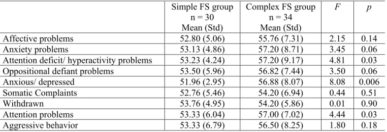
prolonged FS groups ... 93 Table 5. Behavior/ emotion scores on the CBCL for simple and complex FS groups
across both cohorts ... 96 Table 6. School-age cohort behavior/emotion scores on the Conners for simple, multiple ... 98
LIST OF FIGURES
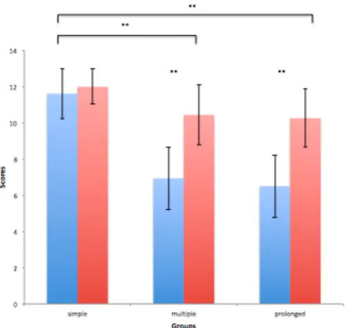
Article 2Figure 1. School-Age group differences according to cognitive domain ... 94
Figure 2. Correlations between executive functioning measures and seizure duration ... 95
Figure 3. Correlations between learning and memory measures and seizure duration ... 95
Figure 4. Significant CBCL group differences across both cohorts ... 97
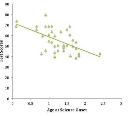
Figure 5. Correlation between Conners Perfectionism Scale and age at seizure onset ... 99
LIST OF ACRONYMS
ADHD: Attention Deficit Hyperactivity Disorder AED: Antiepileptic drugs (treatment)
CBCL: Child Behavior Checklist CNS: Central Nervous System
CVLT-C: California Verbal Learning Test for Children DSM: Diagnostic and Statistical Manual of Mental Disorders EEG: Electroencephalography
ER: Emergency Room
FEBSTAT: The "Consequences of Prolonged Febrile Seizures in Childhood" study FS: Febrile Seizure
FSE: Febrile Status Epilepticus
GABAa: Gamma-Aminobutyric Acid receptor A
GEFS+: Generalized Epilepsy with Febrile Seizures plus ILAE: International League Against Epilepsy
IQ: Intellectual Quotient
MRI: Magnetic Resonance Imaging
NEPSY-II: Developmental Neuropsychological Assessment, 2nd Edition NCPP: National Collaborative Perinatal Project
sAHP: slow afterhyperpolarization SE: Status Epilepticus
WAIS-R: Wechsler Adult Intelligence Scale, Revised
LIST OF ABBREVIATIONS
Bayley-III: Bayley Scales of Infant and Toddler Development, 3rd Edition e.g.: exempli gratia (for example)
et. al.: et alii (and colleagues) i.e.: id est (in other words)
Knowledge comes, but wisdom lingers.
ACKNOWLEDGMENTS
Sarah A very special thank you Sarah, for your guidance throughout this journey; your encouragement, kindness and knowledge and passion for research have made me grow as a future researcher, clinician and individual. Words cannot express my gratitude towards you.
Inga & Fanny A very sincere thank you to you girls, for not only being the "statsperts" that you are, but also for your availability, endless encouragement and sense of solidarity. You knew how to bring laughter to any situation, which was always met with a sense of reassurance and perseverance. Truly, none of this would be possible without you.
Members of the NED lab A huge thank you to all members of the NED lab, which in my biased opinion, are the most wonderful lab mates! Together we were able to talk, laugh, and find solutions to anything and everything.
Participants An incredibly sincere thank you to all participants and their families for their time, effort, and dedication to research; advancements in research would simply not be possible without you.
Funding agencies A particular thank you to the Canadian Institutes of Health and Research (CIHR) and les Fonds des recherche en santé du Québec (FRSQ) for their support.
The most wonderful cohort An indescribable thank you to the most amazing group of colleagues and friends I could have ever possibly asked for! Together we found such an
amazing sense of solidarity, mutual encouragement, and dedication. Our late night group discussions at Tabasco Bar always brought light (and laughter) to any situation. I look forward to working along side all of you for many years to come.
My clinical supervisors A very sincere thank you to all my clinical supervisors, including Elaine De Guise, Elisabeth Perreau-Linck, Maude Lague-Beauvais, Mark Liflan, and all my Hamiltonian supervisors, for your endless encouragement and guidance. I'm very grateful for the countless learning experiences (both professional and in life in general) that were achieved under your supervision, which I will undeniably carry forward in my future life and career.
My parents To both of you; absolutely none of this could have been achieved without your endless support. Words could truly never suffice to express my gratitude. Through thick and thin, through ups and downs, and through the roller coaster ride that defines life as a doctoral student, you were both my rock that, in the end, allowed for me to get through it. Whether it be through phone calls, weekend visits, world-class baked goods or fishing trips, you always knew just what to do, even when I didn't know myself. Thank you, from the very bottom of my heart.
My family A very heart-felt thank you to all my extended family, for their ever-lasting encouragement and support. I feel very lucky to be surrounded by such a supportive group of loving people.
Jen Jen, you are the most wonderful friend; thank you for always being there, not only through the ups and downs of the doctoral journey, but also in life in general. Thank you for having been there through the highest and lowest points over the past decade, and of course for our many late night talks at J&C and summer adventures at Tremblant!
Ericka A wonderfully deep and sincere thank you, Ericka, for being the most dependable, caring and loyal friend. I'm so grateful and thankful for your availability, presence, and fresh outlook on life, that always brings smiles and laughter; it has truly helped me persevere through it all. Thank you also for helping make Hamilton my second home!
INTRODUCTION
Epilepsy encompasses a group of neurological illnesses characterized by epileptic seizures, which are due to abnormal excessive or synchronous electrical activity in the brain (Moorthy et al., 2018). Cognitive impairment is a common comorbidity of epilepsy, which further impacts functioning and quality of life. The most prominent cognitive difficulties observed in this population are memory impairments, mental slowing and attentional difficulties (Aldenkamo, 2006). It has been argued that the cognitive deficits observed largely depend on the pathophysiology of the seizure disorder per se, such that patients suffering from temporal lobe epilepsy are at higher risk of presenting memory impairments, patients suffering from frontal lobe epilepsy are at higher risk of presenting executive impairments, and those with altered thalamo-cortical networks are at higher risk of language and executive functioning impairments (Moorthy et al., 2018). The etiologies of cognitive deficits in epilepsy are multifactorial. Early age at seizure onset has been argued to be the best predictor of cognitive outcome, although several other risk factors have been identified, including seizure type and severity, seizure duration, and use of antiepileptic medication (Strauss, 1995; Aldenkamo, 2006). Cognitive impairments in the context of epileptic disorders are generally considered lasting, particularly when seizure occurrences begin at younger ages (Moorthy et al., 2018).
In considering the pediatric population more specifically, epileptic encephalopathies together form a group of conditions in which epileptic electrical discharges are associated with progressive cerebral dysfunction in the developing brain (Dulac, 2001; Khan & Baradie, 2012). The International League Against Epilepsy (ILAE) has recognized eight age-related
syndromes under the rubric of "epileptic encephalopathies": early myoclonic encephalopathy and Ohtahara syndrome during the neonatal period, West syndrome and Dravet syndrome during infancy and myoclonic status in nonprogressive encephalopathies, Lennox-Gastaut syndrome, Landau-Kleffner syndrome and epilepsy with continuous spike waves during slow wave sleep during childhood (Engel, 2001). These syndromes are commonly characterized by severe and aggressive epileptogenic activity as manifested through EEG paroxysmal activity; seizures that are multiform and intractable, cognitive, behavioral and neurological deficits, as well as occasional early death (Yamatogi & Ohtahara, 1981; Donat, 1992; Dulac, 2001; Michael & Thomas, 2003). In particular, the EEG characteristics of epileptic discharges measured in each syndrome are age-related and vary according to the stage of brain maturity at the time the seizures occur (Yamatogi & Ohtahara, 1981; Donat, 1992; Khan & Baradie, 2012). Specifically, EEG primarily demonstrates burst-suppression patterns in the neonatal period, progressing to hypsarrhythmia in infancy and slow generalized spike-wave discharges in childhood (Yamatogi & Ohtahara, 1981; Dulac, 2001). With increasing age, seizure and epileptogenic features will evolve from one stage to another, and evolutional changes from Ohtahara syndrome to West syndrome to Lennox-Gastaut syndrome are frequently observed with age (Donat, 1992; Michael & Thomas, 2003). Although epileptic encephalopathies are known to attenuate or even stop in adolescence and adulthood, persistent residual neurocognitive sequelae have been well documented (Yamatogi & Ohtahara, 1981; Michael & Thomas, 2003; Khan & Baradie, 2012). Developmental trajectories have been observed to be stunted in all syndromes, including significant psychomotor delays and deficits, as well as mental and cognitive retardation (Khan & Baradie, 2012). Language deficits have been well documented following Dravet syndrome and Landau-Kleffner syndrome. Learning difficulties
have been noted following Ohtahara syndrome. Frontal lobe deficits, including difficulties with judgment and the ability to control and anticipate behavior, have been observed in epilepsy with continuous spike waves during slow wave sleep (Michael & Thomas, 2003; Khan & Baradie, 2012).
A constellation of several different clinical presentations commonly occurs in epileptic encephalopathies, including atonic seizures, astatic seizures, clonic seizures, epileptic spasms, myoclonic seizures, myoclonic-atonic seizures and tonic seizures (Khan & Baradie, 2012). When any of these symptoms occur for a duration longer than 30 minutes, the semiology is consistent with status epilepticus (SE), which is considered the most extreme and severe form of a seizure (Trinka, et al., 2015). Moreover, when any of the symptoms occur as a result of a febrile illness rather than a neurological condition, the semiology is consistent with febrile seizures (FS), the most common form of childhood seizure (Shinnar & Glauser, 2002). SE and FS may occur as an isolated seizure event without defining or being part of any broader syndrome. Although epileptic encephalopathies have been strongly and consistently associated with persistent long-term cognitive sequelae (Michael & Thomas, 2003; Khan & Baradie, 2012), and isolated SE events have generally been shown to impact cognitive development (Sheppard & Lippé, 2012), much less is known about the impact of isolated FS events on cognition. Even though it is a significantly less severe form of seizure, it is the most commonly occurring one, and more research is required to better understand its impact on the developing brain and cognitive development.
Status Epilepticus (SE) is the most severe form of a seizure. It's a transient occurrence of signs and/or symptoms due to abnormal excessive or synchronous neuronal activity in the brain (Trinka, et al., 2015). According to its updated ILAE definition, "SE is a condition resulting either from the failure of the mechanisms responsible for seizure termination or from the initiation of mechanisms which lead to abnormally prolonged seizures" (ILAE, 2015). It is agreed that the seizure in question must persist for at least 30 minutes in order to meet criteria for SE, given that irreversible neuronal injury typically occurs after this length of time. Indeed, it is a condition that can have long-term physiological consequences, including neuronal injury, neuronal death and alterations of neuronal networks (ILAE, 2015). SE can either be classified as convulsive (i.e., with prominent motor symptoms and impairment of consciousness) or non-convulsive (i.e., without motor symptoms or impairment of consciousness), and EEG patterns are non-specific (Lowenstain, et al., 1998). It is not a disease entity, but rather an event with many different etiologies. At least half of patients presenting with SE do not suffer from any particular syndrome or epilepsy, rather the event is due to acute or remote central nervous system (CNS), or systemic illness (Maytal, et al., 1989). Although SE can occur at any age, 40% of SE events occur prior to 2 years of age, owing to the volume of neurons and excitatory connections prior to functional specialization, argued to create an imbalance between excitatory and inhibitory connections, which increases the immature brain's vulnerability to hypersynchronization and SE (Shinnar, et al., 1997; Wasterlain, et al., 1993; Scott, et al., 1998). Febrile SE (FSE) is the most common etiology in children, in which high fever without any other provocation to the CNS induces the SE event (Fountain, 2000).
Cognitive sequelae following SE
Residual cognitive sequelae following SE in early life have been relatively well documented. Alterations in global intelligence, verbal and non-verbal intelligence and motor development have been demonstrated following early-life episodes lasting longer than one hour (Kolfen, et al., 1998; Dam, 1990; Van Esch, et al., 1996, Aicardi & Chevrie, 1970). More specifically, it has been argued that the age at which the SE event occurs will hinder the cognitive abilities under development at the time. Roy, et al. (2011) demonstrated that when an FSE event occurred prior to 11 months of age, hand-eye coordination and motor ability were most affected, but spared in children with FSE onset beyond 12 months of age, who in turn manifested difficulties in language and social behavior. When tested at 2 years of age, these children presented shortcomings in executive functioning, including self-monitoring and inhibition difficulties. Although age at onset is argued to be a principal predictor of subsequent cognitive sequelae, duration and frequency of seizures, as well as etiology are also important risk factors.
Taken together, cognitive sequelae following SE, the most severe form of seizure, has been relatively well documented (Sheppard & Lippé, 2012). Understanding the impact of early life SE and FSE on cognitive development can begin to shed light on understanding cognitive sequelae following early life seizures that are less severe in nature, although the most common, namely, Febrile seizures (FS).
Definition
Febrile Seizures (FS) are the most common form of childhood seizure disorder. The International League Against Epilepsy (ILAE) defines a FS as "a seizure in association with a febrile illness in the absence of a CNS infection or acute electrolyte imbalance in children older than 1 month of age without prior afebrile seizures" (ILAE, 1993; Engel, 2006; Patel, et al., 2017). Since fever is associated with seizure in FS, neurological illness such as meningitis, encephalitis, and others must be excluded upon evaluation. Febrile Seizures usually occur within the first 24 hours of an illness, and fever typically reaches temperatures of 380C or higher (Leung & Robson, 2007; Berg & Shinnar, 1996). With regards to semiology, FS are mostly generalized and convulsive (i.e., generalized tonic-clonic seizures), however approximately 5% present with non-convulsive features, including unconsciousness, staring, eye deviation and atonia. (Pavlidou, 2013; Patel, et al., 2015).
Classification
Febrile Seizures are classified as either simple or complex based on the duration and recurrence of the seizure, as well as the presence of focal features (ILAE, 1993; Shinnar & Glauser, 2002). Simple FS is characterized by an isolated, brief (fewer than 10 minutes) and generalized seizure, whereas complex FS is more severe and quite heterogeneous in its presentation. It is characterized by either focal onset, prolonged duration (lasting between 15 and 30 minutes), occurring more than once during febrile illness, or a combination of different complex features. Febrile status epilepticus (FSE) is the most severe type of complex FS in which the seizure persists for at least 30 minutes. Although research has consistently
demonstrated benign outcomes following the occurrence of a simple FS, such that the affected children have been shown to develop similarly to otherwise healthy children who have not suffered seizures (Chang et al., 2000, Berg & Shinnar, 1996), evidence related to outcomes following complex FS remains unclear and controversial.
Epidemiology
Febrile Seizures occur in 2% to 5% of children between the ages of six months and five years (Verity et al., 1985; Shinnar & Glauser, 2002; Pavlidou, et al., 2013). Epidemiological studies demonstrate that FS onset peaks at 18 months and that onset beyond the age of six is rare (Leung & Robson, 1991; Baumann, et al., 2000). The incidence has been documented to be slightly higher in boys than girls (male to female ratio 1.1:1 to 2:1), although some studies do not show any sex differences (Pavlidou, 2013; Stafstrom, 2002; Chung, 2014). In affected children, approximately 75% will suffer simple FS and 20% to 30% will suffer complex FS (Annegers, et al., 1987; Shinnar & Glauser, 2002; Patel, 2017). The national collaborative perinatal project (NCPP), a study that prospectively followed 1706 children having suffered FS from birth to 7 years of age, revealed that 28% of children presented with an initial complex FS, of which 4% were focal, 7.6% were prolonged and 16.2% were recurrent (Capovilla, et al., 2009). Febrile status epilepticus accounts for 5% of FS events and 25% of overall SE events in children (Berg & Shinnar, 1996; Maytal & Shinnar, 1990; Patel, et al., 2017). Early results of the FEBSTAT study, a prospective and longitudinal multi-center study investigating the long-term impact of FSE on cerebral and cognitive development, reveal that it's presentation is focal in 67% of cases, and its onset peaks in relatively younger children (median age of 1.3 years) (Shinnar, et al., 2008). Moreover, although a seizure lasting at least
30 minutes is required to meet FSE criteria, the FEBSTAT study revealed a median seizure duration of 68 minutes and 24% of durations lasting more than two hours.
Etiology
As per their definition, FS are induced by a fever of at least 380C, typically associated with a systemic illness. The age-specific mechanisms involved in seizure development related to high fever are however quite debated and overall suggest multifactorial etiologies, including both environmental (including the systemic illness and fever itself) and genetic factors (Offringa, et al., 1994; Berg, et al., 1999; Audenaert, et al., 2006).
The pathogenesis of FS remains unclear and has mostly been studied through animal models. Given that FS are age-dependent, there seems to be a temporal association between the immature CNS and the onset of FS, although this association has yet to be clearly established, a situation which is further complicated by the heterogeneity of FS presentations. It is suggested that fever arising from febrile illness is linked to an imbalance between excitatory and inhibitory transmissions leading to seizure activity (Pavlidou, 2007; Heida et al., 2009). More specifically, it is suggested that fever increases brain temperature, which in turn alters neuronal functioning through temperature-sensitive ion-channels and inflammatory processes promoting the secretion of cytokins, which together increase neuronal excitability and increase the probability of generating seizures (Leung & Robson, 2007; Pavlidou, et al., 2013; Reid, et al., 2009; Dubé et al., 2005; 2009). Overarching are on-going "chicken or the egg" debates arguing that either FS, particularly in cases of prolonged complex FS, may lead to hypoxic damage of the CNS and subsequently cause increased vulnerability to seizure activity, or fever
may trigger a seizure in pre-existing CNS disorders (Chang, et al., 2008). Taken together, FS have multifactorial and heterogeneous etiologies, leading to debates as to whether FS arise in otherwise healthy children, or as a result of an underlying although undetected predisposition, or whether this further depends on the type of FS suffered.
Risk Factors Associated this FS Genetic Risk Factors
Population-based studies show that FS tend to occur more frequently in first-degree relatives of children with FS. In particular, 25% to 40% of affected children show a positive family history of FS (Chung, 2014). Moreover, the incidence is 20% to 25% higher among siblings and 10% higher among parents of children with FS (Hauser, et al., 1985; Knudsen, et al., 1996). Twin studies have similarly shown that predisposition to FS is higher among monozygotic twins (22%) than dizygotic twins (11%) (Waruiru, et al., 2004; Pavlidou, 2013).
No single susceptibility gene has been specifically detected for FS (Patel, et al., 2015), although several gene loci have been associated with the onset of FS. In particular, linkage studies have proposed 11 chromososmal locations responsible for FS, including FEB1 to FEB11 (Sghazadeh, 2014). Moreover, mutations in two voltage-gated sodium channel genes (SCN1A and SCN1B) and GABAa receptor gene have been identified in seizure disorders that often initially present as FS, including Dravet syndrome and GEFS+ (a syndrome in which individuals present with complex FS beyond the age of 5 and later develop afebrile seizures) (Kang, et al., 2006; Kira, et al., 2010; Abou-Khalil, 2010).
Environmental Risk Factors
The principle environmental factor involved in FS is the fever event, usually related to systemic infection. The most common causes for fever in FS are those associated with influenza A, gastroenteritis, otitis media, respiratory infection and human herpes simplex virus-6 (Millichap & Millichap, 2006, Kwong et al., 2006; Van Zeij, et al., 2004; Pavlidou, 2013). Other environmental factors related to poor outcome following FS include low family income and parental education (Leaffer, 2013). Developmental delay and neurological abnormalities prior to FS onset are further related to suboptimal outcome (Leaffer, 2013).
Risk of Initial FS
The risk factors associated with the development of an initial FS include a positive family history of FS in first degree relatives, a neonatal stay longer than 28 days, known developmental delay and day-care attendance (Chung, 2014). Children presenting with more than two risk factors have an increased chance of developing a first FS by 28%, and pre-existing developmental delay is most commonly associated with prolonged complex FS (Mastrangelo, et al., 2014).
Risk of FS recurrence
Following an initial FS, up to one third of affected children will have a recurrence (Patel, et al., 2015). Age at onset is the strongest and most consistent risk factor. When seizure onset occurs in the infantile period (younger than 18 months), the risk of recurrence is 50% (Cendes & Sankar, 2011; Pavlidou, 2013). Other risk factors for recurrence include a family history of FS in first-degree relatives, pathological prenatal history, low height and short duration of the
fever episode, focality and recurrence of seizures within the same febrile episode (Waruiru, et al., 2004; Pavlidou, et al., 2008). Children presenting with all these factors have up to an 80% chance of recurrence, whereas children presenting none of these factors have approximately a 4% chance of recurrence (Berg, et al., 1997; Patterson, 2013; Patel, 2015).
Risk of subsequent development of epilepsy
Approximately 2% to 6% of children suffering FS will subsequently develop epilepsy (Baumer, 2004; Abou-Khalil, 2007; 2010). The risk of developing an epileptic syndrome following simple FS is similar to that of the general population (1% to 2% risk), whereas the risk is increased following complex FS (4% to 7% risk) (Verity & Golding, 1991; Vestergaard, et al., 2007; Baumer, 2004; Abou-Khalil, 2010). Moreover, 35% of adults suffering from temporal lobe epilepsy (TLE), the most common focal epilepsy in adults, have a positive history of complex and/or prolonged FS in childhood (Reid, et al., 2009; Abou-Khalil, 2010). The main risk factors for later development of epilepsy include a positive family history of epilepsy, complex and/or prolonged FS and neurodevelopmental impairment (Abou-Khalil, 2010; Vestergaard, et al., 2007; Capovilla, et al., 2009). Causal links between these risk factors and the development of epilepsy are controversial and debated. It is argued that prolonged FS cause acute hippocampal damage resulting in residual hippocampal sclerosis, which represents the hallmark of TLE (Kira, et al., 2010). This hippocampal damage is argued to render the brain more vulnerable to seizures, which can eventually lead to epileptic syndromes (Barr et al., 1997; Harvey, et al., 1995; Wu, et al., 2005).
Although research has shed significant light on the understanding of FS, despite certain links between FS and causes/consequences that remain unclear, outcome following FS is much less understood. Only relatively recently have FS been flagged as not being as benign as previously thought. Further investigations are required to bring better understanding to the impact of FS, given that they are the most common form of childhood seizure, particularly as they occur at a sensitive developmental age.
CEREBRAL DEVELOPMENT
From a structural perspective, neurons are developed and migrate to their final destination at approximately the 16th week of gestation, after which synapses and dendrites form connections, and axons begin to acquire myelin, which helps speed neural transmissions (Sidman & Rakic, 1973; Andersen, 2003). During the period immediately before birth, about 50% of all neurons are eliminated in a process of programmed cell death (i.e., apoptosis), a phenomenon which is believed to increase the efficiency of synaptic transmission (Andersen, 2003). Rapid cerebral development continues after birth, when the brain gains significant weight and volume. Processes involved in neuronal and network development begin to peak around the first year of life. Such processes include dendritic arborization and synaptogenesis, characterized by an explosion in the formation of neuronal connections, as well as myelination and synaptic pruning, characterized by the weeding of unnecessary connections and strengthening of utilized connections. The specialization of neurons and functions continue through infancy, childhood and into adolescence (Casaer, 1993, Andersen, 2003). The processes involved in cerebral development are argued to occur in a hierarchical manner, in
which neurons organize from the deepest layer to the outer-most layer and from posterior to anterior regions of the brain (Jernigan & Tallal, 1990; Andersen, et al., 2000). Moreover, the process of myelination is argued to similarly occur hierarchically from primary and sensory areas, to association areas and cortical regions, such that neurons involved in carrying sensory information are argued to be the first to acquire myelin, followed by neurons involved in carrying motor information, and so on in a hierarchical manner, until the myelination process reaches axons placed in the cortex (Casaer 1993; Hudspeth & Pribram, 1993; Staudt, et al., 1993). Even though anterior cortical regions are argued to be the last to reach maturity, evidence suggests that many areas of the cortex begin to function in infancy, including early specialization of the frontal cortex (Anderson, 2001). In particular, frontal behavior-related metabolic changes have been detected in infants as young as 6 months of age (Chugani, et al., 1987). EEG has been shown to change in relation to improved behavior during the first year of life (Bell & Fox, 1992), even though frontal regions show accelerated development from 7 to 10 years of age.
Understanding cerebral development through cellular and structural maturation can shed light on understanding how structure then relates to behavior and function and further, understanding of the mechanisms of how early life insults to the brain can impact cognitive development. Even though functional specialization occurs throughout childhood, into adolescence and even early adulthood, cognitive capacities begin specializing sufficiently enough at school-age (i.e., approximately 5 years of age) to allow for their specific evaluation through behavior (i.e., neuropsychological measures), in order to assess the impact of early life insults on particular cognitive capacities.
PLASTICITY VERSUS VULNERABILITY THEORIES
Although the extent and location of early life injury to the brain predict severity of residual impairment, timing of the injury will dictate the nature of the impairments. Following insults to the brain in early life, it was traditionally believed that owing to a lack of functional specialization, the young brain was "plastic" and able to adapt to injury, in that abilities subsumed by the damaged region could more readily reorganize and therefore recover function (Kennard, 1936). This is the assumption of Plasticity theory, which predicts that the earlier in life the insult occurs, the better the outcome in later life. This principle was established by early studies demonstrating normally developing intellectual and cognitive capacities in young children following focal brain injuries. In contrast, Vulnerability theory predicts that owing to the lack of functional specialization, the brain will attempt to recover endangered functions from a damaged structure by aberrantly creating faulty connections (Giza et al, 2002). Specifically, if damage occurs at a critical stage of development, cognitive skills already established will be spared, but those emerging and dependent on the damaged region may be irreversibly impaired. As such, a crowding effect will take place such that healthy neurons will take over damaged neurons in an attempt to recover the developing function. However, this phenomenon will limit these neurons' quantitative and qualitative resources, creating a "crowding" of cognitive functions for that particular tissue (Statz et al, 1994). Evidence from brain lesion studies has demonstrated that cognitive functions subserved by the cerebral structures that are under development at the time of the insult are the most affected (Anderson & Moore 1995; Dennis, 1989). In the evaluation of both theories, the
Vulnerability theory has been the most supported to date (Anderson, et al., 1997; Bittigau et al, 2004; Dennis, 1989).
With regards to seizure disorders in early life, it is argued that epileptogenic or abnormal electrical activity will compete with normal brain activity for neural resources (Pavlidou, 2007). If abnormal activity occurs at critical stage of cerebral development, aberrant neuronal connections may be formed and normal brain functions may fail to develop.
OUTCOME FOLLOWING FS
Physiological outcome Animal Models
Animal models of experimentally induced FS, typically using a hyperthermia paradigm in rodents, consistently demonstrate altered hippocampal structure and function following FS. In particular, cytoskeletal changes have been demonstrated in hippocampal neurons within 24 hours following experimentally induced FS, for which altered functional properties of these neurons persisted into adulthood (Toth et al, 1998). Furthermore, MRI studies of experimentally induced FS show abnormally high T2 signal in the hippocampus, demonstrating marked anatomical abnormalities in the acute phase post-seizure, which were long-lasting (Dubé et al, 2004). Additionally, increased cytogenesis in the dentate gyrus and significant dark neuron formation following FS interpreted as marked neuronal injury has been observed, which further proved to be persistent effects of the FS (Nazem, 2012). In the predisposed rat brain, hippocampal damage characterized as atrophy associated with neuronal
loss has also been shown following a single episode of FS (Gibbs et al, 2011). Additionally, a decrease in dendritic spines in hippocampal neurons was found in these rats, which was associated with neuronal hyperexcitability. Indeed, long-lasting neuronal hyperexcitability has been consistently demonstrated in animal models following prolonged FS (Chen, et al., 1999; Brewster, 2002; Notenboom, 2010). This hyperexcitability following FS has been related to learning and memory impairments by a persistent decrease of the slow afterhyperpolarization (sAHP) in hippocampal neurons, characterized as a prolonged afterhyperpolarization that restrains repetitive firing underlying synaptic efficiency and therefore learning and memory (Kamal et al., 2006).
In Children
Loss of hippocampal integrity has also been demonstrated in children having suffered FSE. In the acute phase post-FSE, transient increases in hippocampal volume as well as signals of hippocampal hypertension as revealed by increased T2- weighted MRI relaxation times have consistently been demonstrated (Huang & Chang, 2009; Shinnar, 2003). Although some studies demonstrate resolution of acute abnormalities within the first few months following seizure onset, most argue for persistent residual sequelae (Shinnar, 2003).
Epidemiological studies of TLE have revealed that TLE patients with a prior history of FS demonstrate decreased bilateral hippocampal volume as compared to TLE patients without such prior history (Barr et al, 1997; Harvey et al, 1995). Furthermore, hippocampal sclerosis was found to be strongly associated with prior neurological insult in childhood, mostly characterized as prolonged FS (Harvey et al, 1995).
MRI volumetric analysis studies have similarly demonstrated loss of hippocampal integrity following FS. In particular, follow-up studies performed 4 to 16 months and further at 6 years post-FS have demonstrated that children having suffered from prolonged FS show hippocampal asymmetry, by evidence of a smaller right hippocampus (Scott et al, 2003; Merkenschlager et al, 2009). Furthermore, these asymmetries in hippocampal volume have been shown even when MRI scans done in the acute phase post-FS did not show abnormalities, indicating a progression toward hippocampal injury (Merkenschlager et al, 2009; Lewis, 2014). MRI volumetric analysis studies have also revealed hippocampal atrophy characterized as a decrease in hippocampal volume. In particular, longitudinal studies have demonstrated that most children having suffered prolonged FS showing acute hippocampal injury show hippocampal atrophy two-years after onset (VanLandingham et al, 1999; Provenzale, 2008; Hesdorffer, et al., 2008). Additionally, hypertense hippocampi as evidenced by increased T2-weighted MRI images at time of FS onset were correlated with hippocampal volume loss and even medial temporal sclerosis in some cases (Provenzale, 2008). Results of these longitudinal studies point to evolving hippocampal damage following complex FS, even in children who's initial scans showed no abnormalities.
It is important to note that most imaging research to date has been completed in children having suffered prolonged FS and FSE. The few studies that have investigated the impact of other complex features on cerebral development have revealed persistent MRI hippocampal abnormalities in both prolonged and focal FS (Hesdorffer, et al., 2008), whereas other studies
have demonstrated a greater impact of multiple seizures on hippocampal volume loss as compared to focality and duration of the seizure (Yoong, et al., 2013).
Moreover, the vast majority of imaging studies have focused on the development of the hippocampus. Although it is argued to be the most affected structure in FS and FSE, next to no research has been performed in investigating hippocampal abnormalities in the larger context of brain development, specifically how alterations in the hippocampus might impact the development of other structures, or inversely, how alterations in other structures might impact hippocampal development. Indeed, the hippocampus is a structure that plays an active role in larger networks, the cortico-hippocampal network in particular. The function of this network is known to play an integral part in learning and memory (not just as a function of the hippocampus in isolation). Moreover, given the direct and monosynpatic connections between the hippocampus and frontal/prefrontal areas, including the medial prefrontal cortex known for its involvement in executive functioning, a set of cognitive processes involved in the cognitive control of behavior (e.g., inhibition, self-monitoring, goal-directed behavior), it is possible that damaged hippocampi following FS may result in cognitive challenges beyond learning and memory.
Cognitive outcome following FS Animal models
Animal models of experimental FS in rodents have shed light on cognitive difficulties following FS. Notably, it has been found that hyperthermia-induced FS in predisposed rat brains resulted in impairments on the Morris Water Maze task, a task of learning and memory
(Scantleburry, 2005; Rajab 2014). Furthermore, cognitive testing of adult rats that suffered hyperthermia-induced FS as pups without early cortical lesion demonstrated deficits in working and reference memory in the Morris Water Maze task (Dubé et al., 2009; Rajab 2014). Additionally, these deficits were shown to be related to impaired hippocampal function and structure (i.e., as shown by an abnormally high T2 signal). Taken together these animal models of early life FS demonstrated learning and memory impairments in adult life following an experimental FS event. The FS induced in these animals are considered to be at the "severe" end of the spectrum of FS, and would correspond to FSE in humans (Roper, 2016). Moreover, animal models have focused on spatial learning, as it is challenging to test other types of learning in rodents, and have overall focused on abilities largely dependent on hippocampal functioning. Although these studies provide valuable insight into the deficits observed in the most severe form of FS, they lack evidence related to other possible deficits following different types of FS.
In children
The impact of complex FS on cognitive development and behavior in children remains unclear. Although converging evidence suggests unaltered global intelligence following complex FS and FSE, the impact of these types of seizures on specific cognitive functions, beyond intelligence, is debated. While some studies indicate unaltered development, scholastic achievement and behavior following complex FS, others argue for hindered developmental trajectories and disrupted behavior following the event (Ellenberg & Nelson, 1978; Verity et al., 1998; Hirtz, 2002; Martinos, 2012; 2013; Weiss, 2016). Divergent evidence has typically been the result of inconsistent methodologies and populations studied. Early studies denying
any impact of FS on cognitive development used measures that lacked specificity and objectivity (i.e., surveys). Using more specific, objective and standardized measures, other studies have revealed contrasting evidence regarding the impact of complex FS on cognition and behavior in school-age children, although discrepancies in methodologies are further noted, including populations used (i.e., population versus hospital-based samples), complex seizure type studied (i.e., prolonged versus multiple versus focal), measures used (i.e., objective versus subjective) and time points assessed (i.e., varying time points since seizure onset or last seizure occurrence) (Kolfen et al., 1998; Chang et al., 2000; 2001; Norgaard et al., 2009; Visser et al., 2012).
Understanding early developmental outcome, within the first year-post FS onset, could shed light on the understanding of long-term cognitive outcomes. Few studies to date have investigated development within the first year-post seizure onset. In particular, children having suffered FSE have been shown to develop normally within the first month post-seizure onset, although demonstrated slightly weaker motor development and receptive language one year-post onset (Weiss, 2016). Children having suffered a prolonged complex FS consistently demonstrated worse developmental outcome as compared to controls 6 weeks and 1 year following seizure onset (Martinos, 2013), as well as accelerated forgetting within the first month and 1 year following onset (Martinos, 2012). Weaker development one year-post onset has further been linked to hippocampal anomalies in children having suffered prolonged seizures (Weiss, 2016; Martinos, 2012). These results suggest a possible worsening of the impact of the initial seizure on development over time, particularly in the context of neurodevelopment, as FS occur during a period of rapid cerebral development and functional
specialization (Andersen, 2003). However, studies to date have focused on FSE, forgoing the investigation of the possible impact of focal and recurrent seizures on development, even though they have also been shown to alter structure (Hesdorffer, et al., 2008; Yoong, et al., 2013). Moreover, the impact of seizure duration on cognition can be considered somewhat biased in the FSE studies, as their mean seizure duration varied between 70 and 90 minutes (Weiss, 2016; Martinos, 2012). It is still unknown whether a less prolonged FS, that is, lasting between 15 and 20 minutes, may have a similar or commensurable impact on cognition.
Beyond the impact of complex FS on development, studies investigating their impact on cognition have focused on hippocampus-dependent functions, mainly learning and memory. In particular, infants having suffered FSE have demonstrated reduced memory capacities, as well as accelerated forgetting within the first year post-onset, which were associated with reduced hippocampal volume (Weiss et al., 2016; Martinos et al., 2012). Other studies investigating school-age children having suffered non-prolonged complex FS found that although memory performances were similar between FS and control groups, mechanisms used to achieve similar behaviors were different, evidenced by altered event-related potentials and hemodynamic activity (Kipp et al., 2010; 2012).
With regards to their impact on behavior, complex FS have been associated with increased external behavioral deficits and increased attentional difficulties as measured by parental questionnaires (Kolfen et al., 1998; Lippé et al., 2009; Tsai et al., 2015). More objectively, very few studies to date have examined the impact of complex FS on executive functioning later in life. Roy et al (2011) demonstrated that children of 2 years of age showed reduced
self-monitoring and inhibition abilities following a single episode of SE. They compared these children to two control groups, namely children having suffered a FS and otherwise healthy controls. Their results indicated that although children having suffered from an episode of SE performed worse than healthy controls, children having suffered FS did not differ significantly from either SE or control group, suggesting that an episode of FS may hinder these functions, albeit to a lesser extent. In investigating FS per se, children having suffered complex FS have been shown to demonstrate weaker sustained attention abilities (Hara et al., 1986), although other studies have suggested better performances in children having suffered FS (Chang et al., 2000; 2001). The results of the latter two studies however reveal that although children having suffered complex FS were better at sustaining their attention on complex tasks, they had more difficulty sustaining their attention on simple tasks as compared to controls, which could possibly indicate a need for arousal and challenges in self-monitoring abilities. To our knowledge, no study has yet objectively and specifically assessed executive functioning in children having suffered complex FS beyond working memory and sustained attention abilities.
Overall, studies investigating the impact of complex FS on cognition have to date mainly focused on FSE and hippocampus-dependent functions. Exploring the effects of other complex features (i.e., recurrence and focality) on these functions, as well as on others that could be affected by faulty hippocampal function through cortico-hippocampal networks, including executive functions, could help increase our understanding of the impact of FS on cognition. Moreover, investigating their impact on cognition as a factor of time (i.e., age), could help
understand the evolution of possible challenges through early development, a time when significant maturational changes and functional specializations occur in the brain.
RESEARCH OBJECTIVES AND HYPOTHESES
1. The first objective was to review cognitive sequelae following Status Epilepticus, the most severe form of a seizure in childhood, through a more extensive review of the literature than previously available.
2. The second objective was to investigate development and cognition from onset to school-age following complex FS, as compared to children having suffered simple FS, in the context of known risk factors for poor outcome, including all types of complex features. More specifically, we aimed to transversally evaluate development within the first year-post seizure onset, as well as cognition in a cohort of children old enough for cognitive functions to be sufficiently differentiated (i.e., school-age).
It was hypothesized that infants having suffered complex FS would show hindered development as compared to simple FS controls, within the first year-post onset. It was further hypothesized that school-age children having suffered complex FS would show weaker performances on measures of learning/memory and executive functioning as compared to simple FS controls, given the role of the hippocampus in FS, and the possible impact of this early life insult on the development of
structures subserving executive functions, which are crucial for academic success. Lastly, it was hypothesized that developmental and cognitive measures would be associated with known risk factors for poor outcome, including younger ages at onset and longer seizure durations.
ARTICLE 1
COGNITIVE OUTCOME OF STATUS EPILEPTICUS IN CHILDREN
Emilie Sheppard1,2 & Sarah Lippé1,2
Author Affiliations:
1. Department of Psychology, University of Montreal, CP 6128, succ Centre Ville, Montreal, Qc, H3C 3J7, Canada
2. CHU Sainte-Justine, 3175 Côte Ste-Catherine, Montreal, Qc, H3T 1C5, Canada
Published: Sheppard, E. & Lippé, S. (2012). Cognitive outcome of status epilepticus in children. Epilepsy Research & Treatment; 2012, 1-8.
ABSTRACT
Epileptic encephalopathy encompasses conditions in which cognitive, motor or sensory deficits result as a consequence of epileptic activity defining certain syndromes. It therefore represents a more severe subset of epilepsy, which can be generally characterized as frequent or severe seizures leading to cerebral dysfunction. This disturbance in cerebral functioning can in turn hinder, somewhat dramatically, cognitive development and further impact the future lives of patients. In this review, we describe the cognitive consequences of Status Epilepticus in children and in adults in the context of plasticity theories. Recent studies maintain that consequences of SE may be severe cognitive sequelae, especially in early life. Since the residual consequences of SE in adulthood seem less detrimental and long-lasting, we argue that early life insults, such as those created by SE, during a rapid period of development and functional specialization, result in specific cognitive deficits dependent on the sensitive period at which SE occurred.
1. INTRODUCTION
Epileptic encephalopathy encompasses conditions in which cognitive, motor or sensory deficits result as a consequence of epileptic activity defining certain syndromes (1). It therefore represents a more severe subset of epilepsy, which can be generally characterized as frequent or severe seizures leading to cerebral dysfunction. This disturbance in cerebral functioning can in turn hinder, somewhat dramatically, cognitive development and further
impact the future lives of patients. In this review, we consider Status Epilepticus as an epileptic encephalopathy owing to its impact on cognitive development in early life.
2. STATUS EPILEPTICUS (SE)
Status Epilepticus (SE) is a medical epileptic emergency characterized by either rapidly repeating seizures without recovery or regain of consciousness between episodes, or prolonged continuous epileptic activity, both creating a fixed or lasting condition (2, 3). It is an event rather than a syndrome. It is accepted that the duration of an episode of SE is 30 minutes or more, period after which cerebral functioning is highly probable of being affected and immediate medical attention is needed (4). Recently, the notions of impending SE and established SE have been introduced (5) in order to provide the best possible care for patients presenting with SE. In adults, patients presenting a seizure lasting more than five minutes can be designated as impending SE. In children, impending seizures are considered when seizures last between 5 and 10 minutes (5).
The prevalence of SE varies. Three epidemiologic studies suggest 17 to 108/100000 as being the prevalence of SE (6, 7, 8). Although SE can occur at any age, it is most often encountered in infancy and childhood, 40% of all cases occurring prior to 2 years of age, a period in which the brain is in rapid development (9). Such prevalence is argued to be present in early life owing to the exceeding amount of neurons and excitatory connections prior to functional specialization while undergoing neuronal pruning, which increases the vulnerability of the developing brain to SE (10). In affected children, an imbalance between inhibitory and
excitatory neurotransmissions is argued to lead to anomalies in neuronal impulses leading to prolonged seizures (11). In fact, the pathophysiology of SE seems to involve a loss of inhibitory mechanisms, which result in a deficiency of the neuronal metabolism, which is unable to keep up with the demands of the continuous epileptic activity (12). The seizures are most frequently generalized, but may also be partial and either convulsive or non- convulsive (13). SE is further classified in accordance with its respective etiology (14). Idiopathic SE occurs in otherwise healthy individuals without metabolic dysfunction nor an acute insult to the Central Nervous System (CNS). Furthermore, remote symptomatic SE occurs in patients with a history of insult to the CNS without acute provocation such as in mental retardation. Febrile SE, the most common etiology in children (15), occurs when the only provocation of the CNS is a high fever, usually higher than 38,4 degrees Celsius. In this population, 86% of children demonstrate normal prior development (16). Acute symptomatic SE occurs during an acute illness with a known insult to the CNS such as in meningitis. Although there has been debate on the long-term effects of SE on cerebral functioning, recent research investigating more accurately the cognitive sequelae related to SE demonstrate that cognitive functions under development are exposed to being altered and damaged in children presenting with SE, owing to its high incidence in infancy, a period of marked and rapid cognitive development.
3. PLASTICITY VS. VULNERABILITY IN THE DEVELOPING BRAIN
In considering the impact of an early insult on cerebral and cognitive development, two opposing theories are contradictory in their predictions. The Plasticity theory posits that the young brain is flexible and therefore capable of recovery after insult. As such, since there is
less functional specialization in early life, functions that would depend on a damaged area would simply reorganize to functionally cope with the insult (17, 18). As such, this theory predicts that early brain damage is the most biologically manageable, resulting in less vulnerability to the impact of damage as opposed to an older brain. In contrast, the Vulnerability theory posits that the young brain is the most fragile and therefore vulnerable to early insult. It argues that owing to the lack of functional specialization, the brain will attempt to recover endangered functions, but will do so aberrantly creating faulty connections in early life (19). As such, a crowding effect will take place such that healthy tissue will take over the damaged tissue in attempting to recover the cognitive function at hand, but consequently limiting the tissue's quantitative and qualitative resources (20). This effect was first demonstrated in the context of hemispheric dominance following left hemisphere damage in early life such that an insult to the left hemisphere prior to one year of age resulted in the proper development of language but faulty development of non-verbal skills; owing to brain plasticity, the emerging language functions took over neurons dedicated to non-verbal skills. The reverse effect was observed when the insult occurred after one year of age (20, 21). As such, healthy tissue, although already specialized for a certain function will forgo that specialization for the proper development of the function underlying the insult, creating a “crowding” of cognitive functions for that particular tissue Therefore, the Vulnerability perspective of the developing brain predicts that early life insults are the most difficult to recover from.
In further investigating the opposing predictions of both theories of the impact of early insult on the developing brain, the Vulnerability theory has been the most supported (22, 23, 24, 25).
It has been found that young neurons more readily grow to make new connections, which following an insult, may facilitate aberrant connections (26). As such, the developing brain is the most vulnerable to insult resulting in subsequent damage post-SE potentially persisting in later life. Furthermore, findings demonstrate that not only is the severity of the sequelae following SE predicted by the extent and location of the insult, but the nature of the sequelae itself is determined by the timing of the SE episode (27). As such, the developmental period at which the insult occurs is argued to predict which cognitive functions will be most affected and therefore predict the general outcome of the patient.
4. A MODEL OF HUMAN DEVELOPMENT
In concordance with the Vulnerability theory, early insults to the brain have the most detrimental impact on cerebral and cognitive development persisting in later life. As such, faulty neuronal connections following an early life insult during a critical period of development will hinder the normal development of brain functions, for which the sequelae will persist in later life (28). However, already developed functions at the time of the insult will be spared. The notion of critical periods during infancy through adolescence is widespread and generally accepted (29). Critical periods allow for a logical hierarchy in development such that windows of opportunity allow for the specialization of functions. Furthermore, certain structures and their underlying function must be well specialized prior to others. As such, sensing pathways such as those involved in vision and hearing must develop prior to language pathways, which in turn must develop prior to higher cognitive functioning, including executive functions (29). Critical periods, consequently, expose certain functions as
more vulnerable than others at particular and specific periods during development. The vulnerability of different cognitive functions therefore varies with the developmental process itself. In the presence of an early insult to the brain, the function under development will be hindered, affecting not only that particular function, but also the development of subsequent functions dependent on the hindered one. Healthy development of cognitive functions depends on the integrity of the structure the function underlies. As such, following an early life insult, the integrity of a particular structure is compromised, further compromising the cognitive function that structure is responsible for.
5. PHYSIOLOGICAL ALTERATIONS RESULTING FROM SE
Prolonged and frequent seizures, such as those involved in SE consistently show physiological brain damage. In fact, the physiological properties of cells have been shown to be altered following an SE event (30, 31). The most vulnerable structure to the seizures is the hippocampus, which is involved in learning and memory. Hippocampal edema, cell loss particularly in the Sommer sector, and abnormalities have consistently been detected within this structure following SE (32, 10). Also in human, other structures have been demonstrated to show necrosis following events of epileptic attacks such as the amygdala, dorsomedial thalamic nucleus, medial layers of the neocortex, cerebellum, the piriforme and entorhinal cortices (32, 33, 30, 31). Neuronal degeneration and loss in these areas have been shown to occur rapidly after a SE event (34, 30). Cerebral atrophy has also been demonstrated following SE (35). Animal studies have further supported these physiological alterations. The work of Meldrum involving induced SE in baboons has demonstrated similar neuronal necrosis