HAL Id: hal-01832955
https://hal.archives-ouvertes.fr/hal-01832955
Submitted on 13 Jul 2018
HAL is a multi-disciplinary open access
archive for the deposit and dissemination of
sci-entific research documents, whether they are
pub-lished or not. The documents may come from
teaching and research institutions in France or
abroad, or from public or private research centers.
L’archive ouverte pluridisciplinaire HAL, est
destinée au dépôt et à la diffusion de documents
scientifiques de niveau recherche, publiés ou non,
émanant des établissements d’enseignement et de
recherche français ou étrangers, des laboratoires
publics ou privés.
C-terminal splice variants of P/Q-type Ca(2+) channel
CaV2.1 α1 subunits are differentially regulated by
Rab3-interacting molecule proteins
Mitsuru Hirano, Yoshinori Takada, Chee Fah Wong, Kazuma Yamaguchi,
Hiroshi Kotani, Tatsuki Kurokawa, Masayuki X. Mori, Terrance P. Snutch,
Michel Ronjat, Michel de Waard, et al.
To cite this version:
Mitsuru Hirano, Yoshinori Takada, Chee Fah Wong, Kazuma Yamaguchi, Hiroshi Kotani, et al..
C-terminal splice variants of P/Q-type Ca(2+) channel CaV2.1 α1 subunits are differentially
reg-ulated by Rab3-interacting molecule proteins. Journal of Biological Chemistry, American Society
for Biochemistry and Molecular Biology, 2017, 292 (22), pp.9365–9381. �10.1074/jbc.M117.778829�.
�hal-01832955�
C-terminal splice variants of P/Q-type Ca
2
ⴙ
channel Ca
V
2.1
␣
1
subunits are differentially regulated by Rab3-interacting
molecule proteins
Received for publication, January 29, 2017, and in revised form, March 26, 2017 Published, Papers in Press, April 4, 2017, DOI 10.1074/jbc.M117.778829
Mitsuru Hirano‡, Yoshinori Takada‡, Chee Fah Wong‡§, Kazuma Yamaguchi‡, Hiroshi Kotani‡, Tatsuki Kurokawa‡, Masayuki X. Mori‡, Terrance P. Snutch¶, Michel Ronjat储, Michel De Waard储, and Yasuo Mori‡**1
From the‡Department of Synthetic Chemistry and Biological Chemistry, Graduate School of Engineering, and the
**Department of Technology and Ecology, Hall of Global Environmental Studies, Kyoto University, Kyoto 615-8510, Japan,
the§Department of Biology, Faculty of Science and Mathematics, Universiti Pendidikan Sultan Idris, 35900 Tanjung Malim,
Perak, Malaysia, the¶Michael Smith Laboratories and Djavad Mowafaghian Centre for Brain Health, University of British Columbia, Vancouver, British Columbia V6T 1Z4, Canada, and the储LabEx Ion Channels, Science and Therapeutics, INSERM UMR1087/CNRS UMR6291, Institut du Thorax, Université de Nantes, Nantes F-44000, France
Edited by F. Anne Stephenson
Voltage-dependent Ca2ⴙ channels (VDCCs) mediate
neu-rotransmitter release controlled by presynaptic proteins such as the scaffolding proteins Rab3-interacting molecules (RIMs). RIMs confer sustained activity and anchoring of synaptic vesi-cles to the VDCCs. Multiple sites on the VDCC ␣1 and 
subunits have been reported to mediate the RIMs-VDCC inter-action, but their significance is unclear. Because alternative splicing of exons 44 and 47 in the P/Q-type VDCC␣1subunit
CaV2.1 gene generates major variants of the CaV2.1 C-terminal
region, known for associating with presynaptic proteins, we focused here on the protein regions encoded by these two exons. Co-immunoprecipitation experiments indicated that the C-ter-minal domain (CTD) encoded by CaV2.1 exons 40 – 47 interacts with the␣-RIMs, RIM1␣and RIM2␣, and this interaction was abolished by alternative splicing that deletes the protein regions encoded by exons 44 and 47. Electrophysiological characteriza-tion of VDCC currents revealed that the suppressive effect of RIM2␣on voltage-dependent inactivation (VDI) was stronger than that of RIM1␣for the CaV2.1 variant containing the region
encoded by exons 44 and 47. Importantly, in the CaV2.1 variant
in which exons 44 and 47 were deleted, strong RIM2␣-mediated VDI suppression was attenuated to a level comparable with that of RIM1␣-mediated VDI suppression, which was unaffected by the exclusion of exons 44 and 47. Studies of deletion mutants of the exon 47 region identified 17 amino acid residues on the C-terminal side of a polyglutamine stretch as being essential for the potentiated VDI suppression characteristic of RIM2␣. These results suggest that the interactions of the CaV2.1 CTD
with RIMs enable CaV2.1 proteins to distinguish␣-RIM
iso-forms in VDI suppression of P/Q-type VDCC currents.
Fine regulation of neurotransmitter release is integral to adaptive functions of the nervous system, including learning, memory, and cognition. Neurotransmitter release is triggered by depolarization-induced Ca2⫹influx via voltage-dependent Ca2⫹ channels (VDCCs)2in presynaptic active zones (AZs), where synaptic vesicles (SVs) dock in close vicinity to VDCCs at the presynaptic membrane (1, 2). Among different VDCC types, which are distinguished on the basis of their pharmaco-logical and biophysical properties, L-, N-, R-, and P/Q-types have been reported to mediate Ca2⫹influx responsible for neu-rotransmitter release (3– 6). Different VDCC types show distinct tissue expression patterns, subcellular localizations, activity-dependent properties, and amounts of Ca2⫹influx, all of which contribute to the fine regulation of neurotransmitter release (7–18). In particular, the local Ca2⫹ concentration ([Ca2⫹]local) and spacing between VDCCs and SVs are tightly regulated by the molecular organization of presynaptic AZs and influence the dynamic properties of neurotransmitter release (2, 9, 18 –27). It is also understood that the number of open VDCCs, which determines the [Ca2⫹]
localand release probabil-ity of SVs, depends on the efficiency of targeting and availabilprobabil-ity of VDCCs in the AZ (28). In response to membrane depolar-ization, VDCCs open to evoke [Ca2⫹]localrises and simultane-ously close via inactivation. This negative feedback reduces the number of VDCCs available and restricts the amplitude of Ca2⫹ influx, which is important for the diversification of Ca2⫹ signal-ing (29). Inactivation of VDCCs in the presynapse is largely dependent upon the inward Ca2⫹current magnitude and dis-plays only a weak voltage dependence (13, 30).
In the P/Q-type, VDCCs are composed of the pore-forming ␣1 subunit (CaV2.1) and accessory ␣2␦, , and ␥ subunits. CaV2.1 is the most abundantly expressed VDCC␣1subunit in This work was supported by Grant-in-aid for Scientific Research (B) 16H05140
from the Japan Society for the Promotion of Science. The authors declare that they have no conflicts of interest with the contents of this article. This article containssupplemental Fig. S1.
1To whom correspondence should be addressed: Laboratory of Molecular
Biology, Dept. of Synthetic Chemistry and Biological Chemistry, Graduate School of Engineering, Kyoto University, Kyoto 615-8510, Japan. Tel.: 81-75-383-2761; Fax: 81-75-383-2765; E-mail: mori@sbchem.kyoto-u.ac.jp.
2The abbreviations used are: VDCC, voltage-dependent Ca2⫹channel; VDI,
voltage-dependent inactivation; polyQ, polyglutamine; AZ, active zone; SV, synaptic vesicle; AP, action potential; RIM, Rab3 interacting molecule; CTD, C-terminal domain encoded by the exons 40 – 47 in the CaV2.1 gene;
co-IP, co-immunoprecipitation; IP, immunoprecipitation; WB, Western blotting; I-V, current density-voltage; RIM-BP, RIM-binding protein; PXXP, proline-rich region; EGFP, enhanced GFP.
the mammalian brain (31), and mutations in the CaV2.1 gene, cacna1a, cause several autosomal-dominant neurological dis-orders, including familial hemiplegic migraine type 1, episodic ataxia type 2, and spinocerebellar ataxia type 6 (SCA6) (32–34). Multiple functional P/Q-type VDCC variants are generated by alternative splicing of subunit genes (31, 35), different subunit compositions (36), post-translational processing (37), and asso-ciation with interacting proteins (20, 24, 38, 39). Several types of P/Q-type VDCC complexes can be co-localized in a single neu-ron and are believed to contribute to the fine-tuning of neuro-nal processes, such as neurotransmitter release, because forma-tion of each type of CaV2.1 channel complex is regulated in a different manner (35, 40 – 42).
Rab3-interacting molecules (RIMs) are multidomain scaf-folding proteins expressed in secretory cells (43). Long isoform ␣-RIMs, including RIM1␣ and RIM2␣, contain an N-terminal zinc finger domain, a central PDZ domain, and two C-terminal domains, C2A and C2B. Physiological experiments have shown that␣-RIMs are essential for docking and priming of SVs and for recruiting and tethering VDCCs to the presynaptic AZ, thereby regulating VDCC function and short-term plasticity of neurotransmitter release (20, 22, 44 – 49). We have reported that␣-RIMs increase neurotransmitter release by sustaining Ca2⫹ influx through strong inhibition of voltage-dependent inactivation (VDI) of VDCCs and by anchoring vesicles in the vicinity of VDCCs via interaction with VDCC subunits (Fig. 1A) (20). We have also revealed that the RIM C-terminal C2B domain is essential for RIM- subunit interaction and inhibi-tion of VDI of VDCCs (20, 39). Mutainhibi-tions in the gene encoding RIMs associated with autism and cone-rod dystrophy, CORD7, modify this interaction and/or the regulation of VDCC currents (50, 51). Functional coupling of RIM1␣ to  subunits of VDCCs is also essential for insulin secretion in non-neuronal cells (52). In addition to the subunits, the PDZ domain of ␣-RIMs has been reported to interact with the PDZ-binding motif located at the end of the C terminus of␣1subunits to modulate localiza-tion of P/Q- and N-type VDCC complexes to presynaptic AZs (Fig. 1A) (22). Thus,␣-RIMs may interact with VDCC com-plexes through multiple sites of the constituent subunits. How-ever, the significance of multipoint interactions among VDCC ␣1 subunits,  subunits, and ␣-RIMs, as well as functional effects of interactions between␣-RIMs and the CaV2.1 C ter-minus on VDCC currents, remains unclear.
To quantify the functional significance of multipoint inter-action, it is interesting to focus on alternative splicing of exons 44 and 47, because this generates major CaV2.1 C-terminal splice variants expressed in the human cerebellum (Fig. 1B and Table 1). Introns 42– 44 are flanked by GT/AG splice-site sequences, and alternative splicing leads to either the inclusion or exclusion of exons 43 and 44 (referred to as (⫹43 or ⫺43) and (⫹44 or ⫺44) in Fig. 1B) (53). Insertion of a pentanucleotide GGCAG at the beginning of exon 47 allows in-frame transla-tion of exon 47 to produce a long version of the C terminus (referred to as 47) in Fig. 1B). Otherwise, omission of the GGCAG in transcripts causes a frameshift, leading to stop codon termination near the beginning of exon 47 (referred to as (⌬47) in Fig. 1B) to generate the human homolog of the rabbit VDCC␣1Asubunit BI-I (Fig. 1B) (31, 34, 53). The 12-amino acid
region encoded by exon 44 starts with the arginine residue, which is located 292 amino acids downstream from the transmembrane segment S6 of repeat IV. The exon 44-encoded region is thought to have an AT-hook domain, which is a tripartite DNA-binding motif specific for AT-rich sequences that is typically found in nuclear proteins and DNA-binding proteins (54, 55). The 244-amino acid region encoded by exon 47 with the GGCAG insertion starts with the glycine residue, which is located 451 amino acids downstream from S6 of repeat IV. The exon 47-encoded region has Src homo-logy 3 and PDZ domain-binding motifs, which are targets of syn-aptic proteins such as CASK, Mint1, RIM-binding protein (RIM-BP), and␣-RIMs (19, 22, 56). It is also known that expansion of the polyglutamine tract (polyQ), encoded by CAG trinucleotide repeats in exon 47 of human CaV2.1, causes the neurological dis-ease, SCA6 (34).
Here, we studied the interactions between ␣-RIMs and the CaV2.1 C-terminal regions encoded by exons 44 and 47. We revealed the functional impacts of␣-RIM interaction with differ-ent CaV2.1 C-terminal regions on its VDI. The 17 amino acid res-idues on the C-terminal side of the polyQ stretch play an essential role in the pronounced VDI suppression characteristic of RIM2␣. In the CaV2.1 splice variant lacking exons 44 and 47, VDI suppres-sion remained intact for RIM1␣, but for RIM2␣, it was reduced to a level comparable with that of RIM1␣. These results suggest that the CTD region plays an important role in the␣-RIM isoform-de-pendent potentiation of VDI suppression. Also, our data reveal that the interaction of␣-RIMs with the CTD regions encoded by exons 44 and 47 is not essential for the suppressive effects of ␣-RIMs on VDI, further raising the possibility that interactions of the VDCC subunits with ␣-RIMs underlie their strong suppres-sive effects on the VDI of VDCCs.
Results
Characterization of CaV2.1 C-terminal splice variation in the human cerebellum
We have previously demonstrated that the RIM C-terminal region containing the C2B domain interacts with VDCC sub-units (20, 39, 50). It has also been reported that PDZ domains of ␣-RIMs interact with the PDZ-binding motif located at the C-terminal end of CaV2.1 and CaV2.2 (Fig. 1A) (22, 57). The C-terminal region is highly divergent in VDCC ␣1 subunits because of multiple alternative splice sites (58). In particular, splicing out exon 47 generates CaV2.1 splice variants that lack the most C-terminal region, including the PDZ-binding motif. It is important to quantitatively assess the relative significance of␣-RIM interactions with the C-terminal region of CaV2.1 and the VDCC subunit by comparing ␣-RIM actions on P/Q-type VDCCs containing different CaV2.1 splice variants carrying the C terminus with and without the␣-RIM-interacting region. Previous studies have revealed that exons 43, 44, and 47 con-tribute to C-terminal splice variations in human CaV2.1 (53), but relative levels of splice variants with different combinations of these exons have not been quantified. We performed sequence analysis of PCR products from a cDNA library of the human cerebellum, in which abundant expressions of␣-RIMs and CaV2.1 mRNAs were reported (39, 59, 60). A set of PCR oligonucleotide primers were located in exon 42 (forward) and
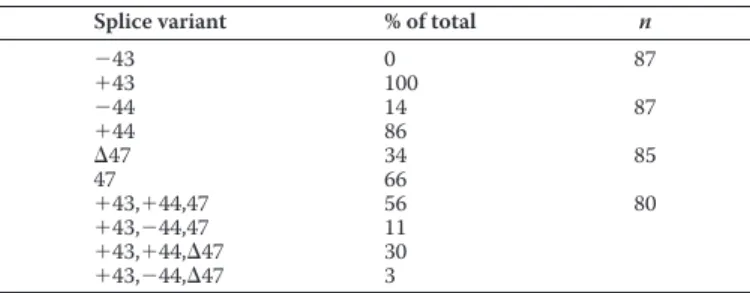
exon 47 (reverse). Agarose gel electrophoresis (1%) revealed a broad band of PCR products of⬃1,000 bp consistent with the predicted sizes ranging from 834 to 989 bp (Fig. 1C). This DNA band was subcloned into a vector, and the relative levels of splice variants were determined by counting the number of clones containing each exon. The relative proportions of indi-vidual splice variants of exon⫹43/⫺43, ⫹44/⫺44, and 47/⌬47 were 100/0, 86/14, and 66/34%, as reported previously (Table 1) (53). CaV2.1-containing regions encoded by exons 44 and 47 (⫹44 ,47) were detected at the highest relative proportion (56%) (Fig. 1A and Table 1). Relative proportions of CaV2.1 (⫹44,⌬47), CaV2.1 (⫺44,47), and CaV2.1 (⫺44,⌬47) were 30, 11, and 3%, respectively (Fig. 1A and Table 1).
Interaction between␣-RIMs and CaV2.1 C-terminal splice variants
We next performed yeast two-hybrid screening of a human brain cDNA library using the C-terminal domain encoded by exons 40 – 47 of human CaV2.1, CaV2.1 CTD (⫹44,47), as bait, and we identified an interaction between CaV2.1 CTD and the amino acid residues 487–1349 of human RIM2␣ (GenBankTM accession number NM_001100117) (Fig. 2A). We also per-formed co-immunoprecipitation (co-IP) experiments to con-firm RIM-CaV2.1 CTD interactions (Fig. 2B). As a control, we chose the VDCC 4 subunit, because 4 is abundantly expressed in the brain and the spontaneous4mutant lethargic
mouse (cacnb4lh) has clear neurological defects, supporting the physiological significance of4in the brain (61, 62). YFP-tagged 4 was co-immunoprecipitated with FLAG-tagged ␣-RIMs, RIM1␣ and RIM2␣, in HEK293T cells (Fig. 2C), as reported previously (20, 39). Next, we performed co-IP between YFP-tagged ␣-RIMs and FLAG-tagged CTDs of CaV2.1 variants derived from alternative splicing of exons 44 and 47 in HEK293T cells. RIM1␣ and RIM2␣ were co-immunoprecipi-tated with the CaV2.1 CTD splice variants except for the variant lacking exons 44 and 47 (Fig. 2D). These results suggest that the two regions encoded by exons 44 and 47 contribute signifi-cantly to the interaction between the CaV2.1 CTD and␣-RIMs. Table 1
Characterization of CaV2.1 C-terminal splice variation in the human
cerebellum
nmeans number of clones sequenced.
Splice variant % of total n
⫺43 0 87 ⫹43 100 ⫺44 14 87 ⫹44 86 ⌬47 34 85 47 66 ⫹43,⫹44,47 56 80 ⫹43,⫺44,47 11 ⫹43,⫹44,⌬47 30 ⫹43,⫺44,⌬47 3
Figure 1. Characterization of splice variants of the CaV2.1 C terminus in the human cerebellum. A, schematic topology of VDCC CaV2.1␣1subunit and
subunit and domain structures of CaV2.1 CTD splice variants. PolyQ is 11 glutamine (Q) repeats in the cDNA construct subjected to functional characterization.
GenBankTMaccession numbers are as follows: U79666 for Ca
V2.1 CTD (⫹44,47) and NM_001127221 for CaV2.1 CTD (⫹44,⌬47). The exon 44-coding region
(GenBankTMaccession number Z80152) is eliminated in Ca
V2.1 CTD (⫺44,47) and CaV2.1 CTD (⫺44,⌬47). The GGCAG sequence at the 5⬘-end of exon 47 is
eliminated in CaV2.1 CTD (⫹44,⌬47) and CaV2.1 CTD (⫺44,⌬47). B, line represents the structure of the region of the human cacna1a gene containing exons
40 – 47 (top), exons 42– 45 (bottom left), and exons 46 and 47 (bottom right). Constitutive exon is shown as black boxes, and alternative exon is shown as gray boxes. The alternatively spliced sequence GGCAG is indicated by dark gray boxes. Stop codons are indicated by asterisks. Alternative splicing patterns are indicated by lines connecting the exons. C, PCR products from human cerebellum cDNA library.
Functional impacts of RIM-CaV2.1 C-terminal interaction on CaV2.1 channel properties
We have previously reported that RIMs strongly suppress VDI of neuronal VDCCs by interacting with the subunits (20, 39, 50). It has also been reported that the RIM-CaV2.1 C-termi-nal interaction modulates localization of VDCCs to presynaptic AZs (22). However, the effects of RIM-CaV2.1 C-terminal inter-action on VDCC properties have not been examined. We char-acterized whole-cell Ba2⫹currents through recombinant P/Q-type VDCCs containing the CaV2.1 splice variants, 4 and ␣2␦-1 subunits, in HEK293 cells. We chose Ba2⫹as a charge carrier, because CaV2.1 splice variants show different Ca2⫹ -de-pendent properties in HEK293 cells (63). Voltage dependence
of inactivation at different voltages (inactivation curve) (Fig. 3A) was first examined in CaV2.1 (⫹44,47)-expressing and CaV2.1 (⫺44,⌬47)-expressing cells. No significant difference was detected between inactivation curves of CaV2.1 (⫹44,47) and CaV2.1 (⫺44,⌬47) channels (Fig. 3B and Table 2), which is consistent with previous reports (63, 64). In cells co-expressing ␣-RIMs and CaV2.1 (⫺44,⌬47), we detected remarkable inacti-vation curve shifts toward depolarizing potentials, as described previously (20, 39). Co-expression of RIM2␣ induced a more significant inactivation curve shift toward depolarizing poten-tials compared with that for RIM1␣ in CaV2.1 ( ⫹44,47)-ex-pressing cells, whereas this difference between ␣-RIMs was not observed in CaV2.1 (-44,⌬47)-expressing cells (the
half-in-Figure 2.␣-RIMs interact with the 4subunit and the CaV2.1 C-terminal splice variants. A, yeast two-hybrid results indicating the interaction level of
CaV2.1 CTD (⫹44,47) with amino acid residues 487–1349 of human RIM2␣ (GenBank
TMaccession number NM_001100117.1) as a function of time. The
interactions are scored by-galactosidase activity judged on a scale of one to five, with one meaning “low activity” and five meaning “high activity.” B, domain structures of mouse␣-RIMs. The arrows indicate molecules interacting with RIM proteins at the following domains: Zn2⫹finger-like domain (Zn2⫹), PDZ domain (PDZ), first and second C2domains (C2A and C2B), and proline-rich region (PXXP). Primary subunit-binding site (RIM1␣(1079–1257)) and  subunit
modulatory region (RIM1␣(1258–1463)) are indicated (20). C, interactions of the YFP-tagged 4subunit with FLAG-tagged␣-RIMs in HEK293T cells. D,
interactions of the FLAG-tagged CaV2.1 CTD splice variants with YFP-tagged␣-RIMs in HEK293T cells. The interactions are evaluated by co-IP with monoclonal
anti-FLAG antibody, followed by WB with polyclonal anti-YFP antibody. Input is 10% of the amount of cell lysate used for co-IP and is analyzed by WB using polyclonal anti-YFP antibody. Immunoprecipitation (IP) of FLAG-tagged CaV2.1 CTD splice variants or␣-RIMs with monoclonal anti-FLAG antibody is analyzed
activation potentials (V0.5) for CaV2.1 (⫹44,47) and CaV2.1 (⫺44,⌬47) with RIM2␣ were ⫺6.7 ⫾ 2.3 and ⫺17.2 ⫾ 2.1 mV, respectively) (Fig. 3B and Table 2). Considering that the previ-ously reported PDZ domain-binding region is not present in the C-terminal end of CaV2.1 (⫺44,⌬47), RIM- subunit inter-action may play major roles in suppressing VDI of VDCCs, whereas RIM-CaV2.1 C-terminal interaction potentiates this regulatory effect exerted by␣-RIMs on VDCCs.
To further assess the importance of interactions via the regions encoded by exons 44 and 47 in this potentiation char-acteristic of RIM2␣ in suppression of VDI, we examined the effect of RIM2␣ on CaV2.1 (⫺44,47) and CaV2.1 (⫹44,⌬47) inactivation curves. A significantly enhanced shift in the inac-tivation curve toward depolarizing potentials was detected in cells co-expressing RIM2␣ and CaV2.1 (⫺44,47), but not in cells co-expressing RIM2␣ and CaV2.1 (⫹44,⌬47), compared with cells co-expressing RIM2␣ and CaV2.1 (⫺44,⌬47) (V0.5 of
CaV2.1 (⫺44,47) and CaV2.1 (⫹44,⌬47) with RIM2␣ was ⫺8.6 ⫾ 2.0 and ⫺15.4 ⫾ 2.4 mV, respectively) (Fig. 3B and Table 2). We could not observe a significant difference between CaV2.1 (⫹44,47) and CaV2.1 (⫺44,⌬47) in current density-voltage (I-V) relationships, with or without RIM2␣ co-expression (Fig. 4 and Table 3). These results suggest that the RIM-CaV2.1 C-terminal interaction via the region encoded by exon 47 is important for the potentiated sup-pressive effect of RIM2␣ on VDI.
Inactivation kinetics of P/Q-type VDCCs was characterized by analyzing the decay phase of Ba2⫹currents evoked by 1-s test pulses in HEK293 cells (Fig. 5A). The decay phase was well fitted by two exponential functions with a non-inactivating component (Fig. 5B) as reported previously (41). The two expo-nential time constants (fastandslow) and the ratio of fast, slow, and non-inactivating components were similar in CaV2.1 (⫹44,47)- and CaV2.1 (⫺44,⌬47)-expressing cells at test
poten-A
-80 mV -90 mV 20 mV 1 s 40 ms 10 mV CaV2.1 (−44,Δ47) Prepulse voltage (mV) 0 0.2 0.4 0.6 0.8 1 -80 -60 -40 -20 0 20 Vector RIM1α RIM2αB
CaV2.1 (−44,47) Ca V2.1 (+44,Δ47) Relative current CaV2.1 (+44,47) Prepulse voltage (mV) 0 0.2 0.4 0.6 0.8 1 -80 -60 -40 -20 0 20 Vector RIM2α Cav2.1 (−44,Δ47) + RIM2α Prepulse voltage (mV) 0 0.2 0.4 0.6 0.8 1 -80 -60 -40 -20 0 20 Vector RIM2α Cav2.1 (−44,Δ47) + RIM2α Prepulse voltage (mV) 0 0.2 0.4 0.6 0.8 1 -80 -60 -40 -20 0 20 Vector RIM1α RIM2α ** * * ** * (−44,Δ47) + RIM2α # ## ## ## # # # #Figure 3. Effects of␣-RIMs on VDI of P/Q-type CaV2.1 channels. A, voltage protocol to determine the inactivation curve and representative traces for Ba2⫹
currents. To determine the voltage dependence of inactivation, currents are evoked by a 40-ms test pulse to 10 mV after the 10-ms repolarization to⫺90 mV following 1-s Vpredisplacements (conditioning pulses) from⫺80 to 20 mV with 10-mV increments. B, effects of␣-RIMs on inactivation curves of Ba2⫹currents
mediated by P/Q-type CaV2.1 splice variants in HEK293 cells expressing4and␣2␦-1 subunits. The inactivation curve in cells co-expressing RIM2␣ and CaV2.1
(⫺44,⌬47) (dashed gray line) is taken from left panel and is shown for comparison. *, p ⬍ 0.05, and **, p ⬍ 0.01, statistical significance of differences between RIM1␣- and RIM2␣-expressing cells. #, p ⬍ 0.05, and ##, p ⬍ 0.01, statistical significance of differences versus cells co-expressing RIM2␣ and CaV2.1 (⫺44,⌬47).
See Table 2 for statistical significance of the differences. Error bars, S.E. Table 2
Effect of␣-RIMs on inactivation properties of P/Q-type VDCCs in HEK293 cells expressing CaV2.1s,␣2/␦-1, and 4
* is p⬍ 0.05; ** is p ⬍ 0.01; ***is p ⬍ 0.001 versus vector. # is p ⬍ 0.05; ## is p ⬍ 0.01 versus RIM1␣. † is p ⬍ 0.05; †† is p ⬍ 0.01 versus CaV2.1 (⫺44,⌬47) for cells co-expressing
RIM2␣. The number of cells analyzed are indicated in parentheses.
Inactivation parameters A V0.5 k mV mV CaV2.1 (⫹44,47) Vector 0.91⫾ 0.02 (5) ⫺30.0 ⫾ 1.9 (5) ⫺7.6 ⫾ 0.4 (5) RIM1␣ 0.63⫾ 0.04 (11)*** ⫺16.9 ⫾ 2.4 (11)** ⫺6.6 ⫾ 0.7 (11) RIM2␣ 0.53⫾ 0.04 (13)*** ⫺6.7 ⫾ 2.3 (13)***, ##, †† ⫺9.9 ⫾ 1.0 (13)# CaV2.1 (⫺44,⌬47) Vector 0.94⫾ 0.01 (5) ⫺27.1 ⫾ 0.6 (5) ⫺6.2 ⫾ 0.2 (5) RIM1␣ 0.67⫾ 0.06 (5)** ⫺16.2 ⫾ 4.2 (5)* ⫺8.4 ⫾ 0.9 (5) RIM2␣ 0.61⫾ 0.02 (15)*** ⫺17.2 ⫾ 2.1 (15)* ⫺7.7 ⫾ 0.6 (15) CaV2.1 (⫺44,47) Vector 0.92⫾ 0.03 (5) ⫺24.8 ⫾ 1.5 (5) ⫺6.2 ⫾ 1.0 (5) RIM2␣ 0.51⫾ 0.07 (7)** ⫺8.6 ⫾ 2.0 (7)***, † ⫺8.4 ⫾ 1.1 (7) CaV2.1 (⫹44,⌬47) Vector 0.92⫾ 0.03 (4) ⫺23.0 ⫾ 0.7 (4) ⫺6.0 ⫾ 0.2 (4) RIM2␣ 0.69⫾ 0.03 (5)** ⫺15.4 ⫾ 2.4 (5)* ⫺6.9 ⫾ 1.1 (5) CaV2.1 (⫹44,47) Gln-40 Vector 0.96⫾ 0.02 (4) ⫺30.6 ⫾ 2.0 (4) ⫺7.1 ⫾ 0.6 (4) RIM2␣ 0.42⫾ 0.08 (8)***, †† ⫺11.0 ⫾ 2.2 (8)*** ⫺8.5 ⫾ 0.8 (8)
tials of 0, 10, and 20 mV (Fig. 5, C and D, and Table 4). Co-ex-pression of␣-RIMs exerted common effects on Ba2⫹currents in CaV2.1 (⫹44,47)- and CaV2.1 (⫺44,⌬47)-expressing cells. The fast-inactivating component was significantly decreased at 0 and 10 mV, whereas the non-inactivating component as well asslowwere significantly increased by␣-RIMs at 0, 10, and 20 mV (Fig. 5, C and D, and Table 4). Thus, it is unlikely that these effects are mediated by interaction of␣-RIMs with the CaV2.1 C-terminal region encoded by exons 44 and 47. In cells co-ex-pressing RIM2␣ and CaV2.1 (⫹44,47), increases in the non-inactivating component at 0, 10, and 20 mV andfastat 0 and 10 mV were more pronounced compared with cells co-expressing RIM2␣ and CaV2.1 (⫺44,⌬47) (Fig. 5, C and D, and Table 4). This effect can be mediated by the RIM2␣-CaV2.1 C-terminal interaction. Furthermore, currents evoked by trains (100 Hz) of action potential (AP)-like waveforms for 3 s, a more physiolog-ical voltage-clamp protocol used to determine closed-state inactivation (65), showed a more rapid decrease in amplitude in cells co-expressing RIM2␣ and CaV2.1 (⫺44,⌬47) compared with cells co-expressing RIM2␣ and CaV2.1 (⫹44,47) (Fig. 5E). These results suggest that RIM2␣-CaV2.1 C-terminal interac-tion is not essential for but enhances the VDI suppression induced by␣-RIMs.
It is important to note that, without the interaction of ␣-RIMs, these splice variants were indistinguishable in channel properties such as inactivation curve (Fig. 3 and Table 2), inac-tivation kinetics (Fig. 5 and Table 4), and I-V relationship (Fig. 4 and Table 3). This underscores the significance of protein-pro-tein interaction in differentiating functional properties of alter-natively spliced CaV2.1 variants.
Biochemical characterization of the RIM2␣-CaV2.1 C-terminal interaction
The CaV2.1 mutant, CaV2.1 (⫹44,47) ⌬DDWC, which lacks four amino acids (DDWC) in the C-terminal end was con-structed, because these four amino acids bind to the PDZ domain of␣-RIMs (22, 57). Inactivation curves of whole-cell Ba2⫹ currents in HEK293 cells co-expressing RIM2␣ and CaV2.1 (⫹44,47) ⌬DDWC were indistinguishable from those in cells co-expressing RIM2␣ and CaV2.1 (⫹44,47), showing shifts toward depolarizing potentials compared with the inactivation curves in cells co-expressing RIM2␣ and CaV2.1 (⫺44,⌬47) (V0.5 of CaV2.1 (⫹44,47) ⌬DDWC, CaV2.1 (⫹44,47), and CaV2.1 (⫺44,⌬47) were ⫺6.7 ⫾ 3.2 mV, ⫺6.7 ⫾ 2.3 mV, and ⫺17.2 ⫾ 2.1 mV, respectively) (Fig. 6A and Table 5). In co-IP experiments, YFP-tagged RIM2␣ showed association with FLAG-tagged CaV2.1 CTD (⫹44,47) ⌬DDWC in HEK293T cells (Fig. 6B). The intensity of the co-IP band for RIM2␣ nor-malized to the IP band for the CTD was significantly decreased in cells co-expressing RIM2␣ and CaV2.1 CTD (⫹44,47) ⌬DDWC, compared with cells co-expressing RIM2␣ and CaV2.1 CTD (⫹44,47) (Fig. 6C). These data suggest that DDWC in the region encoded by exon 47 contributes to the RIM2␣-CaV2.1 C-terminal interaction, as reported previously (22, 57), but not to potentiation of VDI suppression by RIM2␣. To clarify the RIM2␣-interacting regions responsible for the potentiation of VDI suppression by RIM2␣, additional dele-tion mutants of CaV2.1 were constructed (Fig. 7A). In cells
Figure 4. Effects of␣-RIMs on the I-V relationships of P/Q-type CaV2.1 channels. I-V relationships of CaV2.1 splice variants in HEK293 cells expressing4and
␣2␦-1 subunits. Left, representative traces for Ba
2⫹currents on application of test pluses from⫺40 to 60 mV with 10-mV increments from a holding potential
(Vh) of⫺90 mV. Right, I-V relationships of CaV2.1 splice variants. See Table 3 for statistical significance of the differences. Error bars, S.E.
Table 3
Effect of␣-RIMs on the I-V relationships of P/Q-type VDCCs in HEK293 cells expressing CaV2.1s,␣2/␦-1, and 4
The number of cells analyzed are indicated in parentheses. Current densitya V 0.5 k pA/picofarad mV mV CaV2.1 (⫹44,47) Vector ⫺71.7 ⫾ 10.8 (5) ⫺3.0 ⫾ 1.3 (5) 4.5 ⫾ 0.2 (5) RIM1␣ ⫺97.5 ⫾ 21.3 (5) ⫺5.1 ⫾ 1.9 (5) 4.5 ⫾ 0.4 (5) RIM2␣ ⫺83.1 ⫾ 7.5 (7) ⫺3.7 ⫾ 1.1 (7) 3.7 ⫾ 0.6 (7) CaV2.1 (⫺44,⌬47) Vector ⫺111.5 ⫾ 13.8 (4) ⫺4.0 ⫾ 1.6 (4) 3.0 ⫾ 0.3 (4) RIM1␣ ⫺123.8 ⫾ 30.6 (4) ⫺3.2 ⫾ 1.7 (4) 3.6 ⫾ 0.6 (4) RIM2␣ ⫺102.5 ⫾ 7.4 (7) ⫺6.5 ⫾ 1.0 (7) 3.6 ⫾ 0.3 (7)
aBa2⫹currents evoked by depolarizing pulse to 10 mV from a V
hof⫺90 mV are
co-expressing RIM2␣ with CaV2.1 (⫹44,2364X) or CaV2.1 (⫹44,2344X), in which C-terminal residues 2365–2505 or 2345–2505 were deleted, respectively, the inactivation curves were similar to those in cells co-expressing RIM2␣ with CaV2.1 (⫹44,47). In contrast, in cells co-expressing RIM2␣ with CaV2.1 (⫹44,2327X) or CaV2.1 (⫹44,2313X), in which C-ter-minal residues 2328 –2505 or 2314 –2505 were deleted, respec-tively, the inactivation curves were similar to those in cells co-expressing RIM2␣ and CaV2.1 (⫺44,⌬47) (Fig. 7B and Table 5).
These data suggest that 17 amino acid residues, 2328 –2344 (RPGRAATSGPRRYPGPT), on the C-terminal side of the polyQ stretch of CaV2.1 (⫹44,47) is a RIM2␣-interacting region that potentiates the suppressive effect on VDI.
In co-IP experiments, however, YFP-tagged RIM2␣ showed a comparable level of co-IP with FLAG-tagged CTD of CaV2.1 (⫹44,2344X) and FLAG-tagged CTD of CaV2.1 (⫹44,2327X) but a decreased level of co-IP with CTD of CaV2.1 (⫹44,2275X) (Fig. 7C). To eliminate possible contributions of exon
44-en-Figure 5. Effects of␣-RIMs on VDI kinetics of P/Q-type CaV2.1 channels. A, effects of␣-RIMs on inactivation of Ba2⫹currents mediated by P/Q-type CaV2.1
splice variants in HEK293 cells expressing4and␣2␦-1 subunits. The peak amplitudes are normalized for Ba2⫹currents elicited by 1-s pulses to 10 mV from a
Vhof⫺90 mV. B, Ba2⫹current evoked by 1-s test pulse to 10 mV from a Vhof⫺90 mV in HEK293 cells expressing4,␣2␦-1 subunits, and RIM1␣. Current decay
is fitted by a sum of two exponential functions with time constants of 55 and 995 ms, whose fraction of components is 0.19 and 0.61, respectively. The fraction of its non-inactivating component is 0.24. C, voltage dependence of the two inactivation time constants,fastandslow. The mean inactivation time constants
are plotted as a function of test potential from 0 to 20 mV. D, voltage dependence of the fraction of the three components, fast-, slow-, and non-inactivating components. The fractions of the components are plotted against test potentials. †, p⬍ 0.05; ††, p ⬍ 0.01; statistical significance of differences between cells co-expressing CaV2.1 (⫺44,⌬47) and RIM2␣ and cells co-expressing CaV2.1 (⫹44,47) and RIM2␣. See Table 4 for statistical significance of the differences. Error
bars, S.E. E, left, effects of RIM2␣ on Ba2⫹currents mediated by P/Q-type CaV2.1 splice variants in response to 100 Hz AP-like voltage command for 3 s in HEK293
cells expressing4and␣2␦-1 subunits (AP-like waveform, ⫺80 to 33 mV; rise time, 0.4 ms; decay time, 1.1 ms). Right, percentage of currents in response to the
last stimulus compared with currents in response to the first stimulus (CaV2.1 (⫹44,47) RIM2␣, 88 ⫾ 4%, n ⫽ 4; CaV2.1 (⫺44,⌬47) RIM2␣, 67 ⫾ 15%, n ⫽ 8). Data
are presented in scatter plots. The vertical error bars represent standard deviations, and the mean values are indicated with horizontal bars. *, p⬍ 0.05; statistical significance of difference between cells co-expressing CaV2.1 (⫺44,⌬47) and RIM2␣ and cells co-expressing CaV2.1 (⫹44,47) and RIM2␣.
coded amino acid residues to the interaction of RIM2␣ with CTD mutants, we next constructed deletion mutants of the CTD of CaV2.1 (⫺44,47). The co-IP was nearly abolished for the CTD of CaV2.1 (⫺44,2263X), in which 2263X corresponds to 2275X in CaV2.1 (⫹44,47) (Fig. 7D). This result suggests that the amino acid residues 2264 –2280 (GTSTPRRGRRQ-LPQTPS) of CaV2.1 (⫺44,47), which correspond to 2276–2292 of CaV2.1 (⫹44,47), are an important region for the RIM2␣-CaV2.1 CTD interaction. However, the co-IP experiments failed to unveil interaction between the 17 amino acid residues, 2328 –2344, of CaV2.1 (⫹44,47) with RIM2␣, although this region is supposed to be essential for potentiated VDI suppres-sion by RIM2␣.
To demonstrate interaction of the 2328 –2344 amino acid residues, contributions of the exon 44 region and residues 2276 –2292 of CaV2.1 (⫹44,47) were eliminated in deletion mutants based upon CaV2.1 (⫺44,⌬2264–2280). The level of co-IP was markedly diminished for the CTD of CaV2.1 (⫺44,⌬2264–2280,2315X) and CaV2.1 (⫺44,⌬2264–2280, 2301X) compared with the CTD of CaV2.1 (⫺44,⌬2264– 2280,2332X) and CaV2.1 (⫺44,⌬2264–2280,2352X) (Fig. 7E). This result suggests that the 17 amino acid residues, 2316 – 2332, of CaV2.1 (⫺44,47), which correspond to 2328–2344 of CaV2.1 (⫹44,47), indeed constitute an important binding region for RIM2␣ to exert a potentiated suppressive effect on VDI.
It is interesting to note that deletion of the region encoded by exon 44 failed to elicit suppression of interaction of the CaV2.1 CTD containing the region encoded by exon 47 (compare the bands of CaV2.1 CTD (⫺44,47) and CaV2.1 CTD (⫹44,47) in Fig. 2D). In contrast, deletion of the region encoded by exon 47 suppressed the interaction of the CaV2.1 CTD containing the region encoded by exon 44 (compare the bands of CaV2.1 CTD (⫹44,⌬47) and CaV2.1 (⫹44,47) in Fig. 7C). These results sug-gest that the region encoded by exon 47 binds more strongly to ␣-RIMs compared with the region encoded by exon 44. Thus, formation of RIM2␣-CaV2.1 complexes is mediated by multi-point interaction.
Figure 6. Effects of RIM2␣ on VDI of the P/Q-type CaV2.1 deletion mutant
lacking DDWC. A, effects of RIM2␣ on inactivation curves of Ba2⫹currents mediated by P/Q-type CaV2.1 (⫹44,47) ⌬DDWC, which lacks DDWC at the
C-terminal end of CaV2.1 (⫹44,47), in HEK293 cells expressing4and␣2␦-1
subunits. The inactivation curve in cells co-expressing RIM2␣ and CaV2.1
(⫹44,47) (dashed dark gray line) is taken from Fig. 3 and is shown for compar-ison. See Table 5 for statistical significance of the differences. Error bars, S.E. B, interaction of FLAG-tagged CaV2.1 CTD (⫹44,47) or CaV2.1 CTD (⫹44,47)
⌬DDWC with YFP-tagged RIM2␣ in HEK293T cells. The interactions are eval-uated by co-IP with monoclonal anti-FLAG antibody, followed by WB with polyclonal anti-YFP antibody. Input is 10% of the amount of cell lysate used for co-IP and is analyzed by WB using polyclonal anti-YFP antibody. IP of FLAG-tagged CaV2.1 CTDs with monoclonal anti-FLAG antibody is
ana-lyzed by WB using polyclonal anti-FLAG antibody. C, quantification of the data shown in B. The intensity of the co-IP band for RIM2␣ normalized to the IP band for the CTD (CaV2.1 CTD (⫹44,47), 100%; CaV2.1 CTD (⫹44,47)
⌬DDWC, 55 ⫾ 20%) (92). Data for nine experiments are presented in scat-ter plots. The vertical error bars represent standard deviations, and the mean values are indicated with horizontal bars. ***, p⬍ 0.001; statistical significance of difference between CTD (⫹44,47) and CTD (⫹44,47) ⌬DDWC.
Table 4
Effect of␣-RIMs on inactivation kinetics of P/Q-type VDCCs in HEK293 cells expressing CaV2.1s,␣2/␦-1, and 4
* is p⬍ 0.05; ** is p ⬍ 0.01; *** is p ⬍ 0.001 versus vector. # is p ⬍ 0.05; ## is p ⬍ 0.01 versus RIM1␣. † is p ⬍ 0.05; †† is p ⬍ 0.01 versus CaV2.1 (⫺44,⌬47) for cells co-expressing
RIM2␣. The number of cells analyzed are indicated in parentheses.
Fraction of components Time constants
mV Fast Slow Sustained fast slow
CaV2.1 (⫹44,47) Vector (8) 0 0.84⫾ 0.05 0.25⫾ 0.05 0.10⫾ 0.02 0.08⫾ 0.01 0.41⫾ 0.09 10 0.79⫾ 0.10 0.33⫾ 0.06 0.07⫾ 0.01 0.07⫾ 0.01 0.26⫾ 0.05 20 0.44⫾ 0.05 0.61⫾ 0.05 0.03⫾ 0.01 0.04⫾ 0.01 0.16⫾ 0.02 RIM1␣ (11) 0 0.41⫾ 0.04*** 0.38⫾ 0.03 0.32⫾ 0.02*** 0.10⫾ 0.04 0.88⫾ 0.13* 10 0.39⫾ 0.06** 0.50⫾ 0.06 0.19⫾ 0.02** 0.07⫾ 0.02 0.78⫾ 0.09*** 20 0.42⫾ 0.07 0.48⫾ 0.04 0.17⫾ 0.03** 0.09⫾ 0.03 0.74⫾ 0.09*** RIM2␣ (11) 0 0.20⫾ 0.03***, ## 0.28⫾ 0.05 0.53⫾ 0.06***, ##, † 0.71⫾ 0.19*, ##, †† 1.12⫾ 0.11*** 10 0.30⫾ 0.04*** 0.36⫾ 0.04 0.37⫾ 0.04***, ##, † 0.40⫾ 0.14#, † 0.94⫾ 0.08*** 20 0.30⫾ 0.04 0.40⫾ 0.02*** 0.33⫾ 0.03***, ##, †† 0.19⫾ 0.06 0.95⫾ 0.12*** CaV2.1 (⫺44,⌬47) Vector (5) 0 0.95⫾ 0.05 0.22⫾ 0.04 0.06⫾ 0.01 0.09⫾ 0.01 0.39⫾ 0.08 10 0.84⫾ 0.10 0.27⫾ 0.08 0.05⫾ 0.01 0.09⫾ 0.01 0.29⫾ 0.04 20 0.66⫾ 0.12 0.51⫾ 0.08 0.04⫾ 0.01 0.08⫾ 0.01 0.24⫾ 0.05 RIM1␣ (5) 0 0.45⫾ 0.08*** 0.38⫾ 0.05 0.22⫾ 0.03*** 0.16⫾ 0.08 0.88⫾ 0.15* 10 0.45⫾ 0.07* 0.39⫾ 0.05 0.22⫾ 0.04** 0.18⫾ 0.08 0.74⫾ 0.13* 20 0.43⫾ 0.10 0.49⫾ 0.11 0.14⫾ 0.01*** 0.06⫾ 0.01 0.64⫾ 0.18* RIM2␣ (13) 0 0.26⫾ 0.10*** 0.39⫾ 0.05 0.32⫾ 0.04** 0.12⫾ 0.03 1.08⫾ 0.07*** 10 0.26⫾ 0.10** 0.45⫾ 0.04* 0.25⫾ 0.02*** 0.08⫾ 0.01# 0.83⫾ 0.05*** 20 0.29⫾ 0.07* 0.46⫾ 0.04 0.22⫾ 0.02***, # 0.08⫾ 0.01 0.72⫾ 0.09**
Characterization of the RIM2␣ region responsible for interaction with the CaV2.1 C terminus
To identify the region in RIM2␣ that interacts with the CaV2.1 C terminus, we constructed the chimeric RIM, RIM1–2 chimera, with amino acid residues from the N terminus to the C2A domain of RIM1␣ and from the C2A domain of RIM2␣ (Fig. 8A). We examined effects of the RIM1–2 chimera on whole-cell Ba2⫹ currents mediated by CaV2.1 (⫹44,47)- and CaV2.1 (⫺44,⌬47)-containing VDCCs (Fig. 8B). Significant enhancement of inactivation curve shift by the RIM1–2 chi-mera toward depolarizing potentials was observed for CaV2.1 (⫹44,47) compared with CaV2.1 (⫺44,⌬47) (V0.5 for CaV2.1 (⫹44,47) and CaV2.1 (⫺44,⌬47) with RIM1–2 chimera was ⫺4.7 ⫾ 2.6 and ⫺15.9 ⫾ 3.3 mV, respectively) (Fig. 8B and Table 5). This tendency of the RIM1–2 chimera suggests that the region on the C-terminal side of the C2A domain is impor-tant for potentiating the suppressive effect of RIM2␣ on VDI. We also tested a mutant of RIM2␣(1183–1572) composed of the amino acid residues 1183–1572 of RIM2␣ corresponding to the subunit-binding region in RIM1␣ (residues 1079–1463 of RIM1␣) (Figs. 2B and 8A) (20, 39). In cells co-expressing RIM2␣(1183–1572) and CaV2.1 (⫹44,47), we also detected potentiation of the suppressive effect on VDI as observed in cells co-expressing RIM2␣(1183–1572) and CaV2.1 (⫺44,⌬47) (V0.5and the rates of the inactivating component were⫺1.9 ⫾ 3.2 mV and 0.43⫾ 0.06, respectively, for CaV2.1 (⫹44,47) with RIM2␣(1183–1572) and 14.2 ⫾ 4.0 mV and 0.69 ⫾ 0.04, respec-tively, for CaV2.1 (⫺44,⌬47) with RIM2␣(1183–1572)) (Fig. 8C and Table 5). Associations between YFP-tagged RIM2␣(1183– 1572) and FLAG-tagged mutants based upon CaV2.1 CTD (⫺44,⌬2264–2280), which has a deletion of the region corre-sponding to 2328 –2344 of CaV2.1 (⫹44,47), were tested by co-IP experiments. RIM2␣(1183–1572) showed co-IP with the CTD of CaV2.1 (⫺44,⌬2264–2280, 2332X) but not with the CTD of CaV2.1 (⫺44,⌬2264–2280, 2315X) or CaV2.1 (⫺44,⌬2264–2280, 2301X) (Fig. 8D). These data suggest that the C-terminal region containing the C2B domain is critical for RIM2␣ to potentiate suppression of VDI and binds with 2328 –2344 of CaV2.1 (⫹44,47) in addition to the subunits (20, 39).
Effects of polyQ elongation in the CaV2.1 C-terminal region on regulation of VDI by RIM2␣
The polyQ stretch is an interesting characteristic of the CaV2.1 C-terminal primary structure. SCA6 is caused by expansion of the polyQ tract in the human CaV2.1 gene from a normal repeat size range of 4 –17 to a size range of 20 –33 (34, 54, 66). To confirm the effect of polyQ expansion on the inter-action between the CaV2.1 C terminus and RIM2␣, association between YFP-tagged RIM2␣ and FLAG-tagged CTD of CaV2.1 (⫹44,47) Gln-40 with an elongated polyQ stretch of 40 residues was tested by co-IP in HEK293T cells (Fig. 9A). The intensity of the co-IP band for RIM2␣ normalized to the IP band for the CTD was moderately but significantly decreased for CaV2.1 CTD (⫹44,47) Gln-40 compared with that for CaV2.1 CTD (⫹44,47) with a polyQ stretch of 11 residues (Fig. 9B). To exam-ine the effect of polyQ elongation on suppression of VDI by RIM2␣, a recombinant P/Q-type VDCC was expressed as a complex of CaV2.1 (⫹44,47) with polyQ expansion (CaV2.1 (⫹44,47) Gln-40),4, and␣2␦-1 subunits in HEK293 cells (Fig. 9C). Without co-expression of␣-RIMs, inactivation curves of whole-cell Ba2⫹ currents elicited by Ca
V2.1 (⫹44,47) and CaV2.1 (⫹44,47) Gln-40 were indistinguishable (Fig. 9C and Table 2). This contradicts with our previous report that polyQ expansion itself causes a hyperpolarizing shift of the inactiva-tion curve for rabbit CaV2.1 channels carrying the1subunit (67). It is possible that the effect of polyQ expansion on VDI depends on species and subtypes (68). When RIM2␣ was co-expressed, inactivation curves of CaV2.1 (⫹44,47) and CaV2.1 (⫹44,47) Gln-40 were indistinguishable (V0.5of CaV2.1 (⫹44,47) Gln-40 and CaV2.1 (⫹44,47) were ⫺11.0 ⫾ 2.2 mV and⫺6.7 ⫾ 2.3 mV, respectively) (Fig. 9C and Table 2). These data indicate that polyQ expansion reduced the binding affinity of CaV2.1 C terminus for RIM2␣ but not the suppressive effect of RIM2␣ on the VDI of CaV2.1 (⫹44,47).
Functional impacts of␣-RIMs on VDI of N-type CaV2.2 and R-type CaV2.3 channels
CaV2 VDCCs are a major source of presynaptic Ca
2⫹influx (69 –71), and ␣-RIMs also interact with the C terminus of CaV2.2 (22, 57). To explore the generality of our findings using CaV2.1, associations of the C-terminal region of CaV2.2 Table 5
Effects of chimeric RIM and RIM2␣ deletion mutant on inactivation properties of P/Q-type VDCCs in HEK293 cells expressing CaV2.1,␣2/␦-1,
and4
* is p⬍ 0.05; ** is p ⬍ 0.01; *** is p ⬍ 0.001 versus CaV2.1 (⫺44,⌬47) for cells co-expressing RIM2␣. #, p ⬍ 0.05 versus CaV2.1 (⫺44,⌬47) for cells co-expressing RIM1–2
chimera. †, p⬍ 0.05 versus CaV2.1 (⫺44,⌬47) for cells co-expressing RIM2(1183–1572). The number of cells analyzed are indicated in parentheses.
Inactivation parameters a V0.5 k mV mV CaV2.1 (⫹44,47) RIM2␣ 0.53⫾ 0.04 (13) ⫺6.7 ⫾ 2.3 (13)** ⫺9.9 ⫾ 1.0 (13) CaV2.1 (⫺44,⌬47) RIM2␣ 0.61⫾ 0.02 (15) ⫺17.2 ⫾ 2.1 (15) ⫺7.7 ⫾ 0.6 (15) CaV2.1 (⫹44,47) ⌬DDWC RIM2␣ 0.55⫾ 0.05 (7) ⫺6.7 ⫾ 3.2 (7)* ⫺7.2 ⫾ 1.1 (7) CaV2.1 (⫹44,47) 2366X RIM2␣ 0.53⫾ 0.07 (6) ⫺6.3 ⫾ 2.3 (6)** ⫺5.7 ⫾ 0.7 (6) CaV2.1 (⫹44,47) 2344X RIM2␣ 0.54⫾ 0.04 (6) ⫺8.3 ⫾ 3.0 (6)* ⫺8.2 ⫾ 0.9 (6) CaV2.1 (⫹44,47) 2327X RIM2␣ 0.70⫾ 0.03 (6) ⫺14.1 ⫾ 2.4 (6) ⫺8.4 ⫾ 0.9 (6) CaV2.1 (⫹44,47) 2315X RIM2␣ 0.67⫾ 0.04 (8) ⫺13.5 ⫾ 1.2 (8) ⫺6.8 ⫾ 0.5 (8) CaV2.1 (⫹44,47) RIM1–2 chimera 0.49⫾ 0.07 (7) ⫺4.7 ⫾ 2.6 (7)**, # ⫺8.3 ⫾ 0.7 (7) RIM2␣ (1183–1572) 0.43⫾ 0.06 (7)**, †† ⫺1.9 ⫾ 3.2 (7)***, † ⫺5.3 ⫾ 1.2 (7) CaV2.1 (⫺44,⌬47) RIM1–2 chimera 0.59⫾ 0.03 (3) ⫺15.9 ⫾ 3.3 (3) ⫺7.6 ⫾ 1.5 (3) RIM2␣ (1183–1572) 0.69⫾ 0.04 (6) ⫺14.2 ⫾ 4.0 (6) ⫺7.0 ⫾ 1.4 (6)
encoded by exons 41– 47 of the CaV2.2 gene (CaV2.2 CTD) and the C-terminal region of CaV2.3 encoded by exons 41– 48 of the CaV2.3 gene (CaV2.3 CTD) with␣-RIMs were tested by co-IP experiments. Both the YFP-tagged CTD of CaV2.2 and the YFP-tagged CTD of CaV2.3 were co-immunoprecipitated with FLAG-tagged␣-RIMs in HEK293T cells (Fig. 10A). Thus, the C-terminal interaction with␣-RIMs is shared by CaV2.2 and CaV2.3.
We also examined the effect of␣-RIMs on the VDI of N-type VDCCs containing the CaV2.2,4, and␣2␦-1 subunits and of R-type VDCCs containing the CaV2.3,4, and␣2␦-1 subunits. In CaV2.2-expressing cells, ␣-RIMs significantly shifted V0.5 values of the component susceptible to inactivation at high
voltages (V0.5high) in inactivation curves of Ba2⫹ currents toward depolarizing potentials (V0.5
high of Ca
V2.2 co-trans-fected with vector, RIM1␣, and RIM2␣ were ⫺46.2 ⫾ 3.0, ⫺8.2 ⫾ 14.8, and ⫺2.8 ⫾ 5.7 mV, respectively) (Fig. 10B and Table 6), as reported previously (20). RIM2␣ induced signifi-cant reduction of low voltage-inactivated phases (the ratios of low voltage-inactivating phases of CaV2.2 co-transfected with vector, RIM1␣, and RIM2␣ were 0.64 ⫾ 0.11, 0.63 ⫾ 0.12, and 0.29⫾ 0.05, respectively) (Fig. 10B and Table 6). Suppressive effects of␣-RIMs on VDI were also observed for CaV2.3 (the ratios of inactivating components of CaV2.3 with co-transfec-tion of vector, RIM1␣, and RIM2␣ were 0.90 ⫾ 0.03, 0.63 ⫾ 0.09, and 0.67⫾ 0.08, respectively). We failed to detect
signifi-Figure 7. Identification of RIM2␣-binding regions in the CaV2.1 C terminus. A, domain structures of CaV2.1 CTD deletion mutants. B, effects of RIM2␣ on
inactivation curves of Ba2⫹currents mediated by the deletion mutants of Ca
V2.1 (⫹44,47) in HEK293 cells expressing4and␣2␦-1 subunits. The inactivation
curve in cells co-expressing RIM2␣ and CaV2.1 (⫹44,47) (dashed dark gray line) is taken from Fig. 3 and is shown for comparison. *, p ⬍ 0.05; **, p ⬍ 0.01; statistical
significance of differences versus cells co-expressing RIM2␣ and CaV2.1 (⫹44,47). See Table 5 for statistical significance of the differences. Error bars, S.E. C,
interactions of FLAG-tagged CaV2.1 CTD (⫹44) deletion mutants with YFP-tagged RIM2␣ in HEK293T cells. D, interactions of FLAG-tagged CaV2.1 CTD (⫺44)
deletion mutants with YFP-tagged RIM2␣ in HEK293T cells. E, interactions of FLAG-tagged CaV2.1 CTD (⫺44,⌬2264–2280) deletion mutants with YFP-tagged
RIM2␣ in HEK293T cells. The interactions are evaluated by co-IP with monoclonal anti-FLAG antibody, followed by WB with polyclonal anti-YFP antibody. Input is 10% of the amount of cell lysate used for co-IP and is analyzed by WB using polyclonal anti-YFP antibody. IP of FLAG-tagged CaV2.1 CTDs with monoclonal
anti-FLAG antibody is analyzed by WB using polyclonal anti-FLAG antibody. Dashed lines divide mutants in terms of co-IP level (blue) and inactivation curve (red).
cant differences between the effect of RIM1␣ and RIM2␣ on VDI of CaV2.3 channels (Fig. 10B and Table 6). Thus, N-type CaV2.2 channels but not R-type CaV2.3 channels are suscepti-ble to RIM2␣-mediated potentiation of VDI suppression. Discussion
In presynaptic AZs, where SVs dock in close vicinity to VDCCs at the presynaptic membrane, depolarization-induced Ca2⫹influx via VDCCs triggers neurotransmitter release (1, 2). Previous proteomic analysis has shown that P/Q-, N-, and R-type VDCCs are embedded into protein networks assembled from a pool of 200 proteins (72). Unveiling the manner of pro-tein-protein interactions in protein networks and their func-tional consequences is important for understanding the process of synapse formation and the modulation of synaptic transmis-sion (73). Previously, we reported that␣-RIMs increase neu-rotransmitter release by sustaining Ca2⫹influx through strong inhibition of VDI of VDCCs and by anchoring vesicles in the vicinity of VDCCs via the RIM- subunit interaction (20, 39). It has also been reported that␣-RIMs interact with the CaV2.1 C-terminal region and modulate VDCCs targeting to presyn-aptic AZs (22). These previous studies have shown that
RIM-VDCC interactions are key to the protein assembly responsible for stimulus-secretion coupling in presynaptic AZs.
Multipoint interaction plays important roles in the regula-tion of properties such as stabilizaregula-tion of protein complexes. In RIM-VDCC complexes, the significance of multipoint interac-tion has not yet been resolved. To approach this quesinterac-tion, the contributions of each interaction should be quantitatively assessed. Our data strongly indicate that RIM- subunit inter-action is sufficient and necessary for␣-RIMs to exert promi-nent suppressive effects of VDI of VDCCs, because in the CaV2.1 splice variant with the deletion of exons 44 and 47 (CaV2.1 (⫺44,⌬47)), strong VDI suppression remains intact for RIM1␣, but for RIM2␣ it is attenuated to a level comparable with that of RIM1␣. In the presynapse, VDCCs display a weak VDI (13, 30). Moreover, in the calyx of Held nerve terminals of RIM1 and RIM2 conditional double knock-out mice, depolar-ization pre-pulses induced stronger inactivation of VDCCs compared with wild-type mice (48).
CaV2.1 is known to be inactivated through at least two voltage-dependent mechanisms (fast and slow inactivation) (74). The mechanism underlying inactivation is not completely understood but may involve “hinged lid” or pore block-type
Figure 8. Identification of CaV2.1 C-terminal-binding region in RIM2␣. A, domain structures of ␣-RIMs, RIM1–2 chimera, and RIM2␣(1183–1572). B and C,
effects of RIM1–2 chimera and RIM2␣(1183–1572) on inactivation curves of Ba2⫹currents mediated by Ca
V2.1 splice variants in HEK293 cells expressing4and
␣2␦-1 subunits. *, p ⬍ 0.05; **, p ⬍ 0.01; statistical significance of differences between CaV2.1 (⫺44,⌬47)-expressing and CaV2.1 (⫹44,47)-expressing cells. See
Table 5 for statistical significance of the differences. Error bars, S.E. D, interactions of the deletion mutants of FLAG-tagged CaV2.1 (⫺44,⌬2264–2280) with
YFP-tagged RIM2␣(1183–1572) in HEK293T cells. The interactions are evaluated by co-IP with monoclonal anti-FLAG antibody, followed by WB with polyclonal anti-YFP antibody. Input is 10% of the amount of cell lysate used for co-IP and is analyzed by WB using polyclonal anti-YFP antibody. IP of FLAG-tagged CaV2.1
mechanisms (75–77). It has also been reported that fast and slow inactivation represents structurally independent confor-mational changes (78). Our kinetic analyses of current decay showed that both RIM1␣ and RIM2␣ decreased the fast inacti-vation component and increasedslowregardless of the pres-ence of the exon 44 and 47 regions in CaV2.1 (Fig. 5, C and D), although only RIM2␣ (but not RIM1␣) increased fastin the presence of the exon 44 and 47 regions in CaV2.1 (Fig. 5C). These findings may suggest that the interaction between CaV2.1 CTD and RIM2␣ induces conformational changes of the VDCC CaV2.1␣1subunit in addition to those induced by the interaction between subunit and ␣-RIMs.
By focusing on the splice variants of the CaV2.1 C-terminal region, we have deepened our understanding of the interaction
between VDCCs and␣-RIMs. ␣-RIMs interact with the CaV2.1 C-terminal region via the regions encoded by exons 44 and 47, the alternative splicing of which generates major C-terminal variants in the human cerebellum. Our experiments revealed at least four regions in the CaV2.1 CTD involved in interaction with RIM2␣: a site encoded by exon 44 and three sites in the region encoded by exon 47. Analysis of relative mRNA levels of these CaV2.1 C-terminal splice variants indicates that 56, 86, or 67% of the total CaV2.1 mRNA carries either both exons 44 and 47, exon 44 alone, or exon 47 alone, respectively (Table 1). These spliced mRNAs are capable of encoding the CaV2.1 vari-ants that interact with␣-RIMs. The effect of RIM2␣-CaV2.1 C-terminal interaction on VDI can be generated from 67% of the total CaV2.1 mRNA. Interestingly, there is a wide range of fundamental properties for individual synapses, including release probability, unitary response, and effects of previous stimulation on subsequent response (79). This suggests that CaV2.1 C-terminal splice variants may contribute to the hetero-geneous molecular composition and function of VDCCs in
Figure 9. Effects of RIM2␣ on VDI of polyQ-elongated P/Q-type CaV2.1
channels. A, interaction of FLAG-tagged CaV2.1 CTD (⫹44,47) or (⫹44,47)
Gln-40 (Q40) with YFP-tagged RIM2␣ in HEK293T cells. The interactions are evaluated by co-IP with monoclonal anti-FLAG antibody, followed by WB with polyclonal anti-YFP antibody. Input is 10% of the amount of cell lysate used for co-IP and is analyzed by WB using polyclonal anti-YFP antibody. IP of FLAG-tagged CaV2.1 CTDs with monoclonal anti-FLAG antibody is analyzed
by WB using polyclonal anti-FLAG antibody. B, quantification of the data shown in A. The intensity of the co-IP band for RIM2␣ normalized to the IP band for the CTD (CaV2.1 CTD (⫹44,47), 100%; CaV2.1 CTD (⫹44,47) Gln-40,
73⫾ 18%) (92). Data for nine experiments are presented in scatter plots. The vertical error bars represent standard deviations, and the mean values are indicated with horizontal bars. ***, p⬍ 0.001; statistical significance of differ-ence between CTD (⫹44,47) and CTD (⫹44,47) Gln-40. C, effects of RIM2␣ on inactivation curves of P/Q-type CaV2.1 (⫹44,47) Gln-40 currents in HEK293
cells expressing4and␣2␦-1 subunits. The inactivation curves in cells
co-ex-pressing vector and CaV2.1 (⫹44,47) (dashed gray line) and in cells
co-express-ing RIM2␣ and CaV2.1 (⫹44,47) (dashed dark gray line) are taken from Fig. 3
and are shown for comparison. See Table 2 for statistical significance of the differences. Error bars, S.E.
Figure 10. Effects of␣-RIMs on VDI of N-type CaV2.2 and R-type CaV2.3
channels. A, interactions of YFP-tagged CaV2.2 CTD or CaV2.3 CTD with
FLAG-tagged␣-RIMs in HEK293T cells. The interactions are evaluated by co-IP with monoclonal anti-FLAG antibody, followed by WB with polyclonal anti-YFP antibody. Input is 10% of the amount of cell lysate used for co-IP and is ana-lyzed by WB using polyclonal anti-YFP antibody. IP of FLAG-tagged␣-RIMs with monoclonal FLAG antibody is analyzed by WB using polyclonal anti-FLAG antibody. B, effects of␣-RIMs on inactivation curves of N-type CaV2.2
and R-type CaV2.3 currents in HEK293 cells expressing4and␣2␦-1 subunits.
See Table 6 for statistical significance of the differences. *, p⬍ 0.05; **, p ⬍ 0.01; statistical significance of differences between RIM1␣- and RIM2␣-ex-pressing cells. Error bars, S.E.
presynaptic AZs. Importantly, a recent study has suggested that tissue-regulated alternatively spliced exons are significantly enriched in flexible regions of proteins that form conserved interaction surfaces to establish tissue-dependent protein-pro-tein interaction networks (80). It is therefore possible that arrangements of presynaptic proteins that include VDCC sub-units may be precisely coordinated via protein-protein interac-tions through the regions encoded by alternatively spliced exons 44 and 47. In terms of the effect of␣-RIMs on VDCC channel properties, the interaction between RIM2␣ and the 17 amino acid residues, 2328 –2344, of CaV2.1 (⫹44,47) in the region encoded by exon 47 of CaV2.1 is responsible for poten-tiation of VDI suppression (Fig. 7, B and E). In contrast, the interaction between␣-RIMs and the region encoded by exon 44 of CaV2.1 failed to exert any significant effect on VDCC channel properties (Fig. 3B). Alternative splicing of exons 44 and 47 may contribute to the fine adjustment of RIM-VDCC complexes that can affect neurotransmitter release from AZs of different types of presynapses.
The interaction between the CaV2.1 C terminus and RIM-BPs, which bind to ␣-RIMs, is important for the coupling of SVs, VDCCs, and the SV fusion machinery (19, 81). The AZ protein Bassoon interacts with both subunits and RIM-BPs to regulate CaV2.1 targeting to the AZ (9, 82– 84). The subunits are also known to directly interact with the CaV2.1 C terminus (85). These findings may inform the physiological significance of interactions among the CaV2.1 C-terminal region, sub-units, and␣-RIMs. Interestingly, the RIM-BP-binding region in the CaV2.1 C-terminal region is the proline-rich region (PXXP) in amino acid residues 2276 –2292 of CaV2.1 (⫹44,47), which is a RIM2␣-binding region in the region encoded by exon 47 (19). Previous studies have revealed that the amino acid residues 2502–2505 of CaV2.1 (⫹44,47) bind to ␣-RIMs (22) and are essential for interaction with Mint1 (56). These results raise the possibility that the CaV2.1 C-terminal region is the target of AZ scaffolding proteins and that their competitive binding under-lies dynamic properties of AZ protein networks in the molecu-lar processes of neurotransmitter release.
Notably, both RIM1␣ and RIM2␣ bind to the C-terminal regions of CaV2.1, CaV2.2, and CaV2.3 (Figs. 2D and 10A), but potentiation of the suppressive effect on VDI was only observed for CaV2.1 or CaV2.2 combined with RIM2␣ (Figs. 3B and 10B). As already mentioned above, among four regions in the CaV2.1 CTD involved in interaction with RIM2␣, the two regions of CaV2.1 CTD (the amino acid residues 2276 –2292 and 2502– 2505 of CaV2.1 (⫹44,47)) are highly conserved among the three CaV2 isoforms (supplemental Fig. 1). It has been shown that
Bassoon localizes CaV2.1 but not CaV2.2 to AZs via molecular interaction with RIM-BPs (9), despite the fact that RIM-BPs can also interact with the PXXP motif of CaV2.2 (19). Furthermore, mutation of the PDZ-binding motif of EGFP-tagged CaV2.1 failed to affect the localization pattern of CaV2.1 in cultured mouse hippocampal neurons (86). These results indicate that the functions of some protein-protein interactions are de-pendent on other components and that use of appropriate assay systems and neuronal types is necessary to reveal their functionality.
From a pathological point of view, it is interesting that muta-tions associated with genetic diseases in the genes encoding RIMs modify their function in regulating VDCC currents (50, 51). In SCA6 patients, the relative mRNA level of the CaV2.1 splice variant, which possesses exon 47, is increased in cerebel-lar Purkinje cells but not in granule cells (87). In several episodic ataxia type 2 patients, mutations in CaV2.1 result in the loss of the regions encoded by exons 44 and 47 (32). These two dis-eases have similar symptoms, such as ataxic gait and loss of limb coordination. Although our results showed that the polyQ elongation itself does not affect functional regulation of VDCCs by RIM2␣, increase in the relative proportion of the CaV2.1 splice variants that possess the region encoded by exon 47 may cause excessive RIM2␣ functional regulation. Dysregulation of the molecular organization of presynaptic AZs containing VDCC complexes may lead to abnormalities in different func-tional hierarchies of nervous system.
Experimental procedures
cDNA cloning and construction of expression vectors
Mouse RIM1␣, mouse RIM2␣, mouse CaV2.2, rabbit ␣2␦, human CaV2.1 (⫺44,⌬47), and rat CaV2.3 have been described previously (20, 39, 50, 88, 89). Human VDCC 4 subunit (GenBankTMaccession number NM_001005747) was cloned using PCR from human whole-brain Marathon-Ready cDNA (Clontech) and was subcloned into pcDNATM3.1 (⫺) vector (Thermo Fisher Scientific). For production of YFP fusion pro-teins for RIM1␣, RIM2␣, CTD (amino acid residues 1855– 2327) of CaV2.2 (GenBankTM accession number NM_ 001042528), and CTD (amino acid residues 1860 –2295) of CaV2.3 (GenBank
TMaccession number NM_019294), cDNAs for these constructs and the YFP were subcloned together into the pCI-neo vector (Promega). For production of YFP fusion protein for 4, cDNAs for 4 construct and the YFP were subcloned together into the pcDNATM3.1(⫺) vector. CTD (amino acid residues 1966 –2505) of human CaV2.1 (⫹44,47) Table 6
Effect of␣-RIMs on inactivation properties of N-type and R-type VDCCs in HEK293 cells expressing CaV2.2 or CaV2.3,␣2/␦-1, and 4
*, p⬍ 0.05; ***, p ⬍ 0.001 versus vector. #, p ⬍ 0.05 versus RIM1. The number of cells analyzed are indicated in parentheses. Inactivation parameters a V0.5 low Klow b V 0.5 high Khigh mV mV mV mV CaV2.2 Vector 0.64⫾ 0.11 (4) ⫺50.2 ⫾ 1.6 (4) ⫺7.4 ⫾ 0.8 (4) 0.32⫾ 0.11 (4) ⫺46.2 ⫾ 3.0 (4) ⫺3.9 ⫾ 1.7 (4) RIM1␣ 0.63⫾ 0.12 (4) ⫺52.7 ⫾ 5.7 (4) ⫺8.4 ⫾ 2.1 (4) 0.06⫾ 0.14 (4) ⫺8.2 ⫾ 14.8 (4)* ⫺10.6 ⫾ 6.3 (4) RIM2␣ 0.29⫾ 0.05 (5)*, # ⫺46.5 ⫾ 7.6 (5) ⫺11.1 ⫾ 4.4 (5) 0.21⫾ 0.07 (5) ⫺2.8 ⫾ 5.7 (5)*** ⫺2.0 ⫾ 1.6 (5) CaV2.3 Vector 0.90⫾ 0.03 (4) ⫺40.5 ⫾ 2.6 (4) ⫺8.5 ⫾ 0.6 (4) RIM1␣ 0.63⫾ 0.09 (4)* ⫺36.0 ⫾ 2.5 (4) ⫺9.1 ⫾ 1.2 (4) RIM2␣ 0.67⫾ 0.08 (4)* ⫺37.3 ⫾ 3.8 (4) ⫺9.3 ⫾ 1.7 (4)
(GenBankTMaccession number U79666) was cloned using PCR from human whole-brain Marathon-Ready cDNA (Clontech) and was subcloned into the FLAG-tagged vector pCMV-tag2 (Stratagene) and human CaV2.1 (⫺44,⌬47) clone. To construct CTD of CaV2.1 (⫹44,47) Gln-40, the ApaI(7152)–MscI(7196) fragment containing 11 CAG repeats was replaced with syn-thetic oligonucleotides containing 40 CAG repeats. CTD of human CaV2.1 splice variants with different combinations of exons 44 and 47 were constructed by PCR and were subcloned into pCMV-tag2 and human CaV2.1 (⫹44,47) clone. Mutants or chimeras of CTD of CaV2.1 and␣-RIMs were constructed by PCR.
Cell culture and cDNA expression in HEK293 or HEK293T cells HEK293 and HEK293T cells were cultured in Dulbecco’s modified Eagle’s medium containing 10% fetal bovine serum, 30 units/ml penicillin, and 30 g/ml streptomycin at 37 °C under 5% CO2. Transfection of cDNA plasmids was carried out using SuperFect Transfection Reagent (Qiagen). For electro-physiological measurements, recombinant plasmids were co-transfected with pIRES2-EGFP (Clontech), and HEK293 cells with green fluorescence were analyzed. Transfected cells were grown for 36 – 48 h before electrophysiological measurements and co-IP assay.
Characterization of splice variation of CaV2.1 C terminus in the human cerebellum by sequence analysis
We designed PCR oligonucleotide primers as follows: for-ward in the exon 42, 5⬘-GCTGGTCACACCTCACAAG-3⬘, and reverse in the exon 47, 5 ⬘-GCTGGGCTTCCACTTACG-3⬘. Temperature cycles were as follows. Temperature initially 98 °C for 2 min was followed by 25 cycles at 98 °C for 10 s, 64 °C for 30 s, and 68 °C for 1 min. The PCR products from human cerebellum cDNA (Takara, 9523) were electrophoresed on an 1% agarose gel, cut out, purified using the QiaexII gel extraction kit (Qiagen), and ligated into the EcoRV-digested pBluescript II SK(⫺) (Stratagene). The ligation products were transformed into competent Escherichia coli DH5␣ cells and screened on Luria-Bertani plates containing ampicillin, X-Gal, and isopro-pyl -D-1-thiogalactopyranoside for 15–20 h at 37 °C. White colonies were picked randomly and subjected to sequencing analyses using T7 and M13-reverse universal sequencing prim-ers. The sequencing results were analyzed by using BioEdit soft-ware version 7.2.5.
Yeast two-hybrid screening and-galactosidase assay We subcloned CTD of human CaV2.1 (⫹44,47) into pGBK-T7 and used it as a bait to screen a human brain pACT2 library (Clontech) in the yeast strain AH109 (Clontech). We plated transformants (2.5⫻ 106) on synthetic medium lacking adenine, histidine, leucine, and tryptophan and assayed His⫹ colonies for-galactosidase activity with a filter assay. Of the transformants, 56 were His⫹, and 6 of these were also LacZ⫹. We isolated prey clone encoding amino acid residues 487–1349 of RIM2␣ (GenBankTMaccession number NM_001100117). co-IP assay in HEK293T cells
36 – 48 h after transfection, HEK293T cells were solubilized in Nonidet P-40 buffer (150 mMNaCl, 50 mMTris, 1% Nonidet
P-40, and 1 mMPMSF) and then centrifuged at 17,400⫻ g for 20 min. The cell lysate was incubated with anti-FLAG M2 mono-clonal antibody (Sigma, F3165), and then the immunocom-plexes were incubated with protein A-agarose beads (Santa Cruz Biotechnology, SC-2001), and the beads were washed with Nonidet P-40 buffer. The co-immunoprecipitated and immu-noprecipitated proteins were characterized by Western blot-ting (WB) with YFP antibody (Clontech, 632592) and anti-FLAG antibody (Sigma, F7425), respectively. Input was 10% of the amount of cell lysate used for co-IP and was characterized by WB with anti-YFP antibody. The chemiluminescence inten-sities of the bands were measured by Multigauge version 3.0 (Fuji film).
Current recordings
Whole-cell mode of the patch-clamp technique was carried out at 22–25 °C with an EPC-10 (HEKA Elektronik) patch-clamp amplifier as described previously (39, 41, 90). Patch pipettes were made from borosilicate glass capillaries (1.5-mm outer diameter, 0.87-mm inner diameter; Hilgenberg) using a model P-97 Flaming-Brown micropipette puller (Sutter Instru-ment Co.). The patch electrodes were fipolished. Pipette re-sistance ranged from 2 to 4 megohms when filled with the pipette solutions described below. The series resistance was electronically compensated to⬎60%, and both the leakage and the remaining capacitance were subtracted by the ⫺P/4 method. Currents were sampled at 10 kHz after low-pass filter-ing at 3.0 kHz (3 db) in the experiments of inactivation kinetics and AP-like trains, otherwise sampled at 20 kHz after low-pass filtering at 3.0 kHz (3 db). Data were collected and analyzed using Patchmaster (HEKA Elektronik) software. An external solution contained 5 mMBaCl2, 148 mMtetraethylammonium chloride, 10 mMHEPES, and 10 mMglucose (pH 7.4-adjusted with tetraethylammonium-OH). The pipette solution con-tained 95 mM CsOH, 95 mM aspartate, 40 mM CsCl, 4 mM MgCl2, 5 mMEGTA, 2 mMdisodium ATP, 5 mMHEPES, and 8 mMcreatine phosphate (pH 7.2-adjusted with CsOH). Voltage dependence of inactivation
To determine the inactivation curve of VDCCs, Ba2⫹ cur-rents were evoked by 40-ms test pulse to 10 mV after the 10-ms repolarization to⫺90 mV following 1-s (300 ms for CaV2.3) prepulse voltage (Vpre) displacement (conditioning pulse) from ⫺80 to 20 mV with 10-mV increments. Amplitudes of currents elicited by the test pulses were normalized to those elicited by the test pulse after a 1-s Vpre displacement to⫺80 mV. The mean values were plotted against potentials of the 1-s Vpre dis-placement. When the inactivation curve was monophasic, the mean values were fitted to the single Boltzmann equation, h(Vpre)⫽ (1 ⫺ a) ⫹ a/(1 ⫹ exp((V0.5⫺ Vpre)/k)), where a is the rate of inactivating component; V0.5is the potential to give a half-value of inactivation; and k is the slope factor. Otherwise, the mean values were fitted to the sum of two Boltzmann equa-tions: h(Vpre)⫽ (1 ⫺ a ⫺ b) ⫹ a/(1 ⫹ exp((V0.5low⫺ V
pre)/k low))⫹ b/(1⫹ exp((V0.5
high⫺ V pre)/k
high)), where a, b, and (1⫺ a ⫺ b) are the ratios of a low induced phase, a high voltage-induced phase, and a non-inactivating phase; V0.5low and V0.5