HAL Id: hal-01217791
https://hal.archives-ouvertes.fr/hal-01217791
Submitted on 29 Oct 2015HAL is a multi-disciplinary open access archive for the deposit and dissemination of sci-entific research documents, whether they are pub-lished or not. The documents may come from teaching and research institutions in France or abroad, or from public or private research centers.
L’archive ouverte pluridisciplinaire HAL, est destinée au dépôt et à la diffusion de documents scientifiques de niveau recherche, publiés ou non, émanant des établissements d’enseignement et de recherche français ou étrangers, des laboratoires publics ou privés.
Synthesis of Ni-poor NiO nanoparticles for p-DSSC
applications
Baptiste Polteau, Franck Tessier, François Cheviré, Laurent Cario, Fabrice
Odobel, Stéphane Jobic
To cite this version:
Baptiste Polteau, Franck Tessier, François Cheviré, Laurent Cario, Fabrice Odobel, et al.. Synthesis of Ni-poor NiO nanoparticles for p-DSSC applications. Solid State Sciences, Elsevier, 2016, 54, pp.37-42. �10.1016/j.solidstatesciences.2015.10.007�. �hal-01217791�
M
A
N
US
C
R
IP
T
A
C
C
E
P
TE
D
Synthesis of Ni-poor NiO nanoparticles for p-DSSC applications
Baptiste Polteau a, Franck Tessier a, François Cheviré a, Laurent Cario b, Fabrice Odobel c, StéphaneJobic b
a
Institut des Sciences Chimiques de Rennes (UMR CNRS 6226), Université de Rennes 1, 263
avenue du Général Leclerc, 35042 Rennes cedex, France
b
Institut des Matériaux Jean Rouxel, Université de Nantes, CNRS, 2 rue de la Houssinière, 44322
Nantes cedex 3, France
c
CEISAM, Université de Nantes, CNRS, 2 rue de la Houssinière, 44322 Nantes cedex 3, France
ABSTRACT
To improve the performances of p-Dye Sensitized Solar Cell (p-DSSC) for the future, the synthesis
of modified p-type nickel oxide semiconductor, commonly used as photocathode in such devices,
was initiated with Ni3O2(OH)4 as precursor. This specific nickel oxyhydroxide was first
characterized by X-ray photo-electron spectroscopy and magnetic susceptibility measurements.
Then its thermal decomposition was thoroughly studied in order to control the particles size of the
as-prepared NiO nanopowders. Low temperature decomposition in air of this precursor allows the
formation of Ni1-xO nanoparticles with a large amount of Ni vacancies and specific surface areas up
to 250 m2.g-1. Its ammonolysis at 250°C leads to nanostructured N-doped NiO (NiO:N) materials.
M
A
N
US
C
R
IP
T
A
C
C
E
P
TE
D
1. IntroductionOver the past decade, p-type semiconductors (p-SC) have known a renewed interest for applications
for light-emitting diodes, transistors, solar cells, etc. In particular, since the achievement of the first
Dye Sensitized Solar Cells (DSSC) by Grätzel in 1991 [1], a new generation of solar cells has been
developed where the n-type SC is replaced by a p-type one [2]. Such devices are based on the
photoinjection of holes instead of electrons in the external circuit. To date nickel oxide (NiO) is the
reference p-type semiconductor for p-DSSC applications [3]. However yields are still far from those
of n-DSSC and many studies aim to replace NiO by nanoparticles of CuAlO2, CuGaO2, CuCrO2 or
NiCo2O4 to achieve higher photovoltaic performances [4-7]. Following our recent synthesis of
N-doped ZnO nanoparticles with large amount of Zn vacancies (up to 20%) and stabilization of p-type
charge carriers [8], we focus on the preparation of Ni-poor NiO and N-doped NiO nanoparticles to
improve the p-type semiconductivity of NiO. Its origin is still not totally elucidated but is
commonly attributed to a nickel non-stoichiometry inducing a Ni3+/Ni2+ mixed valence in Ni1-xO.
The nanostructuration will also be a key factor to optimize the p-SC/Dye contact surface. Indeed, to
integrate perfectly a p-SC in a p-DSSC, a high specific surface area is necessary to coat the largest
amount of dye molecules on NiO nanoparticles to favor the exchange of charge carriers and high
photogenerated current. The nanostructuration could also promote a change in the chemical
formulas with variation of the Ni3+/Ni2+ ratio that governs the conducting behavior. Moreover, the
substitution of one nitrogen atom for one oxygen atom should allow the increase of the
concentration and the mobility of positive charge carriers (O2- N3- + h+) in order to optimize the
injection of holes in the electrical circuit of the p-type cell, and thus its performances. We discuss in
this work the thermal decomposition of a nickel oxyhydroxide precursor under air and ammonia
that leads to nanostructured Ni-poor NiO and N-doped NiO respectively with very high specific
M
A
N
US
C
R
IP
T
A
C
C
E
P
TE
D
2. Experimental 2.1 MeasurementsX-Ray diffraction. X-Ray diffraction patterns were recorded in the 5-120° 2 range on a
Panalytical X'PERT Powder (Cu K , 40 kV, 40 mA) diffractometer. In situ temperature (30-700 °C
range) X-Ray diffraction patterns were recorded in the 10-90 ° 2 range on a Bruker D8 Advance
diffractometer using Cu K-L2,3 radiation. All phase analyses were performed using the HighScore
Plus software and all data refinements were carried out with the Fullprof suite software [9].
Chemical analysis. Nitrogen and oxygen contents were determined with a Leco TC-600 analyzer
using the inert gas fusion method. Nitrogen was detected as N2 by thermal conductivity and oxygen
as CO2 by infrared detection.
Density measurements. The Micromeritics AccuPyc 1330 system was used for density
measurements by pycnometry under He pressure.
Specific Surface Area measurements. The Brunauer-Emmet-Teller (BET) specific surface area
measurements were carried out with a Micromeritics FlowSorb II 2300 instrument using a mixture
of N2/He (30% / 70%) as gas analyzer.
X-ray Photoelectron Spectroscopy. XPS spectra (Mg K-L3 = 1253.6 eV) were collected in a
Leybold-Heraeus ultrahigh vacuum environment with an analyzer operating in the constant pass
energy mode (31.5 eV). All spectra were calibrated in energy using C 1s = 284.7 eV as a reference.
Transmission electron microscopy. Transmission electron microscopy (TEM) analyses were
realized on a Hitachi H9000NAR (300 kV, Scherzer resolution 0.18 nm) microscope.
Magnetic measurements. Magnetic measurements were performed on a Quantum Design SQUID
magnetometer (MPMS XLS 5). The data were collected in the temperatures range from 2 to 300 K
M
A
N
US
C
R
IP
T
A
C
C
E
P
TE
D
2.2 Synthesis of nickel oxyhydroxide nanoparticles
The nickel oxyhydroxide precursor (Ni3O2(OH)4) was prepared as previously reported [10]. A
strong alkaline solution was prepared by dissolving 5.1 g of sodium hydroxide (NaOH, 98 %
Sigma-Aldrich) in 36 mL of a sodium hypochlorite solution (NaClO, 5 % available chlorine Acros)
used as an oxidizing agent [11]. 15.5 g of nickel sulfate (NiSO4, 6H2O, 99 % Acros) were dissolved
in 35 mL of distilled water. Then the alkaline solution was slowly added dropwise to the nickel
sulfate aqueous solution under vigorous stirring to promote the precipitation according to the
following chemical reaction (1):
3NiSO4 + 6NaOH + NaClO Ni3O2(OH)4 + 3Na2SO4 + NaCl+ H2O (1)
The resulting black suspension was kept under stirring for 90 min at room temperature. The
precipitate was separated from the solution by centrifugation at 2500 rpm for 5 min, dispersed in
distilled water and separated again by centrifugation. These dispersion/separation cycles were
repeated 6 times in order to remove any impurities and the resulting precipitate was dried in an oven
at 70 °C overnight. Finally, the product was crushed in an agate mortar to obtain the nickel
oxyhydroxide powder precursor which was immediately stored in a desiccator to avoid any surface
hydration.
2.3 Synthesis of nickel-poor nickel oxide
600 mg of Ni3O2(OH)4 precursor placed in an alumina crucible was decomposed in a muffle
furnace in air in the temperature range from 250 to 800°C. These temperatures were dwelled for 2
hours with a heating rate of 10 °C.min-1. The samples were cool down to room temperature after
turning off the furnace and stored in a desiccator. Ni1-xO materials prepared at X °C are hereafter
M
A
N
US
C
R
IP
T
A
C
C
E
P
TE
D
2.4 Synthesis of nitrogen doped nickel oxide
200 mg of Ni3O2(OH)4 precursor powder was placed in a tubular furnace under N2 flow. The
furnace was purged during 10 minutes before introducing NH3 flow (10 L.h-1). Then, the
temperature was raised in a the 200-350°C range with a heating rate of 10°C.min-1 and dwelled for
30 minutes. Finally, the tubular furnace was cooled to room temperature by turning it off and the
product was stored in a desiccator. Ni1-xO materials prepared at X °C are hereafter labelled as
NiO-N-X.
3. Results and discussion
3.1 Characterization of nickel oxyhydroxide nanoparticles
In order to prepare undoped and nitrogen-doped nickel oxide nanoparticules with a large amount of
nickel vacancies, we have proceeded as in our previous work on N-doped ZnO (ZnO:N) [8] and
highly Zn-deficient ZnO nanoparticles [12] with zinc peroxide as precursor. Indeed, like ZnO2,
nickel (oxy)hydroxide compounds (Ni2+1-xNi3+xOx(OH)2-x with 0 x 1), are very unstable
oxygen-rich precursors which decompose at low temperature ( 400°C) into the cubic-type NiO structure
[13-15]. Consequently, the thermal decomposition of Ni3+-rich precursors such Ni3O2(OH)4 is
expected to favor the presence of Ni3+ and thus to promote the stabilization of nickel vacancies in
the resulting nickel oxide. The observed XRD pattern of as-prepared oxyhydroxide is displayed in
Figure 1. All the very broad diffraction peaks can be attributed to the Ni3O2(OH)4 phase (JCPDS
file 06-0144) that crystallizes in a structure type related to that of lamellar Ni(OH)2 [16]. To
determine the Ni3+/Ni2+ ratio and thus to confirm the chemical composition of Ni3O2(OH)4, X-ray
photoelectron spectroscopy experiments at the Ni threshold and magnetic susceptibility
measurements were carried out. Figure 2 depicts the Ni 2p XPS spectrum of Ni3O2(OH)4 and its
inverse susceptibility versus temperature. The oxidation states of nickel have been determined from
M
A
N
US
C
R
IP
T
A
C
C
E
P
TE
D
856.1 eV are assigned to Ni0, Ni2+ and Ni3+ species, respectively [17]. The peak deconvolution of
Ni 2p3/2 state (Fig. 2a) clearly indicates two oxidation states of nickel; Ni(II) and Ni(III). Ni3+ 2p3/2
exhibits a BE at 855.8 eV with a peak surface area of 63 %, while Ni2+ 2p3/2 exhibits a BE at 854.3
eV with a peak surface area of 37 %. The resulting Ni3+/Ni2+ ratio equals 2. This ratio can be also
estimated from magnetism measurements (Fig. 2b) with the value of the Curie constant (C)
calculated from the curve 1/ = f(T). Using the slope coefficient represented by a grey dashed line,
we have determined a Curie constant of 0.63 (SI units) and a magnetic moment eff = (8*C) =
2.24 B. Considering the experimental magnetic moments ( eff) of Ni2+ and Ni3+ (3.2 and 1.7 B
respectively), we calculate the ratio Ni3+/Ni2+ 2. According to the nickel oxyhydroxide
formulation NiOx(OH)y, the formula is well balanced with two Ni3+ for one Ni2+ in agreement with
the Ni3O2(OH)4 chemical composition identified from the JCDPS database (Fig. 1). The
Ni3O2(OH)4 powder has also a high specific surface area of 234 m2.g-1 (Table 1) suitable for the
synthesis of nickel oxide nanoparticles [18]. This agrees with particles size estimated to be around
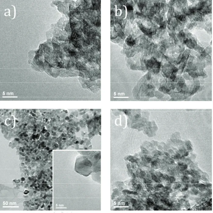
2-3 nm from a TEM analysis (Fig. 3a), that explains the overall feature of the X-ray diffraction
pattern.
3.2 Thermal decomposition of nickel oxyhydroxide nanoparticles 3.2.1 Decomposition of nickel oxyhydroxide in air
Figure 4 represents the temperature evolution of in situ XRD patterns recorded during the
decomposition of Ni3O2(OH)4 in air. The phase transformation from the lamellar oxyhydroxide
structure into the NiO cubic structure (NaCl type) occurs around 150-200 °C. From 200 to 700 °C,
the full width at half maximum (FWHM) of the diffraction peaks decreases which attests a gradual
increase of the crystallite size of NiO with temperature. The picture in inset of Figure 4 shows the
M
A
N
US
C
R
IP
T
A
C
C
E
P
TE
D
becomes green at temperatures typically above 800°C as expected for stoichiometric NiO. Figure 5
shows the evolution of the density of prepared “NiO” samples versus the decomposition
temperature of Ni3O2(OH)4. The densities measured for samples prepared up to 600 °C deviates
from the theoretical density of stoichiometric bulk NiO (6.81). For samples prepared below 350°C
the density is drastically lower than that expected and rises strongly with increasing temperature.
For samples prepared at higher temperature ( 400 °C), the density is still lower than expected but
evolves linearly (see the dashed line) until reaching the theoretical density of stoichiometric nickel
oxide at 750°C. The low density measured for samples prepared below 600°C could indicate the
formation of nickel oxide nanoparticles deficient in nickel (i.e. Ni1-xO) similarly as what was
observed during the preparation of zinc deficient zinc oxide [12]. To estimate the amount of nickel
vacancies the following equations were used (2, 3):
with dexp the measured density; Z the atomic number; Mcomp the experimental molar weight of the
compound; NA the Avogadro's number; Vref the refined cell volume; MO and MNi the oxygen and
nickel molar weights respectively.
The density value measured for Ni1-xO prepared at 250 °C (NiO-250) would almost correspond to a
50 % nickel deficient Ni1-xO material. For NiO-500, the amount of nickel vacancy is estimated to be
20%. Such low densities are correlated to a high specific surface area and low particles size (see
below) and the strong propensity of the material to exhibit oxygen or hydroxide terminated
surfaces. Due to the nanoscale of the particles, surface moisturization and carbonatation are likely to
happen leading to lower measured densities, thus the estimated vacancy rate is overestimated. A
Rietveld refinement on NiO-500 leads to a lower value ( 10%) taking more account of the "bulk"
M
A
N
US
C
R
IP
T
A
C
C
E
P
TE
D
values. Nickel oxyhydroxide seems to be an ideal candidate to prepare Ni-poor NiO samples with
tunable Ni deficiency via the control of the decomposition temperature of the precursor. The
characterization of the nickel oxides morphology was investigated by TEM microscopy (Fig. 3b,
3c), BET specific surface areas and XRD to calculate the crystallites size by Rietveld refinement
(Table 1). TEM images confirm the nanostructuration of NiO-250 (similar case to Ni3O2(OH)4) as
agglomerates of nanoparticles (2-3 nm). As observed by XRD, the increase in temperature allows
the further crystallization of NiO samples with larger particles sizes. In fact, NiO-500 is still
nanostructured with dispersed particles sizes around 10-15 nm. According to Table 1 the crystallites
sizes are very close to the particles sizes determined by TEM analyses. In order to optimize the
performances of NiO in p-DSSCs, high specific surface area is necessary to coat a maximum
number of dye molecules on NiO nanoparticles. Thus, we have also measured the specific surface
areas of Ni-poor NiO samples. The results demonstrate for NiO-250 a very high specific surface
area close to 250 m2.g-1 that is 3 times higher than that of Inframat NiO ( 80 m2.g-1) commonly
used as a reference in p-DSSCs. The thermal treatment at 250 °C maintains the high specific surface
area of the nickel precursor despite the structural change from Ni3O2(OH)4 to NiO. Note that the
specific surface area drops to 42 m2.g-1 for NiO-500, due to the increase in particles sizes.
3.2.2 Decomposition of nickel oxyhydroxide under ammonia
To our knowledge, the stabilization of a nitrogen-doped nickel oxide powder has not been
evidenced yet. Only theoretical study [20] or N-doped NiO film [21] have been recently reported.
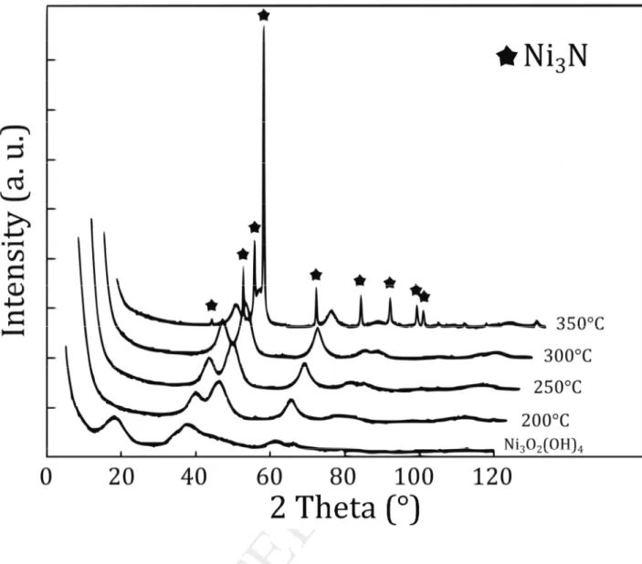
However, the study of the decomposition of Ni3O2(OH)4 under ammonia flow allows the
stabilization of N-doped NiO at low temperatures (see NiO-N-250). Indeed, Figure 6 represents the
evolution of XRD patterns of Ni3O2(OH)4 nitridation at temperatures ranging from 200 to 350 °C.
The investigated temperatures range is narrower than the one for previous Ni1-xO materials because
M
A
N
US
C
R
IP
T
A
C
C
E
P
TE
D
NiO-type cubic phase is formed with very broad peaks. When the nitridation temperature is over
300 °C, a shoulder on the (200) reflection of NiO appears at ~ 44.5° 2 that corresponds to the most
expected intense peak of Ni3N (111 reflection) (JCPDS file 89-5144). Over 350 °C, Ni3N is
unambiguously formed. For higher temperatures, nickel cations are definitely reduced into
elemental nickel. To assert the insertion of N in NiO-N-250, N(1s) XPS experiments were carried
out. Figure 7 displays for NiO-250 no characteristic signal of nitrogen, whereas NiO-N-250 exhibits
a N 1s BE peak at 398.2 eV. It proves unambiguously the presence of nitrogen into the NiO host
lattice [8]. The nitrogen content in NiO-N-250 was determined to be 0.8 wt. % whereas the nitrogen
amount in NiO-250 and Ni3O2(OH)4 was measured to 0.11 and 0.19 wt. % respectively. The
morphology of NiO-N-250 was investigated by TEM analysis (Fig. 3d), XRD and BET specific
surface area (Table 1). The results are quite similar to those of NiO-250, i.e. nanoparticles of 2-3
nm in diameter are observed in agreement with the calculated crystallites sizes. An important result
is the conservation under nitridation of the high specific surface area that shifts from 250 to 200
m2.g-1 from NiO-250 to NiO-N-250.
4. Conclusion
The thermal decomposition of nickel oxyhydroxide nanoparticles was thoroughly studied in order to
control the particles size of the as-prepared NiO nanopowders. The determination of the Ni3+/Ni2+
ratio by XPS and magnetism in the nickel oxyhydroxide confirms the Ni3O2(OH)4 formulation. The
decomposition of this compound in air at temperatures lower than 600 °C leads to
non-stoichiometric NiO nanoparticles with tunable nickel vacancies concentration and high specific
surface areas. Additionally, the stabilization of nitrogen-doped NiO is possible by low temperature
M
A
N
US
C
R
IP
T
A
C
C
E
P
TE
D
AcknowledgementWe are grateful to Eric Gautron, Jonathan Hamon (IMN) and Thierry Guizouarn (ISCR) for their
M
A
N
US
C
R
IP
T
A
C
C
E
P
TE
D
References[1] B. O'Regan, M. Grätzel, Nature 353 (1991) 737.
[2] F. Odobel, L. Le Pleux, Y. Pellegrin, E. Blart, Acc. Chem. Res. 43 (2010) 1063.
[3] S. Powar, T. Daeneke, M. T. Ma, D. Fu, N. W. Duffy, G. Götz, M. Weidelener, A. Mishra, P.
Bäuerle, L. Spiccia, U. Bach, Angew. Chem. Int. Ed. 52 (2013) 602.
[4] H. Kawazoe, M. Yasukawa, H. Hyodo, M. Kurita, H. Yanagi, H. Hosono, Nature 389 (1997)
939.
[5] A. Renaud, B. Chavillon, L. Le Pleux, Y. Pellegrin, E. Blart, M. Boujtita, T. Pauporté, L. Cario,
S. Jobic, F. Odobel, J. Mater. Chem. 22 (2012) 14353.
[6] D. Xiong, Z. Xu, X. Zeng, W. Zhang, W. Chen, X. Xu, M. Wang, Y-B. Cheng, J. Mater. Chem.
22 (2012) 24760.
[7] Z. Shi, H. Lu, Q. Liu, F. Cao, J. Guo, K. Deng, L. Li, Nanoscale Res. Lett 9 (2014) 608.
[8] B. Chavillon, L. Cario, A. Renaud, F. Tessier, F. Cheviré, M. Boujtita, Y. Pellegrin, E. Blart, A.
Smeigh, L. Hammarström, F. Odobel, S. Jobic, J. Am. Chem. Soc. 134 (2012) 464.
[9] T. Roisnel, J. Rodríguez-Carvajal, Mater. Sci. Forum 378-381 (2001) 118.
[10] K. Nakagawa, R. Konaka, T. Nakata, J. Org. Chem. 27 (5) (1962) 1597.
[11] J. Pan, J. Du, Y. Sun, P. Wan, X. Liu, Y. Yang, Electrochim. Acta. 54 (2009) 3812.
[12] A. Renaud, L. Cario, X. Rocquefelte, P. Deniard, E. Gautron, E. Faulques, F. Cheviré, F.
Tessier, S. Jobic. Sci. Rep. (2015). DOI:10.1038/srep12914.
M
A
N
US
C
R
IP
T
A
C
C
E
P
TE
D
[14] M. S. Hamdan, Riyanto, M. R. Othman, Int. J. Electrochem. Sci. 8 (2013) 4747.
[15] M. M. Kashani Motlagh, A. A. Youzbashi, F. Hashemzadeh, L. Sabaghzadeh, Powder
Technol. 237 (2013) 562.
[16] M. Casas-Cabanas, J. Canales-Vázquez, J. Rodríguez-Carvajal, M. R. Palací, J. Am. Chem.
Soc. 129 (2007) 5840.
[17] A.P. Grosvenor, M. C. Biesinger, R. St. C. Smart, N. S. McIntyre, Surf. Sci. 600 (2006) 1771.
[18] T.-L. Lai, J.-Y Liu, K.-F. Yong, Y.-Y. Shu, C.-B. Wang, J. Hazard. Mater. 157 (2008) 496.
[19] A. Renaud, B. Chavillon, L. Cario, L. Le Pleux, N. Szuwarski, Y. Pellegrin, E. Blart, E.
Gautron, F. Odobel, S. Jobic, J. Phys. Chem. C 117 (2013) 22478.
[20] R. Long, N. J. English, D. A. Mooney, Phys. Lett. A 374 (2010) 1184.
[21] F. Lin, D. T. Gillaspie, A. C. Dillon, R. M. Richards, C. Engtrakul, Thin Solid Films 527
M
A
N
US
C
R
IP
T
A
C
C
E
P
TE
D
Figures & table captions
Table 1. Summary of the specific surface areas, crystallites and particles sizes for Ni3O2(OH)4,
NiO-250, NiO-500 and NiO-N-250 samples.
Fig. 1. Powder X-ray diffraction pattern of nickel oxyhydroxide synthesized by precipitation route.
Fig 2. a) Ni 2p XPS spectrum of Ni3O2(OH)4; b) Magnetic properties of Ni3O2(OH)4.
Fig 3. TEM images of a) Ni3O2(OH)4; b) NiO-250; c) NiO-500; d) NiO-N-250 samples.
Fig 4. In situ XRD study patterns of the decomposition of Ni3O2(OH)4 in air. The interval of
temperature is 50°C between each XRD data.
Fig 5. Evolution of the density and Ni vacancies amount in prepared Ni1-xO samples versus
decomposition temperature in air with Ni3O2(OH)4 as precursor.
Fig 6. X-ray diffraction patterns of N-doped NiO materials issued from the ammonolysis of
Ni3O2(OH)4 at 200, 250, 300 and 350°C.
M
A
N
US
C
R
IP
T
A
C
C
E
P
TE
D
Table 1. Summary of the specific surface areas, crystallites and particles sizes for Ni3O2(OH)4,
NiO-250, NiO-500 and NiO-N-250 samples.
Sample Specific surface
area (m2.g-1) Crystallite size (nm) Particle size (nm) Ni3O2(OH)4 234 - 2-3 NiO-250 247 2.0 2-3 NiO-500 42 9.1 10-15 NiO-N-250 198 1.8 2-3
M
A
N
US
C
R
IP
T
A
C
C
E
P
TE
D
M
A
N
US
C
R
IP
T
A
C
C
E
P
TE
D
M
A
N
US
C
R
IP
T
A
C
C
E
P
TE
D
M
A
N
US
C
R
IP
T
A
C
C
E
P
TE
D
Fig 4. In situ XRD study patterns of the decomposition of Ni3O2(OH)4 in air. The interval of
M
A
N
US
C
R
IP
T
A
C
C
E
P
TE
D
Fig 5. Evolution of the density and Ni vacancies amount in prepared Ni1-xO samples versus
decomposition temperature in air with Ni3O2(OH)4 as precursor.
!
"
#
$
M
A
N
US
C
R
IP
T
A
C
C
E
P
TE
D
Fig 6. X-ray diffraction patterns of N-doped NiO materials issued from the ammonolysis of