HAL Id: tel-01391487
https://tel.archives-ouvertes.fr/tel-01391487
Submitted on 6 Jul 2017
HAL is a multi-disciplinary open access archive for the deposit and dissemination of sci-entific research documents, whether they are pub-lished or not. The documents may come from teaching and research institutions in France or
L’archive ouverte pluridisciplinaire HAL, est destinée au dépôt et à la diffusion de documents scientifiques de niveau recherche, publiés ou non, émanant des établissements d’enseignement et de recherche français ou étrangers, des laboratoires
Carcinoma : The heterogeneous immune contextures
accompanying CD8+ T cell infiltration in clear cell
Renal Cell Carcinoma
Nicolas Giraldo-Castillo
To cite this version:
Nicolas Giraldo-Castillo. The Immune Microenvironment in Clear Cell Renal Cell Carcinoma : The heterogeneous immune contextures accompanying CD8+ T cell infiltration in clear cell Renal Cell Carcinoma. Immunology. Université Pierre et Marie Curie - Paris VI, 2015. English. �NNT : 2015PA066321�. �tel-01391487�
Université Pierre et Marie Curie
Ecole doctorale Physiologie et Physiopathologie
Cancer et immunité anti-tumorale
The Immune Microenvironment in Clear Cell Renal Cell Carcinoma
The heterogeneous immune contextures accompanying CD8+ T cell infiltration in clear cell Renal Cell
Carcinoma
Dr. Nicolas GIRALDO-CASTILLO
Thèse de doctorat d’Immunologie
Dirigée par le Pr. Catherine Sautès-Fridman
Présentée et soutenue publiquement le 07 octobre 2015
Devant un jury composé de :
Mme. le Professeur Carole Elbim Présidente du jury
M. le Professeur Eric Tartour Rapporteur
M. le Professeur Antonino Nicoletti Rapporteur
M. le Professeur Pedro Romero Examinateur
M. le Docteur Xavier Cathelineau Examinateur
Acknowledgments
My desire to keep my thesis short will be difficult to accomplish in view of all the persons I would like to thank.
I would like to thank all the people that had made the last 3 years the biggest and more enriching experience I have had in my life.
I shall begin en Español, of course.
A mis Papás, a quienes debo todos mi logros. Quienes me enseñaron la importancia del amor, la rectitud, la confianza en mi mismo, los deseos de auto-superación. Quienes me dieron su ayuda incondicional, su compañía constante, y todos sus consejos. Y quienes, ante todo, me dieron fuerza para superar todos los problemas: siempre levantarse y seguir adelante. Gracias Papás, todos mis triunfos en la vida son gracias a ustedes. Papá, aprovecho esta oportunidad para decir cosas que a veces no decimos por falta de tiempo. Agradezco y admiro muchas cosas en ti. Probablemente la que mas admiro es tu nobleza; los constantes esfuerzos que haces para que todos lo que te rodean se sientan bien. Me alegra saber que este es uno de los mejores valores que he heredado. Además, agradezco tu dedicación con nuestra educación, tus consejos, a veces tu terquedad, tu racionalismo, tu deseos de auto-superación. Gracias papá; has hecho mi (y nuestras) vida (s) un camino cómodo, yendo de logros y de cariño. Mamá, de ti admiro también muchísimas cosas, sobretodo tu dedicación. Eres la segunda columna vertebral de nuestras vidas, nuestra ayuda constante e incondicional, aquella que sabe más de nosotros mismos que nosotros mismos, que responde con cariño cualquier pregunta que hacemos. Haces mi vida muy feliz mamá búha.
To Catherine, from the moment we first met, back in December 2011, until the last day of my thesis, I am deeply grateful. For your wonderful dedication, your patience, your strong but always kind advices and criticisms, and must importantly, your desire in helping me grow as a scientist and as a person, as a student and as a potential boss, as a colleague and as a friend, I am deeply grateful. If I could choose again, I would never hesitate to choose you as my boss.
To Hervé, for his incredibly wise and always pertinent comments, his restless willing to teach, his inspirational passion for research and knowledge, his fantastic skills in convincing people (including myself) of the importance of research, for all his valuable advices and his quick opera lessons, I am deeply grateful.
To the other bosses, thanks to Marie-Caroline for her valuable advices, and her constant disposition in helping me understanding my results. Thanks to Jean-Luc for his kindness, all the valuable discussion, his willing to teach and for his complete support. Thank you Isabelle for your advices, your eagerness and your support. To Lubka, thank you for all your support, your openness and all the interesting discussions. To Sophie Siberil, I write this before going to NY with you, but I’m pretty sure we will have an amazing time there; thanks for being so accessible and opened, for your valuable advices and nourishing discussionsv.
To Pr. Carole Elbim, Pr. Eric Tartour, Pr. Antonino Nicoletti, Pr. Pedro Romero, Dr.
Xavier Cathelineau, thank you for taking the time to read this manuscript and giving constructive criticism.
To Etienne, for his support, his restless willing to help, his not-being-afraid-of-speaking-English, his peacefulness, and for making me feel welcome in France, I would like to thank him.
To Laetitia, Leticia, tia, mitia, myty, la-cruz, lacro-iX. You were my biggest support during these 3 years in the lab, I am not sure where I would be without you. You were my technical and emotional support, my always-reassuring Plan B – SOS!, the person whose motivation inspired all the people in the team, and who best represented the ‘crazy team’. The way you got through all the issues in your life is inspirational, and I have no words to thank you. Thank you my Tia; I will get you back the favor one day (If you know what I
meeeaaan…Boulangerie!)
To the combo latino: Estefania, Claudia y Ana. Por hacerme dar cuenta que los latinos somos mas chidos que todos los demás. Por su calidez, sus sonrisas, su comida, sus risas, las clases de español latino, las risas y las risas. Qué lindo haberlas encontrado, las quiero mucho.
To Benedicte TicTic (or ‘Dic Dic’), thanks you for your jokes, you willing to help, your motivational energy, your rapid style tips and reminding me that I am much much funnier and intelligent when I talk Spanish. If only you’d know how funny I am in Spanish…
To Benedicte BenBen, for her willing to help, her scientific curiosity, for reminding us all how old we are, for the restaurant tips, quick medical reviews and the superb massages, I am very grateful. (More for the massages than anything else ;-)) I’m pretty sure you will be an excellent doctor and teacher one day BenBen; I am proud of you and I am very glad I could participate (a little bit) to your formation as a researcher.
To Ivo, thanks for your craziness, your craziness, your craziness and your craziness. You are the proof that magical thoughts can still make their way into science. Your dedication and eagerness was inspirational, and extremely helpful!
To Sophie Chauvet, la vampira, for her kindness, her generosity, her humbleness, her humor, her tranquility. I felt so welcomed by you, and I am very glad I met you Sophie. You are more than welcome in Colombia (or wherever I will be) when you feel like going for an adventure.
To Priyanka, thank you for your humility Priyankita, your humor, your smile, your vibrant energy and your tranquillity. Your dedication and commitment with work is admirable, and I really hope you’ll fulfill all your dreams (and I am pretty sure you will).
To Claire-la-petite, petite-Claire, for welcoming me in the lab, for your open-mindedness, your willing to help; you were the little voice in my head that render me a bit anxious during my whole thesis.
To Estelle and Helene, for being the funniest, nicest, most helpful people in the CRC. Thank you for teaching me such a great amount of stuff, for reminding me how much I like Flow Cytometry, for making the long cell-sortings into funny moments. Thank you for being so welcoming, I hope we can work again in the future.
To Nathalie Jupiter, pour ton aide, ta gentillesse, ton excellente cuisine, tes éclats de rire, ta bonne humeur et ton envie irrépressible d’aider les gens, j’en suis extrêmement reconnaissant. Je suis content de t’avoir croise sur ma route, tu rends les choses beaucoup plus faciles au laboratoire.
And last but not least, to Sarah for your loud laugh and the 100000 hours you dedicated to correct my English! To Anne for her crazy-sympathetic-funny-open-minded personality. To Yann for his kindness and all his help. To Tessa for her super-crazy craziness, her racist jokes, accusing me instead of Amelie for stealing her Daim during 2 years!, and for the extra-kg I gained because of all the chocolates she gave me. To Lucie for her kindness, her disposition for helping us, her violent massages and all the funny (way too intimate) discussions. To Claire La Grande for all her help, the interesting discussions, her inspirational dedication and commitment with work, her borderline ‘TOC’, for teaching us how important it is to wash our hands and to take a prophylactic morning coffee to avoid the ‘morning mood’. To Myriam for
Moglie, Nathalie Josseaume, Jasmina, Johanna, Tania and Melanie, for your help and valuable discussions.
In my personal life,
To Tristan, a quién escribo en Español para que mas nadie pueda entender. Gracias Lindo por acompañarme durante todos estos años, fuiste mi tranquilidad, mi sustento, mi más grande apoyo, y estoy absolutamente agradecido con la vida porque te encontré cuando más te necesitaba. Sin mayor pretensión, me enseñaste infinitas cosas; que el amor es simple; que la felicidad también; “No need to hurry. No need to
sparkle. No need to be anybody but oneself”. En Francia, debo todos mis triunfos a ti. Te quiero mucho.
To my family, los dejo tan atrás por razones políticas, pero en realidad deberían estar de primeros. A mis tres hermanos, Rommel, Vivi y Pame, mis dos sobrinos, Mateo y Lorenzo, gracias por su apoyo incondicional. Me da alegría que en estos años la distancia nos han vuelto más cercanos, y que ahora conozco el valor de su amistad, y lo importante que es tener una gran familia. Rommelin, por tus consejos, tu pragmatismo, tu compañerismo; demostrarnos que el tamaño de nuestros sueños es el mismo de nuestros logros. Tu fuiste el primero el dar el gran paso, y con ello, diste ejemplo a toda la familia. Gran parte de todos nuestros logros los debemos a ti. Vivi, por tu nobleza, tu incondicionalidad, tu leve y graciosa locura, tu inacabable energía. Por enseñarnos la importancia del autocuidado. Por demostrarme que el pato, por mas fino que sea, no es en realidad tan sabroso. Gracias por ayudarme, te quiero. Pame, alias Burri, gracias por tu apoyo y compañía constante, por sacarme de apuros con los regalos de todos en Colombia, las risas y consejos. Sin darte cuenta, eres la columna vertebral de la familia, y todos estamos tranquilos debido a tu incondicionalidad, y orgullosos por lo lejos que has llegado. Me alegra saber que aunque hemos estado lejos en los últimos años, hemos estado cerquita de corazón (debemos agradecer, entonces, a Apple y FaceTime). Mateo, la persona mas noble y autentica que conozco; estoy orgulloso de ti. Gracias por tu personalidad. Lorenzo, espero que cuando aprendas a leer, leas estos agradecimientos y sepas que nos has alegrado la vida desde que naciste a todos en la familia.
To Amélie, Ma petite Amélie. I would have got crazy without you. My ‘personal teacher’ in so many ways. You taught me French, how to cook, how to ski, all the French Christmas songs, how to love action movies, how to write romantic texts in French, en fin… You are just simply amazing ma petite Amelie, you are one of the most kind, solidary and beautiful person I have ever met. Thank you for being with me “en las duras y en las maduras” (that is the way we say in Spanish, that would translate “dans les dures et les matures”…), understanding me, and letting me understand you back. I hope our friendship lasts until we become old, fat and ugly; we will go to work out together our muffing tops in the pool… Or maybe, just take a sunbath if we are lazy.
To Ana, Anina my love, eres mi persona, mi amiguita, mi soporte, mi alegría, mi compañera de baile, de codazos, mis risas, mi humor negro. Nos construimos y crecimos juntos, y te has convertido en uno de los grandes pilares de mi vida. Me alegra saber qué, a pesar de la distancia, creo que somos mas cercanos que nunca. Te quiero Anina, gracias por estar siempre ‘ahí’ cuando te necesito y llenar de risas (y comida) todos los aspectos de mi vida.
To Vane, fuiste mi gran compañera, con quién descubrí Paris; con quién descubrí tantos restaurantes y tantos tipos de comida; con quien descubrí la fotografía; con quien descubrí lo lindo de mi cultura, la importancia de la amistad en momentos difíciles, la tranquilidad de vivir con alguien luego de estar solo por un tiempo. Me alegra que este viaje nos haya acercado más de los que 6 años de medicina jamás lo hicieron. Descubrí también tu mundo, y creo que tú el mío.
To Andre, Linda Andre. Creo que no sabes lo importante que fuiste en estos años. Fuiste un suporte tan sutil y a la vez tan fuerte. Mi linda Andre, a ti te debo la pequeña línea que me separa de la absoluta ignorancia, mi amor a los arboles y las plantas, mi fijación con los avestruces que viven cerca de mi
por las chucherías y la sensación de qué todo va a estar bien. Linda manitotas, quiero que un día vayamos a celebrar a la playa al lado de tu casa, esa llena de aviones olvidados, y comamos helado mientras vemos las estelas de humo pasar por encima de nuestra cabezas.
To Nico (Barbosini), tanto a ti como a Andre debo la pequeña línea que me separa de la absoluta ignorancia, el amor brusco que me genera Portugal y el Fado, además de la oportuna sensación de tranquilidad que me acompaña a veces. Eres una persona con la que siento puedo crecer y reír al mismo tiempo. Gracias Nico, por acordarme que la vida es mas ligera de lo que parece, que todo duelo es mas fácil si se diluye en medio de risas, que las estrategias hiper-complicadas y basadas en aprendizajes enraizados por las telenovelas, a pesar de tener baja tazas de éxito, son siempre realizables y colorean la vida de un tono que pocos entienden.
To Poly, myvolyvol, Val. Para terminar, my voly, esto ha sido todo un proceso para los dos. Desde que escogimos juntos ciencias en el colegio; cuando fuimos a (modestia aparte) Uniagraria, la Javeriana y los Andes juntos; cuando escogimos nuestra curiosidad científica por encima de cualquier pretensión monetaria. Debo a ti ese amor por la ciencia, ese deseo de auto-superación, la sensación de que los todos los sueños son realizable, de que el mundo es tan grande como queramos que sea. Tu me enseñaste a ser sociable, a sentirme bien dentro de mis zapatos, a estar orgulloso de mi y mis próximos, la alegría que implica ponerse feliz con las triunfos ajenos. My voly, debo gran parte de los logros de mi vida a tu presencia, me alegra que hayamos crecido juntos, y que nos queden tanto años para seguirlo haciendo.
“For it would seem that we write, not with the fingers, but with the whole person. The nerve which controls the pen winds itself about every fiber of our being, threads the heart, pierces the liver.”
Table of Contents
Acknowledgments ... 2 Table of Contents ... 7 Summary ... 9 Abbreviations ... 10 Introduction ... 12Chapter 1 - The Anti-Tumour Immune Response... 12
Bullet points ... 12
Inflammation and Cancer ... 12
Immune Control and Tumour Escape... 14
The Immune Microenvironment as a Prognostic Tool ... 15
The Immune Microenvironment and Other Histopathologic Features ... 19
Therapies that modulate the tumour microenvironment ... 20
Chapter 2 - Renal Cell Carcinoma ... 22
Bullet points ... 22
Epidemiology and Pathophysiology ... 22
Classification ... 23
Diagnosis ... 24
Treatment ... 25
Chapter 3 - The Tumour Microenvironment in Renal Cell Carcinoma ... 28
Bullet points ... 28
Renal cell carcinoma: an inflammatory neoplasia ... 28
Tumour associated macrophages ... 30
Myeloid derived suppressor cells ... 31
Dysfunctional dendritic cells and defects in T cell priming ... 32
T Lymphocytes: Rather a functional defect ... 32
NK cells ... 37
Cytokines shaping the RCC immune microenvironment ... 38
Other mechanisms of immunomodulation: MHC Class I and II ... 39
Shaping the RCC immune microenvironment: Lessons we have learned from immunotherapies? ... 40
Objectives ... 42
Results ... 43
Article 1: <<Orchestration and Prognostic Significance of Immune Checkpoints in the Microenvironment of Primary and Metastatic Renal Cell Cancer>> ... 43
Summary of the results in Article 1: ... 43
Article 1 ... 45
Supplementary data ... 55
Article 2: << The immune contexture of primary and metastatic human tumours>> ... 60
Summary of the results in Article 2: ... 60
Article 2 ... 61
Unpublished results ... 69
Summary of the Unpublished Results: ... 69
Material and Methods ... 69
Results ... 70
DC compartmentalization and orchestration of the immune response in ccRCC ... 84
Immune checkpoints, CD8+ T cells and prognosis in ccRCC ... 86
Towards the identification of the tumours with a suppressive microenvironment in ccRCC ... 87
Revisiting the immunoscore ... 89
Conclusions ... 91
Perspectives and Limitations of the Study ... 92
Summary
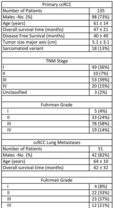
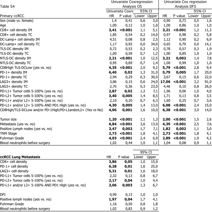
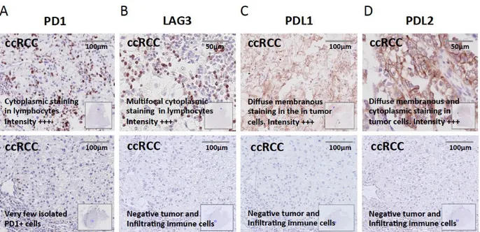
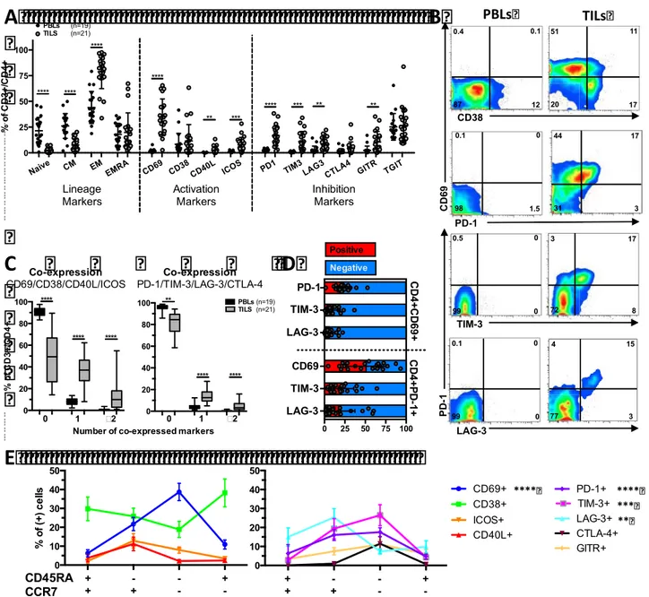
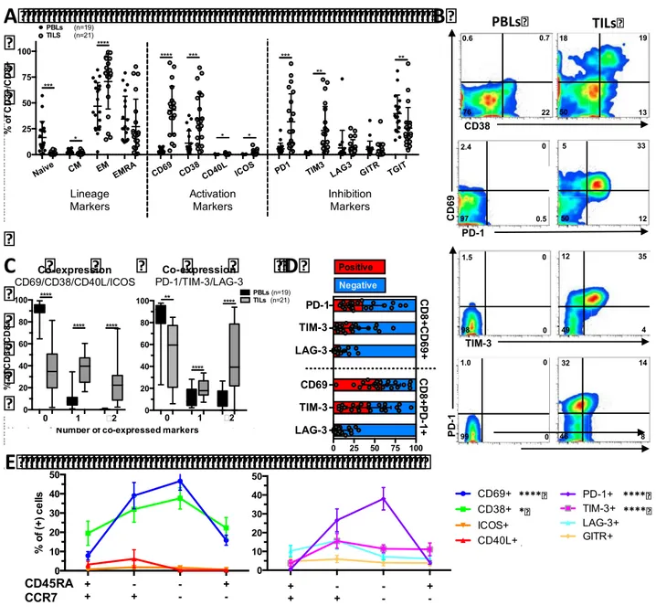
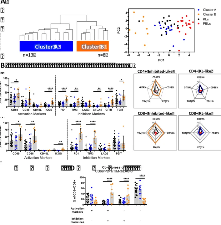
To decipher the potential mechanisms linking increased CD8+ T cell infiltrations with an adverse clinical outcome in ccRCC, in this study we determined: 1) the prognosis associated with the expression of immune checkpoints and its coordination with dendritic cell (DC) and CD8+ cell infiltration, and 2) the phenotypic traits of tumor infiltrating lymphocytes (TIL). The prognosis associated with CD8+ and DC infiltrations, in addition to the expression of immune checkpoints were investigated in a cohort of 135 ccRCC by quantitative immunohistochemistry. We found that the densities of CD8+, PD-1+ and LAG-3+ cells were closely correlated, and independently associated with decreased PFS and OS. In addition, patients whose tumors presented both high densities of PD-1+ cells and PD-L1+ and/or L2+ tumor cells (>5% positive cells), displayed the worst clinical outcome. High densities of immature DC isolated in the tumour stroma were associated with high expression of immune checkpoints and patients’ poor clinical outcome. In contrast, the presence of mature DC within Tertiary Lymphoid Structures identified, among the tumours with high CD8+ TIL densities, those with low expression of immune checkpoints and prolonged survival. To functionally characterize the CD8+ T cell infiltrates, we investigated the phenotype of freshly isolated TIL in 21 ccRCC by flow cytometry. We found a group of tumors (8/21) characterized by the over-expression of inhibitory receptors (PD-1 and TIM-3) and activation markers (CD69 and CD38), the expansion of the effector memory cell subpopulation (CCR7-CD45RA-), and a trend toward more aggressive features. In summary, we demonstrated that the infiltration with CD8+ TIL in ccRCC is accompanied by the enhanced expression of immune checkpoints and a poorly coordinated immune response in a subgroup of aggressive tumors. This immune profile defines a poor prognosis group of patients that should be suitable to receive immune checkpoint inhibitors.
Abbreviations
CRC: Colorectal Cancer.
TME: Tumour Microenvironment. CIN: Cervical in-situ Neoplasia. DC: Dendritic Cells.
TIL: Tumour Infiltrating Lymphocytes. MSI: Microsatellite Instability.
TLS: Tertiary Lymphoid Structures. Treg: Regulatory CD4+ T cells. IM: Invasive Margin.
TAM: Tumour Associated Macrophage. NSCLC: Non-small Cell Lung Cancer. HCC: Hepatocellular Carcinoma. LPS: Lipopolysaccharide.
MHC: Major Histocompatibility Complex. DC: Dendritic Cells.
ccRCC: Clear Cell Renal Cell Carcinoma. VHL: Von Hippel-Lindau.
TT: Target Therapy.
VEGF: Vascular Endothelial Growth Factor. PDGF: Platelet Derived Growth Factor. TGF: Transforming Growth Factor. GLUT: Glucose Transporter. TKI: Tyrosine Kinase Inhibitors.
mTOR: Mammalian Target Of Rapamycin. PB: Peripheral Blood.
MDSC: Myeloid Derived Suppressor Cells. ROS: Reactive Oxygen Species.
RCC-LM: Renal Cell Carcinoma Lung Metastases. CRC-LM: Colorectal Cancer Lung Metastases. nccRCC: Non-Clear Cell Renal Cell Carcinoma.
NTLS-DC: Non-Tertiary Lymphoid Structures Dendritic Cells. TLS-DC: Tertiary lymphoid Structures Dendritic Cells
AM: Activation Markers. InR: Inhibitory Receptors. EM: Effector Memory. CM: Central Memory.
Introduction
Chapter 1 - The Anti-Tumour Immune Response
Bullet points A chronic inflammatory microenvironment promotes the development of cancer.
Once a tumour has emerged, an inflammatory microenvironment also promotes malignant
cells growth, tumour spread and metastasis.
The adaptive immune response controls tumour growth and elimination.
The major cellular mediators of the anti-tumour immune response identified so far are the CD8+ T cells and Th1-oriented CD4+ lymphocytes, and their increased densities are associated with good clinical outcome in most tumours.
There are few exceptions to this rule, including Renal Cell Carcinoma and Hodgkin Lymphoma, where an increased CD8+ cell infiltration has been associated with patient’s poor prognosis.
Tumour cell often develop mechanisms that modulate and/or inhibit the immune response,
which are associated with patient’s poor prognosis.
Inflammation and Cancer
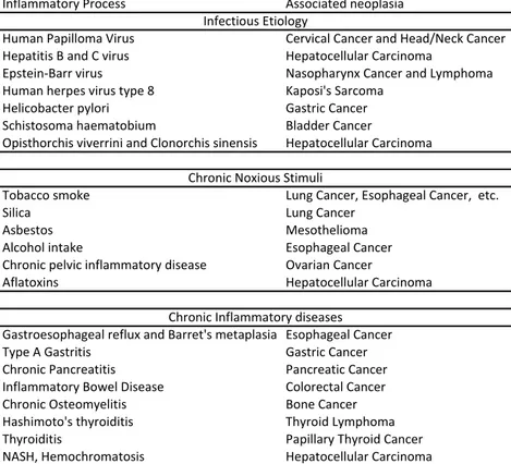
Several lines of evidence have established an association between chronic inflammation and cancer (1). First, approximately 20% of the tumours are linked to inflammation-inducing infectious organisms (2), including Helicobacter pylori and gastric cancer (3), Hepatitis B and C viruses and hepatocellular carcinoma (4) and human papilloma virus and cervical and head/neck cancers (5) (6). Second, chronic noxious stimuli or inflammatory diseases can favor neoplasia, such as cigarette smoke and asbestos/silica for lung carcinoma (7), gastroesophageal reflux for cancer of the esophagus (8), inflammatory bowel disease for colorectal cancer (CRC) (9), chronic pancreatitis for pancreatic cancer (10) and pelvic inflammatory disease for ovarian cancer (11). Third, the chronic intake of nonsteroidal anti-inflammatory drugs inversely correlate with CRC incidence, and recent studies indicate a negative
mediators (e.g. cytokines and pro-inflammatory transcription factors) decreases the incidence and spread of tumours both in mice and in humans (13) (14). Table 1 lists cancers where a chronic inflammation has been implicated in their pathophysiology.
Table 1. Cancers associated with chronic inflammatory conditions
The mechanisms directing inflammation-induced tumourigenesis are well known. DNA damage and extra cellular matrix disruption by inflammatory mediators [e.g. through the production of reactive oxygen species (15) and matrix metalloproteinases (16), respectively], in addition to the stimulation of tumour cell growth by cytokines [e.g. IL-1B for gastric carcinoma (17) and IL-8 for melanoma (18)], are the main recognized tumour-promoting mechanisms.
In several situations in which the pre-cancerous stages can be studied, a shift from an immunological pattern with a Th1 orientation to a pro-inflammatory tumour microenvironment (TME) has been reported. It is illustrated in cervical carcinoma in which high expression of genes encoding Th1 cytokines is found in cervical in-situ neoplasia (CIN), whereas IFN-γ expression is lost, and the expression of pro-inflammatory cytokine-genes and macrophage infiltration is high in invasive and aggressive cervical carcinoma (19) (20). A similar shift has been described in pancreatic cancer, where there is a decrease in the density of CD8+ T cells and mature (DC) cells from low-grade premalignant lesions into invasive ductal adenocarcinoma and also during transformation of benign to malignant head
Inflammatory Process Associated neoplasia
Human Papilloma Virus Cervical Cancer and Head/Neck Cancer
Hepatitis B and C virus Hepatocellular Carcinoma
Epstein-Barr virus Nasopharynx Cancer and Lymphoma
Human herpes virus type 8 Kaposi's Sarcoma
Helicobacter pylori Gastric Cancer
Schistosoma haematobium Bladder Cancer
Opisthorchis viverrini and Clonorchis sinensis Hepatocellular Carcinoma
Tobacco smoke Lung Cancer, Esophageal Cancer, etc.
Silica Lung Cancer
Asbestos Mesothelioma
Alcohol intake Esophageal Cancer
Chronic pelvic inflammatory disease Ovarian Cancer
Aflatoxins Hepatocellular Carcinoma
Gastroesophageal reflux and Barret's metaplasia Esophageal Cancer
Type A Gastritis Gastric Cancer
Chronic Pancreatitis Pancreatic Cancer
Inflammatory Bowel Disease Colorectal Cancer
Chronic Osteomyelitis Bone Cancer
Hashimoto's thyroiditis Thyroid Lymphoma
Thyroiditis Papillary Thyroid Cancer
NASH, Hemochromatosis Hepatocellular Carcinoma
Infectious Etiology
Chronic Noxious Stimuli
reported in the evolution from intraepithelial neoplasia to invasive carcinoma (Bais 2005).
Not surprisingly, once a tumour has emerged, an inflammatory microenvironment can also promote malignant cells growth, resulting in neo-angiogenesis, acquisition of new mutations, extracellular matrix disruption, tumour cell migration and finally metastasis (22). The main cellular mediators of this process are macrophages (and to less extent neutrophils) that are, by far, the major immune cellular component of tumour infiltrates (23). These cells produce high quantities of 1β, IL-6, IL-23 and TNF-α, the key cytokines mediating the inflammatory-induced tumour igenesis (reviewed in (22) and (23)).
Immune Control and Tumour Escape
In addition to the link between inflammation and tumourigenesis, other cellular and molecular mediators of the immune system can contribute to control of tumour growth and elimination. Several epidemiological observations support this fact, including scarce reports of spontaneous cancer regression (24), the augmented incidence of cancer in immunosuppressed individuals (25) and the association between increased tumour infiltrating T cell (TIL) and favorable clinical outcome (26) (27).
The fact that tumour cells express antigens encoded by mutated genes (28) often renders them targets of the immune cells. Indeed, autologous TIL can induce tumour cell lysis in vitro and in vivo (29) and tumour-specific lymphocytes are often detected in patients with cancer. This phenomenon has been well characterized in colorectal cancers, where microsatellite instability (MSI, a genetic defect that impedes DNA mismatch repair) fosters the expression of thousands of new antigens on tumour cells. Characteristically, MSI+ tumours have a prominent CD8+ T cell infiltration and are associated with favorable clinical outcome (30).
The major cellular mediators of the anti-tumour immune response are the CD8+ T cells, in addition to the Th1-oriented CD4+ lymphocytes. The first are in charge of the elimination of tumour cells through the production of apoptosis-inducing molecules or cytotoxic granules (e.g. granzymes, perforin and granulysin) (31), while the latter can provide help to the CD8+ T cells and foster the anti-tumour response by the secreting major cytokines, including IFN-γ (32). Several lines of evidence suggest that mature dendritic cells (DC) orchestrate the T and B cells anti-tumour immune response. Characteristically, these cells are present in highly organized peri-tumour immune cellular aggregates, called Tertiary Lymphoid Structures (TLS) (33) (discussed in next section).
The major antitumour immune response cytokines and chemokines are IFN-γ, IL-12, CXCL9 and CXCL10, mainly involved in CD8+ T cell recruitment (CXCL9 and CXCL10) and activation (IL-12 and IFN-γ) (34) (35).
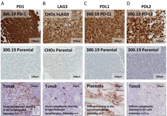
All these processes submit tumour cells to a significant selective pressure. In fact, tumour cells can develop mechanisms that modulate and/or inhibit the immune response, including: first, the production of immunosuppressive molecules (e.g. IL-10 and TGF-β) that hamper the cytotoxic and proliferative capacity of T cells (36); and, second, the expression of ligands for inhibitory receptors expressed on the TIL (37). Of particular relevance, PD-1 is a molecule expressed on activated and exhausted T cells that diminishes the strength of the cellular immune response upon binding to its ligands (PD-L1 and PD-L2) (38). Under physiological conditions, the expression of PD-L1 and PD-L2 is highly regulated and it is limited to dendritic cells, macrophages, activated T cells (PD-L1 only) (39) and certain tissues where immunomodulation is required (e.g. syncytiotrophoblast in the placenta). Nevertheless, certain tumour cells can express these ligands, and subsequently inhibit T cell activity. Similar mechanisms have been reported, including the expression of the ligands for TIM-3 and LAG-3, two additional inhibitory receptors expressed on T cells (reviewed in (37)). Ultimately, this microenvironment induces the development of suppressive immune cells, including regulatory T cells CD4+ (Treg) and myeloid derived suppressor cells (36), that sustain self-tolerance against tumour antigens.
This complex interconnected network of myeloid and lymphoid cells, endothelial and lymphatic vessels and stromal cells –named the tumor microenvironment (TME) (26) (40)– has been largely studied in the last decade. Its influence on patient’s clinical outcome and tumour progression has been of particular interest: patients with tumours that develop immunosuppressive mechanisms have the worst prognosis, and their tumours will often display a higher histologic grade characterized by dedifferentiation, neo-vascularization and an inflammatory infiltrate.
The Immune Microenvironment as a Prognostic Tool
Many studies have described the distribution of the inflammatory and immune infiltrate within different tumours. Overall, the macrophages, mast cells and granulocytes are mostly found infiltrating or surrounding the tumour nests both in the invasive margin and the core of the tumour. On the contrary, the lymphoid infiltration is more precisely distributed, and some locations are enriched in certain cell types: NK cells are mostly found in the stroma and are not in contact with tumour cells; B cells are mostly found in the invasive margin (IM) of the tumours within lymphoid aggregates; and T cells, particularly CD8+ T cells, are mainly located in the IM, but can also infiltrate the tumour core (26) (41).
The analysis of the immune microenvironment in retrospective cohorts across different tumours has established a clear correlation between the density of infiltrating immune cell and patient’s clinical
Tumour associated macrophages
Several studies have found that the clinical outcome associated with increased numbers of Tumor Associated Macrophages (TAM) is mainly determined by their functional orientation, and is heterogeneous across tumor types (42). In fact, while the augmented densities of this population are associated with favorable clinical outcome in colorectal (43) (44) (45) (46) (47), gastric (48), non-small cell lung cancer (NSCLC) (49) (50) (51) (52), hepatocellular carcinoma (HCC) (53) (54), prostate (55) and cervical cancer (56), it has the opposite association in endometrial (57), esophageal (58), gastric (59) (60) (61) (62), urothelial (63), oral (64) (65), HCC (66) (67), melanoma (68), breast (69) (70) (71) (72) (73) (74), ovarian (75), bladder (76) (77), NSCLC (78), thyroid (79), pancreatic neuroendocrine (80) and endometrial (81) tumours.
The heterogeneous association between TAM densities and the clinical outcome across different tumours might reflect the plasticity of this immune population. Overall, two different subtypes of macrophages have been described (82) (83):
1. M1: activated by Toll-like receptor ligands [e.g. Lipopolysaccharide, (LPS)] and IFN-γ; they preferentially express pro-inflammatory cytokines in addition to inducible nitric-oxide synthase, an overall they potentiate the inflammatory and immune response.
2. And M2: stimulated by IL-4 or IL-13; they express arginase 1, CD206, IL-4r, TGF-β1 and PDGF. This population is rather implicated in wound repair, promoting fibroblast proliferation and extracellular matrix deposition.
A protective role in tumourigenesis has been proposed for M1 macrophages (through mechanisms including the activation of the Th1 response and by antagonizing the suppressive activities of regulatory immune cells), while M2 have shown to promote tumour growth, invasion, metastasis, stroma remodeling and angiogenesis (84).
The absence of M1 and M2 specific markers has been the major obstacle in the assessment of the clinical impact of each of theses subtypes. Up to date, this task has been accomplished using CD11c or NOS2 for M1 TAM, and CD163, CD204 or CD206 for M2 TAM. Interestingly, increased M1 TAM densities seem to be associated with a favorable clinical outcome in NSCLC (51) (52), ovarian (85), colorectal (86) and gastric cancer (87), while those of M2 are linked to poor prognosis in several tumours, including: NSCLC (88) (89) (90), mesothelioma (91), esophageal (92), gastric cancer (61) (93), pancreatic (94) (95) (96) (97) (98) (99), CRC (100), HCC (101), Hodgkin lymphoma (102), renal (103) (104) (105), urothelial (106), breast (107), endometrial (108), ovarian (109), melanoma (68), and squamous oral carcinoma (110). Additionally, some studies have demonstrated that, when associated
addition to HIF, VEGF (63) and matrix metalloproteinases expression (62) (69) (111) (112) (76), suggesting they might have an M2 phenotype.
Although not yet conclusive, the evidence linking the infiltration with TAM and the patient’s clinical outcome suggests that high densities of M2-oriented cells are associated with more advanced tumour stages and patient’s poor prognosis. In addition, it also indicates that the biological value of measuring the sole densities of CD68+ cells should be revisited, as it does not provide information of the TAM function or polarization.
NK cells
Natural Killer cells is another major cellular population mediating the anti-tumour immune response (113). These cells express an array of receptors (including activating, inhibitory, adhesion and cytokine receptors) that enable them to identify tumour cells. Overall, the integration of these signals determines whether or not NK cells become activated, and eliminate its target. The two most important mechanisms of cancer cell recognition by NK cells is the down-regulation of major histocompatibility complex (MHC) class I and the expression of stress-induced ligands to NK activation receptors (e.g. MICA or MICB, which bind to NKG2D expressed on the NK cell) by tumour cells.
The prognostic impact of NK infiltration has been studied in some tumours, and their increased densities have been consistently associated with favorable clinical outcome. This association has been demonstrated in CRC (114) (115) (116), gastric (117) (118) (119), vulvar squamous cell (120), esophageal (121), renal (122) (123), HCC (124), NSCLC (125) (126) (127), in addition to CRC and RCC lung metastases (128). Nevertheless, other studies in NSCLC have not found an association between NK infiltration and prognosis (129), where they display an inhibited phenotype and decreased functional capacities (130) (129). More studies assessing the prognostic impact associated with NK infiltration are needed.
Dendritic cells
Upon encounter with an antigen and in the presence of danger signals, immature DC go through a process called maturation, that allows them to migrate into the lymph node, where they can prime naïve CD4+ and CD8+ T cells. The phenotype of the mature DC plays an important role in determining the orientation and strength of the subsequent immune response, and it is determined by the cytokine microenvironment, in addition to the type of antigen being processed (131). The TME takes advantage of the DC plasticity, and can induce a pro-inflammatory and/or tolerogenic DC, or block their maturation at different stages.
Due to the plasticity and heterogeneous DC phenotype, the quantification of infiltrating DC has been a difficult task. The more relevant markers that have been used for their quantification are S-100, CD83, DC-LAMP or CD1a, in addition to CD207 for Langerhans cells, and their clinical impact has been reported in a variety of human solid tumours. Overall, several studies support that the augmented infiltration with DC is associated with increased overall survival in many tumours types, including melanoma (132) (133) (134) (135), HCC (136) (137), gallbladder (138) (139), oral (140), esophageal (141) (142) (143), gastric (144) (145) (146) (119), NSCLC (147) (127) (148), colorectal (149) (150) (151) (152), bladder (153), breast (154) (155) (155) (156) (157), endometrial (158) (159) and ovarian cancer (160) (161). Nevertheless, the infiltration with CD123+ plasmacytoid DC has been associated with shorter overall survival in breast cancer (162) (163), as for the presence of CD208+ and CD1a+ DC in colorectal (164) and gastric cancer (165).
Tertiary lymphoid structures and associated mature dendritic cells
Interestingly, lymphoid aggregates can be detected in the invasive margin of most tumours. Some of them exhibit properties of active immune sites that resemble those arising in other tissues upon infection, or secondary to autoimmune or chronic inflammatory diseases (33). Characteristically, they exhibit a T cell zone (composed mainly of CD4+/CD62L+/CD45RO+ CM an in less extend CD8+/CD62L+/CD45RA+ naïve T lymphocytes) with embedded mature DC, germinal centers with proliferating B cells and they are surrounded by high endothelial venules (166). In addition, laser microdissection of these structures reveled they are enriched in genes associated with T cell chemoattraction molecules, such as CCL19, CXCL13, CCL21, IL16, CCL22 and CCL17 (166).
In view of their similarities with germinal centers in lymph nodes, it has been hypothesized that these Tertiary Lymphoid Structures (TLS) represent a site where in situ antigen presentation and lymphocyte activation can occur under a protected environment (33). Indeed, studies in primary melanoma and NSCLC have correlated the densities of mature DC (DC-Lamp+) within TLS with a strong infiltration with activated T cells and a Th1-oriented response, respectively (132) (167). Moreover, the higher densities of these structures correlated with favorable clinical outcome in NSCLC (168) (167) (169), colorectal (151) (170), melanoma (171) and breast cancer (172). The mechanisms underlying the neogenesis of these structures are still unclear in human tumours.
CD4+ and CD8+ T cells
Overall, a high infiltration by CD8+ T lymphocytes is associated with good clinical outcome in many tumour types, including lung, liver, stomach, CRC, breast, esophageal, bladder, melanoma, ovarian
large B cell lymphoma (173), Hodgkin lymphoma (174), RCC (175) and potentially head and neck cancer (176), where high densities of tumour-infiltrating CD8+ T and/or Th1 cells have been associated with poor prognosis. Overall, Th1 CD4+ T cells show a similar clinical impact to that of CD8+ T cells; nevertheless, there are again exceptions to this rule, including head and neck cancer (177) and RCC (178) (179). The infiltration by other T cell subsets (Th2, Th17 and Treg) is less clear, and seems to be dependent on the cancer type (26).
In view of the clinical impact of infiltrating CD8+ T cells in cancer, sustained efforts are being made to validate and promote their quantification in the routine clinical setting; this approach that has been called Immunoscore (180). The development of automatized software that quantify the densities of immune cells after immunohistochemical staining is promoting the gradual change from semi-quantitative approaches to semi-quantitative, and more powerful, methods.
B lymphocytes
Evidence assessing the clinical impact of tumour infiltrating B cells is scarce. In inflammatory settings other than cancer, B cells enhance T cell responses by producing antibodies, stimulatory cytokines and chemokines, serving as local APCs, and organizing the formation of tertiary lymphoid structures that sustain the immune response. Although studied in less detail, the potential mechanisms of action of B cell in tumours have been divided into a direct (antibodies production and direct cytolitic activity) and an indirect (by presenting antigens to T cells or activating them) (181) effect. In addition, recent evidence suggests that tumor-infiltrating B cells can play an immunomodulatory role through the production of IL-10, among other cytokines (182). Indeed, the role of B cells in cancer is suggested by the fact that the majority of human cancer patients mount tumour-specific antibody responses (183), they often are organized within TLS where they undergo somatic hypermutation (184) (185) (186) (169), and they often correlate with the functionality of T cells (187) (188) (116).
In accordance with this presumption, several studies have reported a positive correlation between the B cell densities and the clinical outcome in different cancers, including NSCLC (189) (169), primary cutaneous melanoma (190), breast cancer (185) (191) (72) and ovarian cancer (192).
The Immune Microenvironment and Other Histopathologic Features
The link between the tumour immune infiltrate and other pathology/clinical parameters has been assessed in independent studies, and there is not yet a consensus on this matter. Overall, tumours poorly infiltrated with CD8+ T cells often display a higher histologic grade, characterized by
higher TIL grade) and thinners lesions (Clark level), smaller radial growth phase, lower stage and negative sentinel lymph nodes (193) (194) (195). A similar picture has been described in CRC, where there is an inverse correlation between the CD8+ and CD45RO+ (memory T lymphocytes) cell densities and the tumour stage (196) and perineural invasion (197). Moreover, the density of innate cells increases, whereas that of most other T cell subsets decreased along with tumour progression in this pathology (116).
Nevertheless, this is not the case for all tumour types. Breast cancer deserves particular attention, because in the basal subtype an increased lymphocytic infiltration has been related with advanced histologic grades (198) (199) (200).
Therapies that modulate the tumour microenvironment
In view of the important immune processes taking place within tumours, many therapies to boost the local immune response and diminish the inflammatory or suppressor molecules are been currently developed.
One of the first successful immunotherapies used in the clinical setting was recombinant IL-2, whose aim was to activate and expand the intra-tumour T lymphocyte (201). The treatment of thousands of patients in the late 80s and 90s established that only metastatic melanoma and metastatic RCC responded to this therapy, and complete response rate was limited to 10% (201). Because of the high rate of adverse effects, this therapy was replaced over the years, but it set a precedent for the development of other immunotherapies: boosting the T cell response could mediate complete destruction of large, vascularized and invasive cancers in humans.
Other immunotherapies used in similar clinical scenarios are IFN-α and anti-angiogenic drugs (e.g. Sunitinib and Bevacizumab). In metastatic RCC, Sunitinib as monotherapy has shown high objective response rates (up to 50%) and currently is the first-line treatment option for metastatic RCC patients (202). In addition to normalizing the tumour vascularization, this drug promote anti-tumour immunity through different mechanisms (203).
New therapies based on the recent understanding of the immune-suppressive cells and T cell inhibitor pathways are being tested. The term checkpoint blockade describes the injection of monoclonal antibodies specific for inhibitory receptors expressed on the surface of lymphocytes (anti-PD-1, anti-CTLA-4 and anti-LAG-3), or their ligands on tumour or other suppressive immune cells (PD-L1 and PD-L2) (37) (204). Several trials on increasing number of malignancies are ongoing; overall they have shown exceptional results in some cancer including melanoma (205) (206), RCC (207) (208) (209), lung cancer (207), Hodgkin lymphoma (210) and bladder cancer (211).
Indeed, the analysis of the TME is becoming a powerful tool to predict the response to immunotherapies. Interestingly, preliminary data from clinical trials of PD-1 blockade suggest that the presence of: 1) infiltrating CD8+ or PD-1+ T cells (212) and/or 2) PD-L1+ tumour (213) (207) (210) or immune cells (211) (214), are the more sensitive parameters to predict the patients’ response to treatment (215).
Chapter 2 - Renal Cell Carcinoma
Bullet points
RCC is the 13th most prevalent tumours worldwide, and the clear cell RCC (ccRCC) histological
subtype accounts for 70% of cases.
70% of the ccRCC displays a lost of function of the von Hippel-Lindau (VHL) gene, which induces the expression of molecules related to cellular hypoxia.
Patients bearing advanced-stage tumours display a very poor clinical outcome (less than 20% for stage IV).
RCC is resistant to chemo and radiotherapy, and currently surgical excision is the first-line treatment for localized disease.
In turn, Target Therapies (TT) are the first-line treatment for advanced RCC, but all tumours will eventually develop resistance.
Checkpoint blockade is becoming a promising treatment for advanced RCC, but no theranostic markers to predict patient’s response are currently available.
Epidemiology and Pathophysiology
Renal cell carcinoma is the 13th most prevalent tumour worldwide (with an estimated incidence
of 209 000 new cases and 102 000 deaths per year), and represents approximately 2.5% of all neoplasias (216). The identified risk factors for developing this cancer are obesity (body max index >30), active or passive cigarette smoking and hypertension. Other factors not conclusively associated are end-stage renal failure, acquire renal cystic disease, exposure to asbestos or trichloroethylene and some dietary habits (216). The risk is similar throughout all races, but it is slightly higher in males than females and the peak of incidence is between the 6th and 7th decade of life (217).
Approximately 2-3% of the RCCs are familiar and expressed as a dominant trait. Notably, the von Hippel-Lindau syndrome is the most prevalent hereditary RCC (1/36 000) and is characterized by the development of several vascular tumours including ccRCC, hemangioblastoma of the central nervous system and pheochromocytoma. In most of the cases, the mutation has been localized in the VHL gene. Physiologically, VHL targets hypoxia inducible factor 1 (HIF-1, a transcription factor involved in the cell response to low oxygen microenvironments) and induces its degradation. The loss of
function of the VHL protein promotes the intra-cellular accumulation of HIF-1, and the subsequent transcription of several hypoxia-related molecules such as Vascular Endothelial Growth Factor (VEGF), Platelet-Derived Growth Factor (PDGF), Transforming Growth Factor alfa (TGF-α) and Glucose Transporter 1 (GLUT-1). More than 300 different mutations have been described in the hereditary form of VHL disease (218).
The rest of the RCC cases (97-98%) are sporadic. The analysis of more than 400 RCC by the TGCA group (219) confirmed that between 55-82% have mutations in the VHL gen (220) (219) (221), while genetic abnormalities on the PBRM1 and SETD2 accounted for 33% and 11% of cases, respectively (219).
Classification
Histologically, RCC has been divided in 6 entities, each deriving from different parts of the nephron and possessing distinct genetic abnormalities (217). The most common –accounting for 70-80% of cases– is the clear cell disease (ccRCC), characterized by the accumulation of lipids in tumour cell cytoplasm that, upon being dissolved during histological preparation, leaves an empty (clear) space. This tumour type often displays a deletion of the chromosome segment 3p, inactivation of the VHL promoter by mutations or hypermethylation or, less frequently, a gain of chromosome 5q (222).
The second most common variant is the papillary subtype that has a frequency of 10-15% among RCC tumours. The lesions with this histology frequently display genetic abnormalities related to trisomy of chromosomes 7 and 17, duplications involving the gene MET or lost of chromosome Y. It has an overall good prognosis (222).
In order of frequency these two phenotypes are followed by the chromophobe, oncocytoma and collecting duct carcinoma (217).
Clear cell renal cell carcinoma
ccRCC probably arises form proximal tubular epithelial cells, as suggested by the shared expression of CD10, villin and intracellular adhesion molecule by tumour and tubular cells (223). Recently, it has been proposed that ccRCC cancer cells arise from primitive regenerating epithelial tubular cells that display a dedifferentiation towards a mesenchymal origin (Vimentin+, E-cadherine) (224). Metabolically, ccRCC tumour cell in ccRCC are characterized by over-active glycolytic and glycogenic pathways, in addition to a deficient lipolysis, that lead to the accumulation of glycogen and lipids in the cell cytoplasm (225). Biallelic loss of VHL function occurs in 90% of the ccRCC, and
cluster (containing, among others, the VHL, PBRM1 and BAP1 genes) is the most common origin of VHL lost in ccRCC (225).
The comprehensive genome-scale mutational and epigenetic analyses of ccRCC have demonstrated that there are at least 4 molecular subtypes of ccRCC (226) (219), with different impacts in patients‘ prognosis. Nevertheless, there is not yet a published consensus on the molecular sub-classification of ccRCC.
Diagnosis
The classic presentation of RCC is hematuria, flank pain and palpable abdominal mass. Nevertheless, this clinical triad is rarely present (around 10% of patients), and currently more that 50% of patients with RCC are incidentally detected when abdominal imaging studies for other pathologies are being carried out. When advanced, this tumour can often cause symptoms derived from paraneoplastic syndromes including hyperparathyroidism, hypereninism and erythrocytosis (217). Currently, the diagnosis is mainly made through computed tomography (enhancing renal mass). Urinalysis, on the other hand, has shown a limited utility.
The staging of RCC masses follows the next parameters (217) (227):
T1: Tumours ≤ 7cm and confined to the kidney.
T2: Tumours >7cm and confined to the kidney.
T3: Tumour invades the adrenal gland (3a), the renal vein (3b) or into the vena cava (3c),
but no beyond Gerota’s Fascia.
T4: Tumour invades beyond Gerota’s Fascia.
N1: Metastasis in one regional lymph node.
N2: Metastasis in more than one regional lymph node.
M1: Distant metastasis present.
Stage I and II (including T1/T2, N0 and M0) are associated with good clinical outcome, with a 5-year survival of 95% and 88%, respectively. Stage III (T3N0/1M0, or T1/2N1M0) is associated with a 59% 5-year survival. Finally, stage IV RCC (T4NxMx or TxNxM1) is associated with very poor clinical outcome, with a 5-year survival inferior to 20% (222) (AJCC Cancer staging Manual. 7th Edition. 2010).
Treatment
Nephrectomy and other surgical approaches
RCC is highly resistant to chemotherapy, with only a 4-6% response rate (228). This is probably due to the expression of multidrug resistant transporter in proximal-tubule cells (cells from which clear-cell and papillary renal-clear-cell carcinoma may originate).
Overall the treatment of most renal masses is surgical excision (through partial or total nephrectomy), although is currently accepted to follow a conservative treatment with masses smaller that 3cm, understanding that surgery can carry serious short and long-term complications. In fact, some studies have found that only 20% of the small renal masses (<4cm) are potential aggressive tumours (229) (230). Additionally, in very selected cases –such as small tumours in patients that cannot be candidate for conventional surgery– thermal ablation is gaining acceptance in clinical setting.
Regarding the choice of the surgical procedure, partial nephrectomy is preferred over total nephrectomy because it carries less risk of chronic kidney disease in the long term (231). Partial nephrectomy is therefore recommended in all tumours measuring between 4-7cm. For local advanced disease, a “radical extirpate” surgery is strongly recommended, with the aim to achieve total tumour surgical excision. With this aggressive approach, up to 40-60% of patients will display a durable tumour remission (232) (233) (234). In addition, complete resection of either synchronous or metachronous solitary metastases from RCC is justified and can contribute to a long-term survival in a selected group of patients (235).
Therapeutic approaches in locally advanced and metastatic RCC
Target therapies
Medical therapies are also generally recommended for locally advanced or metastatic RCC, and those tumours where surgery is not advisable. The growing understanding of the physiopathological mechanisms of RCC has allowed the development of drugs that interfere with specific molecules involved in tumours spread and progression (TT). Until now, therapies targeting the VHL–VEGF pathway have been the main focus of clinical research. Three main categories have been tested in the clinical setting: 1) Tyrosine kinase inhibitors (TKI), 2) mTOR inhibitors and 3) monoclonal antibodies against VEGF.
Apart VEGF receptor 1-3, TKI have multiple targets, including PDGF-receptor, c-KIT, and FLT-3. Four of these drugs are currently available in the United States, including Sunitinib, Sorafenib, Pazopanib, and Axitinib. Sunitinib is the molecule more frequently used in the clinical practice, and
(236). Currently, it is the first-line treatment in advanced and stage IV RCC (237). The survival impact of other inhibitors of the VEGF pathway has been analyzed in clinical trials and overall has shown a benefit on OS (238).
The second class of TT used in RCC are the inhibitors of mammalian target of rapamycin (mTOR), a signaling pathway regulating cell growth, proliferation, metabolism and angiogenesis. The currently available mTOR inhibitors include Everolimus and Temsirolimus. A phase 3 trial established the benefit of Everolimus as mono-therapy over placebo in patients with advanced RCC after progression on Sunitinib and/or Sorafenib (239), and it is approved for Sunitinib/Sorafenib resistant tumours. Also noteworthy, a phase III clinical trial found a beneficial effect on OS and PFS for patients treated with Temsirolimus (another inhibitor of mTOR pathway named) alone versus interferon in advanced ccRCC (240). This agent is currently recommended as therapy for poor prognosis RCC (237).
Finally, Bevacizumab is a humanized anti-VEGF monoclonal antibody that has shown a benefit on OS and PFS in different clinical trials, and is another first-line therapy option, rarely used in clinics (241) (242) (243) (244).
It is worth noting that targeted therapies are not cytotoxic but cytostatic and all tumours will eventually develop resistance and progress. This fact highlights the necessity to develop curative therapies for advanced RCC.
Immunotherapies
The high frequency of chemo/radio-resistant RCC encouraged the development of other therapies to treat the advanced disease. The first medications that displayed a significant impact on tumour recurrence and overall survival in metastatic RCC were IL-2 and IFN-α. The treatment of thousands of patients in the late 80s and 90s with high-dose IL-2 established that complete response rate was limited to 10% patients (201). Because of the high rate of adverse effects, this therapy was replaced over the years. Similarly, treatment of advanced RCC with IFN-α reported response to treatment in up to 14-29% of cases, and an improvement of survival of 3.8 months (245) (222), with a median duration of six months. Currently, it is the drug of choice to use in combination with other agents in experimental approaches, and is recommend as first-line therapy in combination with Bevacizumab.
New therapies based on the recent understanding of the immune-suppressive cells and T-cell inhibition pathways in cancer are being designed, with the ultimate aim of breaking tolerance and boosting anti-tumour immunity.
The term checkpoint blockade describes the administration of monoclonal antibodies that are specific for inhibitory receptors expressed on the surface of lymphocytes (e.g. anti-PD-1, anti-CTLA-4 and anti-LAG-3) or the PD-1 ligands expressed on tumour or other immune cells (PD-L1 and PD-L2)
checkpoint blockade, specifically with antibodies blocking the PD-1 axis. In a phase 1 study, 9 out of 33 patients (27%) with advanced RCC receiving anti-PD1 antibody (nivolumab 0.1 to 10.0mg every 2 weeks, up to 12 cycles) had an objective response (complete of partial). Of the 8 patients under treatment for 1 year or more, 5 had a sustained response (207). A phase I dose escalating trial with an anti-PD-L1 antibody (BMS-936559) was also recently conducted on 17 RCC, from whom 2 had an objective response with a duration of 4 and 17 months (208). Several ongoing clinical trials are evaluating the efficacy of PD-1 blockade in metastatic RCC (https://www.clinicaltrials.gov/).
Theranostic markers
TT and checkpoint blockade are becoming the mainstay of treatment for advanced RCC. Nevertheless, the efficacy of to these therapies is highly heterogeneous across patients, and no markers to predict the response to treatment are currently available.
Although several studies have assessed the utility of different biomarkers to predict the response to TT (reviewed in (246)), there is not yet a consensus on which should be used in the clinical setting.
Nevertheless, the analysis of the TME is becoming a powerful tool to predict the response to checkpoint blockade. Indeed, preliminary data from clinical trials of PD-1 blockade suggest that the presence of: 1) infiltrating CD8+ or PD-1+ T cells (212) and/or 2) PD-L1 expression on tumour (213) (207) (210) or immune cells (211) (214), are the more sensitive parameters to predict the patients’ response to treatment (215).
Chapter 3 - The Tumour Microenvironment in Renal Cell Carcinoma
Bullet points RCC often displays prominent inflammatory microenvironment that is associated with tumour
progression and metastasis.
The inflammatory features in RCC are probably driven by the tumour cells, and amplified in a second instance by tumour-associated macrophages, and probably other innate immune cells. The adaptive immune response in RCC is characterized by functional defects in dendritic-cell
and T-lymphocytes, presumably related to the inflammatory TME.
The T cell dysfunction in RCC is characterized by exhaustion rather than defective recruitment. Scarce publications had reported an association between increased infiltrations of CD45RO+,
CD4+, non-proliferating CD8+ T cell and poor clinical outcome in RCC.
The characteristics of the immune microenvironment associated with a Th1 and CTL immune
response in RCC and their impact on patient’s clinical outcome remains poorly understood.
Renal cell carcinoma: an inflammatory neoplasia
A large amount of evidence indicates that RCC often displays prominent inflammatory features, characterized by the presence of almost all types of chemokines, cytokines and other inflammatory mediators. The strongest evidence supporting this notion comes from gene expression studies in large cohorts of RCC-bearing patients, which have highlighted the central role of molecules such as IL-6, C1q, C1r, GRO1 and MMP9 in the initiation of an intra-tumour inflammatory cascade (247) (248) (249). The presence of several of these cytokines in the supernatant of RCC primary cultures (in addition to IL-8, IL-10, TGF-β, GM-CSF, TNF-α and VEGF) suggests that tumour cells probably orchestrate the inflammatory environment (250) (251) (252) (253) (254). In addition, the fact that some of them can be found in the serum of RCC-bearing patients (e.g. TGF-β, IL-1β, TNF-α and MCP-1), and not healthy individuals (255) (256), supports their active production throughout RCC natural history.
The genetic/epigenetic background inducing the expression of inflammatory mediators by RCC tumour cells is poorly understood. Nonetheless, as the VHL mutation (found in almost 70% of RCC) causes the overexpression of VEGF, PDGF and TGF-α, it seems plausible that it could also promote the transcription of other inflammatory molecules by direct or indirect means (257). Additionally, some studies have suggested that tubular cells (RCC origin cell) have the tendency to acquire a mesenchymal
and highly pro-inflammatory phenotype under stressful conditions (258), a characteristic that tumour cells could possibly retain (259) (260) (261).
Interestingly, the production of inflammatory cytokines in RCC is clinically relevant. Thus, the presence of an inflammatory gene signature in RCC (247) (248) and the plasma concentration of C-reactive protein in RCC-bearing patients (262) (263) (264) (265) (266) (267), are linked to a higher tumour grade, positive metastatic status at diagnosis and poor prognosis. Other markers of systemic inflammation also predict poor survival in patients with RCC, including an augmented erythrocyte sedimentation rate, leukocytosis and thrombocytosis, in addition to the increased plasmatic concentrations of IL-6 and TNF-α (268) (269) (270) (271) (272) (273) (274) (275). In vitro studies reinforce this concept, since several pro-inflammatory cytokines (e.g. IL-6, TNF-α, hypoxia-inducible factor-α and matrix metalloproteinase-2) are preferentially produced by the RCC cell lines exhibiting the highest malignant potential (276) (277) (277) (260).
In view of the correlation between inflammation and adverse clinical outcome in RCC-bearing patients, various clinical trials using drugs that target key molecules in the inflammatory cascade have been carried out. Two sequential Phase II trials using Infliximab (anti-TNF-α monoclonal antibody) after progression with cytokine treatment were conducted in 2007, and induced a partial response or stable disease in 41% of the patients (278), indicating that TNF-α might be implicated in RCC growth and pathogenesis. Nevertheless, the combination of this drug with Sorafenib did not increase the efficacy of Sorafenib alone (279), and therefore clinical trials using this antibody were discontinued. Similarly, a phase I/II clinical trial using Siltuximab (an anti-IL-6 monoclonal antibody) showed response rates of >50% in previously progressive metastatic RCC (280), but similarly no clinical trials assessing its efficacy are currently being conducted.
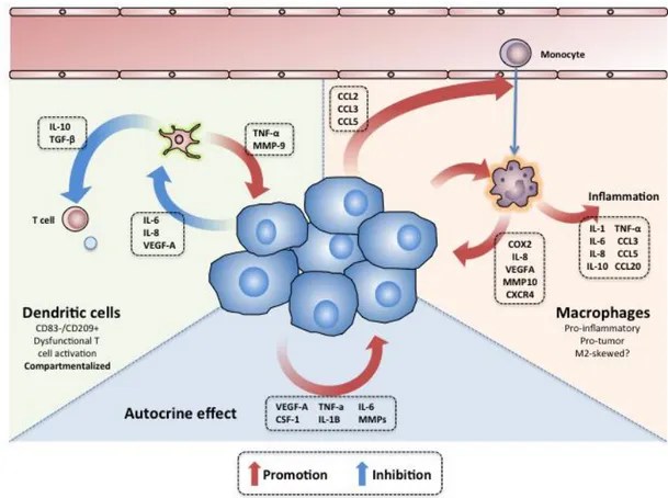
The mechanisms of tumour promotion associated with the RCC inflammatory cascade are diverse, and similar to those described in other types of cancer [discussed in the section “Cancer and Inflammation” (Page 13)]. Other mechanisms specifically described in RCC include: 1) the TNF-α and CSF-1 induction of tumour cell proliferation/migration (281) and Epithelial-Mesenchymal Transition (EMT) (277) (282); 2) the IL-1β-dependent up-regulation of metalloproteinases (283); 3) the IL-6 and IL-8 promotion of angiogenesis; and 4) the TGF-β-dependent stimulation of tumour cell migration and invasion (284). The autocrine effect of tumour-produced molecules in RCC is depicted in Figure 1.
Figure 1. The autocrine and paracrine effects of tumor secreted molecules in RCC microenvironment. Cartoon depicting the inter-talk between the tumour cells, dendritic cells and
macrophages in ccRCC. In the right, the tumour cells can express molecules that induce the infiltration with monocytes; once inside of the tumour, the monocytes/TAM can express a wide arrange of molecules that amplify the inflammatory cascade and promote tumour cell growth and invasion. In the left, several molecules produced by the tumour cells can also render the infiltrating DC tolerogenic, inhibitory and pro-tumoral. In the bottom, distinct cytokines secreted by the tumor cells can have an autocrine effect inducing cell proliferation and spreading.
Tumour associated macrophages
RCC tumour cells produce a wide range of molecules with monocyte-chemoattractive properties (e.g. CCL2, CCL3 and CCL5); not surprisingly, these tumours are often infiltrated with copious amounts of monocytes/TAM (285) (286) (285) (247) (287) (288) (289). In addition, several studies have described that the RCC microenvironment can prompt the differentiation of TAM into highly pro-inflammatory cells, characterized by the production of IL-1, IL-6, IL-8, IL-10, TNF-α, CCL2, CCL3, VEGFA and eicosanoids (290) (291) (292). The mechanisms skewing the RCC TAM into this phenotyope are probably related with the tumour cell expression of TNF, PTGS2, IL-6 and VEGF-A (293).
The overactive monocyte recruitment and activation in RCC turns into a vicious cycle that ultimately promotes tumour inflammation, growth and spreading. A recent work by Chittezhath et al. described the molecular profile of monocytes/TAM in RCC-bearing patients (293); overall, both
peripheral blood (PB) monocytes and TAM exhibited highly inflammatory characteristics (e.g. over-expression of TNF, IL1A, IL1B, IL6, CCL3, CCL5 and CCL20), produced tumour-promoting molecules (e.g. COX2, IL8, VEGFA, MMP10 and CXCR4) and induced angiogenesis (293). In other studies, and consistently with these findings, increased RCC TAM densities have been associated with higher plasma VEGF levels (294), increased tumour microvessels densities (295) (69) (296), presence of necrosis, advanced tumour stages (297) and poor prognosis (104) (298). Altogether these data provide strong evidence supporting the role of RCC monocytes and macrophages in tumour promotion and spreading.
RCC TAM are also capable of producing a series of immunomodulatory molecules that can hamper the cytotoxic anti-tumour immune response. In fact, some studies suggest that RCC TAM are skewed towards an M2-phenotype, characterized by the expression of CD163, interferon regulatory factor 4, fibronectin 1 and IL-10 (103) (104) (299). This phenotype could induce Treg expansion (292), in addition to PD-1 and TIM-3 expression on T lymphocytes (104).
The interaction between the TAM and tumour cells in RCC is depicted in Figure 1.
Myeloid derived suppressor cells
Myeloid derived suppressor cells (MDSC) represent a heterogeneous and complex population of immune cells. Although their function and phenotype has been mainly investigated in animal models, it is now well accepted that they infiltrate human tumours, and play an important role in hampering the T cell responses (300). Several mechanisms of immunoregulation by MDSC have been described, including the depletion of nutrients necessary for T cell function, the production of reactive oxygen species (ROS), the obstruction of T cell trafficking into lymph nodes and the induction of Treg (300). MDSCs originate from immature myeloid circulating cells, and their recruitment is induced by several molecules, including VEGF, TGF-β, GM-CSF, IL-6, IL-10, gangliosides and prostaglandins (300).
Despite the fact that most of the molecules that induce the accumulation of MDSC are abundant in the RCC microenvironment (301), very few studies have assessed the role of this population in the anti-tumour immune response inhibition in this pathology. Nevertheless, evidence suggesting that Sunitinib and other TKI inhibitors induce tumour regression by a mechanism related to the MDSC depletion (discussed below) highlights the possible relevance of this population in RCC pathogenesis. Currently, the only two described mechanisms of MDSC-induced immunosuppression in human RCC are the depletion of L-arginine (302) (303) (304) and the overproduction of ROS (305).