Université de Liège
Faculté de Médecine
Attentional deficits in major depression
A combined approach by clinical and
functional magnetic resonance imaging studies
Martin DESSEILLES
Centre de Recherches du Cyclotron et Service de Psychiatrie
Université et Centre Hospitalier Universitaire de Liège
Promoteurs : Professeurs Pierre MAQUET et Marc ANSSEAU
Thèse présentée en vue de l’obtention du grade de
Docteur en sciences médicales
Cover. Left panel: On the Threshold of Eternity by Vincent Willem van Gogh, 1890; right panel: illustration by Sophie Schwartz adapted from Desseilles, Balteau et al. 2009.
"Si la matière grise était rose, personne n'aurait plus d'idées noires." Pierre DAC, Pensées, 1972.
Université de Liège
Faculté de Médecine
Attentional deficits in major depression
A combined approach by clinical and
functional magnetic resonance imaging studies
Martin DESSEILLES
Centre de Recherches du Cyclotron et Service de Psychiatrie
Université et Centre Hospitalier Universitaire de Liège
Promoteurs : Professeurs Pierre MAQUET et Marc ANSSEAU
Thèse présentée en vue de l’obtention du grade de
Docteur en sciences médicales
Table of Content
Acknowledgements...7
List of publications ...10
Publications reported in the thesis ...10
Thesis ...10 Supplemental thesis ...10 Other publications ...11 Summary ...13 1. Theoretical introduction...16 1.1. Depression...16 1.1.1. Introduction...16
1.1.2. The spectrum of mood disorders...18
1.1.3. Signs and Symptoms of depression ...23
1.1.4. Stress axis and autonomic dysfunctions in depression ...26
1.1.5. Cognitive abnormalities in depression...30
1.1.6. Suicide ideations and attempts...34
1.1.7. Sleep and depression...38
1.1.8. Brain abnormalities in depression...44
1.1.9. Therapies in depression...51
1.1.10. Biological markers of depression...52
1.2. Attentional processes ...53
1.2.1. Attentional networks ...53
1.2.2. Bottom-up and top-down mechanisms ...53
1.2.3. Modular and hierarchical organization of central nervous system ...56
1.2.4. Effects of attention on perception ...59
1.2.5. “Indirect” consequences of depression on perceptual systems...60
2. Objective ...61
2.1. Why and how to explore attentional process in depression? ...61
2.2. Is there an abnormal neural filtering in patients with depression? ...61
2.3. Could a modulation of attentional load impact on mood? ...62
2.4. Exploration of the neurobiological basis of suicidality ...62
3. Methodological considerations about functional magnetic resonance imaging (fMRI) ...63
3.1. Statistical analysis of functional brain imaging data ...65
3.2. Spatial processing ...65
3.3. General linear model, design matrix and regressors...67
3.4. Fixed effects – Random effects...67
3.5. Inferences ...68
4. Reviews and experimental studies ...69
4.1. Introduction...69
4.1.1. Usefulness of neuroimaging in psychiatry...69
4.1.2. Function of attention and emotion in visual perception: implications for neurocognitive dysfunctions in depression. ...70
4.2. Functional magnetic resonance imaging studies...71
4.2.1. Abnormal neural filtering of irrelevant visual information in depression. ...71
5. Conclusions and perspectives ...74
5.1. Construction of an original attentional paradigm ...74
5.2. Contribution of our results to the pathophysiology of depression ...74
5.3. Perspectives in pathology: top-down dysfunction in mental illnesses...78
5.3.1. Top-down dysfunctions in hallucinations...78
5.3.2. Top-down dysfunctions in obsessive-compulsive disorders (OCD) ...79
5.4. Perspective in therapy ...83
5.4.1. Cognitive training in top-down regulation...83
5.4.2. Evaluation of treatment...83
5.5. Perspectives in methodology ...85
5.5.1. Integration with other biological measures...85
5.5.2. Voxel-based morphometry...85
5.5.3. Dynamic causal modelling...86
5.5.4. Independent component analysis and default network ...86
5.6. Concluding remarks ...89
6. References...90
7. Supplemental thesis ...109
7.1. Electroconvulsive therapy in depression ...109
7.1.1. The electroconvulsive therapy in 2008 ...109
7.1.2. Therapy for Depression in a Patient With an Intracranial Arachnoid Cyst ... ...110
7.2. A prominent role for amygdaloid complexes in the Variability in Heart Rate (VHR) during Rapid Eye Movement (REM) sleep relative to wakefulness. ...111
7.3. Neuroimaging insights into the pathophysiology of sleep disorders. ...112
7.4. Obsessive-compulsive disorders ...113
7.5. Aripiprazole diminishes cannabis use in schizophrenia. ...114
7.6. Achalasia may mimic anorexia nervosa, compulsive eating disorder, and obesity problems. ...115
Acknowledgements
This thesis is the result of more than four years of work. During all this time, I have been accompanied and supported by many persons for various aspects of my research, ranging from the development of protocols to the analysis and the writing of results, through to acquisitions of brain imaging in patients and healthy subjects. I wish here to express my sincere gratitude to all the people who conducted, assisted and supported me in my scientific project.
My first and warm thanks go to Pr Gustave MOONEN, head of the department of Neurology and dean of the faculty of Medicine, an outstanding teacher in physiology and in neurology, who gave me the opportunity to work in his neuroscience laboratory and later proposed me to join the Cyclotron Research Centre (CRC). There I was welcomed by Pr Pierre MAQUET, an exceptional fount of knowledge, who introduced me to the exciting field of neuroscience research using functional neuroimaging. I am greatly indebted to him for his continuous availability, insightful advices and scientific perfectionism. I am grateful to Dr Sophie SCHWARTZ, my second mentor working at the Laboratory for Neurology and Imaging of Cognition at the University of Geneva, without her this work would not have been possible. I also thank Pr André LUXEN, director of the CRC, for his continual support and encouragement, and Pr Philippe PEIGNEUX, now head of the Neuropsychology unit at the Free University of Brussels, who helped me through my first analyses of positron emission tomography functional brain imaging data in sleep and then with whom I had constructive discussions on sleep physiology.
I thank the members of the jury, Pr Adelin ALBERT, Eric CONSTANT, Gustave MOONEN (president), Jean-Jacques LEGROS, Marc ANSSEAU (promoter), Marc LARUELLE, Patrick PAPART (secretary), Pierre MAQUET (promoter), Vincent SEUTIN, for accepting to assess my thesis.
This work would not have been possible without the technical contribution of Dr Evelyne BALTEAU, a physicist, and Dr Christophe PHILLIPS and Christian DEGUELDRE, both engineers. I am also grateful to the other PhD students, who began working at the CRC a few years ago like me, for their precious help and for the
nice time spent with them: Dr Thien Thanh DANG VU, an invaluable friend for many years, with whom I started medicine and research, Dr Virginie STERPENICH, Dr Geneviève ALBOUY, Dr Gilles VANDEWALLE, Christina SCHMIDT, Dr Frédéric PETERS, Dr Mélanie BOLY, Maxime BONJEAN, Pierre ORBAN, and Dr Caroline SCHNAKERS. I am particularly grateful to Christel DEVUE from the Cognitive Science Department of the University of Liège. Working at the CRC has been a brilliant experience, which gave me the opportunity to meet exceptional post-doc colleagues and friends: Dr Manuel SCHABUS, Dr Annabelle DARSAUD, Dr Géraldine RAUCHS, Dr Steffen GAIS, Dr Gilberte TINGUELY. I am also grateful for working with fresh PhD students Pierre BOVEROUX, Marie-Aurélie BRUNO, Dorothée FEYERS, Ariane FORET, Yves LECLERCQ, Laura MASCETTI, Luca MATARAZZO and Audrey VANHAUDENHUYSE. I also thank other members of the CRC team with whom I shared interesting discussions on various occasions, Pr Eric SALMON, Dr Fabienne COLETTE, Dr Steven LAUREYS, Dr Gaëtan GARRAUX, Dr Christine BASTIN, and Dr Quentin NOIRHOMME. Finally, I thank secretaries Annick CLAES, Brigitte HERBILLON and Annette KONINGS for their constant assistance.
In parallel with the preparation of this thesis, I have pursued my residency in psychiatry at Liege University Hospital. I would like to thank Pr ANSSEAU, head of department of psychiatry, and Dr Sonia FUCHS, senior physicians of the department, for their enlightened guidance in the clinical practice as well as for their constant support for my research. I thank my colleagues and fellow residents during this residency: Cécile DEBABECHE, Alice MUSELLE, Romain PALLINCOURT, Vladimir ZELINKA, Geraldine WILDEMEERSCH, Laurent FARCY, Nicolas MASSART and Valérie BARBIER, not forgetting the paramedical staff and secretary Christiane GAYETOT and Cécile BROOZE.
I am grateful to new colleagues of the Laboratory for Neurology and Imaging of Cognition at the University of Geneva: Yann COJAN and Camille PIGUET.
I am particularly grateful to the Fonds National de la Recherche Scientifique (FNRS) of Belgium since this research has been mainly supported and funded by the FNRS (Grant 3.4516.05). Additional supports come from the research funds of the
University of Liège, the Queen Elisabeth Medical Foundation, and the Interuniversity Attraction Pole program.
I deeply thank all other people unfortunately not acknowledged here and who contributed to my personal development. In particular, my concluding thanks go to my parents, my family and specifically to my girlfriend for her love and patience during the PhD period.
List of publications
Publications reported in the thesis
Thesis
Muselle A et Desseilles M, Utilité de la neuroimagerie en psychiatrie. Acta Psychiatrica Belgica, 108(6), 2008, 1-9.
Desseilles M, Ansseau M, Maquet P, Schwartz S, Rôle de l’attention et de l’émotion dans la perception visuelle : implications pour les troubles neurocognitifs dans la dépression. Acta Psychiatrica Belgica, 108(4), 2008, 20-24.
Desseilles M, Balteau E, Sterpenich V, Dang-Vu TT, Darsaud A, Vandewalle G,
Albouy G, Salmon E, Peters F, Schmidt C, Schabus M, Gais S, Degueldre C, Phillips C, Luxen A, Ansseau M, Maquet P, Schwartz S. Abnormal neural filtering of
irrelevant visual information in depression. The Journal of Neuroscience, 29(5), 2009,
1395-403.
Desseilles M, Schwartz S, Dang-Vu TT, Sterpenich V, Darsaud A, Vandewalle G,
Albouy G, Schmidt C, Balteau E, Degueldre C, Phillips C, Luxen A, Ansseau M, Maquet P. Neurobiological Bases of Suicidality in Major Depression. (in preparation)
Supplemental thesis
Servais S, Ansseau M, Mikolajczak G, Desseilles M. L'électroconvulsivothérapie en
2008. Rev Med Liege,63(5-6), 2008, 404-410
Desseilles M, Thiry J-C, Monville J-F, Ansseau M and Makhinson M. Electroconvulsive Therapy for Depression in a Patient With an Intracranial Arachnoid Cyst. J ECT, 2009 (in press)
Desseilles M, Dang-Vu T, Laureys S, Peigneux P, Degueldre C, Phillips C, Maquet P. A prominent role for amygdaloid complexes in the Variability in Heart Rate (VHR) during Rapid Eye Movement (REM) sleep relative to wakefulness; Neuroimage, 32(3),
2006, 1008-1015
Desseilles M, Dang-Vu T, Schabus M, Sterpenich V, Maquet P, Schwartz S. Neuroimaging insights into the pathophysiology of sleep disorders. Sleep. 2008 Jun
1;31(6):777-94.
Desseilles M, Mikolajczak G, Devue C, Muselle A, Debabèche C, Les troubles obsessionnels-compulsifs. Acta Psychiatrica Belgica, 2008. 108(3), 1-9.
Desseilles M, Mathot F, Desseilles M. Aripiprazole diminishes cannabis use in schizophrenia. Journal of Neuropsychiatry and Clinical Neurosciences 2008
Winter;20(1):117-8.
Desseilles M, Fuchs S, Ansseau M, Lopez S, Vinckenbosh E, Andreoli A. “Achalasia may mimic anorexia nervosa, compulsive eating disorder, and obesity problems.” Psychosomatics 2006 May-Jun;47(3):270-1.
Other publications
Desseilles M, Dang-Vu TT and Maquet P. Functional neuroimaging in sleep, sleep
deprivation and sleep disorders; In Chokroverty S and Montagna P (Eds.) Handbook of Clinical Neurology (in press)
Desseilles M, Dang-Vu TT, Schwartz S, Peigneux P and Maquet P. Functional
Neuroimaging of Sleep and Sleep Disorders; In Chokroverty S (Ed.) Sleep Disorders Medicine (in press).
Desseilles M, Massart N, Farcy L. Les psychoses délirantes chroniques. Revue
Médicale de Liège, 2009 (in press)
Dang-Vu TT, Desseilles M, Peigneux P, Maquet P. A Role for Sleep in Brain
Plasticity; Pediatric Rehabilitation, 9(2), 2006, 98-118
Dang-Vu TT, Desseilles M, Petit D, Mazza S, Montplaisir J, Maquet P. Neuroimaging
in Sleep Medicine; Sleep Medicine, 8, 2007, 350-373
Dang-Vu TT, Desseilles M, Laureys S, Degueldre C, Perrin F, Philips C, Maquet P, Peigneux P. Cerebral correlates of delta waves during non-REM sleep revisited; Neuroimage, 28 (1), 2005, 14-21
Schabus M, Dang-Vu TT, Albouy G, Balteau E, Boly M, Carrier J, Darsaud A, Degueldre C, Desseilles M, Gais S, Phillips C, Rauchs G, Schnakers C, Sterpenich V, Vandewalle G, Luxen A, Maquet P. Hemodynamic cerebral correlates of sleep
spindles during human nonrapid eye movement sleep; Proceedings of the National
Academy of Sciences of the United States of America, 104(32), 2007, 13164-9 Dang-Vu TT, Schabus M, Desseilles M, Albouy G, Boly M, Darsaud A, Gais S, Rauchs G, Sterpenich V, Vandewalle G, Carrier J, Moonen G, Balteau E, Degueldre C, Luxen A, Phillips C, Maquet P. Spontaneous Neural Activity during Human Slow
Wave Sleep; Proceedings of the National Academy of Sciences of the United States of
America, 105(39), 2008, 15160-5
Dang-Vu TT, Desseilles M, Albouy G, Darsaud A, Gais S, Rauchs G, Schabus M, Sterpenich V, Vandewalle G, Schwartz S, Maquet P. Dreaming: A Neuroimaging
Dang-Vu TT, Schabus M, Desseilles M, Schwartz S, Maquet P. Neuroimaging of
REM sleep and dreaming; In Patrick McNamara and Deirdre Barrett (Eds.) The New Science of Dreaming, Praeger Publishers, Greenwood Press, 2007, Vol. 1, 95-113
Dang-Vu TT, Desseilles M, Peigneux P, Laureys S and Maquet P. Sleep – Definition
and Description: PET activation patterns; In Larry R. Squire (Ed.) New Encyclopaedia of Neuroscience, Academic Press, Oxford (UK), 2009, Vol. 8,
955-961.
Gais S, Albouy G, Boly M, Dang-Vu TT, Darsaud A, Desseilles M, Rauchs G, Schabus M, Sterpenich V, Vandewalle G, Maquet P, Peigneux P. Sleep transforms the
cerebral trace of declarative memories; Proceedings of the National Academy of
Sciences of the United States of America, 104(47), 2007, 18778-83.
Sterpenich V, Albouy G, Boly M, Vandewalle G, Darsaud A, Balteau E, Dang-Vu TT, Desseilles M, D’Argembeau A, Gais S, Rauchs G, Schabus M, Degueldre C, Luxen A, Collette F, Maquet P. Sleep-Related Hippocampo-Cortical Interplay during
Emotional Memory Recollection; PloS Biology, 5(11), 2007, e282.
Vandewalle G, Balteau E, Phillips C, Degueldre C, Moreau V, Sterpenich V, Albouy G, Darsaud A, Desseilles M, Dang-Vu TT, Peigneux P, Luxen A, Dijk DJ, Maquet P.
Daytime light exposure dynamically enhances brain responses; Current Biology,
16(16), 2006, 1616- 21.
Maquet P, Ruby P, Maudoux A, Albouy G, Sterpenich V, Dang-Vu T, Desseilles M, Boly M, Perrin F, Peigneux P, Laureys S. Human cognition during REM sleep and
the activity profile within frontal and parietal cortices. A reappraisal of functional neuroimaging data; Progress in Brain Research, 150, 2005, 219-27.
Maquet P, Sterpenich V, Albouy G, Dang-Vu T, Desseilles M, Boly M, Ruby P, Laureys S, Peigneux P. Brain imaging on passing to sleep; In Parmeggiani and Vellutti (Ed.) The Physiological Nature of Sleep. Imperial College Press, London (UK), 2005, 123-137.
Maquet P, Ruby P, Schwartz S, Laureys S, Albouy G, Dang-Vu T, Desseilles M, Boly M, Peigneux P. Regional organisation of brain activity during paradoxical sleep
(PS); Archives Italiennes de Biologie 142 (4), 2004, 413-9
Maquet P, Peigneux P, Laureys S, Boly M, Dang-Vu T, Desseilles M and Cleeremans A. Memory processing during human sleep as assessed by functional neuroimaging; Revue Neurologique, 159 (11 suppl), 2003, 6S27-6S29.
Maquet P, Laureys S, Perrin F, Ruby P, Melchior G, Boly M, Dang-Vu T, Desseilles M and Peigneux P. Festina Lente: Evidences for fast and slow learning processes and
a role forsleep in human motor skill learning; Learning and Memory, 10 (4), 2003,
237-239.
Maquet P, Peigneux P, Laureys S, Desseilles M, Boly M, Dang-Vu T. Off-line
processing of memory traces during human sleep; Sleep and Biological Rhythms, 1,
Summary
The major depressive disorder (MDD) is a frequent, chronic and debilitating illness characterized by affective, cognitive, motor and autonomic symptoms (DSM-IV-TR) (American Psychiatric Association 2000). Symptoms cause major distress to patients and their families. Invalidity and morbidity, including suicide, associated with this illness constitute important social and economic problems. According to the World Health Organization, depression is the 4th cause of disability and projections predict that depression will be the 2nd cause of disability in 2020 (Murray and Lopez 1997).
However, the pathophysiology of MDD is not yet elucidated. Here, we will use functional neuroimaging methods to study the cerebral mechanisms involved in MDD. We propose that a combined clinical and functional brain imaging approach to MDD will contribute to our understanding of this psychiatric disease, and will open new perspectives on cognitive and affective deficits associated with MDD.
From a cognitive point of view, MDD patients have impairments encompassing several domains of executive, memory, and attentional functioning (Austin, Ross et al. 1992; Beats, Sahakian et al. 1996; Purcell, Maruff et al. 1997; Murphy, Sahakian et al. 1999; Porter, Gallagher et al. 2003). In particular they have deficits for complex tasks including attention and short-term memory (Williams, Hagerty et al. 2000).
In MDD, functional brain imaging at rest showed impairments in polymodal associative cortices (prefrontal and parietal) and in limbic/paralimbic regions (including anterior cingulate cortex, amygdala, hippocampus) (Cummings 1993; Drevets, Price et al. 1997; Goodwin 1997; Drevets 1998; Rogers, Bradshaw et al. 1998; Price 1999; Teasdale, Howard et al. 1999; Drevets 2000; Davidson, Lewis et al. 2002; Drevets 2003; Lacerda, Nicoletti et al. 2003; Phillips, Drevets et al. 2003a; Phillips, Drevets et al. 2003b). At rest, MDD patients typically show a diminution of glucose consumption in prefrontal and parietal areas (Drevets, Price et al. 1997). Functional neuroimaging studies also revealed a deficit of activation in these areas when MDD patients performed cognitive tasks, as compared to normal controls. For instance, there is a significant correlation between frontal hypoperfusion and
cognitive alterations in MDD, in particular for tasks requiring a mental effort (Rogers, Bradshaw et al. 1998).
The brain is organized in a modular, hierarchical and recurrent manner. Firstly, modules at low hierarchical levels code for simple sensorial attributes (e.g. orientation of visual stimulus) and receive information from modules at higher hierarchical levels, which themselves code for progressively more complex attributes (e.g. faces). Secondly, at each hierarchical level of information processing, neuronal activity is modulated by the information provided by inferior and superior levels, forming a system of recurrent loops (Friston and Price 2001; Lee and Mumford 2003). The most extensively studied example concerns how attention can modulate the activity in low-levels sensory cortices (Felleman and Van Essen 1991).
In the context of MDD, this organization implies that dysfunctions at higher cortical levels (associative polymodal) may result in major changes at lower-level of information processing, including primary sensory cortices.
Using attention as a paradigmatic example of such “top-down” effects, we initially proposed and constructed an original attentional paradigm to manipulate and explore these top-down effects in healthy subjects and patients.
Using this attentional paradigm, we measured cerebral activity in 14 MDD patients and 14 healthy controls using functional magnetic resonance imaging (fMRI) while they performed attentional tasks. Our research project aimed at a better understanding of how affective and cognitive dysfunctions interfered with normal functioning in early cortical areas in MDD patients.
Finally, using the same paradigm, we conducted whole-brain correlation with clinical scores derived from the Hamilton Rating Scale for Depression in order to explore the neurobiological bases of suicidality in major depression.
To introduce the experimental studies described in the second part of this manuscript, we will first review the current knowledge about depression, including signs and symptoms of depression, stress axis and autonomic dysfunctions, cognitive abnormalities, suicide ideations and attempts, sleep impairments, brain abnormalities.
We will then propose an overview of human attention in particular how attentional processes interact with other cognitive and perceptual processes.
1. Theoretical introduction
1.1. Depression
1.1.1. Introduction
Major depressive disorder (MDD) or unipolar depression is a psychic disorder which is typically characterized by a pervasive low mood, low self-esteem, and loss of interest or pleasure in normally enjoyable activities (American Psychiatric Association 2000).
The term MDD was selected in 1980 by the American Psychiatric Association (APA) for the third version of the Diagnostic and Statistical Manual of Mental Disorders (DSM-III) and has become widely used since. In the DSM-III, MDD refers to a symptom cluster classified under mood disorders
The term “depression” is often overworked as it is used to describe the disorder (i.e. major depressive episode) and the symptom (i.e. depressed mood). This is the reason why a more specific terminology is favored by clinical and basic neuroscientists.
Major depression is a disabling condition which adversely affects a person's family, work or school life, sleeping and eating habits, and general health. In the United States around 3.4% of people with major depression commit suicide, and up to 60% of all people who commit suicide have depression or another mood disorder (Mann 2003).
As often in psychiatric practice, the diagnosis of MDD is based on the patient's self-reported feelings and behavior, observations by relatives or friends, and a mental status exam by a specialist.
Even if there is no specific laboratory test for major depression, physicians usually request tests for physical conditions that may cause similar symptoms (differential diagnosis) (Halbreich 2006).
While this illness can be present at any age, time of disease onset is most commonly seen between the ages of 30 and 40 years, with a later peak between 50 and 60 years.
Major depression occurs about twice as frequently in women as in men, and men are at higher risk for suicide (Sadock and Sadock 2005).
The majority of the depressed patients are treated in the community with antidepressants, psychotherapy or counseling (Sadock and Sadock 2005). Psychological treatments may be based on psychoanalysis (Blatt 1998), interpersonal communication (Dobson 2008), learning theory (Abrams 1964; Lazarus 1968), and cognitive schemata structuring personality (Beck 2008). In cases with associated self-neglect or a significant risk of harm to self or others, hospitalization may be necessary (Sadock and Sadock 2005). A minority of patients are treated either with electroconvulsive therapy (ECT), under a short-acting general anaesthetic (Pagnin, de Queiroz et al. 2004) or with transcranial magnetic stimulation (Loo and Mitchell 2005), deep brain stimulation (Glannon 2008) or surgery (Juckel, Uhl et al. 2009).
The time course of the disorder varies widely across patients, from a lifelong disorder with recurrent major depressive episodes to a single episode lasting a few months (Benazzi 2006).
Importantly, depressed individuals have shorter life expectancies than those without depression. This is due in part to being more susceptible to serious medical conditions (Carney, Blumenthal et al. 2003; Carney, Blumenthal et al. 2005; Steptoe 2006; Janszky, Ahlbom et al. 2007; Steptoe 2007).
Over the centuries, the understanding of the nature and causes of depression has evolved. Psychological (Beck 2008), biological (Nestler, Barrot et al. 2002; Mann 2005; Belmaker and Agam 2008), social (Klerman and Weissman 1978; Fullilove 2002) and evolutionary (Nesse 1999; Nettle 2004; Hendrie and Pickles 2009) causes have been proposed. An important role for monoamines (serotonin, norepinephrine, and dopamine) in depression has also been suggested, because the efficiency of most antidepressants is mediated by an increase in monoamines levels (Mann 2005; Belmaker and Agam 2008). Nevertheless, many aspects of depression are still now not fully understood.
1.1.2. The spectrum of mood disorders
The term “mood disorder” refers to a cluster of different conditions in the Diagnostic and Statistical Manual of Mental Disorders 4th Revision (DSM-IV-TR) (American Psychiatric Association 2000) classification system. In that cluster, a modification in the person's emotional mood is thought to be the major underlying aspect. The same nosological cluster is known as mood / affective disorders in the chapter V (Mental and behavioural disorders) of the International Statistical Classification of Diseases and Related Health Problems 10th Revision (ICD-10) (World Health Organization 2005).
Affective disorder was first proposed as a category including all emotional disorders. Mood disorder replaced then this term to refer more specifically to the underlying emotional state, whereas affective disorder now mainly refers to the external expression observed by others.
At present, there is a broad recognition of a division between two groups based on whether the patient presented at some point with a manic or hypomanic episode: (i) depressive disorders including major depressive disorder also called major depression, and (ii) bipolar disorder, formerly called "manic depression" and characterized by intermittent periods of manic and depressed episodes, sometimes with mixed states (both states simultaneously).
Depressive disorders correspond a range of clinical presentations such as major depressive disorder (MDD), atypical depression, melancholic depression, psychotic depression, catatonic depression, postpartum depression, seasonal affective disorder and dysthymia (American Psychiatric Association 2000).
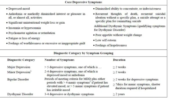
Figure 1 : Depressive symptoms and diagnostic criteria for depressive disorders (American Psychiatric Association 2000). 1 Minor depression is not yet a full categorical diagnosis in the DSM-IV but is included as a research diagnostic category. 2 Manic symptoms include elevated or irritable mood, inflated self-esteem or grandiosity, decreased need for sleep, increased talking or pressured speech, flight of ideas or subjective experience that thoughts are racing, distractibility, increase in goal-directed activity or psychomotor agitation, and excessive involvement in pleasurable activities that may have a high potential for painful consequences.
• According to the DSM-IV-TR, major depressive episode (MDE) happens when (A) Five (or more) of the following symptoms have been present during the same 2-week period and represent a change from previous functioning; at least one of the symptoms is either (1) depressed mood or (2) loss of interest or pleasure. Note: Do not include symptoms that are clearly due to a general medical condition, or mood-incongruent delusions or hallucinations. The nine symptoms are: (1) depressed mood most of the day, nearly every day, as indicated by either subjective report (e.g., feels sad or empty) or observation made by others (e.g., appears tearful). Note: In children and adolescents, can be irritable mood; (2) markedly diminished interest or pleasure in all, or almost all, activities most of the day, nearly every day (as indicated by either subjective account or observation made by others); (3) significant weight loss when not dieting or weight gain (e.g., a change of more than 5% of body weight in a month), or decrease or increase in appetite nearly every day. Note: In children, consider failure to make expected weight gains. (4) insomnia or hypersomnia nearly every day; (5) psychomotor agitation or retardation nearly every day (observable by others, not merely subjective feelings of restlessness or being slowed down); (6)
fatigue or loss of energy nearly every day; (7) feelings of worthlessness or excessive or inappropriate guilt (which may be delusional) nearly every day (not merely self-reproach or guilt about being sick); (8) diminished ability to think or concentrate, or indecisiveness, nearly every day (either by subjective account or as observed by others); (9) recurrent thoughts of death (not just fear of dying), recurrent suicidal ideation without a specific plan, or a suicide attempt or a specific plan for committing suicide. (B) The symptoms do not meet criteria for a Mixed Episode. (C) The symptoms cause clinically significant distress or impairment in social, occupational, or other important areas of functioning. (D) The symptoms are not due to the direct physiological effects of a substance (e.g., a drug of abuse, a medication) or a general medical condition (e.g., hypothyroidism). (E) The symptoms are not better accounted for by bereavement, i.e., after the loss of a loved one, the symptoms persist for longer than 2 months or are characterized by marked functional impairment, morbid preoccupation with worthlessness, suicidal ideation, psychotic symptoms, or psychomotor retardation (Figure 1).
• The diagnosis of Major depressive disorder (MDD, or major depression or unipolar depression) is considered when a person has two or more major depressive episodes. Unipolar depression describes depression without periods of mania because the mood remains at one pole / emotional state.
• Atypical depression is characterized by mood reactivity and positivity, significant weight gain or increased appetite, hypersomnia or somnolence, a sensation of heaviness in limbs known as leaden paralysis, and significant social impairment as a consequence of hypersensitivity to perceived interpersonal rejection.
• Melancholic depression is characterized by anhedonia (a loss of pleasure) in most or all activities, a failure of reactivity to pleasurable stimuli, a quality of depressed mood more pronounced than that of grief or loss, a worsening of symptoms in the morning hours, early morning waking, psychomotor retardation, excessive weight loss, or excessive guilt.
• Psychotic depression is the term for a major depressive episode, particularly of melancholic nature, where the patient experiences psychotic symptoms such as
delusions or hallucinations. These are often mood-congruent (i.e. content coincident with depressive themes such as ruin or death).
• Catatonic depression is a rare and severe form of major depression involving disturbances of motor behavior as a core symptom. The person is mute, stuporous, and either immobile or exhibits purposeless or bizarre movements. Catatonic symptoms also occur in schizophrenia, manic episode, or in neuroleptic malignant syndrome.
• Postpartum depression refers to the intense, sustained, and disabling depression experienced by women after giving birth. It has incidence rate of 10–15%, typically sets in within three months of labor, and lasts as long as three months.
• Seasonal affective disorder happens when at least two depressive episodes happen during the autumn or winter, and resolve in spring with no other episodes at other times over a two-year period or longer.
• Dysthymia, a chronic, milder mood disturbance is diagnosed when a person reports a low mood almost daily over a span of at least two years. Symptoms are not as severe as those for major depression, although people with dysthymia are vulnerable to double depression, which is a secondary episode of major depression.
• Depressive Disorder Not Otherwise Specified (DD-NOS) correspond to depressive disorders that are impairing but do not fit any of the officially DSM specified diagnoses such as Recurrent Brief Depression, and Minor Depressive Disorder.
• Recurrent brief depression (RBD) happens in people who have depressive episodes about once per month, with individual episodes lasting less than two weeks and typically less than 2–3 days. It requires that several episodes occur over the span of at least one year and, for female patients, independently of the menstrual cycle. People with major depression can develop RBD, and conversely. In addition, both illnesses have similar risks. Thus this diagnosis is distinguished from MDD primarily by differences in duration.
• Minor depression refers to a depression that does not meet full criteria for major depression but in which at least two symptoms are present for two weeks.
Bipolar disorders include subtypes such as: bipolar I, II and cyclothymia.
• Bipolar disorder is described by alternating periods of (hypo)mania and depression. For both Bipolar I and II, there are a number of specifiers that indicate the presentation and course of the disorder, including “rapid cycling”, “mixed states”, and “psychotic symptoms”.
• Bipolar I is characterized by at least one manic or mixed episodes with or without major depressive episodes.
• Bipolar II is characterized by recurrent intermittent hypomanic and depressive episodes.
• Cyclothymia is a milder form of bipolar disorder, characterized by recurrent hypomanic and dysthymic episodes, without any more severe ones occurring.
1.1.3. Signs and Symptoms of depression
Major depression is a severe mental illness that affects several domains including personal (familial) and / or professional (work or school) life, major physiological functions such as sleep and eating habits, sexual drive, motor abilities, and cognitive functions. It is a chronic and highly debilitating disease as showed by the study of Hays and collaborators (Hays, Wells et al. 1995). They have conducted a 2-year observational study of 1790 adult outpatients with several mental and physical disorders including depression, diabetes, hypertension, recent myocardial infarction, and/or congestive heart failure. They showed that depressed patients have significant and long-lasting impairments in multiple areas of functioning and well-being that equal or exceed those of patients with chronic medical illnesses (Hays, Wells et al. 1995). Overall, general (mental and physic) health of those patients is highly impaired.
People suffering from major depressive episode (MDE) usually exhibit a low mood diffusing into all facets of life. An inability to experience pleasure in previously enjoyable activities is frequent. They may ruminate over thoughts, feelings of worthlessness, inappropriate guilt or regret, helplessness or hopelessness (American Psychiatric Association 2000).
Additional symptoms include reduced concentration, poor memory, withdrawal from social situations, and abandonment of activities, reduced sexual drive, and negative thoughts about death or suicide. Sleep disorders are common, with a typical pattern of insomnia when patient wakes very early and is unable to get back to sleep. Hypersomnia, or oversleeping, may be present but are less common. Diurnal fatigue is often present in both cases (insomnia or hypersomnia) (American Psychiatric Association 2000). Appetite typically decreases, with consequential weight loss. Nevertheless increased appetite, with resulting weight gain, occasionally occurs.
Physical complaints may be the first presenting problem in several population and cultures such as in Zimbabwe, a developing country, where fatigue and headaches are the most common presentation of depression (Patel, Abas et al. 2001). In fact, nearly all depressions present with various somatic complaints in addition to cognitive and affective ones. About one half of all depressions seen by primary care physicians
initially present either mostly or exclusively with somatic symptoms and are called “masked depression” because affective symptoms are masked by somatic ones. Unfortunately, many of these masked depressions are not recognized or are misdiagnosed and consequently mistreated. Fisch hypothesized that the proportion of depressions that are masked is positively correlated to the tendency of the patients to “somatize” and negatively correlated to the ability of the medical practitioner to recognize depressions that hide behind somatic complaints (Fisch 1987).
Psychomotor symptoms are an important diagnostic clue (Schrijvers, Hulstijn et al. 2008). People close to the patient may observe that his / her behavior is either agitated or lethargic. Theoretically, a mismatch between motor activity and psychic activity may occur. For instance psychic agitation may be concomitant with motor stupor. In addition, impulsive-aggressive behaviors may be present and such behaviors have been shown to increase suicidal risk (McGirr and Turecki 2007). Interestingly, a serotonin deficiency has been implicated in a broad range of psychiatric conditions, including depressive and anxiety disorders, suicidal and aggressive behaviors as well as impulsive control disorders. Once again affective symptoms may be masked by impulsive ones. This raises the question about what should be considered a primary cause in this form of depression: the underlying disturbance of impulsivity, or the mood disorder (Lopez-Ibor 1992).
Patients with depression may experience thought disorders such as crowded thoughts or ruminations. Crowded thoughts may be conceptualized as a pathological thought process characterized by the occurrence of too many thoughts that co-exist almost simultaneously in consciousness, and that give to the subject a sense of constant and unpleasant agitation in his/her own thinking (Koukopoulos and Koukopoulos 1999). Ruminations are generally defined as “a constant preoccupation such as thinking about one single idea or theme” (Sadock and Sadock 2005). When depressed people ruminate, their thoughts are usually focused on the causes, meanings and consequences of depressive symptoms (Nolen-Hoeksema 1991). They have repetitive thoughts concerning their present distress and the circumstances surrounding their sadness (Conway, Csank et al. 2000). Depressive rumination is intimately associated to depressive mood (Papageorgiou 2004). While depressive mood might occur in the absence of ruminations (the subject is feeling permanent emotion of sadness but
his/her mind is so inhibited that he/she is unable to think), depressive ruminations are always associated with depressive mood (the subject is feeling sadness and emotional suffering while he/she is repetitively thinking about the circumstances of his sadness). In depressive ruminations, people report repetitive thoughts about a single, or sometimes a few themes, but they may not report being overwhelmed by numerous ideas dealing with possibly many themes. Moreover, in crowded thoughts patients are not able to control the stream of their thoughts and focus on any single one, while in rumination on the other hand they seem not able to change voluntarily from one topic to another one.
Within the sphere of affective disorders, subtypes of depression will have various presentations. For instance, reactive depression occurs as a result of an identifiable precipitating stress and symptoms include initial insomnia, anxiety, emotional lability, and multiple somatic complaints. Melancholic patients appear profoundly sad, disheveled and malnourished, and slowed in all aspects. Others symptoms according to DSM-IV-TR include diurnal variation of affective symptoms where depression is worse in the morning, early morning wakening, severe psychomotor retardation and severe anhedonia. Symptoms in depression occurring in pregnancy or in postpartum are the same as for major depressive episodes.
Depressed children present more often than adults symptoms as agitation, anxiety, somatic complaints, sad appearance, and even, mood-congruent hallucinations, compared to adults. Adolescent may present delinquency aggressive behavior, substance abuse, rejection hypersensitivity in addition to overeating, oversleeping, poor hygiene, and restlessness (Carlson and Kashani 1988; Ryan and Redding 2004).
Older depressed persons may have cognitive symptoms of recent onset, such as
forgetfulness, and a more noticeable slowing of movements. In severe cases, depressed people may have symptoms of psychosis such as delusions or, less commonly, hallucinations, usually of an unpleasant nature (Koenig 1991; Meyers 1995).
1.1.4. Stress axis and autonomic dysfunctions in depression
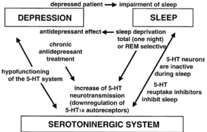
There is a large body of evidence showing that depression is associated with disturbances of hypothalamic-pituitary-adrenocortical (HPA) axis function including the metabolism of cortisol and autonomic function (Steptoe 2006; Pariante and Lightman 2008).
HPA axis is considered to be the “final common pathway” for a considerable part of the depressive symptomatology. A major part of the genetic and environmental risk factors for major depressive disorder appear to correlate with increased HPA-axis activity in adulthood. For instance, child abuse or early maternal separation constitute risk factors for later depression and are accompanied by HPA-axis hyperactivity (Swaab, Bao et al. 2005; Tarullo and Gunnar 2006). Interestingly, a spontaneous remission, the use of antidepressants or electroconvulsive therapy in animal models of depression or in patients with major depression episode is linked with a normalization of the HPA-axis function (Nemeroff 1996).
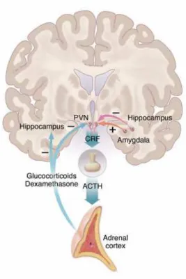
The secretion of adrenocorticotrophic hormone-releasing factor (CRF) and vasopressin (AVP) from the paraventricular nucleus (PVN) of the hypothalamus, through hypophyseal portal vessels, activates the secretion of adrenocorticotrophic hormone (ACTH) from the pituitary gland, which finally causes the secretion of the glucocorticoids from the adrenal cortex (i.e. cortisol in humans and corticosterone in rodents) (Figure 2). These glucocorticoids are responsible for feedback inhibition both on CRF and AVP from the hypothalamus and directly on secretion of ACTH from pituitary corticotropes where they interact with their receptors. In addition, the HPA axis regulates bodily peripheral functions including metabolism and immunity and has deep effects on the brain. For instance, glucocorticoids can regulate neuronal survival, cause neurogenesis, modulate the size of the hippocampus, control the acquisition of new memories and organize the emotional appraisal of events (Herbert, Goodyer et al. 2006). Thus, HPA axis activities play roles at the interface between brain functioning and stress and has been found abnormal in several psychiatric disorders, and in particular in major depression.
HPA dysregulation is observed in approximately 50 % of patients with unipolar depression and it may contribute to the association between depressive symptoms and
many types of physical disorders including, for instance, diabetes, obesity, heart disease, chronic fatigue and cancer (Steptoe 2006). These depressed patients have been found to have increased levels of cortisol in the saliva, plasma and urine, and increased size and activity of the pituitary and adrenal glands (Nemeroff and Vale 2005). These changes in HPA activity may be different depending on the subtypes of depression (Stewart, Quitkin et al. 2005).
On the other hand, oxytocin (OXT) inhibits ACTH release providing an example of the various “ying-yang” actions of AVP and OXT (Legros 2001; Neumann 2008). Moreover, activation of OXT neurons has been related to decreased appetite and weight loss in depression, and particularly in melancholic subtype of depression where loss of weight is one of the key characteristics due to the central effects of this neuropeptide as a satiety hormone (Gimpl and Fahrenholz 2001; Meynen, Unmehopa et al. 2007).
Figure 2 : Regulation of the HPA axis CRF-containing parvocellular neurons of the PVN of the hypothalamus integrate information relevant to stress (Nestler, Barrot et al. 2002). Prominent neural inputs include among other (i) excitatory afferents from the amygdala, (ii) inhibitory afferents from the hippocampus, and (iii) direct and indirect inputs from ascending monoamine pathways including the norepinephrine (from the locus coeruleus or LC) and the serotonin (from the dorsal raphé or DR) (not shown).
In addition to HPA axis function, the paraventricular nucleus (PVN) of the hypothalamus is involved in autonomic nervous system (ANS) regulation. It influences brainstem autonomic control nuclei. The hypothalamus in turn receives inputs from the limbic system, particularly from the central nucleus of the amygdala (the major output way from the amygdala), which operates in tight association with the prefrontal neocortex in order to integrate affective / emotional and motivational behaviours. It has been suggested that the ANS dysregulation underpins the increased risk of heart disease in depressed patients.
Several aspects of the ANS activity have been explored, including heart-rate levels and autonomic control processes governing baroreceptor reflex sensitivity and cardiac sympathovagal balance. This latter aspect has been assessed either with simple temporal measures, or with more complex power spectrum analysis. Heart-rate-variability (HRV) is typically computed by spectral methods. In spectrum analysis, high-frequency power is thought to reflect parasympathetic or vagal tone, while low-frequency power probably reflects sympathovagal balance and baroreceptor receptor reflex modulation of heart rate. Low level of heart-rate variability and low parasympathetic tone predict future cardiac heart disease in apparently healthy individuals (Steptoe 2006).
Comparisons between depressed patients and healthy controls have shown increased sympathetic nervous system activity in patients, but methodological issues have to be pointed out since controls for smoking, body mass and physical activity, altering neuroendocrine function, are often lacking. Sympathetic activation and reduced vagal tone are also known to reduce the threshold for cardiac ventricular fibrillation and to stimulate ventricular arrhythmias. In addition, if there is large evidence that resting heart rate is higher in depressed patients than healthy individuals, data concerning HRV and sympathovagal balance are more mixed and this mechanism has become increasingly difficult to study in clinical sample because of the large use of beta-blockers. Nevertheless, depression was associated with alteration of cardiac autonomic tone towards decreased parasympathetic activity and an increased sympathetic activity (Udupa, Sathyaprabha et al. 2007).
Major depression is a risk factor for medical morbidity and mortality in patients with coronary heart disease (CHD). In addition to elevated levels of plasma catecholamines and elevated heart rate, dysregulation of the ANS as been shown through low heart rate variability, exaggerated heart rate responses to physical stressors, high variability
in ventricular repolarization, and low baroreceptor sensitivity. Interestingly, all of these indicators have been linked to increased risks of mortality and cardiac morbidity in patients with CHD (Carney, Freedland et al. 2005).
Moreover, patients suffering from depression show a two to fourfold increase in sudden death and a sevenfold increase in ventricular arrhythmia (Carney, Freedland et al. 2005). These cardiac risks are hypothesized to result partly from elevated noradrenergic and sympathetic autonomic function, together with reduced parasympathetic tone on the heart rate (Carney, Freedland et al. 2005).
Various autonomic dysfunctions occur according to the subtype of depression (Rechlin, Weis et al. 1994). For instance, patients with reactive depression did not show any differences in auconomic function when they were compared with a control group. Patients with major depression had lower values of the high-frequency peak of spectral analysis than in the other depression subgroups, indicating decreased parasympathetic activity.
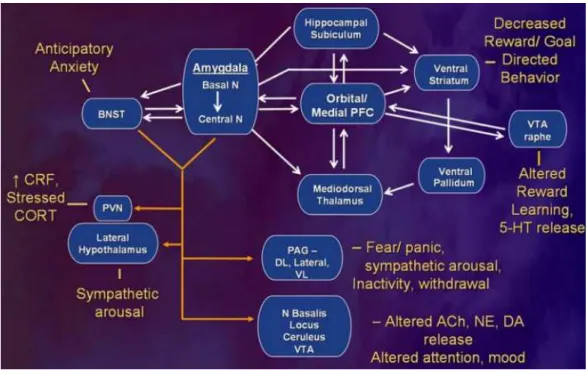
Given the interconnections between autonomic functions and depression, it is likely that a common neurobiological dysfunction contributes to both depression and cardiac autonomic changes in the illness. Indeed, the medial prefrontal cortex (mPFC) and associated limbic structures (including amygdala) give forebrain inflection over visceral control structures in the hypothalamus and brainstem. Dysfunction in this network can account for the neuroendocrine impairment and disturbances in ANS regulation associated with MDD (Drevets, Price et al. 2008). Firstly, amygdala stimulation of the locus ceruleus, lateral hypothalamus and periaqueductal gray matter (PAG) increases sympathetic autonomic arousal in rodents (Gold and Chrousos 2002; LeDoux 2003). Secondly, the parasympathetic tone on the heart-rate is partly regulated by projections from the infralimbic cortex (which putatively forms the posterior segment of the human subgenual anterior cingulate cortex) to the nucleus of the tractus solitarius of the vagus nerve, and structural lesions of this area reduce the parasympathetic tone on the heart in rodents (Frysztak and Neafsey 1994). The combined effect of the hyperactivity within the amygdala and reduced infralimbic cortex function could account for the increased sympathetic tone on the heart rate seen in patients with depression (Carney, Freedland et al. 2005).
1.1.5. Cognitive abnormalities in depression
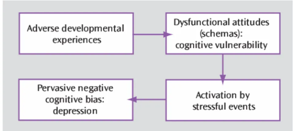
Several cognitive models aimed at describing mechanisms of depression. Beck’s
cognitive model proposes that unpleasant events during childhood lead to the
development of negative automatic cognitive schemata, activated by stressors later in life (Beck, Rush et al. 1979; Wright and Beck 1983; Beck 2008). These schemata drive cognitive distortions that contribute to the maintenance of negativity directed at the self (i.e. self-focused ruminations), world and future (Beck’s negative triad) (Figure 3). The learned helplessness model proposes that adverse experiences lead to subjects accepting that their actions cannot modulate outcomes, i.e. a state of helplessness, which impedes motivation and cognitive learning processes (Seligman 1972; Miller and Seligman 1975; Miller, Seligman et al. 1975; Klein, Fencil-Morse et al. 1976; Miller and Seligman 1976; Seligman 1978; Nolen-Hoeksema, Girgus et al. 1986). The Seligman’s attribution model proposes that patients suffering from depression attribute the causes of perceived failures inappropriately to internal, global, and stable factors (Seligman, Abramson et al. 1979; Raps, Peterson et al. 1982; Alloy, Peterson et al. 1984).
Figure 3 : Developmental model of depression based on vulnerability diathesis and stressful life events, from (Beck 2008).
Cognitive distortions are multiple (Beck, Rush et al. 1979; Kleftaras 2004): (i)
All-or-Nothing Thinking: This type of thinking is characterized by absolute terms like always, never, and forever. However, few situations are ever this absolute and they
present generally more nuances. (ii) Overgeneralization: taking an isolated case or cases and assuming that all others are the same. (iii) Mental Filter: mentally singling out only the adverse events in the life and overlooking the positive. (iv) Disqualifying
the Positive: taking the positive in a situation and turning it into a negative. (v) Jumping to Conclusions: expecting the worst and beginning to prepare early for the
disappointment. (vi) Magnification and Minimization: looking at all the positive through the wrong end of the telescope and the negative through the other end. (vii)
Emotional Reasoning: basing the assessment of the situation on how it makes the
subject feel and not how it really is. (viii) Should Statements: thinking things should be a certain way even if they aren't. (ix) Labeling and Mislabeling: labelling himself / herself as bad, lazy and hopeless. (x) Personalization: taking all the responsibility for how (bad) another person is doing in life or how horrible is a situation in the world.
Psychological approaches based on these models have led to cognitive psychotherapies aiming at (i) training patients to focus on the interactions between affects, cognition and behaviour, (ii) evaluating validity of client's thoughts and beliefs to assess what the client expects and predicts and (iii) assessing client's attributions for causes of events and to try alternative conceptualisations (Socratic questioning and cognitive restructuring) (Beck 1993; Beck 1997).
Early neuropsychological investigations used pen-and-paper approaches and shed light on abnormalities in cognitive flexibility and the retrieval of work lists in patients with mood disorders (Breslow, Kocsis et al. 1980; Calev, Korin et al. 1986). Recent neuropsychological investigations used computerized diagnostic tools such as those in the Cambridge neuropsychological test automated battery (CANTAB) (Cambrige Cognition Accessed february 07, 2009). Attentional deficits during depressive episodes are described in the DSM-IV (American Psychiatric Association 2000), i.e. “reduced ability to think or concentrate”. Similarly, psychomotor retardation during cognitive task is also a criterion of DSM-IV depression. A major issue of neuropsychological experimentation is the development of objective tests aiming at (i) detect the onset, (ii) monitor the course of major depression, and (iii) assess the efficacy of various treatments.
Several neuropsychological studies have shown that several cognitive domains are consistently impaired in patients suffering from major depression, including (i) early information processing, (ii) attention, (iii) memory and (iv) executive functioning (Fossati, Ergis et al. 2002; Ottowitz, Dougherty et al. 2002; Taylor Tavares, Drevets
et al. 2003; Chamberlain and Sahakian 2004; Chamberlain and Sahakian 2006; Drevets, Price et al. 2008) while other studies did not found such abnormalities (Channon, Baker et al. 1993; Purcell, Maruff et al. 1997; Grant, Thase et al. 2001). Discrepancies might come from methodological heterogeneities including heterogeneous patients groups, medication status, and differences in cognitive paradigms used which are thought to assess the same cognitive functions.
Inspection time (minimum stimulus presentation time necessary for near perfect performance on a two-choice visual discrimination task), thought to assess the speed of early information processing independent of motor speed or cognitive strategy, was shown to be longer in patients than in controls (Tsourtos, Thompson et al. 2002).
Even if subjective impairments of attention and concentration are frequently reported in major depressive episodes, standard measures depending on traditional neuropsychological test batteries have failed to identify clear, consistent patterns of deficits in MDD (Elliott, Sahakian et al. 1996; Purcell, Maruff et al. 1997; Grant, Thase et al. 2001; Ravnkilde, Videbech et al. 2002), probably due in part to the absence of tasks designed specifically for this clinical population (Drevets, Price et al. 2008).
Selective attention and working memory (Landro, Stiles et al. 2001; Stordal, Lundervold et al. 2004; Rose and Ebmeier 2006) were shown to be impaired in patients with major depression. Nevertheless some discrepancies remain (Purcell, Maruff et al. 1997). For instance, Purcell and collaborators did not find any impairment in short-term memory capacity, spatial working memory, planning ability and cognitive speed in 20 young patients with unipolar depression, compared with 20 matched healthy subjects (Purcell, Maruff et al. 1997).
Affective processing biases have frequently been described, e.g. patients suffering from depression have excessive recall of negative (autobiographical) material as compared to positively toned information (Williams and Scott 1988; Brittlebank, Scott et al. 1993; Bradley, Mogg et al. 1995; Murray, Whitehouse et al. 1999); present more interference with depression-related negative (compared to happy or neutral) words on emotional stroop tasks (attentional paradigm that works by examining the
response time of the participant to name colours of emotional words) (Gallardo Perez, Banos Rivera et al. 1999; Broomfield, Davies et al. 2007); respond more rapidly to sad versus happy words in an affective Go/No-Go task (affective attention shifting task - requiring a motor response as quickly as possible to affective words fitting with only one category such as sad versus happy) when they are medicated (Murphy, Sahakian et al. 1999), or unmedicated (Erickson, Drevets et al. 2005); attend preferentially to faces with sad expressions in a face dot-probe task (assessing the allocation of attention between neutral or emotional faces) (Gotlib, Kasch et al. 2004a; Gotlib, Krasnoperova et al. 2004b); interpret more negatively ambiguous words (Mogg, Bradbury et al. 2006) and situations (Nunn, Mathews et al. 1997) as compared with healthy participants.
Moreover, abnormal response to negative feedback, leading patients to ruminate over their perceived failures (Elliott, Baker et al. 1997), has been described in depressive patients using the new tower of London test of planning and the delayed matching to sample test of memory from the CANTAB. In the first test there are two boards with pegs and several beads with different colours and the examiner uses the beads and the boards to present the examinee with problem-solving tasks (a computerised variant, known as the Stockings of Cambridge test, is available as part of CANTAB); in the second test, the subject is shown a complex visual pattern (the sample) and then, after a brief delay, four similar patterns. The subject must touch the pattern which exactly matches the sample. Indeed, the probability of failing a problem given that the previous one was failed increases significantly selectively in depressed patients (Elliott, Sahakian et al. 1997). This observation suggests that depressed patients fail to use negative feedback as a motivational (goal directed) encouragement to improve their performance. Thus, unlike controls, depressed patients might not improve their performance after an error, and after the perception of a wrong performance they might allocate their attentional resources more toward themselves than toward the task (Ingram 1990).
Several studies suggest that cognitive deficits in major depression may depend on age, severity of illness and presence of psychotic or melancholic features (Mossner, Mikova et al. 2007). Cognitive deficits in depression could be associated with both trait and state features and raise questions regarding the long-term cognitive
functioning in patients with major depressive disorders. These deficits may be explained by structural or functional changes associated with the severity of illness, ageing effects or a possible cumulative pathologic effect of depression on brain structure and function across recurrent episodes of illness (Mossner, Mikova et al. 2007). These residual cognitive deficits, occurring independently of mood state, include psychomotor slowing, impaired memory and impaired sustained attention (O'Brien, Sahakian et al. 1993; Silverstein, Harrow et al. 1994; Tham, Engelbrektson et al. 1997; Ferrier, Stanton et al. 1999; Rubinsztein, Michael et al. 2000; Clark, Iversen et al. 2002; Clark, Kempton et al. 2005; Paelecke-Habermann, Pohl et al. 2005; Goswami, Sharma et al. 2006).
1.1.6. Suicide ideations and attempts
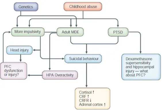
Around one million of fatal suicide (FS) and ten million suicide attempts (SA) occur worldwide each year. Frequently, suicide is consecutive to a psychiatric disorder, most commonly a mood disorder (see for example Beautrais, Joyce et al. 1996). Several other psychiatric conditions or traits have been related to suicide, including schizophrenia (Perenyi and Forlano 2005), alcoholism (Murphy, Wetzel et al. 1992), substance abuse (Murphy 1988), personality disorders (Krysinska, Heller et al. 2006), aggressive/impulsive, hopelessness or pessimistic traits (Brezo, Paris et al. 2006), a history of physical or sexual abuse during childhood (Joiner, Sachs-Ericsson et al. 2007), a history of head injury (Wasserman, Shaw et al. 2008) or neurological disorder (Faber 2003) (Figure 4).
Figure 4 : Effects of genetics, head injury and childhood abuse on mood disorders and impulsivity in relation to suicidal behaviour. Impulsivity in combination with a mood disorder or post-traumatic stress disorder (PTSD) increases the risk of suicidal behaviour. CRF, corticotrophin releasing factor; CRFR, CRF receptor; HPA, hypothalamic pituitary adrenal; MDE, major depressive episode; PFC, prefrontal cortex (Mann 2003).
In addition, various psychosocial factors correlate with suicidal behaviour, including rural areas, high rates of gun ownership, poverty, unemployment, social isolation (Smith, Mercy et al. 1988; Beautrais, Joyce et al. 1996) and imitation (Gould 1990; Johnson, Cohen et al. 2002).
Neurobiological correlates of the diathesis-stress model of suicidal acts involve the serotonergic dorsal raphe (DR), noradrenergic locus coeruleus (LC), and the ventromedial prefrontal cortex (vmPFC).
The diathesis–stress model is a psychological theory explaining behaviours as resulting from both (i) biological and genetic factors ("nature"), and (ii) life experiences ("nurture"). This model assumes that a disposition towards a certain disorder may result from a combination of genetic background and early learning (e.g. as occurring during sexual abuse). The term "diathesis" refers to a genetic
predisposition toward an abnormal condition and this predisposition, in combination with certain kinds of environmental stress, results in abnormal behaviours.
Neurochemical correlates (including indices of neurotransmitters, signal transduction and cellular morphology) of suicidal behaviour have been studied mainly through post-mortem brain tissue analysis. This kind of analysis presents some limitations that include confounding effects of ante-mortem drug treatment and the possibility of examining the brain only at one point in time.
A large majority of post-mortem studies of suicide examined the serotonergic system because early studies (Stanley, Virgilio et al. 1982; Stanley and Mann 1983) identified fewer presynaptic serotonin (5-HT, 5-hydroxytryptamine) transporter sites in several brain areas including vmPFC (Arango, Underwood et al. 1995). In addition, upregulation of 5-HT1A receptor in the vmPFC was observed (Mann and Arango 2001). The same area is involved in behavioural and cognitive inhibition that may be important in order to diminish aggressive or suicidal feelings. There have been fewer studies about noradrenergic neurotransmission. Overall, they showed fewer noradrenergic neurons (Arango, Underwood et al. 1996), lower noradrenaline levels and higher number of alpha-2 adrenergic receptors in the LC of suicide victims (Ordway, Widdowson et al. 1994) and higher beta-1 adrenergic receptor binding (Mann, Stanley et al. 1986) with lower alpha-adrenergic binding (Arango, Ernsberger et al. 1993) in PFC, indicating respectively a brainstem noradrenergic hypoactivity and a cortical noradrenergic overactivity. The latter could have conducted to the former. Although the dopamine system is abnormal in depression, available studies are too scarce to determine whether it can be implicated in suicide.
Genes by environment interactions have been pinpointed. For instance, peer-reared
monkeys have lower serotonergic activity, persisting into adulthood and reflected in greater impulsivity and aggression, as compared to maternally raised monkeys (Higley, Thompson et al. 1993). Stress increases noradrenergic system, HPA axis and cortisol release (Pittenger and Duman 2008). One simple extrapolation may be made with child abuse resulting sometimes in post-traumatic stress disorder (PTSD). This example illustrates the impact of parenting (i.e. neglect, physical or sexual abuse) and