R E S E A R C H A R T I C L E
Open Access
Thalamo-cortical network activity between
migraine attacks: Insights from MRI-based
microstructural and functional resting-state
network correlation analysis
Gianluca Coppola
1*, Antonio Di Renzo
1, Emanuele Tinelli
2, Chiara Lepre
3, Cherubino Di Lorenzo
4, Giorgio Di Lorenzo
5,
Marco Scapeccia
2, Vincenzo Parisi
1, Mariano Serrao
6, Claudio Colonnese
2,7, Jean Schoenen
8and Francesco Pierelli
6,7Abstract
Background: Resting state magnetic resonance imaging allows studying functionally interconnected brain networks. Here we were aimed to verify functional connectivity between brain networks at rest and its relationship with thalamic microstructure in migraine without aura (MO) patients between attacks.
Methods: Eighteen patients with untreated MO underwent 3 T MRI scans and were compared to a group of 19 healthy volunteers (HV). We used MRI to collect resting state data among two selected resting state networks, identified using group independent component (IC) analysis. Fractional anisotropy (FA) and mean diffusivity (MD) values of bilateral thalami were retrieved from a previous diffusion tensor imaging study on the same subjects and correlated with resting state ICs Z-scores.
Results: In comparison to HV, in MO we found significant reduced functional connectivity between the default mode network and the visuo-spatial system. Both HV and migraine patients selected ICs Z-scores correlated negatively with FA values of the thalamus bilaterally.
Conclusions: The present results are the first evidence supporting the hypothesis that an abnormal resting within networks connectivity associated with significant differences in baseline thalamic microstructure could contribute to interictal migraine pathophysiology.
Keywords: Migraine, Thalamus, Resting state, Fractional anisotropy, Magnetic resonance imaging Background
During recent years, various experimental data suggested
that the functional state of the migraineur’s brain is
altered between attacks. This was initially observed with clinical neurophysiology methods that disclosed for instance interictal deficient habituation of sensory responses attributable to abnormal thalamo-cortical interactions [1, 2], and abnormal brain responses to vari-ous neuromodulatory techniques [3]. Interictal func-tional abnormalities were confirmed also with funcfunc-tional imaging methods in response to both noxious [4, 5] and
innocuous stressors [6, 7]. Recently, several independent research groups showed that changes can also be dem-onstrated at rest, i.e. without any sensory input, in the microstructure of several brain areas [8], including the thalamus [9, 10]. The conjunction of neuroimaging and neurophysiological data can be considered as robust evidence favouring morphological and functional brain alterations as prominent features of migraine patho-physiology. Since the brain areas are part of intercon-nected cortical and subcortical networks, it seems of major interest to analyse during the interictal phase the functional connectivity between brain networks at rest and its relationship with the thalamic microstructure.
Among the various neuroimaging procedures, resting state functional MRI (RS-fMRI) analyses the spontaneous
* Correspondence:[email protected]
1Research Unit of Neurophysiology of Vision and Neurophthalmology, G.B.
Bietti Foundation-IRCCS, Via Livenza 3, 00198 Rome, Italy Full list of author information is available at the end of the article
© The Author(s). 2016 Open Access This article is distributed under the terms of the Creative Commons Attribution 4.0 International License (http://creativecommons.org/licenses/by/4.0/), which permits unrestricted use, distribution, and reproduction in any medium, provided you give appropriate credit to the original author(s) and the source, provide a link to the Creative Commons license, and indicate if changes were made.
BOLD signal modulations not attributable to explicit in-puts or outin-puts [11] and allows studying among which distributed brain areas activity at rest is related [12]. A common method to identify spatial patterns of coherent spontaneous BOLD activity, so-called functional connect-ivity, is independent component analysis (ICA). ICA is a high-order statistical method to examine functional connectivity by deconvolving the cerebral signals into components that are maximally independent and that reflect specific interconnected neuroanatomical networks. Compared to other methods, ICA is devoid of anya-priori definition of seed regions of interest [11]. RS-fMRI ICA allows the study of group differences in the temporal rela-tionship among independent spatially distributed networks/ components [12]. With this method, spontaneous brain ac-tivity was shown to be organized in specific and distinct spatial patterns, or sets of resting-state networks [13, 14].
In recent years, several fMRI studies have assessed resting state functional connectivity in various networks in migraine patients. Most of them have used ana-priori selected seed-based analysis [15–21]. Between selected brain areas of the default mode network (DMN) both increased [20, 22] or decreased [23] connectivity was reported. Two studies by the same group of researchers used the single independent component approach with-outa-priori hypothesis [24, 25]. They found evidence for reduced DMN [25] and executive control network [24] connectivity in migraine without aura patients between attacks. To the best of our knowledge, there are no RS-fMRI studies using ICA to determine the functional connectivity between networks (not within) between migraine attacks. Moreover, RS-fMRI studies of subcor-tical and corsubcor-tical nodes were not combined up to now with DTI studies to analyse in migraine patients the connectivity patterns between the thalamus and various functional cerebral networks at rest. We decided, therefore, to use ICA of the whole brain to search for changes in functional connectivity maps at rest in interictal episodic migraine without aura patients. In addition, the thalamo-cortical network was statisti-cally inferred by correlating selected resting state independent component activity strength and thal-amic anisotropy.
Methods
Subjects
We initially enrolled 32 episodic migraine patients with-out aura (MO, ICHD-3beta code 1.1) who attended our headache clinic in a time period of 2 years and agreed to undergo MRI. We discarded recordings of 14 patients who had an attack within 3 days before or after the recording session.
The final analysis dataset comprises thus 18 right-handed MO patients [26] who subsequently participated
in a comprehensive battery of neuroimaging tests, in-cluding RS-fMRI. We have published elsewhere the results of the diffusion tensor imaging and voxel based morphometry studies performed on the initial 14 patients and used these data combined with those of 6 additional patients to search for correlations with RS-fMRI data [9, 27]. Patients underwent MRI scans during the interictal period (MO), defined as an absence of migraine attacks for at least three days before and after MRI. Inclusion criteria were as follows: no history of
other neurological diseases, systemic hypertension,
diabetes or other metabolic disorders, connective or autoimmune diseases, and any other type of primary (in-cluding chronic migraine) or secondary headache. Patients had uni/bilateral migraine headaches, but not fixed pain on the same side. In order to avoid confound-ing effects on neuroplasticity due to pharmacologic treatment, no preventive anti-migraine drugs were allowed during the preceding 3 months. The control group comprised 19 right-handed healthy volunteers (HV) made up of medical school students and health-care professionals of comparable age and gender distri-bution to the experimental group. Control subjects did not have any overt medical conditions, personal or family history of migraine or epilepsy, or take regular medication. Female subjects were always scanned at mid-cycle. All scanning sessions were performed in the afternoon (4.00–7.00 p.m.).
None of the enrolled subjects had sleep deprivation or alcohol consumption the day preceding the scans. Caffein-ated beverages were not allowed on the day of scanning. Further exclusion criteria for both HV and MO were evi-dence of brain lesions on structural magnetic resonance imaging. All participants received a complete description of the study and granted written informed consent. The ethical review board of the Faculty of Medicine, University of Rome, Italy, approved the project.
Imaging protocols
All images were acquired using a Siemens 3 T Verio MRI scanner with a 12-channel head coil and structural anatomic scans were performed using T1-weighted sagit-tal magnetization-prepared rapid gradient echo (MP-RAGE) series (TR: 1900 ms, TE: 2.93 ms, 176 sagittal slices, 0.508 x 0.508 x 1 mm3voxels).
We acquired an interleaved double-echo Turbo Spin Echo sequence proton density and T2-weighted images (repetition time: 3320 ms, echo time: 10/103 ms, matrix: 384 × 384, field of view: 220 mm, slice thickness: 4 mm, gap: 1.2 mm, 50 axial slices).
Functional MRI data were obtained using a T2*-weighted, echo-planar imaging (TR: 3000 ms, TE: 30 ms, 40 axial slices, 3.906 x 3.906 x 3 mm, 150 volumes).
Functional BOLD data were collected in a 7 min 30 s run, during which subjects were instructed to relax, avoid motion and keep their eyes closed.
Data processing and analyses
Image data processing was performed on a personal computer using statistical parametric mapping (SPM8) software package (Wellcome Trust Centre for Neuroim-aging, London, UK; http://www.fil.ion.ucl.ac.uk/spm), GIFT v3.0 and FNC toolbox (http://mialab.mrn.org/) in
Mat-Lab (http://www.mathworks.com/). The overall
image data processing is based on the method already described elsewhere [12].
All images from a single subject were realigned using a 6-parameter rigid body process, replaced by a cubic spline interpolation. We did not perform slice time correction. Slice-timing correction is not mandatory be-cause the haemodynamic response is longer than TR (about 30 s). Moreover, the actual EPI uses multiband sequences with simultaneous echo refocusing and paral-lel imaging (called GRAPPA by Siemens), that makes
slice time correction obsolete. The structural (T1 –
MPRAGE) and functional data were coregistered for each participant dataset, normalized into the standard Montreal Neurological Institute space, and then trans-formed into a common stereotactic space based on Talairach and Tournoux [28]. Finally, functional images were spatially smoothed with an 8 mm full width half-maximum Gaussian kernel on each direction.
Component identification and selection
Grouped spatial ICA was performed for all 37 partici-pants (HV + MO) using the infomax algorithm [29]. Two separate group spatial ICAs were also carried out in HV and MO patients to ensure that the fluctuations of components at rest in each group of subjects were similar to those obtained in the total group of 37 sub-jects. GIFT software automatically decomposed data into 39 components. A modified version of the minimum description length (MDL) criterion was adopted to determine the number of components from the aggre-gate data set [30]. Single subject spatially or temporally independent maps were then back-reconstructed from the aggregate mixing matrix [31].
All 39 components were inspected after plotting to templates in GIFT, usinga priori probabilistic maps, and those of interest whose patterns mainly consisted of gray matter rather than non-gray matter were selected. Com-ponents located in CSF or white matter, or with low correlation to gray matter, can be artifacts, such as eye movements, head motion, ballistic artifacts, and were discarded. With FNC toolbox in MatLab, after removing
all the artifactual components and applying a p-value
threshold of 0.01 (false discovery rate corrected), only
two components survived for further analysis. Before performing correlation analyses, a band-pass Butter-worth filter between 0.033 Hz and 0.13 Hz was applied on the two selected component time courses. Each IC consists of a temporal waveform and an associated spatial map; the latter is expressed in terms of Z-scores that reflect the degree to which a given voxel time-course correlates with the specific IC temporal wave-form, i.e. a way to quantify the strength of the IC [32]. As a further step and in order to search for a correlation between regional RS-fMRI network changes and clinical features, the Z-max scores (voxel-wise analysis) of each IC network were extracted for each participant.
Diffusion weighted imaging of the thalami
Diffusion tensor imaging (DTI) was acquired using single shot echo-planar imaging, with a 12–channel head coil (TR 12200 ms, TE 94 ms, 72 axial slices, 2 mm thickness, isotropic voxels). Images from the same participants and during the same session were obtained with diffusion gradients applied along 30 non-collinear directions, effective b values of 0 and 1000 s/mm2 were used. Image data processing was performed with the FSL 4.0 software package (FMRIB Image Analysis Group, Oxford, England). Diffusion data were corrected for motion and distortions caused by eddy current arti-facts; FMRIB's Diffusion Toolbox (FDT) was used for local fitting of diffusion tensors, and fractional anisot-ropy (FA) and mean diffusivity (MD) maps were created.
Two regions of interest (ROI, from the“Harvard-Oxford
Subcortical Structural Atlas” as distributed with FSL) were defined for each subject, covering totally right and left thalami on each slice (Fig. 1). The medial boundaries were determined on each slice using cerebro spinal fluid as a landmark; lateral limits were verified using FA maps to exclude the internal capsule. Mean FA and MD in each region for every subject were obtained by averaging the values of those voxels contained in the ROI.
We have previously publishedin extenso the results of the DTI analyses performed on the first 29 subjects, 15 HV and 14 MO [9].
Statistical analyses
Group differences for demographic data were estimated using ANOVA test.
We used a 2-sample t-test to detect significant differ-ences in correlation values between the two independent
components for HV vs MO. A conservative p value of
p < 0.05 (correction for multiple comparisons with false discovery rate selected) was used as significance cut-off. Connectivity combinations with statistically significant (p < 0.01) lag values were assessed using a two sample t-test of the difference between averaged HV and MO patient lags. As a further step and in
order to search for a correlation between regional RS-fMRI network changes and clinical features, the Z-max scores of each IC network were extracted for each participant.
Finally, we used Pearson’s test to search for correla-tions between the MR DTI parameters FA and MD, individual IC Z-max scores and clinical variables such as severity of headache attacks [0–10], duration of migraine history [years], monthly attack frequency [n], attack duration [hours], days elapsed since the last
migraine attack [n]. P values ≤ 0.05 were considered
to indicate statistical significance.
Results
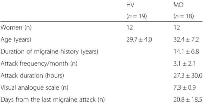
All subjects completed the study. Demographic and clinical data for the two groups are summarized in Table 1. Structural brain MRIs were normal in all partic-ipants. Specifically in migraine patients there were
neither white matter lesions nor features suggestive of cortical atrophy.
Resting state fMRI
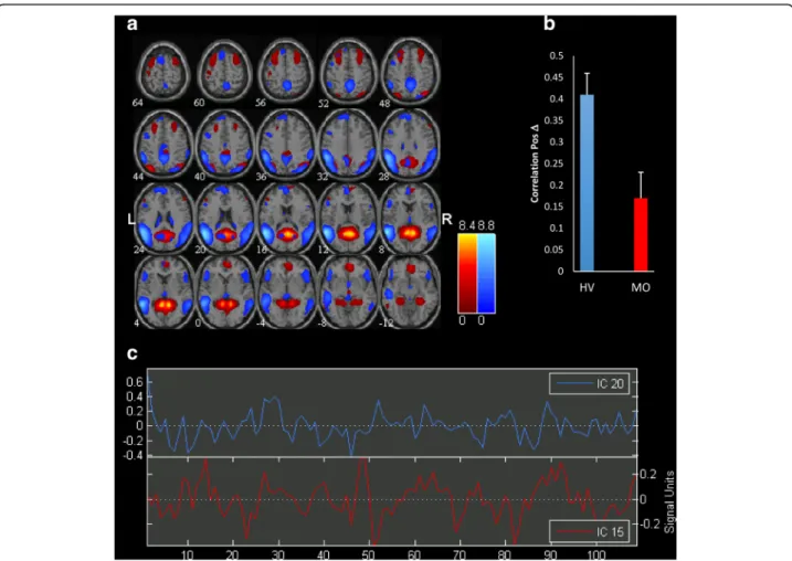
Significantly correlated components are represented in Fig. 2.
We found a significant difference in functional con-nectivity between independent components IC20 and IC15 in migraine patients between attacks respective to healthy volunteers. In fact, the correlation of independ-ent componindepend-ent pair (IC20-IC15) is significantly lower in MO compared to HV (rho = 0.17 in MO vs rho = 0.41 in HV; FDR corrected,p < 0.05).
These components encompass respectively intercon-nected areas of the so-called default mode network (IC20) and a network composed of the visuo-spatial system and medial visual cortical areas (IC15), as seen on GIFT templates.
Although component directions slightly differed be-tween HV and MO patient group, lag difference did not reach the level of significance.
There was no significant correlation between ICs Zmax-scores and clinical data.
Diffusion Tensor Imaging (DTI) data
The DTI results confirmed those published else-where on the first 29 subjects [9]. In MO patients the bilateral thalami have shown a significantly increased fractional anisotropy (F(1,35)= 4.99, p = 0.03
and F(1,35)= 4.86, p = 0.03 for the left and right
thalamus respectively), but only a tendency to mean diffusivity change (F(1,35)= 2.50,p = 0.12 and F(1,35)= 3.44,
p = 0.07 for the left and right thalamus respectively), than in HV.
Thalamo-cortical network correlation analysis
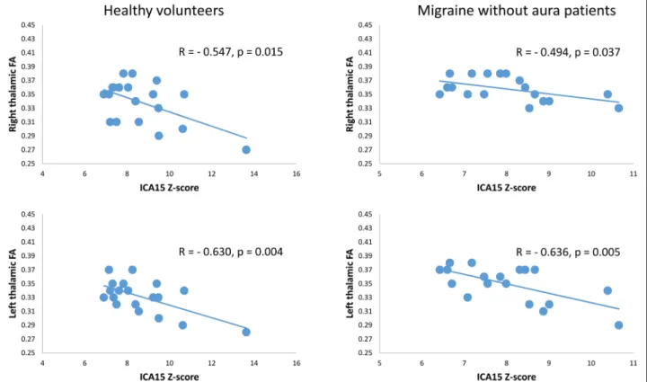
Pearson’s correlation test disclosed that the IC15 Z-score correlated negatively with bilateral thalamic FA values in both HV (right r = − 0.547, p = 0.015; left r = − 0.630, Fig. 1 Exemplary single subject axial representation of the diffusion
tensor imaging FA map with the analyzed thalamic ROIs highlighted in yellow
Table 1 Clinical and demographic characteristics of healthy volunteers (HV) and migraine patients without aura scanned between (MO) attacks. Data are expressed as means ± SD
HV MO
(n = 19) (n = 18)
Women (n) 12 12
Age (years) 29.7 ± 4.0 32.4 ± 7.2
Duration of migraine history (years) 14.1 ± 6.8
Attack frequency/month (n) 3.1 ± 2.1
Attack duration (hours) 27.3 ± 30.0
Visual analogue scale (n) 7.3 ± 0.9
p = 0.004) and MO (right r = − 0.494, p = 0.037; left r = − 0.636, p = 0.005) (Fig. 3).
Discussion
Our study was specifically designed to search for differ-ences in resting state networks and to test whether the resulting networks are correlated with thalamic micro-structure in interictal migraine state. We found that func-tional connectivity of networks involved in in information processing, in cognitive, emotional, and, especially visual, attention processes differ between patients and healthy volunteers. We will discuss the possible neurobiological underpinnings of our findings and their potential rele-vance for migraine pathophysiology.
Resting state functional connectivity between attacks
Functional connectivity between the default mode network (IC 20) and a network composed of the visuo-spatial system and medial visual cortical areas (IC 15) is
significantly reduced in migraine patients between at-tacks compared to HV.
The so-called default-mode network (DMN) encom-passes a set of regions with relatively greater activity
during “rest” than during active conditions [33]. The
DMN includes the posterior cingulate gyrus, precuneus, medial prefrontal cortex, angular gyrus, and medial temporal lobe regions including the hippocampus and the lateral temporo-parietal area [34]. Although the exact functional role of the DMN is not completely understood, it is thought to be involved in retrieval of information from long-term memory and its manipula-tion for optimizing the sensorium and for problem solv-ing and plannsolv-ing. The DMN is thus important in conscious experience as well as in maintaining a general low-level focus of attention for an event [35, 36]. Although the DMN is anatomically and functionally distinct from networks involved in sensory functions, comparative studies in animal and human studies have Fig. 2 a Representation of the two significant Independent Components (IC) functional connectivity networks differing in migraine patients scanned between attacks (MO) compared with healthy volunteers (HV) separated by independent component analysis (ICA). All images have been coregistered into the space of the MNI template. Brain areas are respectively coloured in hot metal scale (IC15) or in azure-blue (IC20). The numbers beneath each image refer to the z coordinate in Talairach space. b The bar graph reflects FDR corrected correlation between the 2 ICs, p < 0.05, in HV and MO. c Time course of spontaneous blood oxygen level dependent (BOLD) activity recorded during resting state and extracted from each of the two significant ICs
shown that it is functionally correlated with all other resting state networks [14]. The visuo-spatial system comprises the posterior parietal cortex at the occipito-parietal junction, the precuneus, the posterior cingulate cortex and the frontal pole [13, 37]. Activity within this network in the resting state is associated with gathering information about our outer, and possibly inner, world [38], in episodic memory recall and orienting attention to salient novel or familiar stimuli and in emotional processing associated with episodic memory [39]. The medial visual cortical areas include the primary visual cortex as well as medial extrastriate regions such as lingual gyrus, inferior division of precuneus and lateral geniculate nucleus [13, 14]. This network is supposed to play a role in episodic memory, visual and visuo-spatial processing, reflections upon self and aspects of consciousness.
Given the brain networks involved and their known functions, the abnormalities we found in interictal migraine suggest a dysfunction of information gathering, evaluation and integration, and impaired short- as well as long- term memory processes. Moreover, because of the predominant involvement of visual areas/systems visuo-perceptual and visuo-spatial integration could be impaired in migraine between attacks. Whether these resting state fMRI abnormalities are the connectivity correlates of the subtle impairments in neuropsychological
performances, such as processing speed, verbal memory, and physiognomy recognition, previously reported in mi-graine between attacks remains to be determined [40–42]. It was shown that resting state spontaneous brain activity can be used to predict the task-response properties of brain regions [43]. It is thus of interest to verify if the reduced spontaneous network activity found here is corre-lated with the abnormal cognitive evoked potentials reported in migraine patients [44–49].
Thalamo-cortical interactions
We previously argued [9] that, in MO patients scanned between attacks, the pattern of increased anisotropy asso-ciated with normal MD may reflect shrinking of neuronal and glial cells and/or gain of directional organization in combination with a preserved cell density [50, 51]. Inter-estingly, data from animal models showed that cell shrink-ing may coincide with a reduced neuronal electric response [52, 53]. Therefore, since grey matter in the single thalamic sub-nuclei does not have a unique oriented fibre structure, the increased FA found in between attacks might also result from a decrease in neuron connections and thus dendritic arborization, which in turn may result in a reduced number of local circuits [54].
Here, the most striking finding is the correlation be-tween MRI diffusion-weighted features of the thalamus Fig. 3 In both HV (left panel) and MO (right panel) groups, Z-score of the independent component (IC)15, encompassing the visuo-spatial system and medial visual cortical areas, correlated negatively with bilateral thalamic fractional anisotropy (FA) values
and the functional connectivity between brain networks. In both HV and migraineurs between attacks, individual Z-scores of IC15, containing the visuo-spatial system and medial visual areas, correlated negatively with FA values of bilateral thalami, suggesting that the lower is the between networks connectivity the higher is the FA in the thalami bilaterally. However, this association between thalamic diffusion parameters and RS-fMRI is evident in examining each individual group, suggesting that the correlation between thalamic microstructure and RS-fMRI is a more general phenomenon related to the connectivity mechanisms. Nevertheless, we found significant differences in baseline thalamic
microstruc-ture – increased FA in MO – and in within networks
connectivity – decreased connection in MO – between
patients and healthy controls.
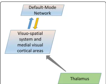
Overall, the results from the correlation analysis fit strikingly with evidence coming from neuroimaging stud-ies showing a distinct functional connectivity between the thalamus and several areas within the visuo-spatial system and medial visual areas (e.g. posterior cingulate cortex, visual cortex, precuneus) [55–57]. Taken together the latter evidence with our present finding of no significant difference in the lag in intrinsic activity, an indirect esti-mation of the direction of the connection, between the pair of less interconnected networks (i.e. IC20-IC15), it is possible that the thalamic relay contributes the most to the cortical networks activity via the thalamocortical loops. Therefore, we hypothesize that a deficient thalamic activity in migraine between attacks, as highlighted by an increased FA [9], activates less the visuo-spatial system and medial visual cortical areas, which in turn leads to less activation of the DMN network (Fig. 4).
Clinical and experimental data indicate that the thal-amus is a key structure in migraine pathophysiology. The thalamus was found to be implicated in many clinical [58–61] and neurophysiological features of migraine [62–64]. From animal experiments, it is known that the vast system of extrastriate and suprasylvian areas comprising the brain’s most important networks receive ex-tensive projections from the lateral posterior-pulvinar thal-amic complex, including the intralaminar nuclei [65, 66], that were recently reported to be reduced in volume in migraine patients scanned when attack-free [10]. It is important to take into account that these nuclei receive the most significant overlap of different sensory modalities [67]. This is of particular interest for migraine since the majority of evoked potential studies between attacks have shown abnormalities, such as deficit of habituation, for most sensory modalities: non-painful and painful somatosensory, auditory and visual [1]. The only notable exception is olfac-tion, the only sensory modality not relayed in the thalamus, for which brain and behavioural responses habituate normally in migraineurs [5].
As with all studies, our findings need to be considered with our study limitations and strengths. The small number of patients could make our study underpowered to reveal more subtle findings, such as correlation between clinical features and functional connectivity, although our cohort was sufficient to disclose strong statistical significance. A strength of the present study is our approach to study the dependencies between pairs of functional networks, since it allowed us to examine weak, but significant, connectivity among strongly con-nected networks.
Conclusions
Overall, these results of RS-fMRI are in line with the con-cept of a global dysfunction in multisensory information processing and integration in migraine. The multimodal MRI data provide specifically structural and functional evidences for the involvement of the thalamus in the abnormal functional connectivity between different brain networks between attacks. Future work should attempt to clarify the role of the different networks with regard to migraine-associated multisensory phenomena, such as photophobia or allodynia, especially during the attack and in chronic migraine. It would also be of particular interest to verify whether the thalamocortical network dysfunc-tions are primary phenomena or secondary to a functional disconnection of the thalamus from the brainstem. Abbreviations
3 T:3 Tesla; DMN: Default mode network; DTI: Diffusion tensor imaging; EEG: Electroencephalography; FA: Fractional anisotropy; HV: Healthy volunteer; IC: Independent component; ICA: Independent component analysis; MD: Mean diffusivity; MO: Migraine without aura; MRI: Magnetic
Fig. 4 Schematic representation of information flow describing the thalamo-cortical neural network model that can encompass the present findings in migraine patients scanned between attacks. During the interictal period, a reduced thalamic activity, as highlighted by an increased FA, activates less the visuo-spatial system and medial visual cortical areas, which in turn deactivate the DMN network
resonance imaging; ROI: Region Of Interest; RS-fMRI: Resting state functional magnetic resonance imaging
Acknowledgment
Italian Ministry of Health and Fondazione Roma financially supported the research for this paper.
Authors’ contributions
GC made substantial contributions to interpretation of data as well as in drafting the manuscript. GDL, VP, JS and FP were implied in the interpretation of data as well as in drafting the manuscript; CDL and MSe gave critical revision of the manuscript for important intellectual content. CC, CL, and MSc were implied in recording data. ADR and ET were implicated in analyzing data. All authors read and approved the final manuscript.
Competing interests
The authors declare that they have no competing interests.
Author details
1Research Unit of Neurophysiology of Vision and Neurophthalmology, G.B.
Bietti Foundation-IRCCS, Via Livenza 3, 00198 Rome, Italy.2Department of Neurology and Psychiatry, Neuroradiology Section,“Sapienza” University of Rome, Rome, Italy.3Department of Medico-Surgical Sciences and Biotechnologies, Neurology Section,“Sapienza” University of Rome, Rome, Italy.4Don Carlo Gnocchi Onlus Foundation, Milan, Italy.5Laboratory of Psychophysiology, Psychiatric Clinic, Department of Systems Medicine, University of Rome“Tor Vergata”, Rome, Italy.6Department of
Medico-Surgical Sciences and Biotechnologies,“Sapienza” University of Rome Polo Pontino, Latina, Italy.7IRCCS Neuromed, Pozzilli, (IS), Italy.8Headache Research Unit, Department of Neurology-CHR Citadelle, University of Liège, Liège, Belgium.
Received: 12 March 2016 Accepted: 18 October 2016
References
1. Coppola G, Di Lorenzo C, Schoenen J, Pierelli F (2013) Habituation and sensitization in primary headaches. J Headache Pain 14:65
2. Magis D, Vigano A, Sava S et al (2013) Pearls and pitfalls: electrophysiology for primary headaches. Cephalalgia 33:526–539
3. Brighina F, Cosentino G, Fierro B (2013) Brain stimulation in migraine. Handb Clin Neurol 116:585–98. doi:10.1016/B978-0-444-53497-2.00047-4 4. Moulton EA, Becerra L, Maleki N et al (2011) Painful heat reveals
hyperexcitability of the temporal pole in interictal and ictal migraine States. Cereb Cortex 21:435–448
5. Stankewitz A, Schulz E, May A (2013) Neuronal correlates of impaired habituation in response to repeated trigemino-nociceptive but not to olfactory input in migraineurs: An fMRI study. Cephalalgia 33:256–265 6. Demarquay G, Royet JP, Mick G, Ryvlin P (2008) Olfactory hypersensitivity in
migraineurs: a H(2)(15)O-PET study. Cephalalgia 28:1069–80. doi:10.1111/j. 1468-2982.2008.01672.x
7. Martín H, Sánchez del Rio M, de Silanes C et al (2011) Photoreactivity of the occipital cortex measured by functional magnetic resonance imaging-blood oxygenation level dependent in migraine patients and healthy volunteers: pathophysiological implications. Headache 51:1520–1528
8. Sprenger T, Borsook D (2012) Migraine changes the brain: neuroimaging makes its mark. Curr Opin Neurol 25:252–262
9. Coppola G, Tinelli E, Lepre C et al (2014) Dynamic changes in thalamic microstructure of migraine without aura patients: a diffusion tensor magnetic resonance imaging study. Eur J Neurol 21:287–e13. doi:10.1111/ene.12296 10. Magon S, May A, Stankewitz A et al (2015) Morphological abnormalities of
thalamic subnuclei in migraine: A multicenter MRI study at 3 Tesla. J Neurosci 35:13800–13806
11. Fox M, Raichle ME (2007) Spontaneous fluctuations in brain activity observed with functional magnetic resonance imaging. Nat Rev 8:700–711 12. Jafri M, Pearlson GD, Stevens M, Calhoun VD (2008) A method for functional
network connectivity among spatially independent resting-state components in schizophrenia. Neuroimage 39:1666–1681
13. Beckmann C, DeLuca M, Devlin JT, Smith SM (2005) Investigations into resting-state connectivity using independent component analysis. Philos Trans R Soc LondonSeries B Biol Sci 360:1001–1013
14. Mantini D, Perrucci MG, Del Gratta C et al (2007) Electrophysiological signatures of resting state networks in the human brain. Proc Natl Acad Sci U S A 104:13170–13175
15. Hadjikhani N, Ward N, Boshyan J et al (2013) The missing link: enhanced functional connectivity between amygdala and visceroceptive cortex in migraine. Cephalalgia 33:1264–1268
16. Jin C, Yuan K, Zhao L et al (2013) Structural and functional abnormalities in migraine patients without aura. NMR Biomed 26:58–64
17. Mainero C, Boshyan J, Hadjikhani N (2011) Altered functional magnetic resonance imaging resting-state connectivity in periaqueductal gray networks in migraine. Ann Neurol 70:838–845
18. Moulton E, Becerra L, Johnson A et al (2014) Altered hypothalamic functional connectivity with autonomic circuits and the locus coeruleus in migraine. PLoS One 9:10
19. Schwedt T, Larson-Prior L, Coalson RS et al (2014) Allodynia and descending pain modulation in migraine: a resting state functional connectivity analysis. Pain Med 15:154–165
20. Xue T, Yuan K, Zhao L et al (2012) Intrinsic brain network abnormalities in migraines without aura revealed in resting-state fMRI. PLoS One 7:10 21. Yuan K, Zhao L, Cheng P et al (2013) Altered structure and resting-state
functional connectivity of the basal ganglia in migraine patients without aura. J Pain 14:836–844
22. Xue T, Yuan K, Cheng P et al (2013) Alterations of regional spontaneous neuronal activity and corresponding brain circuit changes during resting state in migraine without aura. NMR Biomed 26:1051–1058
23. Yu D, Yuan K, Zhao L et al (2012) Regional homogeneity abnormalities in patients with interictal migraine without aura: a resting-state study. NMR Biomed 25:806–812
24. Russo A, Tessitore A, Giordano A et al (2012) Executive resting-state network connectivity in migraine without aura. Cephalalgia 32:1041–1048 25. Tessitore A, Russo A, Giordano A et al (2013) Disrupted default mode
network connectivity in migraine without aura. J Headache Pain 14:89 26. (2013) The International Classification of Headache Disorders, 3rd edition
(beta version). Cephalalgia 33:629–808. doi: 10.1177/0333102413485658 27. Coppola G, Di Renzo A, Tinelli E et al (2015) Evidence for brain
morphometric changes during the migraine cycle: A magnetic resonance-based morphometry study. Cephalalgia 35:783–791. doi:10.1177/ 0333102414559732
28. Talairach J, Tournoux P (1988) Co-planar Stereotaxic Atlas of the Human Brain. Georg Thieme Verlag, New York, Thieme
29. Bell A, Sejnowski TJ (1995) An information-maximization approach to blind separation and blind deconvolution. Neural Comput 7:1129–1159 30. Li YO, Adali T, Calhoun VD (2007) Estimating the number of independent
components for functional magnetic resonance imaging data. Hum Brain Mapp 28:1251–1266
31. Calhoun VD, Adali T, Pearlson GD, Pekar JJ (2001) A method for making group inferences from functional MRI data using independent component analysis. Hum Brain Mapp 14:140–151
32. McKeown M, Makeig S, Brown GG et al (1998) Analysis of fMRI data by blind separation into independent spatial components. Hum Brain Mapp 6:160–188
33. Raichle M, MacLeod AM, Snyder AZ et al (2001) A default mode of brain function. Proc Natl Acad Sci U S A 98:676–682
34. Buckner R, Andrews-Hanna JR, Schacter DL (2008) The brain’s default network: anatomy, function, and relevance to disease. Ann N Y Acad Sci 1124:1–38 35. Binder J, Frost JA, Hammeke TA et al (1999) Conceptual processing during the conscious resting state. A functional MRI study. J Cogn Neurosci 11:80–95
36. Carhart-Harris R, Friston KJ (2010) The default-mode, ego-functions and free-energy: a neurobiological account of Freudian ideas. Brain 133:1265–1283 37. Gusnard D, Raichle ME (2001) Searching for a baseline: functional imaging
and the resting human brain. Nat Rev 2:685–694
38. Vogt B, Finch DM, Olson CR (1992) Functional heterogeneity in cingulate cortex: the anterior executive and posterior evaluative regions. Cereb Cortex 2:435–443
39. Maddock R (1999) The retrosplenial cortex and emotion: new insights from functional neuroimaging of the human brain. Trends Neurosci 22: 310–316
40. Le Pira F, Lanaia F, Zappalà G et al (2004) Relationship between clinical variables and cognitive performances in migraineurs with and without aura. Funct Neurol 19:101–5
41. Suhr JA, Seng EK (2012) Neuropsychological functioning in migraine: clinical and research implications. Cephalalgia 32:39–54
42. Yetkin-Ozden S, Ekizoglu E, Baykan B (2015) Face Recognition in Patients with Migraine. Pain Pract 15:319–22
43. Fox M, Snyder AZ, Zacks JM, Raichle ME (2006) Coherent spontaneous activity accounts for trial-to-trial variability in human evoked brain responses. Nat Neurosci 9:23–25
44. Demarquay G, Caclin A, Brudon F et al (2011) Exacerbated attention orienting to auditory stimulation in migraine patients. Clin Neurophysiol 122:1755–1763
45. Iacovelli E, Tarantino S, De Ranieri C et al (2012) Psychophysiological mechanisms underlying spatial attention in children with primary headache. Brain Dev 34:640–647
46. Mickleborough M, Chapman CM, Toma AS et al (2013) Interictal neurocognitive processing of visual stimuli in migraine: evidence from event-related potentials. PLoS One 8:10
47. Mickleborough M, Chapman C, Toma A, Handy T (2014) Cognitive processing of visual images in migraine populations in between headache attacks. Brain Res 1582:167–175
48. Schoenen J, Timsit-berthier M (1993) Contingent negative variation: Methods and potential interest in headache. Cephalalgia 13:28–32 49. Wang W, Schoenen J, T-B M (1995) Cognitive functions in migraine without
aura between attacks: a psychophysiological approach using the“oddball” paradigm. Neurophysiol Clin 25:3–11
50. Wieshmann UC, Clark CA, Symms MR et al (1999) Reduced anisotropy of water diffusion in structural cerebral abnormalities demonstrated with diffusion tensor imaging. Magn Reson Imaging 17:1269–1274
51. Mandl RC, Schnack HG, Zwiers MP et al (2008) Functional diffusion tensor imaging: measuring task-related fractional anisotropy changes in the human brain along white matter tracts. PLoS One 3:10
52. Tasaki I, Byrne PM (1992) Rapid structural changes in nerve fibers evoked by electric current pulses. Biochem Biophys Res Commun 188:559–564 53. Tasaki I (1999) Rapid structural changes in nerve fibers and cells associated
with their excitation processes. Jpn J Physiol 49:125–138
54. Beaulieu C (2002) The basis of anisotropic water diffusion in the nervous system - a technical review. NMR Biomed 15:435–455. doi:10.1002/nbm.782 55. Zou Q, Long X, Zuo X et al (2009) Functional connectivity between the
thalamus and visual cortex under eyes closed and eyes open conditions: a resting-state fMRI study. Hum Brain Mapp 30:3066–3078
56. Wang X, Xu M, Song Y et al (2014) The network property of the thalamus in the default mode network is correlated with trait mindfulness. Neuroscience 278:291–301
57. Ku J, Cho YW, Lee YS et al (2014) Functional connectivity alternation of the thalamus in restless legs syndrome patients during the asymptomatic period: a resting-state connectivity study using functional magnetic resonance imaging. Sleep Med 15:289–294
58. Burstein R, Jakubowski M, Garcia-Nicas E et al (2010) Thalamic sensitization transforms localized pain into widespread allodynia. Ann Neurol 68:81–91. doi:10.1002/ana.21994
59. Maleki N, Becerra L, Upadhyay J et al (2012) Direct optic nerve pulvinar connections defined by diffusion MR tractography in humans: implications for photophobia. Hum Brain Mapp 33:75–88
60. Noseda R, Kainz V, Jakubowski M et al (2010) A neural mechanism for exacerbation of headache by light. Nat Neurosci 13:239–245
61. Russo A, Marcelli V, Esposito F et al (2014) Abnormal thalamic function in patients with vestibular migraine. Neurology 82:2120–2126
62. Coppola G, De Pasqua V, Pierelli F, Schoenen J (2012) Effects of repetitive transcranial magnetic stimulation on somatosensory evoked potentials and high frequency oscillations in migraine. Cephalalgia 32:700–709
63. Coppola G, Iacovelli E, Bracaglia M et al (2013) Electrophysiological correlates of episodic migraine chronification: evidence for thalamic involvement. J Headache Pain 14:76
64. Coppola G, Bracaglia M, Di Lenola D et al (2016) Lateral inhibition in the somatosensory cortex during and between migraine without aura attacks: correlations with thalamocortical activity and clinical features. Cephalalgia 36:568–578. doi:10.1177/0333102415610873
65. Raczkowski D, Rosenquist AC (1983) Connections of the multiple visual cortical areas with the lateral posterior-pulvinar complex and adjacent thalamic nuclei in the cat. J Neurosci 3:1912–1942
66. Tong L, Kalil RE, Spear PD (1982) Thalamic projections to visual areas of the middle suprasylvian sulcus in the cat. J Comp Neurol 212:103–117
67. Cappe C, Morel A, Barone P, Rouiller EM (2009) The thalamocortical projection systems in primate: an anatomical support for multisensory and sensorimotor interplay. Cereb Cortex 19:2025–2037
Submit your manuscript to a
journal and benefi t from:
7 Convenient online submission 7 Rigorous peer review
7 Immediate publication on acceptance 7 Open access: articles freely available online 7 High visibility within the fi eld
7 Retaining the copyright to your article