HAL Id: pastel-00558455
https://pastel.archives-ouvertes.fr/pastel-00558455
Submitted on 21 Jan 2011
HAL is a multi-disciplinary open access
archive for the deposit and dissemination of sci-entific research documents, whether they are pub-lished or not. The documents may come from teaching and research institutions in France or abroad, or from public or private research centers.
L’archive ouverte pluridisciplinaire HAL, est destinée au dépôt et à la diffusion de documents scientifiques de niveau recherche, publiés ou non, émanant des établissements d’enseignement et de recherche français ou étrangers, des laboratoires publics ou privés.
Nouvelle strategies en Protéomique
Giovanni Chiappetta
To cite this version:
Giovanni Chiappetta. Nouvelle strategies en Protéomique. Ingénierie biomédicale. Université Pierre et Marie Curie - Paris VI, 2009. Français. �pastel-00558455�
THESE DE DOCTORAT DE
L’UNIVERSITE PIERRE ET MARIE CURIE ET DE
L’UNIVERSITÉ FEDERICO II
Spécialité
Ecole Doctorale Inter///Bio (ED387)
Interfaces de la Chimie, de la Physique et de l'Informatique avec la Biologie Ecole Doctorale de Biotechnologies
Présentée par M. Giovanni Chiappetta Pour obtenir le grade de
DOCTEUR de l’UNIVERSITÉ PIERRE ET MARIE CURIE ET
DOCTEUR de L’UNIVERSITÉ FEDERICO II
Sujet de la thèse :
Nouvelle strategies en Protéomique
soutenue le 08/01/2009 devant le jury composé de :
Prof. Jean Rossier Directeur de thèse Prof. Marino Gennaro Directeur de thèse Prof.Giovanni Sannia Rapporteur
Dr. Ariane, Jalila Simaan Rapporteur Dr. Joelle Vinh Examinateur
Prof. Germain Trugnan Examinateur Prof. Pasquale De Santis Examinateur Prof. Claudio Palleschi Examinateur Prof. Vincenzo Scarlato Examinateur
Riassunto pag. 1
Résumé pag. 8
1 INTRODUCTION pag. 9
1.1 Proteomics impact on modern Biotechnologies
pag. 9 1.2 Proteomics analysis
pag 10 1.2.1 Mass spectrometry tools for Proteomics analysis pag 11 1.2.2 The first generation of the Proteomic analysis
pag 17 1.2.3 The second generation of the Proteomic analysis
pag 18
1.2.4 Quantitative Proteomics pag 19
1.3 Oxidative stress
pag 20 1.4 Oxidative stress related PTMs
pag 22 1.4.1 Protein nitration pag 22 1.4.2 Thiol oxydation pag 24 1.6 References pag 26
2 BIDIMENSIONAL TANDEM MASS SPECTROMETRY FOR SELECTIVE
IDENTIFICATION OF NITRATION SITES IN PROTEIN pag 30
2.1 Introduction
pag 30 2.1.1 Proteomic identification of nitrated proteins: the state of the art
pag 30 2.1.2 The hybrid Triple Quadrupole/ Linear Ion Trap mass spectrometer
pag 33 2.1.3 Dansyl-Chloride: the RIGhT reagent
pag 35 2.2 Materials and methods
pag 37 2.3 Results and discussions
pag 38 2.4 Conclusion
pag 44
2.5 References pag 47
3. QUANTITATIVE IDENTIFICATION OF PROTEIN NITRATION SITES BY
USING iTRAQ REAGENTS pag 49
3.1 Introduction
pag 49 3.1.1 Protein nitration quantification: state of the art
pag 49 3.1.2 iTRAQ strategy
pag 51 3.2 Materials and methods
pag 52 3.3 Results and discussions
pag 54
3.4 Conclusion pag 63
3.5 References
pag 66
4. IMPROVED LC-MALDI-MS2 ANALYSIS BY PEPTIDES DANSYLATION
pag 69 4.1 Introduction
pag 69 4.1.1 MALDI-TOFTOF mass spectrometer
4.1.2 Enhanced MALDI-MS analysis by peptide modification: state of the art pag 70 4.2 Materials and methods
pag 71 4.3 Results and discussions
pag 72 4.4 Conclusion
pag 83 4.5 References
pag 85
5 A NEW METHOD TO SELECTIVELY EVALUATE PROTEIN THIOLS
OXIDATION STATE. pag 86
5.1 Introduction
pag 86 5.1.1 Proteomic analysis of oxidation related cysteine: state of the art
pag 86 5.2 Materials and methods
pag 87 5.3 Results and discussions
pag 90 5.4 Conclusion pag 99 5.4.1 Perspective pag 99 5.5 References pag 101
INDEX A: table of the abbreviations
pag A
Riassunto
Nuove metodologie per l’analisi Proteomica
Le “Biotecnologie Moderne” sono l’insieme delle tecniche rivolte alla produzione di prodotti e servizi mediante l’utilizzo di sistemi biologici e moderne tecniche di biologia molecolare. Tipicamente i prodotti finali delle produzioni biotecnologiche sono proteine che trovano largo impiego in campo medico, industriale e agrario. La maggioranza dei prodotti biotecnologici sono ottenuti mediante l’utilizzo di tecniche di DNA recombinante basate su cellule “ospite” geneticamente modificate in sistemi biologici come batteri, lieviti ed anche cellule mammifere. Per l’ottimizzazione di un sistema di produzione biotecnologico, oltre alla messa a punto dei sistemi di fermentazione e crescita cellulare “a monte” del processo, è altrettanto necessario realizzare “a valle” efficienti procedure di estrazione e purificazione dei bio-prodotti dalle matrici impiegate. Tradizionalmente lo sviluppo di un processo prevedeva la progettazione e l’ottimizzazione delle procedure “a monte” e “a valle” mediante l’utilizzo di dati empirici e con un’incompleta o limitata conoscenza dei meccanismi cellulari a livello molecolare. I recenti progressi nel campo della Genomica e Proteomica, tecnologie che permettono di ottenere informazioni sui meccanismi cellulari a livello molecolare, hanno fornito nuovi strumenti e conoscenze utili per la progettazione dei processi biotecnologici. La Genomica è una branca della biologia molecolare che si occupa dello studio della composizione, delle funzioni e dell’evoluzione del patrimonio genetico negli organismi viventi. Tuttavia, è apparso subito evidente che la staticità dei dati genomici non era in grado di spiegare la dinamicità del corrispondente comparto proteico, caratterizzato da eventi di splicing alternativo, modifiche post-traduzionali e formazioni di complessi funzionali proteina-proteina. Per questo motivo negli ultimi anni l’attenzione è stata focalizzata maggiormente sui prodotti del genoma: le proteine. L’analisi Proteomica si occupa proprio della definizione del comparto proteico di un organismo mediante studi di caratterizzazione, variazione dei profili d’espressione, definizione di modifiche post-traduzionali e ricostruzione delle interazioni funzionali tra proteine. L’approccio proteomico ha avuto un forte impatto nel campo delle moderne biotecnologie. Oggi, tali metodologie sono comunemente applicate nelle produzioni biotecnologiche alimentari, nel controllo qualità e anche nel campo delle fermentazioni, per l’ottimizzazione dei bio-processi. La grande innovazione dell’analisi Proteomica è lo studio dell’intero comparto proteico come un unico analita. Differentemente dagli approcci classici di biochimica, le proteine non sono più separate dall’originale contesto di complessità che caratterizza i sistemi cellulari, ma sono piuttosto analizzate contemporaneamente, cercando di mantenere invariata la rete d’interazioni che esse formano in vivo. Attualmente l’analisi proteomica si avvale di numerose tecniche analitiche capaci di fornire una visione globale del comparto proteico di un organismo, tra queste sicuramente le più importanti sono l’elettroforesi bidimensionale (2DE) e la spettrometria di massa (MS) accoppiata a sistemi cromatografici nano-HPLC (LC-MSMS). La versatilità e la sinergia di queste tecnologie hanno permesso di raggiungere innumerevoli risultati in tempi brevi, rendendo oggigiorno l’analisi Protomica una tappa obbligatoria di qualsiasi studio biochimico. Tuttavia, il vasto campo di applicabilità di tale approccio porta inevitabilmente ad incontrare diversi limiti connessi sia alle caratteristiche dei campioni in analisi sia alle peculiarità delle tecniche analitiche utilizzate. Per esempio l’analisi delle proteine di membrana può essere problematica a causa della scarsa
solubilità di questa classe di analiti nei tamponi utilizzati per la separazione elettroforetica. Inoltre l’ampia gamma dinamica di concentrazione in esame rende difficile l’analisi di modifiche post traduzionali sub-stechiometriche anche mediante l’utilizzo di tecniche gel-free. Negli ultimi anni sono stati diversi i lavori metodologici rivolti a superare questi limiti costituzionali dell’analisi proteomica, permettendo di estendere il campo d’analisi perseguibile. Seguendo questa tendenza, lo scopo di questo progetto di tesi è l’ottimizzazione di nuove procedure d’analisi per migliorare l’analisi Proteomica di importanti modifiche post-traduzionali. Ciò è stato realizzato mediante la manipolazione chimica di peptidi e proteine combinata all’impiego di avanzate tecniche di spettrometria di massa.
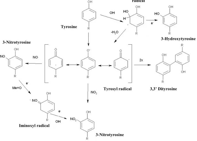
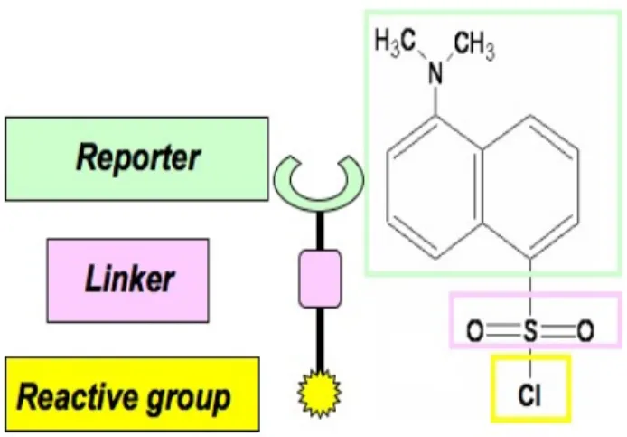
Il primo studio affrontato in questo lavoro di tesi è dedicato alla ottimizzazione di una procedura di analisi per migliorare l’identificazione dei siti di nitrazione nelle proteine. La nitrazione dei residui di tirosina è un’importante modifica post-traduzionale che può verificarsi in vivo durante fenomeni di stress ossidativo associati alla iper-produzione di ossido nitrico (1). Essa consiste nella sostituzione di un idrogeno in posizione orto nel gruppo fenolico dei residui di tirosina con un gruppo nitro (NO2). L’importanza biologica di questa modifica è globalmente accettata sia per le sue funzioni di mediatore della transduzione dei segnali cellulari sia come bio-marker di diverse patologie (2). Quest’ultima caratteristica è oggetto di numerosi studi, infatti la nitrazione delle proteine sembra essere implicata in numerose patologie riguardanti il sistema nervoso come il morbo di Parkinson, il morbo di Alzheimer e l’ischemia (3). Tuttavia, l’approccio classico di Proteomica presenta numerosi limiti nella rivelazione dei fenomeni di nitrazione in vivo. Infatti, anche se l’analisi mediante anticorpi specifici permette di identificare la presenza di proteine nitrate frazionate con tecniche di 2DE (4), il carattere sub-stechiometrico della reazione di nitrazione in vivo rende molto difficile l’identificazione dei siti di modifica. Inoltre, è stato anche dimostrato che l’analisi di spettrometria di massa risente enormemente dell’incorporazione del gruppo nitro nell’anello fenolico dei residui di tirosina (5). Per superare questi limiti sono state realizzate diverse metodologie alternative rivolte ad arricchire proteine o peptidi contenenti residui di nitro-tirosina. Questo è stato realizzato mediante tecniche di immuno-precipitazione (6) o anche di derivatizzazione chimica con reagenti che permettono la purificazione specifica dei residui marcati (7). Mediante quest’ultimo tipo di approccio è anche possibile migliorare l’analisi di spettrometria di massa, in quanto il gruppo nitro è convertito in una specie che favorisce la ionizzazione. Tuttavia le tecniche appena citate consistono di molteplici passaggi cromatografici che possono risultare dispersivi in termini di materiale, tempo e campione. In questo lavoro di tesi è stata ottimizzata una metodologia innovativa che, basandosi sulla modifica chimica dei residui di nitro-tyrosina, permette di identificare proteine nitrate in miscele complesse evitando preventive separazioni cromatografiche. La strategia prevede la riduzione dei gruppi nitro ad ammina e la successiva sulfonazione di quet’ultimi con il reagente cloruro di dansile (DNS-Cl). Le condizioni di reazione e analisi di spettrometria di massa sono state inizialmente ottimizzate utilizzando una proteina modello. A questo scopo è stata scelta l’albumina siero bovina (BSA), costituita da 20 tirosine, che è stata nitrata
in vitro. La selettività della reazione di marcatura è stata controllata dal pH della
soluzione che è stato mantenuto a 5,0, sfruttando in questo modo il peculiare pKa delle ammine aromatiche che è 4,7. L’utilizzo del cloruro di dansile permette di effettuare analisi di spettrometria di massa estremamente selettive con strumenti dotati di analizzatori ibridi triplo quadrupolo-trappola ionica lineare. Infatti, in precedenti studi è stato dimostrato che i peptidi modificati con il dansile generano in
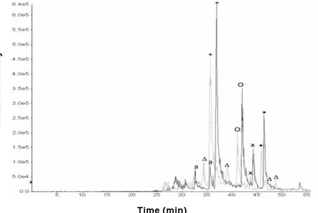
esperimenti di massa tandem frammenti peculiari con rapporto massa carica (m/z) 234 e 170 (8). Inoltre, quest’ultimo frammento può essere anche generato in esperimenti di tipo MS3 dal precursore a m/z 234. Tali caratteristiche di frammentazione sono state utilizzate per ottimizzare una procedura d’analisi LC-MSMS in modalità Precursor Ion Scan (PIS) e MS3. In pratica la miscela peptidica ottenuta dall’idrolisi delle proteine in esame è frazionata mediante cromatografia a fase inversa e analizzata direttamente mediante spettrometria di massa in modalità PIS. Questa modalità permette di ridurre l’analisi ad un numero molto ridotto di specie poiché sono rivelate solo le specie ioniche che generano frammenti a m/z 170 a seguito di collisione con gas neutro (CID), peculiarità tipica dei peptidi modificati con dansile. E’ stato stimato che in questa modalità operativa, per la BSA nitrata il 73% dei peptidi non nitrati non è stato rivelato. Combinando all’analisi PIS anche esperimenti di tipo MS3, che permettono di determinare quali ioni possono generare frammenti m/z 170 da frammenti m/z 234, è possibile definitivamente ridurre l’analisi alle sole specie marcate, con delezione del 100% delle specie di non interesse. La riduzione della complessità analitica permette di guadagnare enormemente in sensibilità, in quanto è incrementato considerevolmente il duty cycle del sistema. Il frazionamento in fase gassosa, realizzato dalla metodica di spettrometria di massa, può considerarsi dunque equivalente ad un passaggio di arricchimento dei peptidi nitrati, senza tuttavia aggiungere ulteriori sistemi cromatografici. L’applicabilità della tecnica è stata verificata in miscele complesse come estratti proteici di E.coli e di latte bovino, confermando la possibilità di individuare proteine nitrate e i corrispondenti siti di modifica anche in campioni di origine biologica. Infine i dati ottenuti da questo lavoro sono stati pubblicati da un’importante rivista di Chimica Analitica (9).
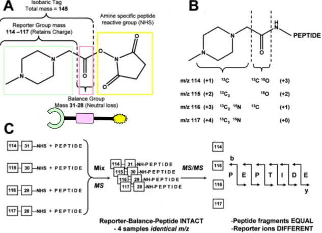
La strategia appena proposta tuttavia non permette di effettuare stime quantitative per valutare variazioni di concentrazione di proteine nitrate in differenti campioni. L’analisi quantitativa di questa modifica post-traduzionale rappresenta un vero limite della Proteomica, al punto che attualmente non ci sono procedure che permettono di identificare e quantificare allo stesso tempo i siti di nitrazione nelle proteine. Le uniche metodologie attualmente disponibili sono il saggio ELISA (10) e il dosaggio mediante gas cromatografia della nitrotirosina in forma aminoacidica ottenuta dall’idrolisi in HCl delle proteine (11). Tale problematica è stata affrontata in questo progetto di dottorato. In particolare sulla base dell’esperienza pregressa è stato ipotizzato che l’utilizzo di un criterio di elevata selettività in spettrometria di massa, accoppiato ad una marcatura altrettanto selettiva dei residui di interesse con un reagente arricchito nella componente isotopica, potesse permettere l’identificazione e quantificazione dei siti di nitrazione. Per questo scopo, non essendo disponibili in commercio isotopi del cloruro di dansile, è stato scelto come reagente la N-metilpiperazina-acetato N-idrossisuccinimide estere che è la molecola utilizzata nella più nota strategia di analisi quantitativa in Proteomica: iTRAQ (12). Il successo di questa metodica è dovuto alla peculiare struttura del reagente iTRAQ che permette l’acilazione delle ammine primarie. Esso è commercializzato in 4 forme isobare che differiscono tra loro per la composizione e distribuzione degli atomi pesanti del C, N, e O, consentendo di modificare i peptidi sempre con lo stesso incremento di massa. La strategia prevede la marcatura di fino a 4 profili d’espressione differenti e successivamente le miscele peptidiche sono unificate prima dell’analisi LC-MSMS. La grande innovazione di iTRAQ è l’indistinguibilità in modalità MS dei peptidi provenienti dai differenti campioni di origine, aumentando di fatto l’intensità del segnale rivelato e soprattutto non introducendo ulteriori segnali nello spettro di
massa. Nei relativi spettri di frammentazione MS/MS sono distinguibili i segnali peculiari del reagente iTRAQ utilizzato, rispettivamente a valori di m/z 114, 115, 116, 117 dalla cui misura dell’intensità è possibile realizzare stime quantitative relative. Differentemente dal cloruro di dansile, la chimica degli esteri della N-idrossisuccinimmide non permette di utilizzare il tipico pKa delle ammine aromatiche per realizzare acilazioni selettive dei residui di nitrotirosina a pH 5,0. Per questo motivo, prima della riduzione del gruppo nitro e della marcatura, le ammine primarie delle lisine e dei residui N-terminali dei peptidi devono essere acetilati. In questo modo è possibile successivamente condurre la marcatura a pH 8,0 senza reazioni aspecifiche. Anche in questo caso le condizioni di reazione sono state ottimizzate utilizzando come proteina modello la BSA. L’analisi di LC-MSMS è stata ancora una volta effettuata mediante uno spettrometro ibrido triplo quadrupolo trappola ionica lineare realizzando esperimenti in modalità PIS, usando come criterio di selettività uno degli ioni frammento peculiari del reagente iTRAQ (114, 115, 116, 117). Mediante questo tipo di analisi è stato possibile per la BSA eliminare il 96% dei peptidi di non interesse. Da un confronto con i risultati ottenuti mediante l’utilizzo del cloruro di dansile, risulta che l’analisi in PIS dei peptidi marcati con iTRAQ è molto più selettiva. Questo perchè la regione dello spettro MSMS dei peptidi compreso tra 114 e 117 è più libera di segnali rispetto a alla regione intorno al valore 170. Tuttavia non è stato possibile realizzare la completa rimozione dei peptidi non nitrati mediante analisi MS3, in quanto non sono state trovate transizioni efficaci. Basandosi solo sull’elevata selettività della PIS la procedura è stata quindi verificata su scala proteomica analizzando miscele complesse di estratti proteici di E.coli e di latte bovino, caratterizzati da differenti concentrazioni di proteine nitrate. E’ stato in questo modo possibile in una singola analisi identificare ed effettuare stime quantitative dei siti di nitrazione anche per proteine poco rappresentate. I dati ottenuti sono stati accettati e pubblicati da un’importante rivista di Proteomica (13).
Un altro aspetto affrontato in questo progetto di tesi riguarda il miglioramento dell’analisi MALDI-MSMS mediante la modifica chimica delle miscele peptidiche in esame. Questa problematica è stata largamente affrontata in altri lavori, dove è stato dimostrato che la frammentazione dei peptidi triptici in sorgenti MALDI generalmente non permette di ottenere una distribuzione omogenea dei segnali per consentire un corretto sequenziamento della regione N-terminale (14). Questo effetto è dovuto alla caratteristiche della ionizzazione MALDI che genera ioni mono-carica nella forma di addotti MH+. La presenza costante di un residuo fortemente basico di arginina o
lisina in posizione C-Terminale, ha un effetto di localizzazione della carica in questa regione, favorendo la generazione di ioni frammento appartenenti alla serie y. Inoltre l’effetto di localizzazione sopra citato, sfavorisce la frammentazione dei legami peptidici nella regione N-Terminale come predetto dalla teoria del protone mobile (15). Per superare questo limite sono state realizzate diverse strategie che prevedono la marcatura chimica dell’ammina N-Terminale inserendo un ulteriore “protone mobile” o una carica fissa, capaci di favorire la frammentazione dei legami e la ritenzione della carica nella regione N-Terminale dei peptidi (14). Queste strategie hanno permesso di migliorare la qualità degli spettri di frammentazione ottenuti con spettrometri di massa MALDI, tuttavia diminuendo l’intensità dei segnali in modalità MS. Inoltre tali strategie non sono mai state applicate su scala proteomica. In questo progetto di tesi è stato valutato l’utilizzo del cloruro di dansile per la marcatura del residuo N-Terminale dei peptidi con lo scopo di migliorare l’analisi MS e MSMS. Le proprietà in modalità MALDI-MS dei peptidi dansilati sono state già studiante in un precedente lavoro (16). E’ stato dimostrato che l’introduzione di un gruppo
dimetilammino-naftalenico, che ha un massimo di assorbimento alla lunghezza d’onda del laser utilizzato nelle comuni sorgenti MALDI, permette di migliorare il rapporto segnale rumore dei peptidi modificati in modalità MS. Inoltre è stato osservato che la marcatura degli idrolizzati triptici cambia la tipica “impronta digitale” di una proteina ottenuta mediante analisi MALDI-MS. Quest’ultimo effetto è dovuto alla natura della modifica introdotta, che cambia la gerarchia dei profili di ionizzazione, permettendo l’identificazione di peptidi non rivelati mediante un approccio classico, tuttavia a discapito di altri normalmente osservati. Gli autori hanno dunque proposto una strategia che prevede l’idrolisi delle proteine in esame, la marcatura con DNS-Cl di un’aliquota delle risultanti miscele peptidiche ed infine l’analisi in parallelo delle due miscele ottenute mediante Peptide Mass Fingerprint (16). La complementarietà dei risultati generati dall’analisi delle miscele marcate e non marcate, permette di ottenere considerevoli miglioramenti nell’attendibilità dei dati statistici e nella copertura di sequenza. In questo lavoro di tesi sono state migliorate le condizioni di reazione avvantaggiandosi dell’utilizzo delle micro-onde. Inoltre i dati sopra citati sono stati confermati mediante l’utilizzo di uno spettrometro di massa d’ultima generazione MALDI-TOFTOF. E’ stato realizzato uno studio più approfondito rivolto a comprendere l’effetto di variazione dei profili di ionizzazione dopo la marcatura, mediante l’utilizzo di un numero elevato di peptidi e di dati ottenuti da esperimenti MSMS. Da questo studio risulta che la modifica dell’ammina N-Terminale con un gruppo che assorbe la radiazione UV, aumenta il trasferimento di energia dalla sorgente laser al sistema matrice/campione, favorendo il desorbimento e la ionizzazione soprattutto dei peptidi dotati di m/z più bassi. Tuttavia la sulfonazione dei gruppi amminici causa una netta diminuzione del pKa di questi residui, comportando nei peptidi una minore tendenza a formare addotti MH+. Mentre
questo effetto è irrilevante per la conversione dell’ammina N-teminale (pKa ≃ 8,0), è stato osservato che il 91% dei peptidi non identificati in seguito alla marcatura conteneva un residuo di lisina (pKa ≃ 12,0), in posizione C-Terminale. I dati ottenuti spiegano la complementarietà osservata a seguito della marcatura con DNS-Cl, che ha permesso di identificare per la BSA 18 peptidi normalmente non osservati. In questo studio sono anche state valutate le caratteristiche di frammentazione dei peptidi dansilati in modalità Post Source Decay mediante uno spettrometro di massa MALDI-TOFTOF. Risulta che la presenza del residuo di dansile in posizione N-terminale permette di ottenere una migliore copertura della sequenza aminoacidica ricavata dall’interpretazione degli spettri MSMS. Infatti in fase di ionizzazione, il gruppo dimetilammino-naftalenico assorbe discrete quantità di energia che è ridistribuita nei nei vari sottolivelli vibro-rotazionali dei legami chimici lungo la catena peptidica. Questo comporta un aumento della probabilità di frammentazione del legame peptidico in prossimità del sito di dansilazione, infatti lo spettro MSMS risulta essere caratterizzato da una maggiore presenza di segnali relativi alla regione N-Terminale. In definitiva l’analisi complementare mediante la dansilazione dei peptidi triptici è vantaggiosa per aumentare la copertura di sequenza e l’attendibilità dei dati statistici anche per l’analisi LC-MALDI/MSMS. Ciò è stato verificato su scala proteomica dall’analisi di miscele complesse di estratti proteici di E.coli.
L’ultimo oggetto di studio affrontato in questo progetto di tesi è rivolto allo sviluppo di una procedura in grado di permettere l’analisi dei differenti stati di ossidazione dei residui tiolici in vivo. Tale problematica è attualmente di grande interesse in quanto la transduzione di importanti segnali biologici è regolata dalla modifica ossidativa reversibile dei residui di cisteina (17). Inoltre è stato osservato che diversi fenomeni patologici sembrano relazionati a una incorretta ossidazione di tali amminoacidi a
seguito di eventi di stress ossidativo (18). I metodi dell’analisi Proteomica classica non sono sempre efficaci per la definizione dello stato di ossadazione delle cisteine. Questo perchè la più parte delle modifiche è reversibile e quindi fortemente suscettibile ai tamponi normalmente impiegati per l’estrazione delle proteine e il frazionamento elettroforetico. Per questi motivi sono stati studiati approcci alternativi che consistono nell’alchilazione differenziale dei tioli, in tamponi riducenti a selettività decrescente (19). La pletora dei reagenti alchilanti impiegati permette oggigiorno di valutare le variazioni dello stato ossidativo dei tioli mediante le classiche tecniche di Proteomica. Tuttavia i reagenti utilizzati non sempre favoriscono tutti i passaggi analitici che caratterizzano l’approccio proteomico. In questo progetto di tesi sono stati effettuati studi preliminari per valutare l’applicabilità dei derivati del dansile nella caratterizzazione delle modifiche post-traduzionali connesse allo stato d’ossidazione dei tioli. Infatti, oltre alle eccezionali caratteristiche di spettrometria di massa mostrate nei precedenti studi di questo progetto di tesi, i dansil derivati possono essere impiegati per la rivelazione di fluorescenza di proteine frazionate per elettroforesi (20). Inoltre in bibliografia sono presenti diverse metodologie che prevedono l’utilizzo dei dansil-derivati nelle tecniche di istochimica (21) e citometria a flusso (22). Dunque sembrerebbe possibile sviluppare per la prima volta una metodologia che prevede l’utilizzo di un unico reagente per la determinazioni dello stato ossidativo dei tioli nei tessuti e cellule compatibile con il frazionemento elettroforetico e l’analisi di spettrometria di massa. La strategia prevede la marcatura dei tioli con il reagente dansil-aziridina (DAZ) e successiva alchilazione con iodoacetammide in un tampone riducente. Per raggiungere questo scopo sono state ottimizzate le condizioni di reazione ed è stata verificata la selettività del reagente utilizzando come proteina standard la BSA, caratterizzata da 34 cisteine ossidate in 17 ponti disolfuri e una in forma ridotta. Verificata l’efficienza della reazione, la BSA modificata con DAZ è stata frazionata mediante SDS-PAGE per valutare la sensibilità della rivelazione di fluorescenza che è stata stimata compresa tra 1 e 10ng di proteina marcata. E’ stata inoltre valutat la possibilità di apprezzare ossidazioni delle cisteine mediante variazione dell’intensità di fluorescenza in proteine frazionate mediante elettroforesi. Questo è stato realizzato facendo reagire la BSA con una soluzione 1 mM di H2O2 e successivamente applicando il protocollo
di marcatura ottimizzato. E’ stato osservato che la banda corrispondente alla BSA ossidata presentava un’evidente diminuzione della fluorescenza dovuta alla minore disponibilità di tioli liberi dopo la reazione di ossidazione. Questo tipo di analisi è stata anche estesa ad un estratto proteico di E.coli permettendo di confermare la specificità e l’applicabilità del metodo su scala proteomica. In questo progetto di tesi sono state anche ottimizzate le condizioni di reazione per realizzare la marcatura dei tioli intra-cellualre. Questo è stato possibile perchè i dansil derivati sono permeabili alla membrana cellulare. E’ stato verificato che la quantità di reagente utilizzato non fosse stata nociva per le cellule batteriche ed umane mediante test del blu tripano, microscopia in fluorescenza e citometria a flusso. Dunque, cellule batteriche e umane sono state sottoposte a stress ossidativo indotto da H2O2 ed inseguito trattate
con dansil-aziridina. Mediante citometria a flusso è stato possibile discriminare la popolazione di cellule sottoposte a stress in quanto presentava una minore emissione fluorescenza rispetto al controllo. Inoltre dall’analisi dell’immagine dei gel 2DE degli estratti cellulari relativi ai campioni sopra citati è stato possibile identificare le proteine coinvolte nei processi ossidativi. Allo stato attuale la strategia presentata deve ancora essere ottimizzata in alcuni punti. Infatti, devono essere valutate eventuali differenze tra la marcatura in vitro ed in vivo, dovute alla differente
accessibilità dei residui di cisteina da parte del reagente. In caso affermativo si potrebbe considerare la possibilità di estendere la procedura anche a studi conformazionali e di interattomica. Inoltre allo stato attuale la strategia, come tutte le altre metodologie presenti in letteratura, anche se permette di identificare la proteina coinvolta nel processo di ossidazione non consente sempre in maniera univoca la determinazione del sito di modifica. Questo scopo sarà raggiunto, nel prosieguo di questo progetto, quando saranno sintetizzati analoghi del dansile arricchiti della loro componente isotopica.
Bibliografia
1. Radi R., Proc. Natl. Acad. Sci. U S A 2004, 101(12):4003-4008.
2. Ohshima H., Friesen M., Brouet I., Bartsch H., Food Chem. Toxicol. 1990 28(9): 647-52
3. Sacksteder C.A., Qian W.J., Knyushko T.V., Wang H., Chin M.H., Lacan G., Melega W.P., Camp D.G., Smith R.D., Smith D.J., Squier T.C., Bigelow D.J.,
Biochemistry 2006, 45(26):8009-22
4. Kanski J., Behring A., Pelling J., Schöneich C., Am. J. Physiol. Heart Circ.
Physiol. 2005, 288: H371–H381;.
5. Sarver, A., Scheffler N.K., Shetlar M.D.; Gibson B.W., J. Am. Soc. Mass
Spectrom. 2001, 12:439–448.
6. Thomson L., Christie J., Vadseth C., Lanken P.N., Fu X., Hazen S.L., Ischiropoulos H., Am J Respir Cell Mol Biol. 2007, 6(2):152-7
7. Nikov G., Bhat V., Wishnok J.S. and Tannenbaum S.R., Anal. Biochem. 2003, 320, 214-222.
8. Amoresano A., Monti G., Cirulli C., Marino G., Rapid Commun. Mass Spectrom.
2006, 20:1400-1404.
9. Amoresano A., Chiappetta G., Pucci P., D'Ischia M., Marino G., Anal. Chem.
2007, 79(5):2109-17
10. Zheng, L., Nukuna, B., Brennan, M. L., Sun, M., Goormastic, M., Settle, M., Schmitt D., Fu X., Thomson L, Fox P. L., Ischiropoulos H., Smith J. D., Kinter M., and Hazen S. L., Clin. Invest. 2004, 114: 529–541
11. Leeuwenburgh C., Hardy M.M., Hazen S. L., Wagner P., Oh‐ishi S., Steinbrecher U. P., J. Biol. Chem. 1997. 272: 1433–1436.
12. Ross P.L., Huang Y.N., Marchese J.N., Williamson B., Parker K., Hattan S., Khainovski N., Pillai S., Dey S., Daniels S., Purkayastha S., Juhasz P., Martin S., Bartlet-Jones M., He F., Jacobson A., Pappin D.J., Mol. Cell. Proteomics. 2004, 3(12):1154-69.
13. Chiappetta G., Corbo C., Palmese A., Marino G., Amoresano A, Proteomics 2008 in press
14. Pashkova A., Chen H.S., Rejtar T., Zang X., Giese R., Andreev V., Moskovets E., Karger B.L., Anal. Chem. 2005, 77(7): 2085-2096
15. Wysocki V.H., Tsaprailis G., Smith L.L., Breci L.A., J. Mass Spectrom. 2000, 35: 1399–1406
16. Park S.J.. Song J.S.. Kim HJ.. Rapid Commun. Mass Spectrom. 2005. 19(21):3089-96
17. Biswas S., Chida A.S., Rahman I., Biochem. Pharmacol. 2006, 71:551-564
18. Dafre A.S., Artenib N.S., Siqueirab I.R. and Netto C.A., Neuroscience Letters
2003, 345 (1): 65-68
20. Hsi K.L, O’Neill S.A., Dupont D.R., and Yuan P.M., Anal. Biochem. 1998, 258: 38-47
21. Braga-Vilela A.S., de Campos Vidal B., Acta Histochem. 2006, 108(2):125-32. 22. Jones R.J., Barber J.P., Vala M.S., Collector M.I., Kaufmann S.H., Ludeman
S.M., Colvin O.M., Hilton J., Blood. 1995, 85(10):2742-6
Résumé
Le travail de thèse présenté dans ce mémoire consiste en la mise au point de nouvelles méthodologies d’analyse pour les études protéomiques. Le sujet principal de ce travail de thèse a été l’optimisation de procédures innovantes qui se basent sur la manipulation chimique de mélanges peptidiques et sur l’ analyses avancée par spectrométrie de masse MS3, et qui ont pour objectif d’améliorer les capacités d’identification et quantification des modifications post-traductionelles par une approche protéomique.
Une nouvelle procédure d’identification des sites de nitration des protéines a été mise au point, par le moyen d’une modification sélective des peptides nitrés après dansylation et par l’analyse par spectrométrie de masse en modalité Precursor Ion Scan/MS3. Une telle stratégie pemet de surmonter quelques unes des limites classiques de l’analyse des sites de nitration des protéines, comme la nécessité d’utiliser des techniques d’enrichissement chromatographique mais permet aussi de surmonter les difficultés d’identification des peptides nitrés par spectrométrie de masse. Il a également été optimisé une procédure pour l’identification et la quantification des sites de nitration des protéines par le biais d’une modification sélective des peptides nitrés avec le réactif iTRAQ et par une analyse successive par spectrométrie de masse en modalité Precursor Ion Scan. Cette technique peut etre considérée comme étant la première stratégie capable simultanément d’identifier et de quantifier les sites de nitration des protéines par spectrométrie de masse. En outre, différentes conditions d’analyses pour l’identification des phospopeptides dans des mélanges biologiques ont été évaluées par analyse LC-MSMS en modalité neutral loss et precursor ion scan. De plus, une nouvelle procédure qui utilise la chimie des micro-ondes afin d’améliorer les analyses LC-MALDI-MSMS a également été optimisée, par un marquage extensif des mélanges peptidiques analysés avec du chlorure de dansyle. Cette étude a également permis de comprendre les caractéristiques de la fragmentation des peptides dansylés dans des éxpériences de post source decay. Enfin, des expériences préliminaires ont été réalisées afin de vérifier la possibilité d’utiliser les dérivés de la dansylation pour développer une nouvelle procédure finalisée à l’identification des états d’oxydations des résidus de cystéine des protéines séparées par une électrophorèse bi-dimensionelle. Les rèsultats obtenus indiquent que les dérivés du dansyle permettent de déterminer les variations des états d’oxydation des cysteines par des technniques de cytométrie en flux, électrophorèse bidimensionelle et spectrométrie de masse.
1. INTRODUCTION
1.1 Proteomics impact on modern Biotechnologies
In modern terms, Molecular Biotechnology has come to mean the use of cells and tissue cultures, molecular biology, and in particular recombinant DNA technology, to generate unique organisms with new features leading to produce specific products, that many times are proteins. On this basis, the fields of the Industrial (white) Biotechnology, Medical (red) Biotechnology, Vegetal (green) Biotechnology, can be distinguished if these tools are applied to the development of industrial processes, molecular studies of the diseases and drug discovery, agricultural and food production improvement respectively. Most of the biotechnological products result from the use of recombinant DNA technology, where a recombinant production system is set up, based on a genetically modified host cell, using either microbial fermentation or mammalian cell cultures. In addition to fermentation, the extraction and subsequent purification of the desired bio-product from the fermentation broth is an important part of the overall production process. These downstream procedures form a major part of the overall process development and are important in the determination of the final productivity and characteristics of the product. Traditionally, process development involves designing and optimizing upstream and downstream processes based on empirical data, with only incomplete or limited understanding at the cellular level. The advent of Genomic and Proteomic technologies, which can provide details at the molecular level, has generated interest in the application of these tools for the development of biotechnological processes.
Genomics is the comprehensive analysis of the genetic content of an organism. It also often refers to genome wide studies of mRNA expression (1). Already during the “genomic era” that ended with the sequencing of Human Genome in the year 2003, the scientific community realized that the identification of coding sequences is insufficient to understand the molecular mechanisms of cell activity. Therefore, the attention increasingly focused on the products of the genome: the proteins and enzymes that determine cellular architecture and function. According to the current annotation, the human genome consists of about 25000 genes, scattered among 3 billions nucleotides of chromosome-based DNA code (2). This represents a huge amount of static information, which needs to be correlated with dynamic information coming from gene products and their interactions. In contrast to the genome, the proteome is dynamic and is in constantly modulated because of a combination of factors, which include mRNAs differential splicing, post translational modifications (PTMs), as well as temporal and functional regulation of gene expression as well as the formation of multi-protein complexes.
Proteomics provides methods for correlating the vast amount of genomics information that is becoming available with the equally vast protein information that is being produced through analysis of cells under normal versus altered states (3). In the last few years Proteomics has become a powerful tool for the investigation of complex biochemical processes and protein-protein interactions (4-5). In medical sciences, the discovery and the use of disease biomarkers, especially in cancer and neurodegenerative diseases, has became a routine analysis for a better and more targeted medical treatment (5–8). Currently, Proteomic methods are widely used in food biotechnology as quality control so the terms “industrial process proteomics” (9) and “industrial proteomics” (10) are currently used. It has also been shown that proteomic technology can be very useful in the development of production processes
for therapeutic proteins by using genetically engineered animal cells (11-12) or human stem cells (13). In “classical” fermentation industry, Proteomic methods can also be used for the identification of targets for bioprocess improvement (14). Unlike mammalian cells that are used almost exclusively for production of protein-based therapeutics (15), microbial cells are also widely used for production of small molecules (14). Microbial fermentation in food processing has been a tradition for thousands of years (16). Besides optimization of the fermentation process, purification, optimization of yield and purity, the characterization of the final process are crucial steps in integral process development.
In the near future, in parallel with genomics and metabolomics, proteomics will play an important role in process development. These technologies will be used to enhance productivity of microbial cells or to influence targeted properties of the final product. Knowledge of the cell proteomics will be helpful to predict the cellular response to environmental change and its adaptation to different substrates. Finally, proteomics are important for optimization of downstream processing and for thorough analytical investigation of the final products.
1.2 Proteomics analysis
The terms Proteomics is associated to the set of analytical tools used to depict the protein compartment of a cell. This innovative “biotechnology” is the natural continuation of Genomics approach. It moves away from classical Protein Chemistry, also if from the latter, taking advantages of all the heritage of knowledge and methods developed from the latter. In fact, the great innovation of the Proteomics analysis is the idea that to study the cellular molecular mechanisms, in which proteins play a key role, it is necessary to study the entire proteome as a “single analyte”. Meaning that, the proteins target of the analysis are no more purified and isolated from their highly complexity context found in living systems. Indeed, they are analyzed all together in order to obtain a real snapshot of the proteome, related to a particular cell state. From this general definition it may distinguish two main area of interest: i) “expression proteomics” that is focused to the characterization of the change in protein expression levels and definition of PTMs (17), ii) “functional proteomics” aimed to understand protein-protein interactions (18), signal pathways (19) and structure function relations.
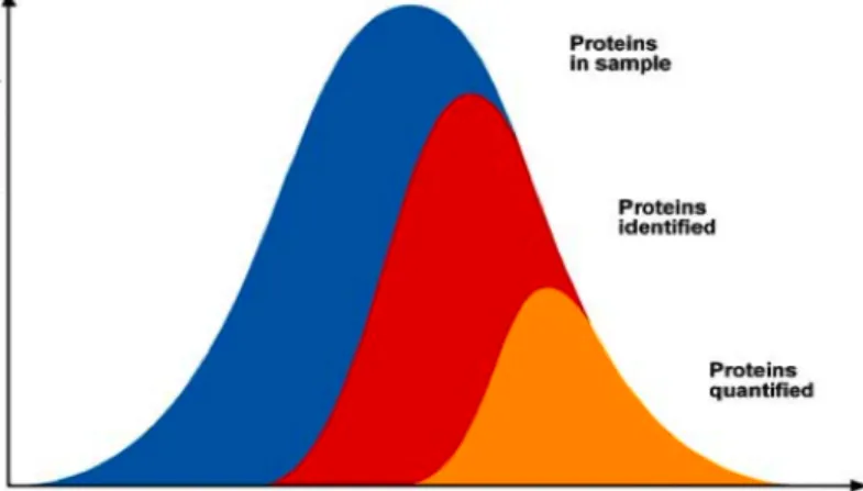
However, the proteome analysis is hindered by several analytical problems. First of all is the large range of protein concentration present in the samples. For example, in the human serum the 50 most abundant proteins represent about 99% of the total amount of protein mass but only less than 0.1% in number (20). Another important challenge is surely the detection of PTMs. In fact only a minor part of the proteins of interest are post translationally modified, for instance just 1 in 106 tyrosines are nitrated (21). The high sample complexity, in terms of number of analytes, is also a feature that has to be taken in account, in fact for about 21000 human protein-encoding genes are estimated around 106 human proteins (22). For these reasons the Proteomics analysis needs a pool of methodologies and technologies that are high throughput, sensitive, selective toward the proteins target of the analysis and with large dynamic range effectiveness.
Currently, Proteomics may rely on many chromatographic and electrophoresis tools to fractionate the analytes. However, if different approaches are in relation to these techniques of separation, all the strategies have a common essential final step: the mass spectrometry (MS) analysis of peptides or proteins.
-1.2.1 Mass spectrometry tools for Proteomics analysis
a) Principles and instrumentation
Mass spectrometers consist of three basic components: an ion source, a mass analyser, and an ion detector. Molecular mass measurements are carried out indirectly, calculating the mass charge ratio (m/z), by the analysis of kinetics behaviour of the ionized analytes in the gas phase and in electromagnetic fields. The need of a method that transfers efficiently intact molecules from solution or solid phase into the gas phase, was the limit that for many years excluded proteins and peptides from this powerful technique. With the development and the commercialization of mass spectrometers based on the ion source technologies of
“Matrix-Assisted Laser Desorption/Ionisation” (MALDI) (23) and “Electrospray Ionisation” (ESI) (24) the gas phase ionization of polar analytes has also become
possible. Both MALDI and ESI are considered as soft ionisation techniques with which the generated ions undergo minor fragmentation.
In MALDI technique, samples are co-crystallised with a weak aromatic acid matrix on a metal target. A pulsed laser is used to excite the matrix, which causes rapid thermal heating of the molecules and desorption of ions into the gas phase in a
pulsed beam fashion. After the laser activation, the weak acid nature of the matrix
drives to generate single-charged ions in the form of MH+ adduct. The one-one analyte-MS signal relation in MALDI-MS makes possible to analyze also multi-component mixtures without any interpretation difficulties caused by the peaks overlapping.
ESI creates ions by spraying an electrically generated fine mist of ions into the inlet
of a mass spectrometer at atmospheric pressure. By creating a potential difference between the capillary, through which the liquid flows, and the inlet of the mass spectrometer, small droplets of liquid are formed. These are transferred into a heated device to induce efficient evaporation of solvent. Once the droplets have reached the Rayleigh limit, ions are desorbed from the droplet generating gas-phase ions in a
continuous beam fashion. The ions produced by ESI sources are multiply charged
adducts MHnn+, where the number of protons incorporated depends on the statistical
acid/basic equilibrium of the analytes. This situation complicates the spectra interpretation because each analyte may give rise to many signals in the spectrum. For this reason it is not possible to analyze complex mixture without a previous fractionation. However, because the ionization is carried on a liquid flow, ESI source may be simply coupled to liquid chromatography systems. The most notable improvement of ESI technique has come from the reduction of the liquid flow rate used to create the electrospray to nano-scale level. This device leads to create ions more efficiently (25) because the charge density at the Rayleigh limit increases significantly with decreasing droplet size (zR α 8R3/2 where R is the droplet radius).
Another advantage using separation techniques with low flow rate ESI is the increase in the concentration of the analyte as it elutes off the column.
After ionisation, the analytes reach the mass analyzer, which separates ions by their mass-to-charge (m/z) ratios. Ion motion in the mass analyser can be manipulated by electric or magnetic fields to direct ions to a detector, usually an electro-mcultiplier, which records the numbers of ions at each individual m/z value converting the signals
in current. In Proteomic research, four basic kinds of mass analysers are currently
Fig.1.1 Main components of Proteomics commonly used mass spectrometers
used: time-of-flight (TOF), ion trap (IT), quadrupole (Q), and Fourier transform ion
cyclotron resonance (FTICR) analysers (Fig.1.1). All four differ considerably in
sensitivity, resolution, mass accuracy and the possibility to fragment peptide ions. By using a TOF analyzer the mass spectrum is measured by determining the flight time of ions after the extraction and the acceleration from the source by an electric potential. The ions are separated in a field-free flight tube. The ions time-of-flight is related to their m/z values:
m/z= 2Et2/d2, E= potential of extraction, t= time of flight, d=length of the tube
The experimental arrival times of each ion to the end of the flight tube can be a mass spectrum can be acquired. The two main improvements to enhance the resolution of this technique were the “delayed extraction” that corrects the initial velocity vectors and the “reflectron” (26) that increases the ions path, curving their trajectories. TOF based instruments have high resolution and accuracy characteristics and they are usually coupled to pulsed beam type sources.
The simpler mass analyzer is the quadrupole that consists of four precisely straight and parallel rods that are aligned in the axial direction of ion beam. A voltage combining a DC component and a radio frequency (RF) component is applied between adjacent rods but with inverse polarity, opposite rods are electrically connected. Once inside the quadrupole the ions will oscillate in the (x) (y) direction as a result of the electric field. The differential equation which describes the motion of the charged particle along the x or the y-axis is:
(with ω the frequency of RF, r0 the radius between the electrodes, U is the constant
DC voltage, V the maximum RF voltage, m the ion mass and e its charge). The general solution of this differential equation was found by Mathieu and has the form:
f =AeµτF(τ) + B-µτ (-τ), with µ (a, q)=α +iβ
A and B are the constant of integration while α and β are real function of a and q
(27). It is possible to demonstrate that for a given m/z value ion oscillations are stable only for a restricted couple of a and q values that are operative depending respectively by the DC and RF voltage. Otherwise the ions have very large x,y coordinates and strike the rods and dissipate. The family of Mathieu stable solutions may be graphically represented in function of RF and DC. The resulting diagram helps to understand for a given m/z for what couple of RF and DC values it would have stable trajectories (Fig.1.2A). The symmetry around the DC voltage =0 means that in only RF mode the quadrupole lets pass all the ions. The mass filtering is realized by changing in time the DC voltage and the RF frequency, keeping constant their ratio (Fig.1.2B), therefore instantly varying the ions that are in their boundary stability condition.
The three-dimensional ion traps (IT) are composed of three hyperbolic electrodes: two end-caps and one ring. The ring electrode defines the radial direction and the end-cap electrodes define the axial direction. One end-cap electrode will usually have a hole in it to allow ions to enter the trapping volume. The ions are injected axially with kinetic energies that lead to pass the fringing field barrier near the end-cap hole. However, to overcome the exit of the ions, their high kinetic energy is dissipated by cooling them by collision with neutral gas by filling the trap with helium (28). In the IT ion trajectories undergo the same quadrupole rules and motion
Fig. 1.2 Panel A shows a RF voltage vs DC voltage diagram of Mathieu stable solution for the ion
219 m/z in a 9.5 mm diameter quadrupole. All the RF-DC couples of voltage values inside the curve give to the ion stable trajectories and may be detected. Panel B shows the effect of the quadrupole scanning. In the RF voltage VS DC voltage diagram are represented three stability curves for the ions 28 m/z, 69 m/z and 219 m/z. The “operative line” starting from the origin represents the RF and DC ratio used during the scan in function of the time. When the line crosses the stability region of an ion it means that it is actually scanned. By this representation it is showed that with a linear scan line through the origin, the peak width increase geometrically with increasing mass. By using higher DC/RF ratio the operative line increase its slope and it passes through less stability regions resulting in higher resolution scan.
equations showed before. However, the geometry of the electric field used gives to the ions an elliptical trajectory in the radial plane so they are confined inside the volume of the electrodes, and maintains a stable periodic motion in the axial direction with a peculiar secular frequency ωz for each m/z (29). By applying a supplementary
voltage at the frequency ωz,the axial oscillation is increased causing the ions ejection
from the trap. By timely changing the frequency it is possible to scan the mass range. Mass spectrometer equipped with IT technology has good sensitivity but low resolution and mass accuracy.
The most common FTICR analyzer consists of a cell composed of six plates arranged in the shape of a cube (30). This cell is oriented in a magnetic field (4,7-13 Tesla) so that one opposing pair of plates is orthogonal to the direction of the magnetic field lines. It is common for trapping electrodes of cubic cells to have openings that permit electrons or ions to enter the cell along the magnetic field lines. The four remaining plates are used for ion excitation and ion detection. When the ions pass into the magnetic field they are bent into a circular motion in a plane perpendicular to the field by the Lorentz Force. The cyclotron frequency of the ions rotation is dependent on their m/z ratio
ωc=eB/2πm, ωc, secular frequency, B=magnetic field
At this stage, no signal is observed because the radius of the motion is very small. The excitation of each individual m/z is achieved by a RF pulse across the plates of the cell. Each individual excitation frequency, corresponding to the peculiar ωc value,
will excite the ions natural motion to a higher orbit inducing an alternating current between the cell plates. When the RF goes off resonance for that particular m/z value, the ions relax to their natural orbit and the next m/z packet is excited. Although the RF sweep is made up of a series of stepped frequencies, it can be considered as all frequencies simultaneously. This results in the measurement of all the ions together producing a complex frequency vs. time spectrum containing all the signals. The deconvolution of this signal by Fourier transform methods results in a frequency vs intensity spectrum, which is finally converted to the mass vs. intensity spectrum. FTICR mass spectrometers are the most accurate and the highest resolving power instruments currently available. (31)
b) Tandem Mass Spectrometry
Conventional MS produces ions that are separated by m/z and analyzed directly. If a soft ionization method is used, the mass spectrum will lead to calculate the molecular weight values of compounds present in the analyte but with little or no structural information. In a tandem mass spectrometer, ions of a particular mass (parent or precursor ion) are selected by the first mass analyzer (MS1) can be subjected to collisions with neutral gas atoms or molecules. This process is known as Collision
Induced Dissociation (CID). The ionic fragments (daughter ions) are then separated
in a second analyzer (MS2), giving structural information such as the amino acid sequence of a peptide. Therefore, to realize the multi-stage MS analysis many mass spectrometers equipped with hybrid combinations of analyzer were commercialized. Triple quadrupole (QqQ) (32), quadrupole/ time of flight (QqTOF) (33) and time of flight/ time of flight (TOFTOF) (34) may perform “space separated” tandem MS. Ion trap analyzer by itself may realize tandem MS experiments. This is accomplished by the expulsion of the ions trapped in MS mode, isolating the parent ion. This one is further fragmented by collision and the produced ions are first accumulated, then
scanned and detected. This “time separated” operative mode allows to realize very sensitive MSn experiments with simple equipments. However, the usage of a unique
quadrupole trap analyzer leads to record MSn spectra with low mass range cut-off. In fact, during their generation the daughter ions have experience of the RF and DC values used to trap the precursor ion.
These couple of potential values are in the stable region of Mathieu graphic for ions with m/z within 60% of the parent ion m/z, thus resulting in the complete loss of the smaller fragments (28). The chemistry of the fragmentation depends on many factors as the energy transferred to the ions, the time elapsing between the activation and the dissociation, the distribution of the internal energy (35). The amount of energy that can be deposited in an ion depends on the relative collision energy of the colliding ion/neutral pair under the classical impact rules. Considering that the neutral gas molecules have no velocity component the maximum centre-of-mass energy is:
Ecom= (N/N+m) Ekin
with N the neutral gas the mass, m the ion mass, Ekin the parent ion kinetic energy
(36-37). It results that heavier ions are more difficult to fragment and, by using heavier neutral gas, higher amount of energy are transferred to the parent ion. The Ekin value depends on the mass spectrometer optics and the operative mode. It is
also related to the energy transfer kinetics and to the distribution in internal degrees of freedom. CID experiments of peptides and proteins are realized in three different modes: “High Energy”, “Low Energy” and “Very Low Energy”. High Energy CID processes involve keV kinetic energy of the parent ion and they are carried out by sector-based instruments (38). The gas pressure inside the spectrometer is kept low, thus 1-5 collisions take place between the fragmentation events. Consequently a large part of ion population increments the internal energy of few eV while the remaining has high energy favouring only a restrict number of critical sequential dissociation (Fig.1.3A). Therefore the efficiency of the process is low (10%) and the spectra consist principally of d and w ions characterized by side chain fragmentations
Fig.1.3 Panel A shows the internal energy distribution for an ion population after a rapid activation
High Energy CID (7keV). In these condition the collision are rare and high energetic. Thus, it appears that only a restricted number of ions increases their internal energy with high increment resulting in poor sample utilization and drastic fragmentation. Panel B shows the ions internal energy distribution after a slow activation Low Energy CID (28 eV). In these conditions the collisions are more probable and the ions are cooled by the higher operative pressure (0,5 mtorr). Thus, it results that a large number of ions increases their internal energy favoring peptide bn,, yn and
immonium ions generation. Panel C shows the ions internal energy distribution after a slow activation Very Low Energy CID (26 eV). The higher operative pressure (2 mtorr) and the lower ions kinetic energies lead to a very slow activation compatible with the formation of a reaction intermediate. It results in a Gaussian curve without high energy tail. Thus, the fragmentations follow the Arrhenius theory producing mainly bn,, yn ions
only useful to discriminate isobaric amino acids (38). Low Energy and Very Low Energy CID processes involve 1-100 eV kinetic energy realized by using QqQ and QTOF and Ion
Trap instruments
respectively. Higher gas pressures are applied leading to 10-100 collisions between dissociations events. Under multiple collision conditions, energy is deposited in small increments, thus such activation effectively gives rise to a statistical internal energy distribution without
higher component
(Fig.1.3B and C). Consequently, no d or w ions or sequential dissociation is observed. The difference between Low Energy and Very Low Energy CID is essentially due to the “beam type” and “no beam type” features of the spectrometers. In fact the ions in QqQ and QTOF instruments have higher kinetic energy that may causes with the classical b and y ions also internal fragments as immonium ions (Fig.1.4) or a and x ions generated by the cleavage of α-C and carbonyl-C. On the contrary in the Ion Trap the ions are “cooled” resulting in lower energy fragmentations with enhanced efficiency (50-100%)(39). The peptide bond cleavage is the principal event using Low and Very Low Energy CID, giving rise to b and y ions if the charge is retained on the N-Terminal or C-N-Terminal containing moiety respectively (Fig.1.4). It is commonly accepted that the cleavage occurs predominantly through charge-directed pathways. In practice the fragmentation is initiated by a charge that is transferred to the vicinity of the cleavage site after the hydrogen rearrangement as described by the mobile proton model (40). This interaction of the charge and the heteroatoms is more important in the gas phase because the solvent molecules that typically stabilize the molecules in the solution phase are not present in the gas. However, a charge-remote component is also observed in peptides CID fragmentation by which the acquired proton is localized on the more basic sites and takes no part to the fragmentation mechanism. In this pathway the peptide bond cleavage takes place by internal proton rearrangements, with charge retention on the initial protonated moiety. During this hybrid fragmentation pathway, singly-charged tryptic peptides give rise predominantly to y ions, by the presence of a strong basic lysine or arginine in C-Terminal position. The ions of b series are better visualized in multiply-charged peptides where the probability to have a N-Terminal charge retention is higher. The resulting Low Energy CID fragmentation spectra recorded with the common mass
Fig.1.4 Principal fragmentations of Low and Very Low Energy
spectrometer consist of a large number of peaks related mainly to the b and y series ions and also to eventual neutral loss of NH3, CO and H2O. By the interpretation of
these spectra it is very simple to reconstruct the peptide sequences calculating the difference between the mass values relative to the ions belonging at the same series. This procedure may be also automated by using special algorithms (41-42) leading to the de novo sequencing or the protein database searching (43-44).
-1.2.2 The first generation of the Proteomic analysis
Proteomic analysis is aimed to analyze the whole proteome as a single analyte. However, the high complexity of the mixture examined imposes to use fractionation procedures in order to simplify the analysis without losing a global view of all the protein component of the samples. Bidimensional electrophoresis was the first technique leading to study the proteins on Proteomic scale and it is currently the leader technique in this field. Although 2DE development was realized many years before the post-genomic era (O’farrel et al. 1975) (45) the usage of this technique is still of high value because it allows to separate protein with high resolution (up to 10000) (46) giving rise at the same time to protein maps of the biological matrix examined. 2DE consists of a first fractionation step by Iso-Electric Focusing (IEF) leading to separate proteins by their typical iso-electric point. In the second step, co-focalized proteins are orthogonally separated by their hydrodynamic volume by SDS-PAGE. 2DE conversion from only-imaging technique to Proteomic leader technique was favoured both by the commercialization of the first MALDI-MS instruments and by the enrichment of sequence database using genomic data. Thus, the synergy between 2DE separation features, MALDI-TOF accurate and sensitive mass measurement, the sequencing database software versatility, lead to develop in 1993 the Peptide Mass Fingerprinting (PMF) (47-49) strategy that may be considered the first Proteomic high throughput methodology. PMF is based on the idea that a protein digested by an enzyme with known specificity produces a peptides pool that may be used as discriminatory for its identification. MALDI-TOF analysis produces a unique spectrum giving the accurate monoisotopic mass of all the peptides produced by the protein digestion, producing peculiar molecular masses map for each protein. This map is then compared to the ones generated in silico by the virtual digestion of all the protein sequences present in the genomic database. The results are statistically analyzed to find the best match. Therefore, such gel based strategy consists of six analytical steps: I) protein recovery from biological matrix II) 2DE fractionation III) Image analysis IV) in gel protein digestion V) MS analysis VI) Data base searching. The statistic nature of the protein identification, without any information about the amino acid sequence, implies a high hydrolysis yielding optimize the sequence coverage. In addition, in order to avoid misunderstanding results high protein purification and high mass accuracy of the mass measurements is also important. While the last two features are achieved by coupling 2DE and MALDI-TOF, it must be considered that the in gel hydrolysis leads to poor sequence recovery. This characteristic was the most important limitation of the first generation Proteomic analysis. The introduction of MS2 This problem was in part overcome with the
introduction of nanoLC-MS2 technique in 1996, leading to determinate peptide amino
acid sequence (50-51). So, the association of molecular weight and amino acid sequence for each given peptide is a milestone of Proteomic evolution. The statistic relevance of the sequence data coupled to the mass measurement allows to the un-ambiguous protein identification, even if a reduced number of peptides is detected. In
the last 10 years many technological progresses have changed the materials and methods of the gel-based Proteomics. New gel dyes were introduced in order to enhance the 2DE sensitivity (Brillant Blue Coomassie, Sypro, DIGE) (52-53) and the selectivity toward the post-translational modifications (Pro Q diamond, Pro Q emerald) (54-55). Mass spectrometry analysis has been improved by using new instruments with higher resolution power and faster scan rates. The electrophoresis techniques are radically changed with the introduction of the first dimension gel strip with pre-formed gradient. Also research algorithms were powered and protein database were enriched, in particular considering the complete human genome sequencing in 2003. This overall evolution of modern Proteomic is driven by the methodological revolutions in the protein analysis over the years 1993-1995. This is particularly true if we consider that the six steps procedure is still currently un-varied in many Proteomic applications of the more recent research lines (56-57).
-1.2.3 The second generation of the Proteomic analysis
The great results reached by the coupling of the 2DE high resolution power and MS flexibility were the driving force for the development of modern technologies and high throughput strategies. In fact, the analysis carried out by 2DE-MS presents many constitutive limitations for the study of some kinds of Biological aspects. For example, the analysis of membrane proteins is still a challenge, because of the poor solubility of the sample and the low recovery after the extraction procedures. To avoid 2DE high conductivity, that may induce the gel over-warming, low quantity of sample must be analyzed. Under this condition a high percentage of less abundant proteins never reaches the detection limits and remains un-analyzed. For the same reason the PTMs identification is a challenge because of the sub-stoichiometric characteristics of the biological modifications. In addition the 2DE-MS strategy is not a high throughput approach. To identify the detected proteins a large number of spots have to be processed, however the method is time consuming and it requires an intensive work of skilled operators. This is very limiting because the modern Proteomic studies relies on reproducible analysis of large numbers of proteins and samples that is realizable only with a high automation of the procedures. Moreover, the 2DE has also low reproducibility and triplicates of the analysis are required. In order to overcome this problem and make accessible all the fields of analysis, a new generation of Proteomic strategies was implemented. It consists in the extensive hydrolysis of protein extracts without gel-based separation. By this “shotgun” method, theoretically all the proteins present in the sample may be identified, also the ones with low solubility, if enzymes capable to achieve heterogeneous phase digestion as trypsin are used. However, the resulting peptide mixtures are too complex to be directly analyzed by RP/LC-MS2 (58). The optimization of the multidimensional chromatography techniques followed by improved MS methods makes possible to realize this approach. The most common one employs µLC column packed with strong cationic exchange (SCX) and RP materials. Peptides are separated first by their electrostatic charge features then by their hydrophobicity followed by online MS2
analysis. In parallel the evolution of the MS technique played a key role in the achievement of the gel free approach. In fact the introduction of high scan rate mass spectrometers, the realization of routinely data dependent methods to survey the LC separations make possible to manage a large number of samples and to analyze large amount of peptides in a full automated mode, resulting in a definitive high throughput strategy. However the large dynamic range of protein concentration in