1351
Competition between Bactericidal/Permeability-Increasing Protein and
Lipopolysaccharide-Binding Protein for Lipopolysaccharide Binding to
Monocytes
Didier Heumann, Philippe Gallay, Sally Betz-Corradin, Catherine Barras, Jean-Daniel Baumgartner,
and Michel Pierre Glauser
Division of Infectious Diseases. Department of Medicine. Centre Hospitalier Universitaire Vaudois. Lausanne. and Institute of Biochemistrv, University of Lausanne, Epalinges, Switzerland
The bactericidal/permeability-increasing protein (BPI) inhibits the lipopolysaccharide (LPS)-mediated activation of monocytes. Due to its inhibitory activity for various LPS, BPI has thera-peutic potential in endotoxic shock. To be efficient in vivo, BPI should overcome the action of LPS-binding protein (LBP), a serum molecule that increases the expression of LPS-inducible genes via CD 14 of monocytes. rBPI23 , a recombinant fragment of BPI, prevented in a dose-de-pendent manner the binding and the internalization of LPS mediated by LBP. Consequently, rBPI23 also inhibited LPS-induced tumor necrosis factor (TNFa) synthesis from monocytes. LPS- and LBP-mediated activation of monocytes was totally inhibited when LPS was preincu-bated with rBPI23 •Adding rBPI23at the same time as LBP resulted in an important but partial inhibition of TNFa release, but this inhibition vanished with delaying the time of addition of rBPI23 •These studies suggest that the inhibitory activity of BPI is related to its ability to compete with LBP for LPS.
Living organisms have developed several protective mech-anisms against toxic lipopolysaccharide (LPS) of gram-nega-tive bacteria. Very recently, two members ofa family of pro-teins have been recognized that possess LPS-binding sites. Indeed, the LPS-binding protein (LBP) and the bactericidalf permeability-increasing protein (BPI) share the ability to bind to smooth and rough LPS as well as to lipid A [1, 2].
There is a striking homology in the DNA sequence of the two human, rabbit, or bovine proteins [3]. However, despite their structural similarities, the functions of LBP and BPI appear an tagonistic.
Most circulating LPS binds to plasma LBP, and LPS linked to LBP is transferred to CD 14 on the surface of cells of the monocytic lineage and neutrophils, resulting in increased ex-pression of LPS-inducible genes [3, 4]. LBP-mediated pro-cesses include increased tumor necrosis factor-a(TNFa) pro-duction by monocytes [3-5], accelerated priming of neutrophils for the oxidative response to FMLP [6], and in-creased adhesive capacity of complement receptor CR3 of neutrophils [7]. LBP was originally described as an acute-phase reactant in the rabbit [8], but its concentration in nor-mal serum of rabbits, calves, mice, and humans [5] suggests
Received 17 November 1992: revised 8 February 1993.
Grant support: Fonds National Suisse de la Recherche Scientifique (32-30265.90): XOMA Corp.. Berkeley, California.
Reprints or correspondence: Dr. Didier Heumann, Division oflnfectious Diseases. CH UV -10 I I. Lausanne, Switzerland.
The Journal ofInfectious Diseases 1993;167:1351-7 © 1993 by The University of Chicago. All rights reserved. 0022-1899/93/6706-0013$01.00
that constitutive LBP is sufficient to bind concentrations of LPS usually found in infected hosts [4].
BPI, a 55-kDa cationic protein found essentially in the azurophilic granules of polymorphonuclear leukocytes[9],is specifically cytotoxic for gram-negative bacteria[10]. Solu-ble BPI, recombinant BPI, or its 25-kDa N-terminal frag-ment have been shown to inhibit endotoxin activity by neu-tralizing LPS, as detected by the inhibition of the proteolytic cascade in the limulus amebocyte lysate assay, by the tion of the priming of neutrophils by LPS, and by the inhibi-tion of LPS-mediated producinhibi-tion of TNFa by monocytes [11-13].
Because of the apparent antagonistic properties of these two proteins that bind LPS, it is important to better define the respective roles of LBP and BPI in the LPS-induced acti-vation process, inasmuch as BPI has therapeutic potential in endotoxic shock. We characterized in vitro the direct compe-tition between LBP and BPI for binding ofLPS to cells of the monocytic lineage and investigated the competition between these two molecules in the activation process of monocytes resulting inTNFa release.
Materials and Methods
Materials. rBPI23 [2], a human recombinant NlIj-terminal
fragment of BPI possessing LPS-neutralizing activity [14], was obtained from XOMA (Berkeley, CA). Thaumatin (XOMA), used as a control protein, has a molecular weight and isoelectric point similar to rBPI23 [2]. Murine LBP was prepared from serum. Murine LBP is a 61-kDa molecule that shares NlL-termi-nal sequence homology and functioNlL-termi-nal properties with rabbit LBP and is able to present LPS to human monocytes [15].
Rab-intensity of fluorescence
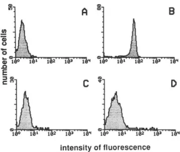
Figure 1. Effects of recombinant bactericidal/permeability in-creasing protein (rBPI23) on LPS-binding protein-mediated bind-ing of fluorescein-labeled Escherichia coli 0 I I I LPS by human pe-ripheral blood mononuclear cells. Incubation medium contained 4% albumin (A), 10% plasma (B), 10% plasma after CDI4 was blocked on monocytes with anti-CD 14 monoclonal antibody (C), or 10% plasma with 10 J.Lg of rBPI23(D). Fluorescence was assessed
by flow cytometry.
was thus of interest to evaluate whether LPS-BPI complexes would also bind to CO 14 on monocytes or would interfere with the LBP-mediated binding of LPS through CD 14. Hu-man monocytes incubated with 1 Jlg/mL FITC-0 III LPS in medium containing 4% albumin showed only a modest fluo-rescence (figure lA). In contrast, when cells were incubated with FITC-OIII LPS in the presence of 10% plasma, mono-cytes showed a distinctly brighter fluorescence (figure 1B). Preincubation of monocytes with MAb MY4 completely suppressed the plasma-mediated LPS binding (figure 1C) to the level of control monocytes incubated in the presence of albumin.
In our observations with 10% plasma, the concentration of LBP was 2 Jlg/mL. We added rBPI23to 10% plasma at a final concentration of 10 JLg/mL together with FITC-Olli LPS. The presence of rBPI2 3resulted in a decreased fluorescence (figure 10) compared with that observed with 10% plasma alone, indicating that less LPS had bound to monocytes. We then tested the effect of increasing concentrations of rBPI2 3 in 10% plasma containing LBP on FITC-Olil LPS binding to monocytes, using FITC-0 III LPS concentrations from 10 ng/ml.to 1 JLg/mL, as described [5] (table I). The con-centration ofrBPI2 3required for an efficient inhibition ofthe plasma-mediated binding of FITC-0 II 1 LPS to monocytes was dependent on the LPS concentration. With a weight ra-tio of 10: I (10 JLg/mL rBPI2 3with I JLg/mL FITC-O III LPS or I JLg/mL rBPI23with 100ng/rnl.FITC-O Ill), a 75%
de-crease in fluorescence intensity compared to control without
B
c
.!!
Q) u-
o
. . CSIi CI) 1 0 .QE
~~bits were immunized with purified murine LBP, and immune IgG was purified by protein G chromatography. A control prepa-ration of IgG was obtained from a polyclonal rabbit antiserum raised against murine recombinant interleukin-I.
LPS isolated from Escherichia coli 0 III, either unlabeled or labeled with fluorescein (FITC-O 111 LPS), was purchased from Sigma (St. Louis). MY4, a specific monoclonal antibody (MAb) recognizing human CDI4 [16], was purchased from Coulter Immunology (Luton, UK).
Plasma was prepared from a pool of donors. The concentra-tion of LBP in this pool was 20 Jlg/mL as measured by ELISA [17] (done by P. Tobias, Scripps Clinics, La Jolla, CA).
Test for LPS binding to monocytes. Heparinized human
blood samples obtained from normal donors were used to pre-pare peripheral blood mononuclear cells (PBMC) by centrifuga-tion over Ficoll-Paque (Pharrnacia, Uppsala, Sweden). PBMC ( (06
) were incubated with IJlg(alternatively, 10 or 100 ng) of FITC-01 11 LPS in I mL of RPM I enriched with 10%plasma or with 4% albumin as control. For CD 14 blockade, monocytes were pretreated with 10 Jlg/mL MY 4 MAb at 37°C After incu-bation for I h, the cells were washed twice with cold RPMI and analyzed using a FACScan flow cytometer with the LYSYS soft-ware package (Becton Dickinson, Basel, Switzerland). To re-strict the analysis to monocytes, side scatter parameters were used for gating of the signal [18].
Confocal laser microscopy. Human monocytes were purified
from PBMC by adherence to plastic in 10%human serum for I h and recovered using a rubber policeman. Purity was >95% as assessed by morphology and nonspecific esterase staining. Monocytes were incubated with I Jlg/mL FITC-0 I 11 LPS in RPMI containing 4% albumin or 10% plasma for 30 min. After two washes, they were spun down using a cytocentrifuge (Cyto-spin 2; Shandon, Basel, Switzerland) for 3 min at 700 rpm. Slides were mounted in TRIS-glycerin-saline buffer and directly examined by fluorescence microscopy. Optical slices were gen-erated in a Laser-Sharp MRC 500 confocal microscope (Bio-Rad Laboratories, Glattbrugg, Switzerland).
TNFa assays. PBMC (0.5 X (06
) in 200 JlL of RPMI enriched with 10% plasma were added to each well of 96-well flat-bottom microtiter plates (Becton Dickinson). These prepara-tions were stimulated with unlabeled 0111 LPS at various con-centrations. Thaumatin or rBPI2 3 at 10 Jlg/mL was added at various times relative to LPS exposure in plasma. Supernatants were collected for TNFa measurements after 4 h of culture at 37°C. Under the conditions used in this study,TNFarelease by lymphocytes present in the PBMC fraction was negligible [19]. The cytotoxic activity ofTNFa was measured on WEHI clone
13 cells as described [20, 21].
Results
Effects of rBPIn on the LBP-mediated binding of
FITe-DIll LPS to monocytes. We previously reported that
plasma enhanced the binding of FITC-0 I I I LPS to human monocytes compared with binding observed with albumin alone. The enhancement in LPS binding was due to plasma LBP and was blocked by the MY4 anti-CD 14 MAb [5]. It
JID 1993:167 (June) Binding of LPS to Monocytes 1353
Figu~e 2. E~ects of recombinant bactericidal/permeability in-creasing protem (rBPI23 ) on binding of fluorescein-labeled
Esche-richia coli0 III LPS to human peripheral blood mononuclear cells
after antibody-mediated blockade ofLPS binding protein (LBP) in plasma. Anti-LBP MAB, rabbit IgG directed against mouse LBP. Data are fluorescence units measured on monocytes.
o
CONTROL PLASMA• PLASMA
+
ANTI-LBP MAB~ ALBUMIN Table1. Effect of recombinant bactericidal/permeability
increas-ing protein (rBPI23 ) on the LPS binding protein (LBP)-mediated
binding of fluorescein-labeled Escherichia coli 0 III LPS to human peripheral blood mononuclear cells.
LPS concentration (ngjmL) Medium, rBPln (~gjmL) 10 100 1000 4k albumin 2.0 2.3 2.4 10'1;· plasma 0 4.2 16.2 40.1 0.1 4.1 16.6 39.9 1.0 2.8 3.6 25.7 10.0 2.0 2.5 9.7 10k plasma+anti-CD 14 monoclonal antibody
a
2.3 2.2 2.5 10 2.1 2.3 2.5NOTE. Data are mean fluorescence units measured on monocytes in one experiment. Experiment was repeated many times with similar observa-tions (data not shown).
ANTI-CD14
+
+
BPI+
+
o
FLUORESCENCE UNITS 48
12
16
ExperimentNOTE. Data are mean fluorescence units measured on monocytes by flow cytometry in two separate experiments.
!able 2'. Effect of recombinant bactericidal/permeability increas-mg protem (rBPI23) on the binding to peripheral blood mononu-clear cells of fluorescein-labeled Escherichia coli 0 I I I LPS me-diated by purified murine LPS-binding protein (LBP).
observed in albumin was seen when rBPI23was added after
antibody-mediated LBP blockade in plasma. These experi-ments thus confirmed that rBPI23did not promote the
bind-ing of FITC-O III LPS to monocytes but rather interfered with the LBP-mediated binding ofFITC-O III LPS to mono-cytes.
Effects of competition between purified murine LBP and rBPI23forthe binding ofFITC-Olll LPS to monocytes. We
further investigated whether rBPI23could interfere with
puri-fied LBP for FITC-0 I I I LPS binding to monocytes. We used purified murine LBP for these experiments (table 2). When I
J.Lg
of rBPI23(43 nM) and IJ.Lg
of purified murineLBP (16 nM) were added to monocytes in the presence of
100 ng/rnl. LPS, rBPI23suppressed the LBP-mediated
bind-ing of FITC-0 I I I LPS to monocytes.
Effects ofrBPI2J on the internalization of FITC-Olll LPS
into human monocytes. To distinguish between cell surface
binding and intracellular localization ofLPS, preparations of adherent monocytes were incubated with FITC-OIII LPS
2 3.1 11.8 12.3 3.2 3.2 15.5 13.9 3.1 4% albumin I~gmurine LBP I~gmurine LBP+100 ng rBPI2J I~gmurine LBP+ I~grBPI2 3
rBPI2 3was observed. By increasing the weight ratio to 100: I
(10 JLg/mL rBPI23 with 100 ng/rnl. FITC-O III LPS), the
plasma-mediated binding of LPS to monocytes was abol-ished.
In the presence or absence of rBPI23 , the observed LPS
binding was totally suppressed by CD 14 blockade by anti-CD 14 MAb. Control thaumatin did not affect the LBP-me-diated binding of FITC-O III LPS by human monocytes (data not shown).
Effect of rBPI23 on the binding of FITC-Olll LPS to
hu-man monocytes after LBP blockade in plasma. The
observa-tions reported above indicated that rBPI23 inhibited the
plasma-mediated binding ofLPS by monocytes in a dose-de-pendent manner. When FITC-OIII LPS was used at a 1-JLg/mL concentration, residual binding was found despite the presence of 10 JLg/mL rBPI23 (table I). This binding was
suppressed by CD 14 blockade. We then investigated whether this residual binding was due to LBP or to BPI. We used murine plasma for these experiments, because we had an anti-murine LBP antibody readily available and because murine LBP is able to present LPS to human CDI4 in a fashion identical to that of human LBP [5, 15]. LBP was blocked in murine plasma with a rabbit anti-murine LBP IgG fraction. An irrelevant rabbit IgG fraction directed against murine interleukin-I was used as control. In the pres-ence of control murine plasma, LBP was able to increase the binding of FITC-0 I I I LPS to human monocytes over that to control monocytes incubated in albumin (figure 2). This binding was suppressed by antibody-mediated LBP blockade and, t.o a lesser extent, by the addition of rBPI23 •The prein-cubation of monocytes with anti-CD 14 MAb MY 4 sup-pressed LBP-mediated binding of FITC-0 III LPS. No in-crease in binding of FITC-0 I I I LPS to monocytes over that
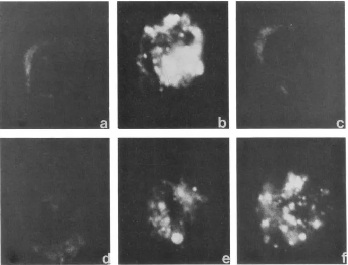
Figure 3. Effects of recombinant bactericidal/permeability increasing protein (rBPI23 )on LPS binding protein (LBP)-mediated
internal-ization of fluorescein-labeled Escherichia coli 0 III LPS by human monocytes. Incubation medium contained4%albumin (a); 10%human plasma (b); 10%plasma after pretreatment ofmonocytes with anti-CD14monoclonal antibody (c); 10%human plasma and rBPI23that was
preincubated with LPS for 5 min (d); rBPI23added concurrently with 10%plasma and monocytes (e); rBPI23added 5 min after mixture of
monocytes, 10%plasma, and LPS (f). Fluorescence of monocytes was visualized in confocal microscope after 1 h of incubation.
and analyzed by confocal microscopy, which allows observa-tion of subsurface structures. Monocytes were scanned in depth at various focal planes. Through-focus images were similar, and figure 3 is representative of one focal plane. When monocytes were incubated for I h with I Jlg/mL FITC-OIII LPS in 4% albumin (figure 3a), no fluorescence was detectable. When incubation medium contained 10% human plasma, the fluorescence observed was cytoplasmic (figure 3b). When FITC-OIII LPS was added to anti-CD 14-treated monocytes in the presence of 10% plasma, fluorescence was abolished (figure 3c). When FITC-Olll LPS was preincubated with 10Jlg/mL rBPI23 before adding
human plasma as a source of LBP, no fluorescence was de-tectable (figure 3d). However, when rBPI23was added at the
same time as plasma LBP (figure 3e) or 5 min thereafter (figure 3f), the uptake of FITC-O III LPS into monocytes was only partially prevented. At concentrations
<
10Jlg/mL,rBPI2 3was unable to suppress the fluorescence in these ex-periments (data not shown).
Time-dependent inhibition by rBPI23 of the LBP-mediated
binding ofFITC-Olll LPS to human monocytes. We
previ-ously reported that the LBP-mediated binding of FITC-0111 LPS by monocytes is a rapid process largely completed in 5-15 min [5]. Thus, in the next set of experiments, we added rBPI2 3 at various times relative to FITC-0 I I I LPS exposure in plasma (figure 4). When LPS was first preincu-bated with rBPI2 3 30 min or 5 min before being added to monocytes in medium containing plasma, LBP-mediated binding ofFITC-O III LPS to monocytes was virtually abol-ished. When LPS and rBPI23were added without preincuba-tion together with monocytes in 10% plasma, rBPI23was still able to inhibit LPS binding to monocytes. The addition of rBPI23from 5 to 30 min after the initiation of the reaction of plasma LBP with FITC-O III LPS progressively reduced the
JID 1993:167 (June) Binding of LPS to Monocytes 1355
Discussion
The focus of this investigation was to assay the direct com-petition between BPI and LBP for LPS binding to mono-cytes. Indeed. since BPI appears to be an efficient neutraliz-ing agent of LPS [11- 13], it should in vivo be able to overcome the action of LBP, which amplifies the effects of LPS on both monocytes and neutrophils [3-7].
The experiments were first conducted in plasma contain-ing LBP uscontain-ing a high LPS concentration (I tLg/mL). By both flow cytometry and confocal microscopy, we observed that rBPI2 3 •a recombinant Nl-ly-rerminal fragment of BPI [2, 14], reduced LPS binding to monocytes when added to normal plasma containing LBP. Low residual LPS binding was ob-served. which was suppressed by CD I 4 blockade. Since bind-ing of LPS to monocytes in the presence of plasma is me-diated by the formation of LPS- LBP complexes that bind to CD 14 receptors [3]. these observations demonstrate that the small residual LPS binding in the presence of a suboptimal concentration of rBPI23 was most likely due to LBP. Thus,
rBPI23 was able to prevent the formation of LPS-LBP
com-plexes and their subsequent binding to human monocytes, although the inhibition was not complete at a high concen-tration of 1 tLg/mL LPS. Furthermore, when LBP in plasma was functionally blocked by specific antibodies. rBPI2 3was not able to present LPS to CD I 4 of monocytes. Confocal microscopy, which allows the observation of subsurface structures. confirmed that rBPI23 prevented the
LBP-me-diated internalization of LPS through CD I 4.
At LPS concentrations of 100 and 10 ng/rnl., a 100: I weight ratio of rBPI23to LPS appeared necessary to suppress
the LBP-mediated binding of LPS in plasma. The relative concentrations of LPS. BPI, and LBP appeared thus to play an important role. When similar amounts ofrBPI2 3(43 nM) and purified murine LBP ( I 6 nM) were added to monocytes in the presence of 100 ng/mL LPS, rBPI2 3appeared able to completely suppress the LBP-mediated binding of LPS to monocytes. These data indicate that a 3-fold molar excess of rBPI23has a potent inhibitory effect on LPS in direct compe-tition with LBP. The concentration ofLBP in pooled plasma used in these experiments was 20 tLg/mL. as measured by ELISA [17]. In our observations with 10% plasma, the con-centration of LBP was 2 tLg/mL, that is. 33 nM. To com-pletely suppress the LBP-mediated binding of 10 ng/mL LPS, 10 tLg/mL rBPI23 , or 430 nM, was necessary. This
ob-servation thus suggests that in plasma at least a IO-fold molar excess of rBPI23should be necessary to overcome totally the action of LBP.
Our experiments showed in addition that the rBPI
2rme-data not shown) after exposure to LPS resulted in unham-pered TNFa release compared with that seen in controls in-cubated with thaumatin.
B
A
-30 -5 0 +5 +30
e
E
Time of addition of reagents(A:rBP~;.a
B: thaumatin) relative to FITC-lPS 'ii exposure in plasma..
-~ l- I-r---, 120 100 8060
40
20
OL-_J....-_..L...._..._~----_....L.._..._....
120 100 80 60 40 20o
inhibitory effect of rBPI23 . Furthermore, rBPI23 could not
displace the LBP-mediated binding of FITC-0 I I I LPS. This was shown by adding rBPI2 3for a period of I h, 1 h after FITC-0 I I I LPS had reacted with monocytes in 10% plasma. In this setting, the addition of rBPI2 3for a prolonged period did not modify the fluorescence pattern (data not shown). Control thaumatin had no effect on the LBP-me-diated binding of FITC-O I II LPS by human monocytes.
Effects ofrBPI23on the LPS-mediatedTNFa releaseby
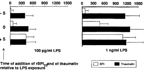
hu-man nionocvtes in medium containing plasma. Having
ob-served that rBPI2 3efficiently blocked LPS binding in a
time-dependent manner, we added rBPI2 3 to 10% plasma at various times relative to LPS exposure, and TNFa was mea-sured in the supernatants after 4 h (figure 5). When LPS (either 100 pg/rnl. or 1 ng/mL) was first incubated with rBPI2 3for 5 min before being added to plasma, virtually no
TNFa was detectable in the supernatant after 4 h. rBPI23was
still able to inhibit TNFa production, although partially, when added together with LPS. This inhibition of TNFa vanished when LPS concentrations> 1ng/ml. were used. Delaying the time of addition of rBPl2 3 for 5 min (or more, Figure 4. Time-dependent inhibition of LPS binding protein (LBP)-mediated binding of fluorescein-labeled Escherichia coli 0111 LPS (FITC-LPS) by human peripheral blood mononuclear
cells (PBMC) in presence of recombinant bactericidalj
permeability-increasing protein (rBPIn ) . Binding was assessed by flow cytometry after I h of incubation with FITC-LPS; results are%
TNF (pg/ml)
o
300 600 900 1200 1500o
300 600 900 1200 1500-5
o
+5
100 pg/ml LPg 1 ng/ml LPgFigure 5. Effects of recombi-nant bactericidal/permeability in-creasing protein (rBPI23 ) and of thaumatin on tumor necrosis factor
(TNF)release of human peripheral blood mononuclear cells stimu-lated with LPS.TNFwas measured by bioassay in supernatants after 4 h of culture. Data are mean
+
SD of5experiments.Time of addition of rBPI211nd of thaumatin
relative toLPg exposure
I
0
BPI • ThaumatlnI
diated inhibition of LBP-LPS binding to monocytes was more efficient when rBPI23 could interact with LPS before
in terference with LBP. Indeed, rBPI23was unable to reverse
the LBP-mediated binding of LPS to monocytes. This was observed in experiments using flow cytornetry, confocal mi-croscopy, and monocyte activation measured by TNFa re-lease, thus extending and confirming a similar study with the native nonrecombinant protein [12]. This observation indi-cated that a concentration of 10 jLg/mL rBPI23was sufficient
to totally inhibit the production of TNFamediated by either 100
pg/rnl.
or I ng/mL LPS. rBPI23was still able to inhibitTNFa release when added at the same time as LBP, but the inhibition was not complete. However, delayed addition of rBPI23 respective to LBP failed to inhibitTNFarelease, an
observation consistent with the hypothesis that a few min-utes of interaction of the LPS-LBP complex with monocyte CD14 could be sufficient to trigger cytokine production.
In conclusion, under the conditions of our assays-I 0% plasma containing LBP-we can conclude that rBPI23
com-peted with but did not totally suppress the LBP-mediated effects of LPS. Indeed, if rBPI23appeared able to block the
LBP-mediated binding of LPS to monocytes as revealed in our binding assays, this apparent blockade did not totally suppress TNFa production. Additional studies should be done in vitro using whole blood or other sources of LPS to confirm these observations. Thus, our studies showed that rBPI23 was very effective as an inhibitor of LPS on
mono-cytes, provided it was used prophylactically. Further studies in vivo are needed to investigate whether rBPI23could
over-come the LPS-mediated activation of the host.
Acknowledgments
We thank G. Centeno, J. Smith, andC.Knabenhans for tech-nical assistance.
References
I. Tobias PS. Mathison JC, Ulevitch RJ. A family of lipopolysaccharide binding proteins involved in responses to gram-negative sepsis. J Bioi Chern 1988;263: 13479-81.
2. Gazzano-Santoro H. Parent JB, Grinna L. et al. High affinity binding of the bactericidal/permeability increasing protein and a recombinant amino-terminal fragment to the lipid A region oflipopolysaccharide. Infect Immun 1992;60:4754-61.
3. Schumann RR. Leong SR. Flaggs GW. et al. Structure and function of lipopolysaccharide binding protein. Science 1990;249: 1429-31. 4. Wright SO. Ramos RA, Tobias PS. Ulevitch RJ. Mathison Jc. CO 14, a
receptor for complexes oflipopolysaccharide (LPS) and LPS-binding protein. Science 1990;249: 1431-3.
5. Heumann O. Gallay P, BarrasC. et al. Control of LPS binding and LPS-induced TNF secretion in human peripheral blood monocytes. J Immunol 1992; 148:3505-12.
6. Vosbeck K. TobiasP,Mueller H. et al. Priming of polymorphonuclear granulocytes by lipopolysaccharides and its complexes with lipopoly-saccharide binding protein and high density lipoprotein. J Leukoc Bioi 1990;47:97-104.
7. Wright SD. Ramos RA, Herrnanowski-Vosatka A. Rockwell P. Detmers PA. Activation of the adhesive capacity ofCR3 on neutro-phils by endotoxin: dependence of lipopolysaccharide binding pro-tein and CDI4. J Exp Med 1991; 173: 1281-6.
8. Tobias PS. Soldau K. Ulevitch RJ. Isolation of a lipopolysaccharide-binding acute phase reactant from rabbit serum. J Exp Med 1986; 164:777-93.
9. Weiss J. OlssonI.Cellular and subcellular localization of the bacteri-cidal/permeability-increasing protein of neutrophils. Blood 1987; 69:652-9.
10. Mannion BA, Kalatzis ES. Weiss J, Elsbach P. Preferential binding of the neutrophil cytoplasmic granule-derived bactericidal/permeabili-ty-increasing protein to target bacteria. Implications and use as a mean of purification. J Immunol 1989; 142:2807-12.
II. Ooi CE, Weiss J, Doerfler ME, Elsbach P. Endotoxin-neutralizing prop-erties of the 25 kD N-terminal fragment and a newly isolated 30 kO C-terminal fragment of the 55-60 kD bactericidal/permeability-increasing protein of human neutrophils. J Exp Med 1991; 174:649-55.
12. Marra MN, WildeCG, Collins MS, Snable JL, Thornton MB, Scott R W. The role of bactericidal/permeability-increasing protein as a
110 1993; 167 (June) Binding of LPS to Monocytes 1357
natural inhibitor of bacterial endotoxin. J Immunol 1992; 148:532-7.
13. Marra MN, Wilde CG, Griffith JE,SnableJL, Scott RW. Bactericidal/ permeability-increasing protein has endotoxin-neutralizing activity. J Immunol 1990; I44:662-6.
14. Weiss J, Elsbach P, Shu C. et al. Human bactericidal/permeability-increasing protein and a recombinant NH2-terminalfragment cause
killing of serum-resistant gram-negative bacteria in whole blood and inhibit tumor necrosis factor release induced by the bacteria. J Clin Invest 1992;90: 1122-30.
15. Gallay P, Carrel S, Glauser MP, et al. Purification and characterization of murine LPS-binding protein. Infect Immun 1993;61 :378-83. 16. Griffin JD, Ritz J, Nadler LM, Schlossman SF. Expression of myeloid
differentiation antigens on normal and malignant myeloid cells. J Clin Invest 1983;68:932-41.
17. Leturcq D, VanHook P, Smith R, Tobias P, Ulevitch R, Moriarty A. Generation of monoclonal antibodies to human LBP and their use in
the detection of LBP protein in serum. J Cell Biochem 1992;suppl 16C: 161.
18. Thompson JM, Gralow JR, Levy R, Miller RA. The optimal applica-tion of forward and ninety-degree light scatter in flow cytometry for the gating of mononuclear cells. Cytometry 1985;6:401-6. 19. HenterJI, Soder0,Andersson U. Identification of individual tumor
necrosis factor cachectproducing cells after lipopolysaccharide in-duction. Eur J Immunol 1988; 18:983-8.
20. Espevik T, Nissen-Meyer J. A highly sensitive cell line, WEHI 164 clone 13, for measuring cytotoxic factor/tumor necrosis factor from human monocytes. J Immunol Methods 1986;95:99-105. 21. Baumgartner JD, Heumann D, Gerain J, Weinbreck P, Grau GE,
Glauser MP. Association between protective efficacy ofanti-li po poly-saccharide (LPS) antibodies and suppression of LPS-induced tumor necrosis factora and interleukin 6. Comparison of0 side chain-spe-cific antibodies with core LPS antibodies. J Exp Med 1990; 171 :889-96.