HAL Id: hal-02972031
https://hal.archives-ouvertes.fr/hal-02972031
Submitted on 20 Oct 2020HAL is a multi-disciplinary open access archive for the deposit and dissemination of sci-entific research documents, whether they are pub-lished or not. The documents may come from teaching and research institutions in France or abroad, or from public or private research centers.
L’archive ouverte pluridisciplinaire HAL, est destinée au dépôt et à la diffusion de documents scientifiques de niveau recherche, publiés ou non, émanant des établissements d’enseignement et de recherche français ou étrangers, des laboratoires publics ou privés.
Incorporation of Ru(II) Polypyridyl Complexes into
Nanomaterials for Cancer Therapy and Diagnosis
Nancy Soliman, Gilles Gasser, Christophe Thomas
To cite this version:
Nancy Soliman, Gilles Gasser, Christophe Thomas. Incorporation of Ru(II) Polypyridyl Complexes into Nanomaterials for Cancer Therapy and Diagnosis. Advanced Materials, Wiley-VCH Verlag, In press, �10.1002/adma.202003294�. �hal-02972031�
1
Incorporation of Ru(II) Polypyridyl Complexes into
Nanomaterials for Cancer Therapy and Diagnosis
Nancy Soliman,a,b Gilles Gasser,b,* and Christophe M. Thomasa,*
a Chimie ParisTech, PSL University, CNRS, Institut de Recherche de Chimie Paris, 75005 Paris,
France. E-mail: christophe.thomas@chimie-paristech.fr; WWW: https://www.ircp.cnrs.fr/la-recherche/equipe-cocp/
b Chimie ParisTech, PSL University, CNRS, Institute of Chemistry for Life and Health Sciences,
Laboratory for Inorganic Chemical Biology, 75005 Paris, France. E-mail: gilles.gasser@chimieparistech.psl.eu; WWW: www.gassergroup.com
ORCID-ID:
Nancy Soliman: 0000-0002-3031-8584
Gilles Gasser: 0000-0002-4244-5097
Christophe M. Thomas: 0000-0001-8014-4255
KEYWORDS: Anticancer; Medicinal Inorganic Chemistry; Metals in Medicine; Nanomaterials, Nanomedicine.
2 Abbreviations
AFM = atomic force microscopy, AuNPs = gold nanoparticles, AgNPs = silver nanoparticles, BCPs = block copolymers, CNAs = cross-linked nanoassemblies, CNTs = carbon nanotubes, DLS = dynamic light scattering, EPR = enhanced permeability and retention, FDA = Food and Drug Administration, HSA = human serum albumin, Mn = number average molecular weight, MOFs =
metal-organic frameworks, MSNs = mesoporous silica nanoparticles, MWCNTs = multi-walled carbon nanotubes, PACT = photoactivated chemotherapy, PEG = poly(ethylene glycol), PDI = polydispersity index, PDT = photodynamic therapy, PLA = polylactide, PLGA = poly(lactide-co-glycolide), PS = photosensitizer, PTT = photothermal therapy, RAFT = radical addition-fragmentation transfer, rGO = reduced graphene oxide, ROS = reactive oxygen species, SEC = size exclusion chromatography, SEM = scanning electron microscopy, SeNPs = selenium nanoparticles, SiNPs = porous silicon nanoparticles, SQ =squalene, SWCNTs = single-walled carbon nanotubes, TEM = transmission electron microscopy UCNPs = upconversion nanoparticles, 2P = two-photon, 1P = one-photon
Abstract
Ru(II) polypyridyl complexes are compounds of great interest in cancer therapy, due to their unique photophysical, photochemical and biological properties. For effective treatment, they must be able to penetrate tumor cells effectively and selectively. The development of nanoscale carriers capable of delivering Ru(II) polypyridyl complexes has the potential to passively or selectively enhance their cellular uptake in tumor cells. Many different strategies have been explored to incorporate Ru(II) polypyridyl complexes into a variety of nano-sized constructs, ranging from organic to inorganic materials. This review serves to highlight recent developments in nanomaterials loaded with Ru(II) polypyridyl complexes. Their rational design, preparation and physicochemical properties are described, and their potential applications in cancer therapy are eventually discussed.
1. Introduction
Cancer is a disease caused by the uncontrollable growth of abnormal cells. It is one of the leading causes of death in the world, accounting for around 10 million deaths each year.[1] This alarming
3
data brings up the necessity to develop efficient methods for the diagnosis and treatment of cancer. In this regard, metal-based drugs have been highly investigated for different medicinal applications against various cancer-related issues, especially since the discovery of cisplatin in the late 1960s. Among them, Ru(II) complexes and in particular Ru(II) polypyridyl complexes have drawn increased attention in recent years due to their unique and appealing photophysical and photochemical properties, which can be tuned by varying the nature and the numbers of the polypyridyl ligands around the Ru(II) metal center.[2] They absorb light and emit wavelength within the red and the near
infrared (NIR) spectral regions and possess large Stokes shift, long luminescence lifetime, and potential two-photon (2P) absorption properties. These characteristics have made them highly desirable across numerous research fields such as catalysis,[3] solar energy,[4] sensors[5] and now
across the biomedical field either in diagnostics as cellular imaging tools, luminescent probes of DNA structures or in therapy as new classes of anticancer drugs for chemotherapy and photosensitizers (PSs) for photodynamic therapy (PDT) or photoactivated chemotherapy (PACT).[6-11] Worthy of note,
a Ru(II) polypyridyl compound (TLD1433) has recently entered phase II clinical trials in Canada for the treatment of non-muscle invasive bladder cancer as a PS for PDT.[12] However, these complexes
still suffer from some drawbacks such as poor water solubility, lack of targeting capability, nonspecific distribution, systemic toxicity and hence, low therapeutic index hampering their translation into the clinic. One strategy to address those medical challenges is to use nanomedicine. Nanomedicine is the application of sub-micron particles (i.e., nanoparticles) in the field of medicine. The materials used for the synthesis and/or formulation of nanoparticles are extremely diverse, ranging from organic to inorganic molecules (Figure 1).[13] The unique features of nanoparticles including small
size, high surface area, surface chemistry, water solubility and multi-functionality make them highly interesting for drug delivery purposes.[14,15] Owing to their size, nanoparticles can passively
accumulate to solid tumors while sparing healthy cells thanks to the enhanced permeation and retention (EPR) effect.[16] The EPR effect exploits the abnormalities of tumor vasculature, namely
hypervascularity, atypical vascular architecture, leaky vasculature, and lack of lymphatic drainage. This results in the efficient extravasation of nanoparticles from the tumor vasculature and their retention in the tumor interstitium. Therefore, drug incorporation into nanoparticles provides significant improvements in pharmacokinetics, solubility, toxicity and biodistribution when compared
4
to freely administered molecules, reducing adverse side effects observed with conventional medicine. However, to take full advantage of the EPR effect, nanoparticles must remain in circulation long enough for tumor accumulation. PEGylation, which refers to modification with poly(ethylene glycol) (PEG) chains or PEG copolymers (e.g., Jeffamine), is the most common strategy for imparting stealth properties to nanoparticles.[17] The presence of PEG enables steric stabilization preventing
particle interactions, protein adsorption, phagocytic uptake by macrophages, and interactions with immune cells. Nanoparticles can be further modified with ligands that have binding affinity for receptors overexpressed in tumor cells to allow active targeting.[18] Although it is widely held to
improve delivery of nanodrugs to tumors, the EPR effect remains controversial and subject to debate.[19] Among materials available for constructing nanomedicines, liposomes and polymers are
the most widely used and the promising ones.[20] The early discoveries in polymer encapsulation of
metal complexes for biological and medicinal applications have been extensively and remarkably covered and will not be duplicated herein.[21,22] The present review is concerned with recent selected
papers that have described methodologies and strategies that allow incorporation of Ru(II) polypyridyl complexes into a variety of nanomaterials for cancer-related biomedical applications.
5
Figure 1. Schematic representation of the nanosized constructs reported for the incorporation of Ru(II) polypyridyl complexes
2. Organic nanoparticles
2.1. Polymeric nanocarriers
Polymer-based nanocarriers have been adopted as the preferred system for drug delivery because of their ease of synthesis, their great diversity of composition, architecture and functionalization and their ability (for most of them) to degrade in physiological media. Thanks to the great diversity of polymer architectures, a wide range of polymeric nanomaterials - comprising micelles, nanoparticles, nanogels, vesicles and dendrimers – is accessible, which make them an important class of drug
6
delivery systems. Polymeric micelles are obtained by the self-assembly of amphiphilic polymers where the hydrophobic core of the micelles creates a microenvironment for the encapsulation of therapeutic compounds and the hydrophilic shell provides a stabilizing interface between the hydrophobic core and the aqueous medium. Nanogels are made of three-dimensional cross-linked networks of hydrophilic polymers while dendrimers are highly branched macromolecules that form a tree-like structure. As to polymer nanoparticles, they are solid particles composed of macromolecular polymers regardless of their structure.
Drugs can be encapsulated into polymer matrix using two different strategies: (i) physical encapsulation relying on non-covalent interactions between drugs and polymer matrix and (ii) covalent encapsulation where the drug is covalently conjugated to the polymer. In most cases, drugs are physically encapsulated into the polymer matrix. However, this type of encapsulation suffers from important limitations such as (i) “burst release”, which consists in the abrupt release of a large amount of drug post administration; (ii) the difficulty to encapsulate drugs that are poorly miscible to the polymer matrix and (iii) poor drug loading (less than 10 %), thus requiring a high concentration of drug delivery systems to obtain a therapeutic effect. To overcome these drawbacks, a covalent encapsulation has been considered. Three synthetic strategies can be used to obtain drug-polymer conjugates: (i) the drug can be conjugated to a pre-synthesized polymer or (ii) to a monomer prior to polymerization, or (iii) the drug can, after appropriate functionalization, serve as a polymerization initiator.[23] When drugs are transition metal complexes, drug-polymer conjugates can also be
prepared by chelation of a polymeric macroligand to the metal precursor. Either way, polymeric nanosystems can be formulated using the following techniques: (i) emulsification and solvent evaporation/extraction, (ii) nanoprecipitation (also known as solvent-displacement method),[24] (iii)
supercritical anti-solvent method, (iv) dialysis, and (v) salting-out method.[25] Among these
techniques, emulsification, solvent evaporation/extraction, and the nanoprecipitation methods are by far the most widely employed.
2.1.1. Physical encapsulation
Aliphatic polyesters such as FDA-approved polylactide (PLA) and poly(D,L-lactide-co-glycolide) (PLGA) are the most used biodegradable and biocompatible polymers in drug delivery systems. The
7
physical properties, and degradation of these polymers, which proceeds through the hydrolysis cleavage of ester bonds, can be tuned by varying the molecular weight, the stereochemistry, and the copolymer composition. The degradation of PLA and PLGA affords acidic products, namely lactic acid and glycolic acid, which are non-toxic and can be metabolized to give CO2 and H2O as benign
by-products. [26]
Lemercier and co-workers reported the encapsulation of two 5-substituted-1,10-phenanthroline-based Ru(II) 2P-PDT PSs, Ru1 and Ru2 (Figure 2) in PLGA nanoparticles by nanoprecipitation in the presence of Poloxamer 188 (P188), from a commercially available acid terminated PLGA with a 50:50 D,L-lactide to glycolide ratio and a number average molecular weight Mn of 24 000 – 38 000
g.mol-1.[27] A drug loading of around 1 % could be achieved. As confirmed by dynamic light scattering
(DLS) and atomic force microscopy (AFM), the authors obtained spherical 100 nm and narrowly dispersed nanoparticles with a polydispersity index (PDI) lower than 0.15. These nanoparticles were much less toxic in C6 glioma cells than the related free Ru(II) polypyridyl complexes after irradiation with a white light source. The release of Ru payloads was accelerated after irradiation: 50 % of Ru was released after two days as opposed to 10 % after six days without irradiation.
Chen and co-workers reported the encapsulation of [Ru(phen)2-p-MOPIP](PF6)2.2H2O (Ru3,
p-MOPIP = 2-(4-methoxyphenyl)imidazo[4,5-f][1,10]phenanthroline, phen = 1,10-phenanthroline, Figure 2), a potential anticancer agent,[28] in PLGA nanoparticles (PLGA@Ru) using the
nanoprecipitation technique from a commercially available PLGA with a 75:25 ratio of D,L-lactide to glycolide and a Mn = 50 000 g.mol-1.[29] The resulting 130 nm nanoparticles were then coated with a
polyethylenimine (Mn = 10 000 g.mol-1), previously functionalized with biotin and poly(ethylene glycol)
(PEG, Mn = 5 000 g.mol-1), Bio-PEI-mPEG. They obtained 150 nm spherical Bio-PLGA@Ru
nanoparticles with active targeting ability towards cells overexpressing biotin receptors. Cytotoxicity of Bio-PLGA@Ru was evaluated in different cancer cell lines (i.e., HepG2 human hepatocellular carcinoma cells, A375 human malignant melanoma cells, HeLa, Siha and Caski human cervical carcinoma cells) and in a non-cancerous cell line (i.e., NIH3T3 mouse fibroblast cells) and compared to the uncoated PLGA@Ru and the free Ru complex, Ru3. Bio-PLGA@Ru nanoparticles enhanced the accumulation of Ru3 in cancer cells by the lipid raft-mediated endocytosis pathway causing a
8
cytotoxicity against HepG2 cells 3-4 folds higher than those of Ru3 and PLGA@Ru. This might be explained by a higher intracellular ROS levels, hence a higher level of DNA damage. Biodistribution in HepG2 xenograft nude mice revealed that Bio-PLGA@Ru was mainly accumulated in the liver and tumor regions while Ru3 was distributed around the whole mice body, showing how the nanoparticle formulation could improve the biodistribution of Ru3 in the body.
Glazer and co-workers focused on improving the efficacy of Ru complexes for PDT by employing cross-linked nanoassemblies (CNAs) as a nanogel delivery platform.[30] They used polymeric CNAs
obtained from poly(ethylene glycol)-block-poly(L-aspartate) copolymers (PEG-b-PASP, Mn = 6 100
g.mol-1 as determined by 1H NMR spectroscopy), two Ru-based photoactivatable prodrugs with
varying levels of hydrophobicity, namely [Ru(bpy)2(dmbpy)]Cl2 (Ru4, bpy = 2,2′-bipyridine, dmbpy =
6,6′-dimethyl-2,2′-bipyridine) and [Ru(dmbpy)2(dip)]Cl2 (Ru5), as well as one photostable
[Ru(dip)3]Cl2 (Ru6, dip = 4,7-diphenyl-1,10-phenanthroline, Figure 2). The Ru complexes were
physically entrapped in PEG-b-PASP CNAs (Mn = 340 000 g.mol-1) via the attractive ionic interactions
between positively charged complexes and negatively charged aspartate (ASP) groups allowing drug loading up to 20 %. The CNAs, before and after Ru loading, were defined as relatively monodisperse with an average diameter of ca. 20 nm and a PDI of ca. 0.25. The release rate and the amount of Ru(II) complex released from PEG-b-PASP CNAs were found to depend on complex hydrophobicity and solution ionic strength. However, the cytotoxicity of Ru-loaded CNAs in A549 human lung adenocarcinoma, before and after irradiation (λ > 400 nm, 5 min), was similar to the one obtained with the parent Ru(II) complexes. Ru4, loaded or not in PEG-b-PASP CNAs, was not toxic in the dark with an EC50 > 300 µM, while photocytotoxicity was observed after irradiation with an
EC50 of 8.3 ± 1.2 µM for the free Ru complex and 12.9 ± 1.1 µM when loaded in PEG-b-PASP CNAs.
As to Ru5 and Ru6, loaded or not, they were already toxic in the dark (EC50 = 9.7 ± 1.3 µM and EC50
= 0.6 ± 1.1 µM, respectively) and had EC50 values in the micromolar range after irradiation (EC50 =
3.9 ± 1.1 µM and EC50 = 0.1 ± 1.1 µM, respectively).
Gallei, Gasser and co-workers described the encapsulation of a Ru(II) polypyridyl PDT photosensitizer, [Ru(bpy)2-dppz-7-hydroxymethyl](PF6)2 (Ru7, dppz =
9
methacrylate) (PDMAEMA) as the hydrophilic segment and either poly(methyl methacrylate) (PMMA) or a statistical copolymer PMMA-co-PDMAEMA as the hydrophobic segment.[31] These
polymers showed a low critical solution temperature (LCST) behavior in water: micelles were formed below LCST and shrinkage of micelles occurred above LCST allowing the release of Ru payloads. This temperature could be tailored by varying ionic strength and the ratio of MMA to MAEMA. Not only the release of Ru(II) complex could be triggered by temperature, it could also be triggered by pH variation and ultra-sound. However, as the LCST was too low (i.e., around room temperature), no biological tests were reported. Therefore, more research will need to be performed for the synthesis of these copolymers with LCST around 37 – 43 °C to allow the controlled release of the Ru complex in pharmaceutically relevant temperature using hyperthermia for example.[32]
10
Figure 2. Chemical structures of Ru1-Ru7. 2.1.2. Covalent encapsulation
Covalent encapsulation is the formulation of polymer-drug conjugates into nano-sized constructs. The concept of polymer-drug conjugates, in which the drug is covalently linked to a hydrophilic polymer, was inspired by the prodrug approach and first introduced in 1975 by Ringsdorf.[33]
Depending on where the Ru center is conjugated to the polymer (e.g., the backbone, a lateral pendant group, the center of a star polymer), different synthetic strategies have been described.[34]
11
Ranucci, Maggioni and co-workers investigated the use of linear polyamidoamine (PAMAM) bearing pendant phenanthroline groups that could coordinate to Ru(phen)2(OTf)2 (Ru8, OTf = triflate anion,
CF3SO3-) for optical imaging and PDT applications. PAMAM copolymers were obtained by Michael
polyaddition of 4-(4′-aminobutyl)-1,10-phenanthroline (BAP) with piperazine derivatives and bisacrylamide derivatives, followed by complexation with Ru8, which resulted in [Ru(phen)3]2+ lateral
pendant groups linked to the PAMAM chain via butyl segments (Figure 3a). First, they reported the synthesis of an amphoteric PAMAM copolymer, namely PhenISA, with a Mn value of 46 800 g.mol -1, M
w/Mn of 1.78 and 6 % of phenanthroline-containing repeating unit, which could coordinate to
Ru8.[35] The resulting polymer Ru8-PhenISA could self-assemble into nanoparticles with a diameter
of ca. 20 nm that were able to internalize in HEK-293 cells via an endocytic pathway while displaying no cytotoxicity up to 50 µM, suggesting that this system could be used for imaging. They then decided to vary the structure of PAMAM by synthesizing Ru8-PhenAN with a Mn value of 34 600, Mw/Mn of
1.38 and 2 % of Ru-containing repeating units that could self-assemble into ca. 10 nm nanoparticles.[36] The PDT efficiency of Ru8-PhenAN was evaluated in HeLa cells and compared to
that of Ru8-PhenISA and [Ru(phen)2(BAP)](OTf)2 (Ru9), a complex used as a model of the
photoactive units. Ru8-PhenAN was found to be more cytotoxic than Ru8-PhenISA and Ru9 in HeLa cells with an EC50 of 0.7 µM as opposed to 9 µM for the free complex upon visible light
irradiation (40 min, 23.7 mW.cm-2). The reason is that Ru8-PhenAN accumulated preferentially in
the nucleus unlike the free complex, which was found in the cytosol.
Wu and colleagues reported the synthesis of three well-defined amphiphilic Ru-containing block copolymers (BCPs) P1, P2 and P3 with varying molecular weights (Mn = 11.2, 15.7 and 16.9 kg.mol -1) and high Ru loading (%Ru = 41, 55 and 51 wt%) that could self-assemble into different sub-150
nm nanostructures including micelles, hollow spheres and large compound micelles (Figure 3b).[37]
[Ru(tpy)(biq)(H2O)](PF6)2 (Ru10, tpy = 2,2′:6′,2′′-terpyridine and biq = 2,2′-biquinoline) was
coordinated to a preformed polymer, poly(ethylene glycol)-block-poly(6-(4-cyanophenoxy) hexyl methacrylate) (PEG-b-PCPH), which was synthesized by reversible-addition fragmentation chain transfer polymerization (RAFT) polymerization of a methacrylate monomer, 6-(4-cyanophenoxy) hexyl methacrylate (CPH), using a PEG-based macro-RAFT agent. P1, in comparison with P2 and
12
P3, showed a higher anticancer effect after light irradiation (656 nm, 30 mW cm-2, 30 min) in HeLa
cells attributed to a higher cellular uptake (hence, higher Ru intracellular concentration), the light-triggered release of Ru10 – an anticancer therapeutic agent, IC50 = 3.5 µg.mL-1 in HeLa cells - and
the generation of intracellular 1O 2.
This strategy is suitable for treatment of hypoxic tumor as it does not uniquely rely on the generation of 1O
2. Therefore, they used the same approach with a slightly different Ru complex
[Ru(CHLtpy)(biq)(H2O)](PF6)2 (Ru11), where the commercially available anticancer drug
chlorambucil (CHL) was conjugated via an ester bond, for phototherapy against hypoxic tumors in vitro and in vivo (Figure 3b).[38] As explained above, Ru11was coordinated to a preformed block
copolymer PEG-b-PCPH to give P4 with a Mn value of 20 300 g.mol-1 and a Ru loading of ca. 45 wt%
that could self-assemble into micelle-like nanoparticles with a diameter of ca. 15 nm as determined by TEM. The micelles showed negligible toxicity in the dark in HeLa cells under normoxic and hypoxic conditions. However, upon light irradiation (656 nm, 60 J.cm−2), they were found to be toxic in both
conditions with an EC50 of 25 µg.mL-1 attributed to the release of the aqua adduct Ru11. The
phototoxicity was O2-independent.
The same authors later investigated the potential use of a Ru-containing ABA-type triblock copolymer (PolyRu) for combined photodynamic therapy and photochemotherapy (Figure 3c).[39]
PolyRu was prepared through a multi-step synthesis: the polyol-containing Ru complex [Ru(Biq)2(Hob)2][PF6]2 (Ru12, Biq = 2,2′-biquinoline, Hob = 4-[(6- hydroxyhexyl)oxy]benzonitrile)
was synthesized to react with 2,4-diisocyanato-1-methylbenzene via polycondensation to form Ru-containing polyurethanes before being functionalized by poly(ethylene glycol) methyl ether resulting in the formation of monodisperse PolyRu of Mn = 22 000 g.mol-1 as determined by 1H NMR
spectroscopy, and further confirmed by size exclusion chromatography (SEC), Mn,SEC = 15 kg mol-1,
Mw,SEC/Mn,SEC = 1.22). PolyRu could self-assemble, using a nanoprecipitation technique, into
spherical 180 nm stealth nanoparticles as confirmed by TEM and scanning electronic microscopy (SEM). The biological activity of PolyRu nanoparticles was evaluated in vitro in HeLa, PC3 and HepG2 and in vivo in HeLa tumor bearing mice. They showed that PolyRu could accumulate in tumor sites after 12 h and inhibit tumor growth after irradiation at 656 nm (50 mW.cm-2, 30 min), a
13
wavelength in the therapeutic window (650 – 800 nm), ideal for the treatment of deep-seated or large tumors. This high anticancer effect was attributed to the light-triggered release of [Ru(Biq)2(H2O)2](PF6)2 (Ru13), a potential anticancer agent, which was reported to inhibit malignant
tumor growth with a IC50 of 25 µg.mL-1 in the dark and 2.5 µg.mL-1 after irradiation due to the
generation of 1O 2.
14 a) O O O NC O O O N S S S O O O N N N N N N Ru2+ n x y P1: R = H, n=113, x=13, y=9 P2: R = H, n=45, x=26, y=22 P3: R = H n=113, x=22, y= 22 P4: R = CHL, n=113, x=7, y=15 (PEG-b-P(CPH-co-RuCHL) b) R H2O, light O N N N N N Ru2+ R H H R = H OH O O N Cl Cl Ru10 Ru11 N N N N N O O H N O H N O O O113 113 O O O HN O H N O O N Ru2+ n PolyRu c) H2O, light N N N N O O Ru2+ H H H H Ru13 N N N N N O O N Ru2+ HO OH polym eriz ation 6 Ru12 6 N N N N N N Ru2+ NH2 N O N O N N N O N O N N N N N N N Ru2+ H N O H N O HO O N N HN O O HO N N N N N N N Ru2+ 0.5 0.6 Ru8-PhenAN Ru8-PhenISA Ru9 9.5 9.4 N OTf OTf N N N Ru Ru8
Figure 3. a) Structures of Ru8 and the three [Ru(phen)3]2+ derivatives: Ru9, Ru8-PhenAN and Ru8-PhenISA. b) Chemical structures of PEG-b-PCPH of different chain lengths coordinated with Ru10 or Ru11 to give P1 – P4. c) Structure of Ru12, PolyRu and Ru13. Ru(II) polypyridyl complexes are represented in black, polymers in purple with the
15
In most of the articles reported so far, the Ru(II) complex is coordinated to the pendant group of a water-soluble polymer. This strategy possesses one key drawback, which is the lack of control over functional groups. To overcome this, drug-initiated polymerization, first described by Kricheldorf, in which a polymer chain is grown from the drug itself, can be considered to eliminate any possible heterogeneities.[40,41] Recently, we reported the one-pot synthesis of ruthenium-containing PLA by
ruthenium-initiated ring opening polymerization of lactide from a non-cell penetrating PDT PS, namely [Ru(bipy)2-dppz-7-hydroxymethyl](PF6)2 (Ru7).[42] These polymers Ru7-PLA, shown in
Figure 4a, could further be formulated into size tunable nanoparticles from 100 to 300 nm by nanoprecipitation using poly(vinyl alcohol) as a stabilizer. All nanoparticles showed improved photophysical properties including luminescence, singlet oxygen generation, and enhanced cellular uptake in HeLa cells. Capitalizing on this, an improved photo-toxicity towards HeLa cells (λ = 480 nm, 3.21 J.cm−2) with IC
50s as low as 4.4 ± 0.8 µM after 48 h was determined while the free Ru
complex showed no significant photocytotoxicity (IC50 > 500 µM).
To obtain high drug loading, dendrimers can be used.[43] Velders and co-workers demonstrated that
functionalization of PAMAM dendrimers with two Ru(II) polypyridyl complexes, Ru14 and Ru15 (Figure 4b), could afford 5 nm sized materials in water, as characterized by Diffusion-Ordered Spectroscopy (DOSY) NMR based on Pulse-Field Gradient Spin-Echo (PFGSE) measurements in D2O, with improved fluorescent imaging properties.[44] The luminescence of these materials was
16-fold higher than that of the free Ru complexes, with no self-quenching observed. They mainly accumulated in the lysosomes of CT26 colon carcinoma cells, implying an internalization via passive endocytosis. The phototoxicity of these materials was evaluated qualitatively by confocal fluorescence microscopy, showing disruption of membrane integrity of CT26 cells treated with Ru14-PAMAM (carrying 32 positive charges) after irradiation at 405 nm for 30 min, while no change was observed with Ru15-PAMAM (carrying 32 negative charges). Because of their small size (ca. 5 nm), dendrimers are known to be rapidly cleared from the blood through the kidneys, which eliminate the need for biodegradability. Although promising, dendrimers are more expensive than other nanoparticles and require many repetitive synthetic steps, posing a challenge for large-scale production.
16
Figure 4. a) Structure of Ru7-PLA. b) Chemical structures of Ru14 and Ru15. Ru(II) polypyridyl complexes are represented in black, polymers in purple.
2.2. Lipid-based nanocarriers
2.2.1. Liposomes
Liposomes are nano-sized to micro-sized vesicles consisting of an aqueous core surrounded by a phospholipid bilayer.[45] They were the first nanomedicine delivery system to make the transition from
concept to clinical application making them an established technology platform with considerable clinical acceptance.
Shen, Mao and co-workers reported the encapsulation of the well-studied “light-switch” and active compound, [Ru(phen)2dppz](ClO4)2 (Ru16, Figure 5a), in PEGylated sub-100 nm liposomes formed
from dipalmitoylphosphatidylcholine (DDPC), PEG-modified phospholipid, 1,2-Distearoyl-sn-glycero-3-phosphoethanolamine-poly(ethylene glycol) (DSPE-PEG, Mn = 2 000 g mol-1) and
cholesterol with a Ru loading of 4 %.[46] The cytotoxicity of the resulting liposomes LipoRu was
evaluated in MDA-MB-231 human breast cancer cells and compared to that of the free complex. LipoRu was much more cytotoxic than Ru16 with an IC50 value lower than 4 µM while Ru16showed
no toxicity up to 200 µM. This higher anticancer effect was attributed to a higher intracellular accumulation of LipoRu, which was 15-fold higher than that of the free Ru complex. For tumor specific imaging and therapy, a nanocarrier based on a 220 nm folate-conjugated liposome incorporating Ru16 was proposed by Xie and co-workers.[47] The interaction between the nanocarrier
17
which was attributed to the effective light-triggered release of Ru16 from the liposome carrier into the cytoplasm. No cytotoxicity was observed in the dark up to 100 µM.
Bonnet’s research team examined the potential of ruthenium-decorated vesicles in the context of light-triggered release drug delivery.[48] By synthesizing a thioether-cholestanol hybrid ligand that
could coordinate to Ru via its sulfur atom, they were able to decorate liposomes with Ru(II) polypyridyl complexes using either negatively charged lipids (1,2-dimyristoyl-sn-glycero-3-phospho-rac-(1-glycerol) sodium salt, DMPG) or neutral lipids (2‐dimyristoyl‐sn‐glycero‐3‐phosphocholine, DMPC) to build up liposome membranes. They showed how it was possible to release [Ru(trpy)(bpy)(H2O)]2+ (Ru18) using light irradiation (Figure 5b). As NIR light is of great interest for
treating deep-seated tumors, they focused on the preparation of liposomes capable of generating photons of blue light in situ by triplet-triplet annihilation (TTA) upconversion of either green or red light to trigger the photodissociation of [Ru(tpy)(bpy)SRR’]2+ (Ru17, SRR’ = a monodentate
thioether-cholestanol hybrid ligand) with a clinical grade PDT laser at 630 nm.[49] TTA principle relies on the
conversion of low-energy protons into higher energy photons by a bimolecular mechanism, involving a photosensitizer and an annihilator molecule (i.e., in this case, palladium tetraphenyltetrabenzoporphyrin and perylene, respectively). Two liposomal formulations were prepared separately and mixed together afterwards: one was made of DMPC, 4 mol% of DSPE‐ PEG, the photosensitizer and the annihilator molecule while the other was a Ru-functionalized liposomes made from the same lipids with a diameter ranging from 130 to 170 nm (Figure 5c). In 2015, the same authors combined all the components in one liposomal formulation with an average diameter of 130 – 180 nm and a PDI of 0.05 – 0.20, as determined by DLS (Figure 5d).[50] The clinical
grade PDT laser at 630 nm could trigger the photodissociation of the Ru complex into its cytotoxic aqua species and in a faster way than the formulation described above. However, no biological evaluation was reported for both liposomal systems.
2.2.2. Lipid-drug conjugates: example of squalenoylation
Couvreur and his research team developed an original approach based on the use of squalene (SQ), a natural and biocompatible lipid involved in the cholesterol’s biosynthesis, as a building block for the synthesis of SQ-drug conjugates.[51] The resulting SQ-drug conjugates could self-assemble in an
18
aqueous medium as nanoparticles, without the need for any surfactant and/or stabilizer. This concept called “squalenoylation” has been applied to commercially available anticancer agents, whether hydrophilic (i.e., gemcitabine) or hydrophobic (i.e., paclitaxel and cisplatin) and extended to a potential imaging agent, [Ru(bpy)2BIPPBI](PF6)2 where BPPBI =
1-methyl-2-(4-methylpyridine-2-yl)-1H-benzoimidazole (Ru19). As SQ has no functional group, the SQ carbon core must be derivatized to react with drugs. For example, 1,1’,2-trisnorsqualenoic acid, a SQ derivative, can be obtained from squalene via a three-step synthesis involving squalene monobromohydrine and squalene-2-3-epoxide as intermediates. However, this process suffers from a very low yield of 5 %.[52]
1,1’,2-Trisnorsqualenoic acid can react with N-hydroxysuccinimide ester (NHS), SQ-NHS, for subsequent peptide coupling with a higher yield.
Gobetto and co-workers described the conjugation of the luminescent Ru(II) polypyridyl complex, [Ru(bpy)2BPPBI-hx](PF6)2 (Ru20, BPPBI =
6-[2-(4-methyl-pyridin-2-yl)-1H-benzoimidazol-1-yl]-hexylamine) to SQ-NHS via an amide bond (Figure 5e).[53] The resulting conjugate, Ru20-SQ, could
self-assemble into narrowly dispersed 300 nm nanoparticles using a nanoprecipitation technique. Ru20-SQ was found to be non-toxic towards HT-29 colorectal (IC50 = 380 µM) and MCF-7 breast
cancer cell lines (IC50 = 110 µM). While remaining non-toxic, this technique could improve the cellular
uptake of Ru19, particularly in the nucleus, and diffuse into a more organized cell system: 3D multicellular tumor spheroids (MCTSs). This is ideal for cell imaging.
19 PS A A PS A A Ru17 Ru17 Ru17 Ru17 Ru17 NIR irradiation UV/blue emission Ru18 Ru18 NIR irradiation UV/blue
emission
Figure 5. a) Chemical structure of [Ru(phen)2dppz]2+ (Ru16). b) Photoactivation of Ru17 leading to the formation of the aqua adduct Ru18. c and d) Schematic representation of the liposomal formulations capable of generating blue light by
TTA-UC. e) Chemical structures of Ru19, Ru20 and Ru20-SQ with SQ derivative represented in purple.
2.3. Protein-drug conjugates
Protein-drug conjugates are proteins directly conjugated to drug molecules by a biodegradable bond that can self-assemble into nano-sized materials. Protein-drug conjugates are typically very small (< 50 nm), allowing the nanoparticle to have a long half-life in vivo and thus facilitating its delivery to the target tumor site. An inherent issue with protein-based nanoparticles is that the structural sensitivity of some drugs makes them difficult to bind to a protein base. As a result, certain drugs may not be suitable for this nanoparticle delivery system.
20
Weil and co-workers reported the conjugation of a PDT PS, [Ru(bpy)2(Hipa)2]Cl2 where Hipa is
4-(1H-imidazo[4,5-f][1,10]phenanthroline-2-yl)-aniline (Ru21) to a PEGylated human serum albumin (HSA) bearing triphenylphosphonium (TPP) units for mitochondria targeting (Figure 6).[54] HSA, the
most abundant protein in plasma, was functionalized with PEG (Mn = 2000 g mol-1) to impart water
solubility and reduce non-specific interactions, and with TPP units to give cHSA-PEO-TPP. Then, about 10 Ru complexes were conjugated to cHSA-PEO-TPP by reacting with the tyrosine side chains of HSA via a Mannich type reaction, resulting in the formation of a cHSA-PEO-TPP-Ru21 nanomaterial with an average hydrodynamic radius of ca. 40 nm in water as well as in cell media. cHSA-PEO-TPP-Ru21 exhibited significantly improved photophysical properties, enhanced 1O
2
quantum yields as well as excellent mitochondria specific colocalization in HeLa cells compared to the free Ru(II) complex. As a result, efficient phototoxicity (470 nm, 5 min, 20 mW cm-2) of
cHSA-PEO-TPP-Ru21 was achieved at nanomolar concentrations in different cancerous cell lines: HeLa (IC50light = 34.9 ± 2 nM, PI = 250 as opposed to IC50light = 7.7 ± 1.3 µM, PI = 27 for the free Ru
complex), CHO (IC50light = 135.2 ± 1 nM), MCF-7 (IC50light = 114.3 ± 1 nM), and A549 (IC50light = 119.1
± 1 nM). In the absence of TPP units, the phototoxic effect of the drug was reduced by approximately 8-fold in HeLa cells (IC50light = 265 ± 1.2 nM). They also showed that cHSA-PEO-TTP-Ru21 exhibited
2P luminescence with σ = 50 GM, attributed to the ruthenium complex. This feature was exploited by the Chao research team (see gold nanoparticles part).
21
Figure 6. a) Chemical structure of [Ru(bpy)2Hipa]2+ (Ru21), b) Schematic illustration of a part of cHSA-PEO-TPP-Ru21 showing how PEG (in blue), TPP (in pink) and Ru21 (in black) are conjugated to HSA (in purple).
3. Inorganic nanoparticles
Inorganic nanoparticles are an important class of drug-delivery systems because of their rich variety, precision in size/shape control, excellent physicochemical properties, and multi-functionality. However, their inability to degrade has limited their scope of applications.
3.1. Carbon-based nanomaterials
Graphene consists of a layer with π-conjugated structure of six-atom rings forming a planar aromatic macromolecule.[55] This planar structure provides excellent ability to immobilize many substances,
including drugs and fluorescent probes. Therefore, graphene has generated great interest in nanomedicine and biomedical applications, where suitably modified graphene can be used as a drug delivery platform for cancer treatment. Also, graphene is a basic building block for other graphitic materials with different geometries, such as carbon nanotubes (CNTs). Graphene-based nanomaterials have been shown to be able to destroy cancer cells by photothermal heating. This inherent feature makes them highly interesting for multi-modal therapy.
22
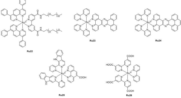
Among graphene-based nanomaterials, reduced graphene oxide sheet (rGO) is one of the most effective photothermal therapy (PTT) agents that can absorb NIR light and induce a temperature increase in the local environment, causing irreversible cell damage. Mao and co-workers developed a nano-theranostic platform, in which a PDT PS and a phosphorescent PEG-modified Ru(II) complex, Ru22 were loaded onto the PPT agent rGO surface via π-π and intermolecular hydrophobic interactions for the combination of PDT, PTT and imaging (Figure 7).[56] The resulting
nanomaterials rGO-Ru22, with a thickness of 25 nm as characterized by AFM, showed a higher photothermal effect than naked rGO (ΔT = 33 °C as opposed to ΔT = 22 °C for rGO at 808 nm, 10 min, 1 W,cm-2) and enhanced cellular uptake in human lung cancer A549 cells (15.40 ± 0.69 ng 10 -6 cells as opposed to 9.46 ± 0.43 ng 10-6 cells for Ru22). However, the ability of rGO-Ru22 to produce 1O
2 upon irradiation at 450 nm was lower than that of Ru22, which could be due to the quenching
effect caused by the photoinduced electron transfer between Ru22 and rGO. Complete release of Ru22 from rGO-Ru22 after irradiation at 808 nm for 5 min (0.5 W.cm-2) could restore the ability of
PS to produce 1O
2. By capitalizing on these, rGO-Ru22 showed higher anticancer efficacy in a
combined PTT−PDT treatment compared to PTT or PDT treatment alone. At 6.25 µM, cell viability for PTT treatment (808 nm, 0.5 W.cm-2, 5 min), PDT treatment (450 nm: 20 mW.cm-2, 2 min), and
successive PTT−PDT treatment was 17.6 ± 3.6 %, 51.8 ± 1.0 % and 8.3 ± 0.5%, respectively, although no significant toxicity was observed in the dark. These results were further confirmed in vivo in mice bearing A549 tumors: combined PDT-PTT could significantly inhibit the growth of tumors.
3.1.2. Carbon nanotubes
Carbon nanotubes (CNTs) are well ordered tubular allotropes of carbon with a diameter in the nanometer range and a length reaching up to several centimeters.[57a] They can be single graphene
sheet rolled up to form hollow tubes with walls one atom thick (single-walled carbon nanotubes, SWCNTs) or a multiple layer of graphene rolled simultaneously to form concentric tubes (multi-walled carbon nanotubes, MWCNTs). However, the apparent toxicity of CNTs has caused concerns for their use in drug delivery explaining its little use.[57b] To date, only one example for each CNTs
23
potential drug delivery systems, CNTs have physical intrinsic properties including photothermal ones that could facilitate therapy and be used to develop innovative multi-modal therapies.
Chao and co-workers reported the loading of two photon-absorbing Ru(II)-based PDT PSs, Ru23 and Ru24, onto commercially available SWCNTs of 0.7−1.3 nm diameter by π-π interactions after sonication in water.[58] The resulting nano-systems Ru@SWCNTs, stable up to three months in
solution, were characterized with a length ranging from 20 nm to several micrometers by TEM imaging. The photothermal effect triggered the release of Ru(II) complexes, which produced 1O
2
upon two-photon laser irradiation (808 nm). Ru@SWCNTs showed a higher cancer killing effect than that of the free Ru(II) complexes and pristine SWCNTs upon 2P 808 nm light irradiation (0.25 W cm -2, 5 min) in HeLa monolayer cancer cells as well as in 3D multicellular tumor spheroids (MCTSs) and
in vivo on nude mice bearing a HeLa tumor model. Half of Ru complexes were released after 5 min of irradiation. They were found to localize in lysosomes, implying endocytosis as the mode of cellular uptake Ru@SWCNTs.
Chen and co-workers described the use of MWCNTs as a radiosensitive nanodrug delivery system for Ru3 (Figure 2) in liver cells, including the multidrug resistant hepatocellular carcinoma R-HepG2, HepG2 and human hepatic L02 cell lines.[59] Approximately 9.8 % of Ru3 could be loaded into
Jeffamine-functionalized MWCNTs (Mn = 1 900 g.mol-1) with an average diameter of 225 nm via
π-π interactions. The resulting Ru3@MWCNTs nanoparticles were found to enhance the cellular uptake of Ru3 in R-HepG2 cells via endocytosis and to have some cytotoxicity (IC50 = 40 ng.mL-1).
No significant toxicity (>80 % cell viability) was observed with unloaded MWCTs up to 160 ng.mL-1.
Ru3@MWCNTs showed significantly lower cytotoxicity towards healthy cells (i.e., L02 cell lines) due to 1-2 fold lower cellular uptake. After being taken up by the cells, Ru3@MWCNTs were found in lysosomes, where Ru3 could be released more rapidly due to the acidic nature of the lysosomes. Moreover, the sensitivity of R-HepG2 cells to ionizing radiation at a dose of 8 J.kg-1 was increased
due to ROS overproduction: at 40 ng.mL-1, cell viability went down to 20 % instead of 44.8 % without
irradiation.
24
Due to their intrinsic upconversion fluorescence, carbon nanodots (CDs) hold a great advantage in imaging and therapy that requires the use of NIR irradiation.
Mao, Tan and co-workers designed Ru complex modified CDs for lysosome-targeted 1P and 2P imaging and PDT.[60] Ru25 was incorporated onto the surface of CDs using a typical EDC/NHS
coupling reaction. Nanoparticles with a diameter of ca. 100 nm were obtained with a Ru loading of 4 %. The nanoparticles exhibited a higher 2P absorption cross-section than the free Ru complex at 810 nm (σ = 1118 GM as opposed to σ = 810 GM for Ru25) attributed to CDs. In addition, conjugation to CDs could improve the cellular uptake of Ru25, resulting in a higher inhibitory effect on cancer cell growth and an enhanced photodynamic efficiency in both 2D A549 cell (IC50,light = 3.0 ± 0.2 µM,
PI > 20 after irradiation with visible light for 5 min, 20 mW cm-2) and 3D multicellular tumor spheroid
models (IC50,light = 2.2 ± 0.2 µM, PI > 45.4 after 810 nm irradiation for 20 min). These nanoparticles
were also suitable for 2P imaging as shown on zebrafish.
Qu and co-workers developed a biocompatible imaging nanoplatform to monitor the oxygen level in HeLa cells.[61] An oxygen sensitive Ru(II) polypyridyl complex Ru26 was conjugated to CDs using a
EDC/NHS coupling reaction. The modified CDs were then PEGylated to form stealth and stable spherical nanoparticles in aqueous solution with diameters of 37 ± 5 nm, which could serve as an oxygen sensitive probe upon 720 nm NIR irradiation. While remaining non-toxic up to 0.4 µmol mL -1, the PEGylated CDs could be imaged in HeLa cells after being excited at 720 nm, confirming the
upconverted imaging capacity of these nanoparticles. The luminescence intensity was higher in hypoxic environment than in normoxic environment. In vivo imaging in mice bearing HeLa tumors showed that these nanoparticles could accumulate preferentially in the tumor rather than in other organs.
25
Figure 7. Chemical structures of Ru22-Ru26.
3.2. Porous nanomaterials
3.2.1. Porous silicon-based nanomaterials
Porous silicon-based nanoparticles refer to two types of nanomaterials: porous silicon nanoparticles (SiNPs) and mesoporous silica nanoparticles (MSNs).[62]
3.2.1.1. Porous silicon nanoparticles
SiNPs typically have a size around a few hundreds of nanometers or several microns. Their size, porosity, pore size, pore morphology, and surface chemistry can be tuned during the synthesis to achieve the optimal performance in drug delivery. However, these materials, which are inherently fluorescent, have only limited control on the three-dimensional porous structures distinguishing them from MSNs. Cunin, Lemercier and co-workers reported the multi-functionalization of SiNPs, including with the 2P Ru(II) luminescent complex [Ru(5-Fluo-Phen)2(5-E-Phen)]2+ (Ru27).[63] SiNPs were
synthesized through electrochemical etching of silicon wafer, followed by sonication. They were then functionalized in a step-wise manner; the hydrosilylation of Ru27 to the surface through the terminal alkyne moiety giving SiNPs-Ru27 was followed by the silanization of surface hydroxyls with amine-terminated PEG to yield SiNPs-Ru27-PEG and finally the attachment of mannose (as cancer
26
targeting ligands) resulted in SiNPs-Ru27-PEG-Man with diameter ranging from 50 - 450 nm as characterized by TEM. The cytotoxicity of all the nanoparticles were evaluated against MCF-7 breast cancer cells and compared to that of the free complex: SiNPs-Ru27 could reduce the cell viability down to 43 % with 1P excitation (420 – 440 nm, 14 J cm-2, 20 min) and 36 % with 2P irradiation (800
nm, 3 scans of 1.57 s). Surprisingly, the incorporation of PEG and mannose did not improve significantly the 2P PDT efficacy.
3.2.1.2. Mesoporous silica nanoparticles
MSNs are solid materials with sizes ranging from 50 to 300 nm, containing hundreds of empty channels - called mesopores - in a 2D honeycomb-like network of porous structure. These nanoparticles possess a high surface area and pore volume, a stable meso-structure, a tunable pore diameter (ca. 2–10 nm), and facile surface modification (including the channels). This ordered pore network defines the size homogeneity, allowing a high loading capacity of drugs and enabling their controlled release. The loading of drugs into its matrix can be achieved by using different strategies: (i) entrapment by mixing the MSN precursors with the drug in a sol-gel phase before removing the liquid phase, (ii) encapsulation where the drug is loaded inside the pores as described for the entrapment strategy, followed by capping the pores, (iii) dissolution, (iv) adsorption relying on the attractive interaction between the silica surface and the drug molecules (physisorption) or (v) attachment taking advantage of the surface functionalization of MSNs by attaching drug molecules covalently on the surface (chemisorption), requiring a stimulus to cleave the covalent bond to release the drug.
Gómez-Ruiz, Gasser and co-workers reported the preparation of three Ru(II)-functionalized MSNs from the PDT PS Ru7: MSN-CL-Ru7, MSN-CNO-Ru7 and MSN-TRI-Ru7 with a diameter of around 77 nm.[64] They were prepared by functionalizing the surface of MSNs with two different silane
coupling agents, 3-chloropropylethoxysilane and 3-isocyanatopropylethoxysilane, to give MSN-CL and MSN-CNO, respectively. The resulting materials reacted with the Ru complex to give MSN-CL-Ru7 and MSN-CNO-MSN-CL-Ru7 with a Ru loading of 2.1 and 3.1 wt%, respectively. MSN-CL could also react with tri(2-aminoethyl)amine to give MSN-TRI, in which 7.5 wt% Ru complex was physically absorbed to give MSN-TRI-Ru7. Unfortunately, due to poor drug loading and quenching effect, these
27
nano-systems did not improve the photo-properties of the Ru(II) complex and were found to be not toxic towards HeLa cells.
Stoddart, Sauvage and Cryns developed a drug delivery platform obtained by grafting the surface of mesoporous silica nanoparticles with ruthenium(II) complexes.[65] A monodentate benzonitrile ligand
was grafted onto the surface of MCM-41 (average nanopore diameter of 2 nm), followed by coordination of [Ru(tpy)(dppz)(H2O)]2+ (Ru29) under dark conditions at room temperature. Since the
coordination between the Ru(II) complex and the monodentate ligand linked covalently to the nanoparticles can be cleaved under irradiation with visible light, Ru29 can be released from the surface of the nanoparticles and can act as a potential anticancer agent. Moreover, other drugs such as the chemotherapeutic agent paclitaxel were loaded into MSNs pores for multi-modal therapy. In that case, the Ru(II) complex acted as a capping agent. Therefore, paclitaxel was first loaded into the pores and Ru29 was then coordinated to block the pore openings. Light triggered first the release of Ru29 before paclitaxel. The resulting nano-systems were toxic towards MDA-MB-468 breast cancer. The toxicity was mainly attributed to paclitaxel.
Chen and co-workers described the encapsulation of Ru3 within ca. 100 nm arginine-glycine-aspartic acid (RGD) peptide-decorated MSNs.[66] The RGD peptide is the most used and effective
tripeptide in targeting delivery because it can specifically bind to an integrin receptor, overexpressed in a wide range of tumor cells. The cytotoxicity of the resulting MSNs was evaluated in different cancer cells (i.e., A375 melanoma, HepG2 hepatocellular carcinoma, MCF-7 breast adenocarcinoma, Neuro2a neuroblastoma and RHepG2 drug resistant carcinoma cells) and in HK-2 normal cells. The best results were obtained with A375 cells. Incorporation of RGD led to a significant enhancement in the uptake of the nanoparticles in A375 cells, which have the highest expression level of integrin receptors, via receptor-mediated endocytosis. This allowed a much higher anticancer effect with an IC50 of 65.8 nM while the IC50 value for Ru3 was 5.9 µM and the
unloaded RGD-functionalized MSNs showed no significant toxicity up to 400 µg mL-1. It also
improved the selectivity between cancer and healthy cells as the toxicity in HK-2 healthy cells were much lower with an IC50 higher than 75 µM. The release of Ru3 from the nanoparticles were much
28
7.4. This is of interest since tumors are known to have an acidic environment. The cell death was attributed to apoptosis through ROS production.
3.2.2. Metal-organic frameworks
Metal-organic frameworks (MOFs) are organic–inorganic crystalline hybrid materials with permanent pores that are formed by the self-assembly of organic linkers and metal ions or clusters through coordination bonds. Like porous silicon-based nanoparticles and especially MSNs, MOFs have the advantages of high porosity, adjustable pore size, easy modification and good biocompatibility. Among the large family of MOFs, only zirconium-based MOFs have so far been used to incorporate Ru(II) polypyridyl complexes.
Lee and co-workers developed a zirconium-based MOF as a theranostic nanoplatform for combined two-photon imaging and PDT applications.[67] Ru(II) complexes have even been incorporated into
MOF nanoparticles, with average diameters of 92 nm. Incorporation of [Ru(bpy)3]2+ (Ru30, Figure 8)
into the pores of the MOFs resulted in an overall enhancement of the photophysical properties, including luminescence quantum yield, 2P absorption cross-section (σ2 = 21.9 GM at 880 nm), 1O2
generation and photostability compared to that of the free complex. Ru(II)–MOFs showed low toxicity towards HeLa cells in the dark at a concentration of 200 µg mL-1. However, upon white light
irradiation (200 mW cm-2, 10 min), a significant reduction in the viability of the treated cells was
observed.
Shi, Yao and co-workers reported a multifunctional MOF nanoplatform to combine PDT, PTT and chemotherapy.[68] Zirconium-based MOF, namely UiO-66, was functionalized with an azide group to
perform a click reaction with a Ru(II) polypyridyl complex-based PDT PS bearing an alkyne group, Ru31, resulting in the formation of nanoparticles with a diameter of ca. 110 nm and a Ru loading of 10.2 %. The pores (1.64 nm) within these MOF nanoparticles acted as a host to 13.5 wt% of doxorubicin (DOX), and 6.8 wt% of copper sulfide (CuS) nanoparticles, a potential PTT agent, with a diameter of ca. 10 nm were loaded onto the surface of the nanoplatform by physical absorption. The structure of the framework was preserved throughout the chemical transformation. UiO-Ru27-DOX-CuS could generate 1O
29
Figure 8. Chemical structures of Ru27-Ru31. Light-induced release of the monodentate ligand L leading to the formation of [Ru(tpy)(dppz(H2O)]2+ (Ru29).
3.3. Metal-based nanoparticles
3.3.1. Gold and silver nanoparticles
Gold and silver nanoparticles are very interesting nano-systems as they have intrinsic physical properties that could be used to construct a unique platform for potential multi-modal therapy. These nanoparticles are characterized by their surface plasmon resonance, which corresponds to a rise of plasmons when their surface is irradiated with an electromagnetic wave in the UV-visible region, leading to oscillation of conduction electrons. This feature can be used for detecting molecular interactions. For Ru(II) polypyridyl complexes, gold nanoparticles have so far been utilized to visualize and characterize biological processes at a molecular and cellular level.
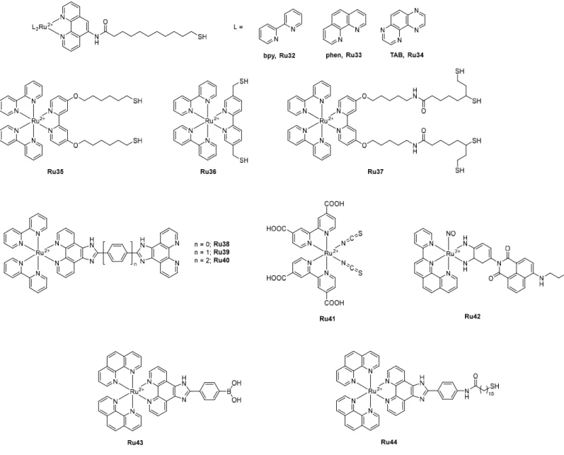
Gunnlaugsson and co-workers reported the synthesis of three alkyl thiol group terminated Ru(II) polypyridyl complexes [Ru(L)2L’](PF6)2 (L = bpy, phen or 1,4,5,8-tetraazaphenanthrene (TAB) and L’
= 11-mercapto-N-(1,10-phenanthrolin-5-yl)undecanamide, Ru32, Ru33, Ru34) to react with surface-functionalized AuNPs (Figure 9).[69] The resulting sub-5 nm water-soluble AuNPs offered attractive
30
photophysical properties, ideal for applications in cellular imaging: they showed higher DNA affinity compared to the free Ru(II) complexes and no cytotoxicity towards HeLa cells after internalization and localization in the cytoplasm, allowing their potential use for cellular imaging. However, because of their small size, it was difficult to further investigate and characterize their uptake mechanism in cancer cells. The use of larger AuNPs of ca. 15-20 nm allowed visualization in cells using TEM.[70]
They were observed within single membrane vesicles in the cytoplasm, suggesting a cellular uptake by endocytosis. Two of these Ru complexes, Ru32 and Ru33, were also used by Shi and co-workers for functionalization of silver nanoparticles (AgNPs), resulting in monodisperse and homogeneous spherical nanoparticles with an average diameter of 20 nm.[71] AgNPs are known to induce
cytotoxicity by an excessive generation of reactive oxygen species (ROS). Functionalization with Ru(II) complexes allowed cell imaging and enhanced anticancer activity. The Ru(II)-functionalized AgNPs were more toxic towards HeLa cells than the naked AgNPs, while the parent Ru complexes were not toxic up to 50 µg.mL-1. This improved cytotoxicity was attributed to an enhanced cellular
uptake, hence an increase in ROS generation.
Pikramenou, Hodges and co-workers reported the functionalization of two Zonyl 7850 non-ionic fluorinated surfactant pre-coated AuNPs of different sizes (13 and 100 nm) with Ru35, a Ru(II) complex bearing two thiol groups and a hexyl spacer group to distance the Ru center from the nanoparticles’ surface.[72] Pre-coating with a non-ionic fluorinated surfactant allowed a higher loading
of Ru on the AuNPs surface by preventing irreversible aggregation. Attachment to AuNPs surface resulted in an enhancement in luminescence lifetime (from 260 ns for Ru35 to 340 ns for Ru35@AuNP13 and 360 ns for Ru35@AuNP100). Their strong luminescence allowed imaging in A549 cells with no significant cytotoxicity. The same authors then investigated the effect of the distance of the thiol-functionalized ruthenium luminescent center from the gold surface on the luminescence properties of the nanoparticles.[73] Three ruthenium probes with different length spacer
units (Ru35, Ru36 and Ru37) were attached on Zonyl pre-coated AuNPs of different sizes (13, 50 and 100 nm). An enhanced luminescence lifetime was obtained upon attachment with an increase of 20 %, 40 % and 70 % depending on the spacer length: the longer the spacer chain was, the higher the luminescence lifetime was. By contrast, AuNPs size did not affect it.
31
Chao and co-workers developed a strategy combining the 2P luminescence of Ru(II) polypyridyl complexes (Ru38, Ru39 and Ru40, shown in Figure 9) and the photothermal properties of AuNPs in one single theranostic nanoplatform for image-guided PTT.[74] While the luminescence of Ru
complexes was quenched upon attachment to AuNPs due to an energy transfer from the Ru(II) center to the AuNPs, the AuNPs still displayed significant 2P luminescence, which could be used in 2P lifetime imaging in cells, especially with Ru39 (σ = 187 – 308 GM depending on the AuNPs size at 808 nm, 1 GM = 1 x 10-50 cm4 s-1 photon-1). The resulting AuNPs exhibited prominent
photothermal conversion efficiency (18.3 % - 33.3% % as opposed to 7.3 % - 10.2 % for the naked AuNPs) and excellent photothermal stability. An effective tumor ablation was observed after photothermal treatment (808 nm, 0.8 W cm-2, 5 min) in vivo on HeLa tumor-xenograft model mice.
As the best results were obtained with Ru39, they investigated the attachment of this Ru(II) complex on AuNPs with different morphologies: nanorods (AuNRs) with an average length of 40 ± 2.0 nm and width of 13 ± 0.8 nm and nanostars (AuNTs) with four tips of 15 – 25 nm long and a center sphere sizing 20 ± 1.3 nm.[75] The resulting AuNRs@Ru37 and AuNTs@Ru37 exhibited higher
photothermal conversion efficient (η = 33.7 % for AuNRs@Ru37, η = 34.5 % for as opposed to ηAuNRs = 22.0%, ηAuNTs = 18.9%). Their cytotoxicity was evaluated in HeLa cells as well as in MCTSs
and in vivo in a HeLa tumor-xenograft model. They showed similar results as of the ones obtained with AuNPs and good photothermal ablation at 808 nm using 0.25 W.cm−2 laser irradiation, which is
lower than the maximal permissible exposure of skin by the American National Standard Institute (ANSI) regulation (0.33 W.cm−2 at 808 nm). The enhanced photothermal property was not dependent
on the nanoparticle morphology.
Then they took advantage of the luminescence quenching effect of AuNPs to construct an off-on fluorescence probe for the detection of thiol-containing amino acids including glutathione (GSH), homocysteine (Hcy) and cysteine (Cys) in living cells.[76] AuNPs surface was modified with Ru21
(shown in Figure 6) by reducing HAuCl4 with NaBH4 in the presence of the Ru(II) complex.
Monodisperse and spherical Ru19@AuNPs, with diameters ranging from 4 to 11 nm, were obtained, in which the luminescence of the complex was completely quenched. Luminescence was restored when the nanoparticles reacted with thiol-containing compounds.
32
Figure 9. Chemical structures of Ru(II) polypyridyl complexes Ru32 – Ru44 incorporated in metal-based and selenium nanoparticles.
3.3.2. Metal oxide nanoparticles
In addition to metal nanoparticles, which are predominantly gold and silver nanoparticles, metal oxide nanoparticles such as iron oxide (magnetite, Fe3O4) and titanium oxide (TiO2) have also been
investigated for cancer-related biomedical applications.
Sun, Zeng, and co-workers reported the preparation of fluorescent magnetic nanoparticles Fe3O4
-Ru for simultaneous optical and magnetic resonance imaging applications.[77] Ru(dcpy)
2(NCS)2
(Ru41) was coupled to Jeffamine-functionalized (Mn = 1900 g.mol-1) magnetic Fe3O4 nanoparticles
via the reaction of the amine end groups of Jeffamine with the isothiocyanate ligand of the Ru complex. Around 36 Ru complexes were incorporated onto the surface of each 8 nm Fe3O4
33
nanoparticle. Functionalization with Jeffamine not only afforded nanoparticles stable in biological media but also helped prevent the fluorescence quenching of the Ru complex by Fe3O4. Fe3O4-Ru41
showed excellent colloidal, photochemical and magnetic stability. Fe3O4-Ru41 nanoparticles were
found in the cytoplasm of SK-BR-3 human breast cancer cells while showing no significant cytotoxicity.
Liu and co-workers developed a tumor-targeting system based on carbon-doped TiO2 (C-TiO2)
nanoparticles for PACT applications.[78] Receptor-targeting folate molecules and 6.64 wt% of Ru(II)
polypyridyl-nitrosyl complexes Ru42, bearing a lysosome-targeting morpholine moiety (Figure 9), were incorporated onto the surfaces of the 4 nm C-TiO2 nanoparticles. The resulting nanoparticles
exhibited excellent liposome specific colocalization in HeLa cells. NIR irradiation at 808 nm (600 mW cm-2, 10 min) then led to the release of nitric oxide and ROS from the as prepared nanoparticles,
resulting in a significant decrease in the viability of HeLa treated cells, with an IC50light value of 20 µg
mL-1. No significant toxicity was observed in the dark towards HeLa cells at the concentrations tested
(10 – 200 µg mL-1).
3.4. Selenium nanoparticles
Selenium, naturally present in trace amounts in the human body and required for the biosynthesis of selenocysteine-containing selenoproteins, has proven to be a potential anticancer agent in its nanoscale form. Decorating SeNPs with Ru(II) polypyridyl complexes could not only provide fluorescence for imaging but also an enhanced anticancer effect.
Tumor angiogenesis is the proliferation of a network of blood vessels, which provides the tumor with oxygen and nutrients required to grow. Blocking angiogenesis could be a strategy to inhibit and stop tumor growth. This strategy has been exploited by Liu and his research team. They reported the anti-angiogenesis and anti-tumor behavior of SeNPs decorated with [Ru(phen)2(p-BPIP)](PF6)2 (Ru43,
p-BPIP = (4-(1H-imidazo[4,5-f][1,10]phenanthrolin-2-yl)phenyl)boronic acid).[79] Fluorescent
Ru-SeNPs were obtained, by reduction of Na2SeO3 with gallic acid prior to the addition of the Ru
complex, as monodisperse homogeneous spherical particles with an average diameter of ca. 100 nm, characterized by TEM and an atomic ratio Se/Ru of 1/8. Ru43-SeNPs, compared to SeNPs or RuBP, could strongly inhibit at low concentrations (5 – 10 µg mL-1) human umbilical vascular
34
endothelial cell (HUVEC) proliferation, migration, and tube formation, which are key steps for angiogenesis. The cytotoxicity of Ru39-SeNPs was examined against various cancerous cell lines and compared to that of the free Ru complex and naked SeNPs. Ru43-SeNPs showed a higher anti-tumor effect than the two counterparts: 4.8 ± 0.8 µg mL-1 for HUVEC, 3.1 ± 0.9 µg mL-1 for HepG2,
12.5 ± 1.2 µg mL-1 for SW480, 17.4 ± 1.5 µg mL-1 for PC-3 and 20.2 ± 2.3 µg mL-1 for MCF-7.
The same authors then described the functionalization of SeNPs with the thiol-based Ru(II) complex, [Ru(phen)2MUA](PF6)2 (Ru44, MUA =
2-(4-11-mercaptoamide-N-phenyl)-1H-imidazo[4,5f][1,10]phenanthroline).[80] The resulting monodisperse and homogenous spherical
nanoparticles of less than 100 nm, Ru44@SeNPs, enhanced the cellular uptake of Ru44 in HepG2 cells with nuclear and cytoplasm localization, whereas Ru44 was mainly localized in the cytoplasm. This resulted in significant cell damage at a relatively low concentration, IC50 = 18.5 µg mL-1.
3.5. Upconversion nanoparticles
The biological application of Ru(II) polypyridyl complexes for PDT and PACT can somehow be limited by the need of the use of poorly penetrating blue or green light. Lanthanide-doped upconversion nanoparticles (UCNPs) can overcome this limitation as they can convert NIR photons to visible or UV light. Most reported UCNPs are hexagonal-phase NaYF4 nanocrystals and the most
common lanthanide ions used in photon upconversion are the pairs erbium(III)-ytterbium(III) or thulium(III)-ytterbium(III) (Figure 10).[81]
Salassa and co-workers were, to the best of our knowledge, the first to use UCNPs for the light-triggered release of photoactivated Ru(II) polypyridyl complexes with NIR irradiation. [Ru(bpy)2(py)2]Cl2 (Ru45), where py is pyridine, was absorbed at the surface of UCNPs with a
diameter of ca. 80 nm.[82] Irradiation at 980 nm could trigger photolysis to afford the aqua adduct
[Ru(bpy)2(py)H2O]Cl2. However, only one of the two monodentate ligands was able to dissociate
from the complex, preventing potential interaction with DNA.
Liu and co-workers designed a drug delivery system using HSA coated UCNPs to efficiently deliver a photoactivated Ru(II) polypyridyl complex, Ru4.[83] The HSA coating made the particles highly
biocompatible and well‐dispersed in aqueous solution. HSA coated UCNPs were prepared by crosslinking HSA in the presence of UCNPs and Ru4using glutaraldehyde in ethanol/water solution.
35
Uniform spherical nanoparticle Ru-HSA-UCNPs with an average hydrodynamic diameter of 120 nm and a PDI of 0.092 were obtained. While the fluorescence was quenched after encapsulation of the Ru complex, HSA coating provided green fluorescence for cell imaging. Ru-HSA-UCNPs showed photo‐induced cytotoxicity (white light, 10 min, 10 mWcm-2) to HepG2 cells and HeLa cells attributed
to the formation of [Ru(bpy)2(H2O)2]Cl2, which was shown to interact with DNA. No significant toxicity
was observed in the dark (IC50 > 300 µM).
Wang and co-workers reported the encapsulation of a photoactivable Ru complex Ru46 in UCNPs coated with PEG-modified lipid (DSPE-PEG, Mn = 5000 g mol-1).[84] The resulting nanosystem had a
core-shell structure with an average diameter of 670.4 nm. From the nanoparticles, 80 % of Ru complex was released as [Ru(bpy)2(H2O)2]2 after 2 h irradiation with visible light (λ > 400 nm, 0.07
W.cm-2) or after 40 min irradiation with NIR light (980 nm, 3 W.cm-2). The use of UCNPs could trigger
the release of the DNA binding agent [Ru(bpy)2(H2O)2]2+(Ru47) by NIR irradiation, suitable for
deep-seated tumors, as shown in Figure 10b. However, no biological evaluation has been reported to date.
Due to the competing presence of a water absorption peak at the same wavelength requiring prolonged irradiation, the use of 980 nm light raises some concerns as it can deeply penetrate cells, therefore causing severe burns to biological systems. Bonnet and Natile described the conjugation of [Ru(bpy)2(3H)](PF6)2 (Ru48), a Ru(II) polypyridyl complex where 3H is a photocleavable
bis(thioether) ligand modified with two phosphonate moieties, onto the surface of a core-shell-shell UCNPs (CSS-UCNPs) through its thioether groups.[85] They demonstrated that blue light could be
generated from the resulting 115 nm particles after 796 nm irradiation at 50 W.cm-2, allowing the
photodissociation of the sulfur ligand from the Ru center. However, the photoreaction was not complete under therapeutically relevant conditions. Therefore, no biological evaluation was reported.


![Figure 3. a) Structures of Ru8 and the three [Ru(phen) 3 ] 2+ derivatives: Ru9, Ru8-PhenAN and Ru8-PhenISA](https://thumb-eu.123doks.com/thumbv2/123doknet/7771105.256913/15.892.40.830.70.1069/figure-structures-ru-ru-phen-derivatives-phenan-phenisa.webp)

![Figure 5. a) Chemical structure of [Ru(phen) 2 dppz] 2+ (Ru16). b) Photoactivation of Ru17 leading to the formation of the aqua adduct Ru18](https://thumb-eu.123doks.com/thumbv2/123doknet/7771105.256913/20.892.106.773.81.737/figure-chemical-structure-phen-photoactivation-leading-formation-adduct.webp)
![Figure 6. a) Chemical structure of [Ru(bpy) 2 Hipa] 2+ (Ru21), b) Schematic illustration of a part of cHSA-PEO-TPP-Ru21 showing how PEG (in blue), TPP (in pink) and Ru21 (in black) are conjugated to HSA (in purple)](https://thumb-eu.123doks.com/thumbv2/123doknet/7771105.256913/22.892.54.834.88.489/figure-chemical-structure-schematic-illustration-showing-conjugated-purple.webp)
![Figure 8. Chemical structures of Ru27-Ru31. Light-induced release of the monodentate ligand L leading to the formation of [Ru(tpy)(dppz(H 2 O)] 2+ (Ru29)](https://thumb-eu.123doks.com/thumbv2/123doknet/7771105.256913/30.892.91.853.101.575/figure-chemical-structures-induced-release-monodentate-leading-formation.webp)