_____________________________________________________________________________________________________ *Corresponding author: E-mail: [email protected], [email protected], [email protected],
26(3): 1-13, 2018; Article no.ARRB.41297
ISSN: 2347-565X, NLM ID: 101632869
In vitro Antioxidant Activities of Various Extracts
from Flowers-Leaves Mixture of Algerian
Cytisus triflorus
Soraya Madoui
1*, Noureddine Charef
1, Lekhmici Arrar
1,
Abderrahmane Baghianni
1and Seddik Khennouf
21
Laboratory of Applied Biochemistry, University Ferhat Abbas, Setif 1 19000, Algeria. 2
Laboratory of Phytotherapy Applied to Chronic Diseases, University Ferhat Abbas, Setif 1 19000, Algeria. Authors’ contributions This work was carried out in collaboration between all authors. All authors read and approved the final
manuscript. Article Information DOI: 10.9734/ARRB/2018/41297 Editor(s): (1) George Perry, Dean and Professor of Biology, University of Texas at San Antonio, USA.
Reviewers: (1) Noriah Bidin, Universiti Teknologi Malaysia, Malaysia. (2) Yves B. Nyamien, University of Abidjan, Côte d’Ivoire. (3) Dike Charles Chijioke, Nnamdi Azikiwe University-Awka, Nigeria. Complete Peer review History:http://www.sciencedomain.org/review-history/24544
Received 28th February 2018 Accepted 7th May 2018 Published 10th May 2018
ABSTRACT
Background: Antioxidants from plants are preferred due to their multiple mechanisms of actions. In this study, the mixture of flowers and leaves (according to traditional use) of Cytisus triflorus (Fabaceae) was assessed for their in vitro antioxidant activities.
Materials and Methods: The mixture leaves-flowers powder was macerated in methanol, filtered, and then dried to give the crude extract. The crude extract was successively extracted with different solvents of increasing polarity giving: petroleum ether extract, chloroform extract and ethyl acetate extract. Total phenol content was determined by Folin-ciocalteau assay and the AlCl3 method was used for determination of the total flavonoids. The mixture was assessed for its in vitro antioxidant activity using spectrophotometric methods like DPPH, β-carotene/linoleic acid bleaching, Ion chelating, reducing power, Superoxide anion radical scavenging, ABTS radical scavenging,
Hydrogen peroxide and Anti-Hemolytic Assays. Antioxidant activities were compared with BHT and ascorbic acid as standard antioxidants.
Results: Quantitative analysis of antioxidative components showed that ethyl acetate extract is the richest fraction in term of polyphenols (69.78 ± 2.97 µg GAE/mg of extract) when chloroform extract has the highest amount of flavonoids (17.4 ± 0.46 µg QE/mg extract). Results showed that the crude extract and fractions of this plant exhibited high antioxidant activities. Crude extract showed the strongest effect in almost all tests (DPPH, ion Chelating, β-carotene/linoleic acid bleaching and Anti-Hemolytic assays), when petroleum ether extract was the weakest one. Aqueous extract exhibited the highest activity in reducing power test with IC50 value of 320 µg/mL while chloroform extract was the most effective in hydroxyl scavenging assay (IC50 = 440.45 µg/mL). In hydrogen peroxide scavenging test, ethyl acetate extract was the most effective with a similar effect to that of ascorbic acid (IC50 = 1.54 µg/mL).
Conclusion: Results obtained indicated that extracts from Cytisus triflorus exhibited a potential effect to prevent disease caused by the overproduction of radicals.
Keywords: Antioxidants; Cytisus triflorus; flavonoids; polyphenols; reactive oxygen species. 1. INTRODUCTION
New sources of antioxidants are continually sought from a variety of plants, animals and microorganisms. Among them, plants are of particular interest because they may produce raw materials or preparations containing phytochemicals which have significant antioxidant capacities and health benefits [1]. Reactive oxygen species (ROS) are various forms of activated oxygen, which include free radicals such as superoxide ions (O2-.) and hydroxyl radicals (OH.) as well as non-free radical species such as hydrogen peroxide [2]. These ROS have been found to play an
important role in the initiation and/or progression of various diseases such as atherosclerosis, inflammatory injury, cancer, and cardiovascular disease [3]. Antioxidants are known to play an important role in the protection against disorders caused by oxidant damage. Antioxidants can delay or inhibit the initiation or propagation of oxidative chain reactions and thus prevent or repair damage done to the body’s cells by oxygen [4]. Plants have been the basis of traditional medicines throughout the world for thousands of years and continue to provide new remedies to humankind [1]; a great deal of effort has therefore focused on using available experimental techniques to identify natural
antioxidants from plants. Several authors have reviewed the beneficial uses of these plant species [5]. Cytisus triflorus is a perennial shrub of the Fabaceae family within the tribe Genisteae. It is known in North of
Algeria for its medicinal properties [6]. It is used for treating abdominal pain, wounds healing and as haemostatic, antifungal and
hypotensor. Additionally, the leaves are used as “henna” to treat and dye the hair [6].
In this work, we studied the antioxidant and free radicals scavenging effects of plant extracts from Cytisus triflorus. The contents of total polyphenols and flavonoids in these extracts were also
determined.
2. MATERIALS AND METHODS 2.1 Plant Materials
Cytisus triflorus was gathered in April 2014 from the municipality of Chemini (Bejaia). The
sample was authenticated by Pr. Oudjhih Bachir, university Elhadj Lakhdar, Batna. The
leaves and the flowers were separated from other parts. The plant samples were air dried in shadow and finely powdered in a rotating knife grinder. The plant powder of the mixture leaves-flowers was then used for the extraction procedure.
2.2 Polyphenols Extraction Procedures
100 g mixture flowers-leaves of Cytisus triflorus are macerated in 1 L methanol, the extract was filtered by the cotton and the filter paper of Wattman, the solvent (methanol) is eliminated from the filtrate by rotary evaporation in Rotavapor (BÜCHI). The milk white crude extract was coded as CrE. A defined portion of total CrE volume was lyophilized and stored at until use [7].
CrE was subjected to fractionation using liquid-liquid extraction. CrE was successively extracted with different solvents of increasing polarity: petroleum ether for defatting, chloroform for aglycone flavonoids extraction and ethyl acetate for glycoside flavonoids extraction. The obtained organic layer of each partition was evaporated under reduced pressure on a rotavapor below 45°C to dryness and to afford petroleum ether, chloroform, ethyl acetate and aqueous fractions coded as PeE, ChE, EAE and AqE, respectively.
2.3 Determination of Total Phenolic
Content
In order to measure phenolic compounds in plant extracts (the methanol, chloroform, ethyl acetate, aqueous fractions and petroleum ether extracts) at different occasions, we used the Folin– Ciocalteu assay. The reagent of Folin–Ciocalteu consists of a mixture of acid phosphotungstic and phosphomolybdic acid. During oxidation, it is reduced to a mixture of blue oxide. The color produced is proportional to the amount of polyphenols present in the analyzed extract [8]. According to the method of Li and their collaborators [9], 50 µL of each plant’s extract (CrE, AqE, ChE, PeE and EAE) are diluted and mixed with 250 µL Folin-Ciocalteu reagent (2 M, 10 times diluted) and 250 µL of sodium carbonate (7.5 g/ 100 mL). The absorbance is measured at 765 nm against a methanol blank, after incubation for 1 hour and 30 min at ambient temperature. A standard curve of gallic acid was created using an adequately range of gallic acid concentration from 5 to 150 µg/mL. The results were expressed as mg gallic acid equivalent/ gram of plant extract.
2.4 Determination of Total Flavonoids Content
The aluminium chloride (AlCl3) method was used for determination of the total flavonoids content of the C. triflorus extracts, employing the reaction of complex formation between flavonoids and AlCl3 [10]. Aliquots of 1ml of each extract were added to equal volumes of a solution of 2% AlCl3. The mixture was vigorously shaken, then it incubated for 10 min, in room temperature. The absorbance was read at 430 nm. Quercetin and rutin (0-40 mg/mL) were used as standards. Flavonoids contents were expressed as µg quercetin and rutin equivalent (QE and RE, respectively)/mg of each extract.
2.5 Determination of the Antioxidant Activity of Plant Extracts
2.5.1 DPPH scavenging assay
The DPPH assay measures hydrogen atom donating activity and hence provides a measure of free-radical scavenging antioxidant activity. DPPH is a purple-colored stable free radical; it becomes reduced to the yellow-colored, diphenyl picryl-hydrazine. Briefly, 50 µL of various dilutions of each extract or standards were mixed
with 1250 µL of DPPH methanol solution (0.004%). After an incubation period of 30 min in dark and at room temperature, the absorbance of the samples was read at 517 nm [11]. BHT, gallic acid, quercetin and rutin were used as standards. The lower the absorbance, the higher free radical-scavenging activity. The ability to scavenge the DPPH radical was calculated by using the following equation:
Scavenging effect %=(Ac-Ae )/Ac x100 Where Ac: control absorbance and Ae: absorbance in the presence of extracts. IC50 values of the extract i.e., concentration of extract necessary to decrease the initial concentration of DPPH by 50 % were calculated.
2.5.2 β-carotene/linoleic acid bleaching assay
In this assay, the antioxidant capacity of the extracts is given by measuring the inhibition of the decomposition oxidative of β-carotene (discolouration) by the products of oxidation of the linoleic acid [12]. The emulsion of β-carotene/ linoleic acid is prepared by solubilization of β-carotene 0,5 mg in 1 ml of chloroform, 25 µL of the linoleic acid and 200 mg of Tween 40 are added, chloroform is completely evaporated with the rotavapor, thereafter 100 mL of distilled water saturated with oxygen are added, the resulting emulsion is agitated vigorously. 350 µL of solution of extracts or antioxydants of reference (BHT) solubilized in methanol (2 mg/mL) is added with 2,5 mL with the preceding emulsion. The kinetics of discoloration of the emulsion in presence and absence of antioxidant (negative control in which the sample is replaced by 350 µL methanol and distilled water) is followed to 490 nm with intervals of regular times during 48 hours (after: 1 h, 2 h, 3 h, 4 h, 6 h, 24 h, and 48 h) of incubation at room and in the darkness. The inhibition percentage of β-carotene oxidation by extracts was measured as follows:
AA% = ABS test / ABS BHT ×100
AA%: Percentage of the antioxidant activity; ABS test: Absorbance in the presence of the extract (test);
ABS BHT: Absorbance in the presence of positive control BHT.
2.5.3 Ferrous ion chelating assay
Ferrous ion chelating activity was measured by inhibition of the formation of iron (II)–ferrozine
complex after adding the test sample [13]. The reaction mixture contained 250 µL sample, 50 µL Fe2Cl2 (0.6 mM in water) and 450 µL methanol. The control contained all the reaction reagents except the sample. The mixture was shaken and allowed at room temperature for 5 min. An aliquot of 100 µL of ferrozine (5 mM in methanol) were then added, the mixture shaken again, followed by further reaction at room temperature for 10 min to complex the residual Fe+2 ion. The absorbance of the Fe+2–ferrozine complex was measured at 562 nm against a blank contained all the reaction reagents except ferrozine. Lower absorbance indicates a higher chelating power. EDTA, quercetin and rutin were used as reference chelators. The chelating activity was calculated as following:
Chelating activity %=(Ac-Ae )/Ac*100 Where Ac: control absorbance and Ae: absorbance in the presence of extract. Concentration corresponding to chelating of 50 % of Fe+2 ions was calculated from curve of chelating percentage in function of C. triflorus extracts concentrations.
2.5.4 Determination of reducing power The reducing capacity of a compound serves as a significant indicator of its potential antioxidant activity. The reducing power assay is based on the mechanism of electron donating activity, which is main mechanism of phenolic antioxidant action [14]. The reaction mixture contained 400 µl of extract in an appropriate solvent was mixed with phosphate buffer and ferrocyamide solution. After 20 min at 50°C, 10% TCA was added and the mixture was centrifuged for 10 min at 3000 rpm. Finally, a 200 µL supernatant was mixed with 200 µL distilled water and 40 µL of 0.1% FeCl3, the absorbance was recorded at 700 nm, and the results were represented as ascorbate equivalents.
2.5.5 Hydroxyl scavenging activity
The reaction mixture contained 500 µL of FeSO4 (1.5 mM) and 350 µl of H2O2 (6 mM). After 5 min incubation, 150 µL of sodium salicylate (20 mM) were added. The mixture was incubated at 37°C for 30 min, then; the absorbance of samples was read at 562 nm [15]. Ascorbic acid (Asc. A) was used as standard. The ability to scavenge the Hydroxyl (OH•) radical was calculated by using the following equation:
Where Ac: control absorbance and Ae: absorbance in the presence of extracts.
IC50 values (Concentration of extract necessary to decrease the initial concentration of Hydroxyl (OH•) radical by 50%) were
calculated.
2.5.6 ABTS radical scavenging activity The ABTS scavenging test is used to determine the antioxidant activity (by estimating peroxide formation) of both hydrophilic and hydrophobic compounds. The spectrophotometric analysis of 2,2-azinobis (3-ethyl-benzothiazoline-6-sulfonic acid, ABTS) radical scavenging activity was determined according to Pavithra and Vadivukkarasi [16]. The ABTS•+ was produced by reacting 7 mM ABTS in H2O with 2.45 mM potassium persulfate (K2S2O8), stored in the dark at room temperature for 12 h. Before use, the ABTS•+ solution was diluted with methanol to get an absorbance of 0.850 ± 0.025 at 734 nm. Then, 2.3 ml of ABTS•+ solution was added to 100 μL of the solution at different concentrations. After 30 min, the inhibition percentage was calculated for each concentration. Solvent blanks were run in each assay. The extent of decolorization is calculated as percentage of the reduction of absorbance. The scavenging capability of ABTS•+ radical was calculated using the following equation:
ABTS•+ scavenging effect %=(Ac-Ae )/Ac*100 Where AC is the initial concentration of the ABTS•+ and Ae is absorbance of the remaining concentration of ABTS•+ in the presence of varying concentrations of tested compound or BHT.
2.5.7 Superoxide anion radical scavenging activity
In this experiments, the reaction mixture containing 0.1 ml of Nito-blue tetrazolium chloride (1 mg/mL solution in DMSO) and 0.3 mL of the various concentration of the extracts in DMSO, 1 mL of alkaline DMSO ( 1 mL DMSO contain 5 Mm NaOH) was added to give a final volume of 1.4 mL and the absorbance was measured at 560 nm [17]. The same procedure was repeated for the standard ascorbic acid. Pure DMSO was used as blank instead of alkali DMSO. The percentage inhibition of superoxide anion generation was calculated using the following equation:
Inhibition %=(Ac-Ae )/Ac*100
Where Ac: control absorbance and Ae: absorbance in the presence of extracts.
IC50 values of the extracts (i.e. concentration of extract necessary to decrease the initial concentration of Superoxide anion (O2•-) radical by 50 %) were calculated.
2.5.8 Hydrogen peroxide scavenging activity Hydrogen peroxide scavenging potential of plant extracts was determined in this experiment, 250 µL of ferrous ammonium sulphate (2 mM) were added to 1.5 mL of different concentrations of extract and then mixed with 62.5 μL of Hydrogen peroxide (5 mM), thereafter incubated at room temperature in dark for 5 minutes. Then, 1.5 mL of 1,10-phenanthroline (1 mM) was added to each tube, mixed and incubated for 10 minutes at room temperature. Finally the absorbance was read at 510 nm [18]. The same procedure was repeated for the standard ascorbic acid. The percentage inhibition of Hydrogen peroxide generation was calculated using the following equation:
Inhibition %=(Ac-Ae )/Ac*100
Where Ac: control absorbance and Ae: absorbance in the presence of extracts. IC50 values of the extract i.e., concentration of extract necessary to decrease the initial concentration of hydrogen peroxide by 50 % were calculated. 2.5.9 Anti-Hemolytic assay
Blood was obtained by venipuncture from healthy human volunteers collected in heparinized tubes and centrifuged at 3500 rpm for 15 min. Plasma was removed and Red blood cells (RBCs) were suspended in 10 volumes of 0.9% NaCl and centrifuged at 2500 rpm for 10 min. The RBCs were washed three times with the same solution. RBC suspension was incubated with 5 μM ferrous sulfate and various concentrations of the extracts at 37°C for 30 min. After incubation, further centrifuged at 2500 rpm for 5 min was conducted. Hemolysis was determined by measuring the optical density of the supernatant at 540 nm. The reaction without extract was used as a control sample [19]. BSA was used as a standard. Percentage of anti-hemolysis was calculated from following equation:
Where Ac: control absorbance and Ae: absorbance in the presence of extracts. IC50 values of the extracts were calculated.
2.6 Statistical Analysis
The results were expressed as mean ± standard deviation of three replicates. Where applicable, the data were subjected to one-way analysis of variance (ANOVA) and the differences between samples were determined by Duncan’s multiple comparison and Tukey’s multiple comparison, using Graph Pad program.
3. RESULTS AND DISCUSSION 3.1 Polyphenols Extraction
This extraction allows obtaining five extracts: CrE, PeE, ChE, EAE, and AqE. Yields of extraction are shown in Table 1. AqE gave the highest yield with 38% followed by PeE (28.06%), ChE (17.2%) and EAE (8.2%).
3.2 Total phenols and flavonoids content
As phenolic compounds constitute one of the major contributors to the antioxidant capacity of plants, it was reasonable to determine their total amount in the selected plant extracts. The total phenol content was estimated by the Folin– Ciocalteu method [9], and the amount of polyphenols in the extracts was expressed as µg GAE/mg of extract. According to the results shown in Table 1, the total phenol contents in decreasing order were 98.1, 79.85, 69.78, 54.36 and 53.89 µg/mg in EAE, CrE, ChE, PeE and AqE, respectively.
The choice of Folin-Ciocalteau method as total phenols determination method is due to it’s advantages: convenient, simple and reproducible. Although FCR (Folin-Ciocalteu reagent) is nonspecific to phenolic compounds as it can be reduced by many non-phenolic ones, the measurement of color changes after two
hours storage and under basic conditions could be used to determine the existence of phenol in extracts [20]. The determination of phenolic level is not based on absolute measurements of the amounts of phenolic compounds, but is in fact based on their chemical reducing capacity relative to gallic acid [21].
Since flavonoids are probably the most important natural phenolic, their amount in each Cytisus
triflorus extract was measured using aluminium
chloride method [8]. This method is simple, inexpensive and offer high sensitivity, this is why preferred in quality control and analytical laboratories. In addition, this method allows the total flavonoid content determination even in the presence of other polyphenolic compounds not forming complexes with aluminum chloride [22]. Results are expressed as µg QE /mg of extract presented in Table 1.
C. triflorus CrE showed a similar polyphenols
content (79.85 µg GAE/mg of extract) to that of
Bituminaria bituminosa L. (78.38 µg GAE/mg of
extract), plant which also belongs to the Fabaceae family [23]. ChE showed the highest value of flavonoids content (17.4 ± 0.46 µg QE/mg extract), followed by AqE, PeE, CrE and EAE in order.
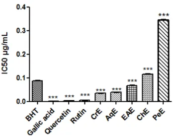
3.3 DPPH-scavenging Assay
DPPH radical scavenging activity of samples increased with concentration (Fig. 2). The CrE was found to exhibit the greatest scavenger activity with IC50 of (34 µg/mL). The radical scavenging activity in plant extracts decreased in the following order: CrE ˃ AqE ˃EAE ˃ ChE ˃ PeE which correspond to 34 µg/mL, 39 µg/mL, 67 µg/mL, 115 µg/mL and 344 µg/mL respectively. All extracts showed lower DPPH scavenging activities than gallic acid, rutin and quercetin (1.26 µg/mL, 5.58 µg/mL and 2.65 µg/mL respectively). While only CrE, AqE and
EAE exhibited higher DPPH scavenging effect than BHT (87 µg/mL). The CrE
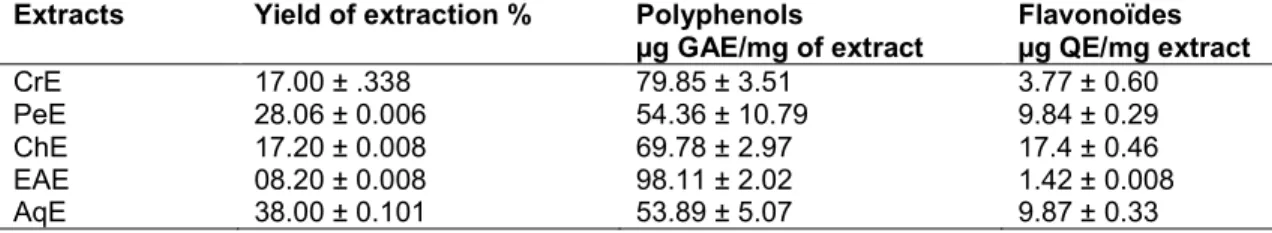
Table 1. Yields of extraction, total polyphenols and flavonoids content of Cytisus triflorus extracts
Extracts Yield of extraction % Polyphenols
µg GAE/mg of extract Flavonoïdes µg QE/mg extract CrE 17.00 ± .338 79.85 ± 3.51 3.77 ± 0.60 PeE 28.06 ± 0.006 54.36 ± 10.79 9.84 ± 0.29 ChE 17.20 ± 0.008 69.78 ± 2.97 17.4 ± 0.46 EAE 08.20 ± 0.008 98.11 ± 2.02 1.42 ± 0.008 AqE 38.00 ± 0.101 53.89 ± 5.07 9.87 ± 0.33
Fig. 2. DPPH scavenging activity of C. triflorus extracts and some phenolic compounds
Values are expressed as means ± SD (n=3). CrE: crude extract, PeE: petroleum ether extract, EAE: Ethyl acetate extract, ChE: chloroform extract and AqE: Aqueous extract showed a strong scavenging activity compared to
methanolic extract of Adenocarpus mannii (Fabaceae) leaves [24].
Surveswaran and their collaborators [25] reported that the radical scavenging activity of plant extracts depends on the amount of polyphenolic compounds in the extracts. A high radical scavenging activity of phenolics can be attributed to their high degree of aromatic rings hydroxylation, the arrangement of the hydroxyl group as well as the number of galloyl groups and ortho-hydroxyl groups on benzene nucleus structure [26]. However, in our study, the order of decreasing scavenging activity among the C.
triflorus extracts was found to be CrE ˃ AqE
˃EAE ˃ ChE ˃ PeE.
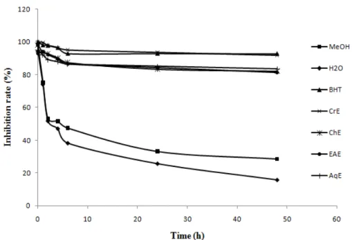
3.4 β-carotene/linoleic Acid Bleaching Method
The inhibition of β-carotene bleaching in a coupled oxidation with linoleic acid is a well-known methodology used for evaluating the antioxidant activity [27]. In order to compare the results of extracts, the AA of all extracts, BHT, methanol and water was calculated at 1 h, 2 h, 4 h, 6 h, 12 h, 24 h and 48 h. CrE exhibited the highest AA (91.93 ± 4.12%), which was similar to that of BHT (92.79 ± 1.05%) used as positive control, following by AqE, ChE, EAE, and PeE with AA values of 83.43 ± 1.88%, 82.29 ± 1.68%, 81.32± 1.28% and 17.70 ± 3.40%, respectively (Fig. 3).
All extracts of Cytisus triflorus exhibited a higher AA than that of methanolic extract of Ramorinoa
girolae Speg (Fabaceae) (46.9 ± 2%) [28]. It was
probable that the antioxidative components in these extracts were found to hinder the extent of β–carotene bleaching by neutralizing the linoleate free radical and other free radicals formed in the system.
3.5 Ion Chelating Assay
In this study, ferrous chelating activity was measured by inhibition of the formation of Fe+2– ferrozine complex after treatment of test material with Fe+2 [29]. The chelating effects of the plant extracts decreased in the following order: CrE ˃ AqE ˃EAE ˃ ChE ˃ PeE, which correspond to 226 µg/mL, 290 µg/mL, 560 µg/mL, 630 µg/mL and 800 µg/mL respectively (Fig. 4). The chelating ability of the CrE and AqE fraction reached over 90% in this assay. Although polyphenols were considered as potent chelators agents. Our study on C. triflorus extracts showed a weak relation between polyphenols contents and chelating activity.
3.6 Reducing Power
The antioxidant activity has been reported to have a direct, positive correlation with the reducing power [30]. The reducing properties are generally associated with the presence of reductones, which have been shown to exert antioxidant action by breaking the free radical
chain by donating a hydrogen atom [27]. Reductones are also reported to react with certain precursors of peroxide, thus preventing peroxide formation [31]. The reducing capacity of a compound may serve as a significant indicator of its potential antioxidant activity. The reducing power of all samples is shown in Fig. 5. Among
the samples, the scavenging ability was found to be decreased in the order of AqE ˃ CrE ˃ChE ˃ EAE ˃ PeE, which correspond to 320 µg/mL, 460 µg/mL, 510 µg/mL, 960 µg/mL and 1800 µg/mL respectively. However, ascorbic acid only showed higher activity with a reducing power of 50 µg/mL.
Fig. 3. Antioxidant activity of C. triflorus extracts compared with water, methanol and BHT at 2 mg/ml using a β-carotene/linoleic acid bleaching assay
Values are expressed as means of triplicate. CrE: crude extract, PeE: petroleum ether extract, EAE: Ethyl acetate extract, ChE: chloroform extract and AqE: Aqueous extract
Fig. 4. Ferrous chelating ability of C. triflorus extracts
CrE: crude extract, PeE: Petroleum ether extract, EAE: Ethyl acetate extract, ChE: Chloroform extract and AqE: Aqueous extract
Fig. 5. Reducing power assay of the C. triflorus extracts and Ascorbic acid. Comparison was realized against Ascorbic
acid
Values are expressed as means ± SD (n=3) ***: p ≤ 0.001. Asc.A: ascorbic acid, CrE: crude extract, PeE: Petroleum ether extract, EAE: Ethyl acetate
extract, ChE: Chloroform extract and AqE: Aqueous
extract
Reducing ability is generally associated with the presence of reductones such as ascorbic acid that can react directly with peroxides and with certain precursors, thus preventing the formation of peroxide. Also, reducing redox potential could decrease proton motive force, which is linked to a decrease of intracellular pH and thus a deactivation of the growth of microorganisms in food [32].
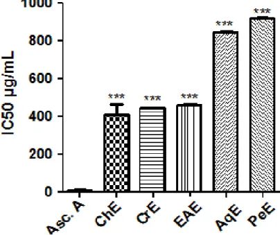
3.7 Hydroxyl Scavenging Activity
Hydroxyl radicals are highly potent oxidants, which can react with biomolecules in living cells and cause severe damage [33]. OH• radicals were generated using a system containing FeSO4 and H2O2 and detected by their ability to hydroxylate salicylate. Hydroxyl scavenging activity of samples increased with concentration (Fig. 6). The ChE, CrE and EAE were found to exhibit almost the same scavenger activities with IC50 of (440.45 µg/mL, 442.51 µg/mL and 458 µg/mL) respectively, which are twice better than AqE and PeE (840.84 µg/mL and 921.38 µg/mL). However, ascorbic acid only showed higher activity with a reducing power of 10.03 µg/mL. These results are in agreement with previous studies of Nagarajan and Sellamuthu [34].
Fig. 6. Hydroxyl scavenging assay of the C. triflorus extracts and Ascorbic acid. Comparison was realized against Ascorbic acid
Values are expressed as means ± SD (n=3) ***: p ≤ 0.001. Asc.A: ascorbic acid, CrE: crude extract, PeE:
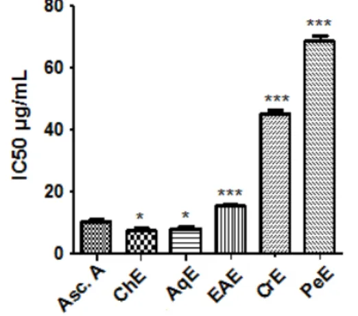
3.8 ABTS Radical Scavenging Activity
ABTS assay was the most popular spectrophotometric method because they are simple, rapid, sensitive and reproducible. Its reaction involves an electron-transfer process. Bleaching of ABTS cation has been extensively used to evaluate the antioxidant capacity [35]. The plant extract showed antioxidant activity, proving their capacity to scavenge ABTS (Fig. 7). The results showed that ChE, AqE and EAE extracts presented a good ability to scavenge the ABTS radical, with IC50 of 7.61 µg/mL, 7.71 µg/mL and 15.52 µg/mL, respectively. However, CrE and PeE have lower antioxidant activity against ABTS (IC50 = 45.09 µg/mL and 68.7 µg/mL, respectively). The EAE showed activity almost similar to that of ascorbic acid (13.79 ± 1.05 %) used as a positive control. All extracts of Cytisus triflorus have IC50 higher than those of
Indigofera tinctoria L. (Fabaceae), a study that
was carried according to Anusuya and Manian [36].
Fig. 7. ABTS scavenging assay of the C. triflorus extracts and Ascorbic acid. Comparison was realized against Ascorbic
acid
Values are expressed as means ± SD (n=3) ***: p ≤ 0.001. Asc.A: ascorbic acid, CrE : crude extract, PeE: Petroleum ether extract, EAE: Ethyl acetate
extract, ChE: Chloroform extract and AqE: Aqueous
extract
3.9 Superoxide Anion Radical
Scavenging Activity
Superoxide anion assay was carried out by alkaline DMSO assay. Superoxide is generated by the addition of NaOH to air saturated dimethyl sulfoxide (DMSO).The generated superoxide remains stable in solution, which reduces
Nitroblue tetrazolium in to formazan dye at room temperature [37]. C. triflorus extracts showed good scavenging potential of superoxide anions (Fig. 8). Percentage of scavenging activity increases with rising concentration. IC50 values for CrE, EAE, AqE, ChE and PeE were 36.35 µg/mL, 38.15 µg/mL, 64.38 µg/mL, 182.57 µg/mL and 432.76 µg/mL respectively. Antioxidant activity of extracts was near to the antioxidant activity of ascorbic acid (39.59 µg/mL). All extracts of C. triflorus (flowers and the leaves) showed a similar to the extracts of Mucuna
pruriens L. which belongs to the same family of
Fabaceae [38].
Fig. 8. Superoxide anion scavenging acivity of the C. triflorus extracts and Ascorbic acid.
Comparison was realized against Ascorbic acid
Values are expressed as means ± SD (n=3) ***: p ≤ 0.001. Asc.A: ascorbic acid, CrE: Crude extract, PeE: Petroleum ether extract, EAE: Ethyl acetate
extract, ChE: Chloroform extract and AqE: Aqueous
extract
3.10 Hydrogen Peroxide Scavenging
Hydrogen peroxide is highly important because of its ability to penetrate biological membranes. H2O2 itself is not very reactive, but it can sometimes be toxic to cell because of it may give rise to hydroxyl radical in the cells [39]. The scavenging ability of different extracts of C.
triflorus on hydrogen peroxide is shown in Fig.9.
The results of EAE are comparable to that of ascorbic acid (1.54 µg/mL). On the other hand, ChE and CrE are also comparable to the IC50 value 4.36 µg/mL, but fractions of AqE and PeE were found to be less potent compared to EAE extract with values of 7.57 µg/mL and 9.24 µg/mL respectively. All extracts of Cytisus
triflorus have an IC50 higher than those of Cicer
arietinum L. which belongs to the same family of
Fig. 9. Hydrogen peroxide scavenging activity of the C. triflorus extracts and Ascorbic acid.
Comparison was realized against Ascorbic acid
Values are expressed as means ± SD (n=3) ***: p ≤ 0.001. Asc.A: ascorbic acid,CrE: Crude extract, PeE: Petroleum ether extract, EAE: Ethyl acetate
extract, ChE: Chloroform extract and AqE: Aqueous
extract
In this study, we used different assays, each test confirmed and completed the other test of antioxidant activity of C. triflorus extracts, which indicate the antioxidant potential of various extracts with different mechanisms and showed that C. triflorus as a natural source of antioxidants.
3.11 Anti-Hemolytic Effect of Plant
Extracts
Anti-hemolytic inhibitory effect of extracts on ferrous ion induced hemolysis was illustrated in Fig. 10. In the present study, all extracts exhibited satisfactory inhibitory properties against hemolysis at low concentrations. Higher antihemolytic potential was exhibited by EAE and the CrE (HT50= 23.65 µg/mL). This might be attributed to the presence of high concentration of phenolic substances in these extracts (Table 1). The phenolic compounds in these extracts may probably have an important role in participating in biological pathways in the protection from radical-induced hemolysis. For the other extracts, the ChE represented the strongest efficiency followed by AqE and PeE, which correspond to 39.37 µg/mL, 54.24 µg/mL and 78.69 µg/mL respectively. However, ascorbic acid only showed higher activity with Anti-hemolytic of 7.36 µg/mL. A similar observation of hemolysis inhibition have also been reported for the extracts of Indigofera
tinctoria L. [36].
Fig. 10. Anti-hemolytic activity of the C. triflorus extracts and Ascorbic acid. Comparison was realized against Ascorbic
acid
Values are expressed as means ± SD (n=3) ***: p ≤ 0.001. Asc.A: ascorbic acid, CrE: Crude extract, PeE: Petroleum ether extract, EAE: Ethyl acetate
extract, ChE: Chloroform extract and AqE: Aqueous
extract
4. CONCLUSIONS
Medicinal plants always remain a reliable source of active ingredients known for their therapeutic properties. This study examined the antioxidant properties of various extracts of flowers-leaves mixture of Cytisus triflorus. Quantification of total polyphenols showed that the plant contains a considerable amount of phenolic compounds and flavonoids. The antioxidant activity of plant extracts was also investigated using different methods: the DPPH scavenging assay, ferrous ion chelating, reducing power, the β-carotene bleaching, hydroxyl scavenging, ABTS scavenging, superoxide anion scavenging, hydrogen peroxide scavenging and anti-Hemolytic assays. These results indicate that various extracts of this plant possess remarkable antioxidant properties and could be potential resource of natural antioxidants and could contribute very effectively in the prevention from several free radicals related diseases.
COMPETING INTERESTS
Authors have declared that no competing interests exist.
REFERENCES
1. Heimler D, Isolani L, Vignolini P, Tombelli S, Romani A. Polyphenol content and antioxidative activity in some species of
freshly consumed salads. J Agric Food Chem. 2007;55:1724-1729.
2. Nita M, Grzybowski. The role of the reactive oxygen species and oxidative stress in the pathomechanism of the age-related ocular diseases and other pathologies of the anterior and posterior eye segments in adults. Oxid Med Cell Longev. 2016;3(1):647-654.
3. Lobo V, Phatak A, Patil A, Chandra N. Free radicals, antioxidants and functional foods: Impact on human health. Pharmacogn Rev. 2010;4(8):118-126. 4. Luchtemberg M, Petronilho F, Constantino
L, Gelain D, Andrades M, Ritter C, Moreira J, Streck E, Dal-Pizzol F. Xanthine oxidase activity in patients with sepsis. Clin Biochem. 2008;41:1186-1190.
5. Ghulam M, Rawaba A, Asia A, Sumaira S, Amer J. Bioactive compounds from medicinal plants and their importance in drug discovery in Pakistan. Mat. Sc. Pharm. 2017;1:17-26.
6. Ait-KaciAourahoun K, Fazouane F, Benayache S. Pharmacological potential of
Cytisus triflorus l’Hérit. extracts as antioxidant and anti-inflammatory agent. Pharm. Lett. 2015;7:104-110.
7. Sharma U, Sharma K, Sharma N, Sharma A, Singh H, Sinha A. Microwave-assisted efficient extraction of different parts of
Hippophae rhamnoides for the
comparative evaluation of antioxidant activity and quantification of its phenolic constituents by reverse-phase high-performance liquid chromatography (RP-HPLC). J. Agric. Food Chem. 2008;56:374-379.
8. Liu L, Sun Y, Laura T, Liang X, Ye H, Zeng X. Determination of polyphenolic content and antioxidant activity of kudingcha made from Ilex kudingcha C. J. Tseng. Food Chem. 2009;112:35-41.
9. Li H, Cheng K, Wong C, Fan K, Chen F, Jiang Y. Evaluation of antioxidant capacity and total phenolic content of different fractions of selected microalgae. Food Chem. 2007;102:771-776.
10. da Silva LA, Pezzini BR, Soares L. Spectrophotometric determination of the total flavonoid content in Ocimum basilicum L. (Lamiaceae) leaves. Pharmacogn Mag. 2015;11(41):96-101. 11. Prior R, Wu X, Schaich K. Standardized
methods for the determination of antioxidant capacity and phenolics in foods
and dietary supplements. J Agric Food Chem. 2005;53:4290-4302.
12. Kartal N, Sokmen M, Daferera D, Polissiou M, Sokmen A. Investigation of the antioxidant properties of Ferula orientalis L. using a suitable extraction procedure. Food Chem. 2007;100:584-589.
13. Luo W, Zhao M, Yang B, Ren J, Shen G, Rao J. Antioxidant and antiproliferative capacities of phenolics purified from
Phyllanthus emblica L. fruit. Food Chem.
2011;126:277-282.
14. Kalaivani T, Mathew L. Free radical scavenging activity from leaves of Acacia
nilotica (L.) Wild. ex Delile, an Indian
medicinal tree. Food Chem Toxicol. 2010;298-305.
15. Ates B, Abraham L, Ercal N. Antioxidant and free radical scavenging properties of N-acetyl cysteineamide (NACA) and comparison with N-acetylcysteine (NAC). Free Radic Res. 2008;42:372-377.
16. Rubalya Valantina S, Neelamegam P. Selective ABTS and DPPH- radical scavenging activity of peroxide from vegetable oils. FRJ. 2015;22(1):289-294. 17. Gowri R, Ramaiah M. Evaluation of
antioxidant activity of ethanolic extract of
Sphaeranthus amaranthoides Burm. Int
J Drug Dev Res. 2013;5:0975-9344. 18. Mukhopadhyay D, Dasgupta P, Sinha Roy
D, Palchoudhuri S, Chatterjee I, Ali S, Dastidar S. A sensitive In vitro spectro-photometric hydrogen peroxide scavenging assay using 1,10 phenanthroline. Free Radicals and Antioxidants. 2016;6:124-132.
19. Thephinlap C, Pangjit K, Suttajit M, Srichairatanakool S. Anti-oxidant properties and anti- hemolytic activity of
Psidium guajava, Pandanous odorus and Rhinacanthus nasutus.J Med Plants Res. 2013;7:2001-2009.
20. Huang D, Ou B, Prior L. The chemistry behind antioxidant capacity assays. J Agric Food Chem. 2005;53:1841-1856.
21. Amzad M, Shah D. A study on the total phenols content and antioxidant activity of essential oil and different solvent extracts of ndemic plant Merremia borneensis. Arab J Chem. 2011;4(9):717-721.
22. Matyushchenko N, Stepanova T. Quantitative determination of the total content of flavonoids in the new phytopreparation Elima. Pharm Chem J.
23. Azzouzi S, Zaabat N, Medjroubi K, Akkal S, Benlabed K, Smati F, Dijoux-Franca M. Phytochemical and biological activities of
Bituminaria bituminosa L. (Fabaceae).
Asian Pac J Trop Med. 2014;7:S481-S484. 24. Ndjateu F, Tsafack R, Nganou B, Awouafack M, Wabo H, Tene M, Tane P, Eloff J. Antimicrobial and antioxidant activities of extracts and ten compounds from three Cameroonian medicinal plants:
Dissotis perkinsiae (Melastomaceae),
Adenocarpus mannii (Fabaceae) and Barteria fistulosa (Passifloraceae). S Afr J
Bot. 2014;91:37-42.
25. Surveswaran S, Cai YZ, Corke H, Sun M. Systematic evaluation of natural phenolic antioxidants from 133 Indian medicinal plants. Food Chem. 2007;102:938-953. 26. Cai YZ, Mei S, Jie X, Luo Q, Corke H.
Structure, radical scavenging activity relationships of phenolic compounds from traditional Chinese medicinal plants. Life Sciences. 2006;78:2872-2888.
27. Reis FS, Ferreira IC, Barros L, Martins A. A comparative study of tocopherols composition and antioxidant properties of
in vivo and in vitro ectomycorrhizal fungi. J
Food Sci Technol. 2011;44:820-824. 28. Luna LC, Pigni NB, Torras-Claveria L,
Monferran MV, Maestri D, Wunderlin DA, Feresin E, Bastida J. Ramorinoa girolaeSpeg (Fabaceae) seeds, an Argentinean traditional indigenous food: Nutrient composition and antioxidant activity. J Food Compost Anal. 2013;31: 120-128.
29. Ebrahimzadeh MA, Pourmorad F, Bekhradnia AR. Iron chelating activity, phenol and flavonoid content of some medicinal plants from Iran. Afr. J. Biotechnol. 2008;7(18):3188-3192.
30. Osman H, Nasarudin R, Lee S. Extracts of cocoa (Theobroma cacao L.) leaves and their antioxidation potential. Food Chem. 2004;86:41–46.
31. Xing RE, Yu H, Liu S, Zhang W, Zhang QB, Li Z. Antioxidant activity of differently regioselective chitosan sulfates in vitro. Bioorg Med Chem. 2005;13:1387-1392. 32. Lim DH, Choi D, Choi O, Cho K, Kim R,
Choi H, Cho H. Effect of Astragalus sinicus L. seed extract on antioxidant activity. J Ind Eng Chem. 2011;17:510-516.
33. Yang J, Guo J, Yuan J. In vitro antioxidant properties of rutin. J Food Sci Technol. 2008;41:1060-1066.
34. Nagarajan A, Sellamuthu M. Antioxidant and free radical scavenging potential of different solvent extracts of Indigofera
tinctoria L. leaves. Int J Pharm Pharm Sci.
2013;5:0975-1491.
35. Schlesier K, Harwat M, Böhm V, Bitsch R. Assessment of antioxidant activity by using different In vitro methods. Free Radic Res. 2002;36:177-187.
36. Anusuya N, Manian S. Antioxidant and free radical scavenging potential of different solvent extracts of Indigofera
tinctoria l. leaves. Int J Pharm Pharm Sci.
2013;1: 142-147.
37. Lobo VC, Phata K, Chandra N. Antioxidant and free radical scavenging activity of
Hygrophila schulli (Buch.-Ham.) Almeida
and Almeida. seeds. Adv Biomed Res. 2010;1:72-78.
38. Satheesh Kumar D, Kottai Muthu A, Anton Smith A, Manavalan R. In vitro antioxidant activity. Int J Pharm tech Res. 2010;2: 2063-2070.
39. Muthu Lakshmi T, Radha R, Jayshree N.
In vitro antioxidant activity, total phenolic
and total flavonoid content in extracts from the bark of Dalbergia sissoo Roxb. Int J Pharm Sci Res. 2014;5:0975-9492. 40. Vadnere GP, Patil AV, Wagh SS, Jain SK.
In vitro free radical scavenging scavenging
and antioxidant activity of Cicer arietinum L. (Fabaceae). Int J Pharm Tech Res. 2012;4:0974-4304.
_________________________________________________________________________________ © 2018 Madoui et al.; This is an Open Access article distributed under the terms of the Creative Commons Attribution License (http://creativecommons.org/licenses/by/4.0), which permits unrestricted use, distribution, and reproduction in any medium, provided the original work is properly cited.
Peer-review history:
The peer review history for this paper can be accessed here: http://www.sciencedomain.org/review-history/24544