HAL Id: tel-02408332
https://tel.archives-ouvertes.fr/tel-02408332
Submitted on 13 Dec 2019
HAL is a multi-disciplinary open access archive for the deposit and dissemination of sci-entific research documents, whether they are pub-lished or not. The documents may come from teaching and research institutions in France or abroad, or from public or private research centers.
L’archive ouverte pluridisciplinaire HAL, est destinée au dépôt et à la diffusion de documents scientifiques de niveau recherche, publiés ou non, émanant des établissements d’enseignement et de recherche français ou étrangers, des laboratoires publics ou privés.
Hypothesis of a Non-SNARE-Function of Syntaxin-5
Stefan Rathjen
To cite this version:
Stefan Rathjen. Hypothesis of a Non-SNARE-Function of Syntaxin-5. Molecular biology. Université Paris-Saclay, 2017. English. �NNT : 2017SACLS450�. �tel-02408332�
Hypothesis on a non-SNARE function
of syntaxin-5
Thèse de doctorat de l'Université Paris-Saclay
Préparée à « Institute Curie »
École doctorale n°568
Biosigne – Signalisations et reseaux integratifs en biologie
Aspects moleculaires et cellulaires de la biologie
Sciences de la vie et de la santé
Thèse présentée et soutenue à Paris, le 12/12/2017, par
Stefan Jan-Matthias RATHJEN
Composition du Jury :
Marc le MAIRE Président
Professeur Émérite, Université Paris-Sud, CEA – UMR 9198
Thierry GALLI Rapporteur
Directeur d'unité, Center of Psychiatry and Neuroscience – INSERM U894
Catherine JACKSON Rapporteur
Chef d’équipe, Instutute Jaques Monod – UMR 7592 CNRS
Jost ENNINGA Examinateur
Chef d’équipe, Institut Pasteur
Anne HOUDUSSE Examinatrice
Chef d'équipe, Institute Curie –UMR144 CNRS
Ludger JOHANNES Directeur de thèse
Directeur d'unité, Institute Curie – UMR3666
NNT : 2 0 1 7 S A C L S 4 5 0
1
F
RENCH
A
BSTRACT
Titre : Hypothèse d'une fonction non-SNARE de la syntaxine-5
Mots clés : Syntaxin-5, GPP130, Retro-2, Sec16A, transport rétrograde, Shiga toxine
Abstract :
L’introduction commence avec la description de toxines d’origines bactérienne et végétale, en particulier la toxine Shiga ainsi que les toxines de la même famille (chapitre 9.1.2). Les petites molécules inhibitrices de ces toxines sont ensuite résumées dans le chapitre 9.1.3, en particulier le composé Retro-2. L’efficacité de ces toxines à atteindre leurs cibles reposant sur le trafic intracellulaire, un aperçu général de l’endocytose et du trafic endosomal sont présentés (chapitre 9.2). Puis, l’entrée de la voie rétrograde est décrite (chapitre 9.2.5), avec un intérêt particulier porté sur la clathrine, le rétromère et GPP130, une protéine qui circule de manière continue entre le Golgi, la membrane plasmique et les endosomes. Les protéines SNARE, en particulier la syntaxine-5 et le syntaxine-16, sont ensuite introduites (chapitre 9.2.6). Après une brève section sur les micro-ARNs de la famille 199 (chapitre 9.3), l’introduction se termine avec la description des techniques clés utilisées au cours de mon travail, tels que la chimie click bio-orthogonale, la synchronisation du trafic antérograde par rétention grâce à des hameçons spécifiques (RUSH), et la ligation par proximité basé sur des anticorps (chapitre 9.4).
Ci-inclus, mon article en cours de soumission ouvre la partie résultats (chapitre 10.1), dans laquelle je présente l’intérêt de la chimie click bio-orthogonale pour identifier les cibles cellulaires de Retro-2. Je décris un des candidats potentiels, Sec16A, et illustre comment grâce à la technique de RUSH, perturber la fonction de Sec16A conduit à la relocalisation partielle de la syntaxin-5 au niveau du reticulum endoplasmique via l’inhibition du transport antérograde de la syntaxine-5. La seconde partie de l’article décrit comment la relocalisation de la syntaxine-5 induit l’inhibition du trafic de la toxine Shiga des endosomes au TGN. Je présente une nouvelle interaction entre la syntaxine-5 et la protéine TGN GPP130, qui ont déjà été caractérisées en relation avec le trafic de la toxine Shiga. Mon travail connecte à la fois les facteurs de trafic avec le trafic rétrograde au niveau de l’interface endosome-TGN. De manière frappante, cette interaction est très probablement basée sur une fonction non-SNARE de la syntaxine-5 car le domaine de fixation sur GPP130 est structurellement non lié à toute fonction SNARE.
En collaboration avec Juan Francisco Aranda et Carlos Fernandez aux Etats-Unis, nous avons placés des micro-ARNs dans un contexte de régulation endogène du trafic rétrograde de la toxine Shiga (chapitre 11.2). Une discussion plus approfondie sera apportée dans le chapitre 12.
Enfin, une vue d’ensemble des projets en cours est apportée dans la section des perspectives (chapitre 12), dans laquelle les collaborations plus approfondies sont mises en lumière.
Mots clés : transport rétrograde, toxine Shiga, toxine de la famille Shiga, STxB, syntaxin-5, Sec16A, GPP130, Retro-2, Retro-2.1, chimie click sans cuivre, identification des cibles de petites molécules, spétrométrie de masse, function non-SNARE, inhibition du trafic antérograde, miARN, miR199, rétromère, VPS26
2
E
NGLISH
A
BSTRACT
Title: Hypothesis on a non-SNARE-Function of Syntaxin-5
Keywords: Syntaxin-5, GPP130, Retro-2, Sec16A, retrograde transport, Shiga toxin
Abstract:The introduction of my PhD manuscript starts with describing plant and bacterial toxins (chapter 9.1), in particular Shiga toxin and Shiga-like toxins (SLTs) (chapter 9.1.2). Small molecule inhibitors of these toxins are summarized afterwards in chapter 9.1.3, notably the Retro-2 compound. Since these toxins rely on intracellular trafficking to reach their molecular targets, a general overview of endocytosis and endosomal trafficking is provided (chapter 9.2). Next, the retrograde route entry is presented (chapter 9.2.5), with focus on clathrin, the retromer and GPP130, a protein that constantly cycles between Golgi, plasma membrane, and endosomes. SNARE proteins, particularly syntaxin-5 and syntaxin-16, are then introduced (chapter 9.2.6). After a brief section of the micro RNA family 199 (chapter 9.3), the introduction finishes with the description of some salient techniques that were used in my work, such as - bio-orthogonal Click-Chemistry, anterograde trafficking synchronization with the retention using selective hooks (RUSH) assay, and the antibody-based proximity ligation assay (chapter 10.6.1, 0, 10.11.1). Herein, my submitted publication opens the results part (chapter 11.1), in which I present the utility of biorthogonal click chemistry for the search of the cellular targets of Retro-2, a small molecule inhibitor that was previously shown to protect cells and animals against Shiga toxin and ricin. I describe that Sec16A is a likely cellular target candidate, and illustrate using the RUSH approach how interfering with Sec16A functions leads to the partial relocalization of syntaxin-5 to the endoplasmic reticulum (ER) by slowing-down its anterograde transport. The second part of the paper describes how syntaxin-5 relocalization causes the inhibition of Shiga toxin trafficking from endosomes to the TGN. I present a novel interaction between syntaxin-5 and the Golgi protein GPP130, which both have been already described in relation to Shiga toxin trafficking. My work connects both trafficking factors in retrograde trafficking at the endosomes-TGN interface. Strikingly, I demonstrate that this interaction is most probably based on a non-SNARE function of syntaxin-5.
In collaboration with Juan Francisco Aranda and Carlos Fernandez in the US, we put micro RNAs into an endogenous regulation context of Shiga toxin retrograde trafficking (chapter 11.2). An extended discussion will be given in chapter 12.
Last, a general outlook of ongoing projects is given in the perspectives section (chapter 13), in which further collaborations are highlighted.
Keywords: Retrograde transport, Shiga toxin, Shiga-like toxin (SLT), STxB, syntaxin-5, Sec16A, GPP130, Retro-2, Retro-2.1, azide-functionalized Retro-2, copper-free click chemistry, small molecule target identification, mass spectrometry, non-SNARE function, anterograde trafficking inhibition, miRNA, miR199, retromer, VPS26
3
A
CKNOWLEDGEMENTS
It is finally done! After 3 years of doing top-notch research at the Institut Curie in Paris, I could assemble this manuscript with the help of many people who deserve to be mentioned here. These words cannot express nearly my gratitude for them. More importantly, I cannot mention everyone by name. Thus, I hope that everyone will find himself between the lines.
First of all, I would like to thank Ludger who gave me the opportunity to work with him on this research project and who guided and formed me professionally through this thesis period. Regarding this project I have to stress the work and passion of Maria Daniela Garcia-Castillo, who taught me a lot and initiated this work. It has been a pleasure working with her. I also want to mention everyone else on related to this project. The current and previous members of the unit: Henri-Francois Renard, Valérie Chambon, Alison Forrester and Raphaël Rodriguez. Our collaborators within the institute and beyond: The Nikon imaging facility of the Institute Curie, the mass spectrometry platform around Damarys Loew, the groups of Christophe Lamaze, Franck Perez, Anne Houdusse, Graça Raposo, Adam Linstedt, Ben Glick, Jean-Christophe Cintrat and Daniel Gillet. We appreciated very much your expertise, kindness and responsiveness.
The UMR3666! All of you! Thanks for the support on a professional but also on a personal level! In no particular order: Marco, Anne (Thanks for all the translation of texts into French), Julio, Raphael, aka Philippe, Siau, Joanna, Lavaniya, Veronica, the other Anne, Thomas, the other Thomas, Estelle, Massi, Dhiraj, Christian, Sebastian, Steve, Bhanu, Bibhuti, Satish, Senthil, Vesela, Tatiana, Antoine, Stephanie, Christine (both), Sylvie, Frederic, Yannick, Barbara, Yoane, Katharina, Daniela, Melissa, Nicolas, Cedric, Ulrike, Haifei, Alena, Cesar and everyone else, I shamefully forgot!
Basically everyone else who crossed my path in the past years, here at the campus! The people of the ADIC. They are such a great bunch of people. I enjoyed involving myself with the many events we organized. I hope to keep as many connection throughout the upcoming years and decades.
All my friends! The ones in Germany and in France! The ones involved in science and also the ones outside academia. If a mention you here, I’ll expand the scope of this manuscript. Yes, you are mentioned here, for sure.
Thanks to my family, especially my parents, Bernd and Astrid, my siblings, Melina and Björn! I love you! Thank you for everything. I want to mention also the family that I gained though my beloved Amorcita! I has been a pleasure to get so many lovely people into my live.
Last and by far the most important to me within the past years, my Amorcita, aka Pelusa or Mari! I cannot express my gratitude, appreciation and love. I don’t want to know how much harder all the struggles would have been without you! I consider myself blessed and lucky to have “discovered” (found) you within my research period here in Paris. I love you! I want to thank you all from the bottom of my heart!
Sincerely yours, Stefan
PS: I also want to thank Bluetooth technology and my headphones for shutting off all the daily noise and distractions! Much appreciated! Hooray to technology!
4
T
ABLE
O
F
C
ONTENTS
Page 1 FRENCH ABSTRACT... I 2 ENGLISH ABSTRACT ... II 3 ACKNOWLEDGEMENTS ... 1 4 TABLE OF CONTENTS ... 2 5 LIST OF TABLES ... 6 6 TABLE OF FIGURES ... 7 7 ABBREVIATIONS ... 19 8 SUMMARY ... 22 9 INTRODUCTION ... 19.1 PLANT AND BACTERIAL RIBOSOMAL INACTIVATING PROTEIN-TOXINS ... 2
9.1.1 Bioterrorism, bio threat agents and biodefense ... 4
9.1.2 Shiga toxin and its Shiga-like toxins ... 4
9.1.3 Small molecule inhibitors of the retrograde route... 7
9.1.3.1 Retro compounds ... 7
9.1.3.1.1 Structural evolution of Retro-2 ... 10
9.1.3.1.2 Retro-2 effect on viral infections ... 11
9.1.3.1.3 Retro-2 effect on intracellular parasites ... 12
9.1.3.1.4 Retro-2 effect on intracellular bacteria ... 13
9.1.3.1.5 Bio threats that are not affected by Retro-2 ... 13
9.1.3.2 Other small molecules... 13
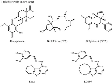
9.1.3.2.1 Inhibitors with identified target... 14
9.1.3.2.1.1 Ilimaquinone (IQ) ... 15
9.1.3.2.1.2 Brefeldin A (BFA) ... 15
9.1.3.2.1.4 Golgicide A (GCA) ... 16
9.1.3.2.2 Compounds with unknown target... 17
9.2 INTRACELLULAR TRAFFICKING ... 18
9.2.1 Anterograde trafficking via the biosynthetic/secretory pathway ... 18
9.2.1.1 ER Exit Sites (ERES) ... 21
9.2.1.1.1 Sec16A and the COPII coat ... 22
9.2.1.1.2 COPII coat dynamics: Two models for Sec16A function ... 22
9.2.1.1.2.1 Model one: Sec16 as a scaffolding protein ... 24
9.2.1.1.2.2 Model two: Sec16A as a regulator of COPII vesicle release ... 26
9.2.2 Endocytosis ... 27
9.2.2.1 Clathrin-mediated endocytosis (CME) ... 28
9.2.2.2 Clathrin-independent Endocytosis (CIE) ... 30
9.2.3 The recycling pathways ... 34
9.2.3.1 Fast recycling – direct pathway ... 35
9.2.3.2 Slow recycling – Rab11 mediated... 35
9.2.4 The lysosomal/degradation pathway ... 35
9.2.5 Retrograde trafficking ... 36
9.2.5.1 GPP130: A cycling Golgi protein involved in Shiga toxin trafficking. ... 37
9.2.5.2 Clathrin... 38
9.2.5.3 Retromer ... 38
9.2.6 SNAREs ... 40
9.2.6.1 The general mode of vesicle fusion ... 41
9.2.6.2 Syntaxin-5 (STX5) ... 43
9.2.6.3 Syntaxin-16 ... 43
9.3 MICRO INTERFERING RNA FAMILY MIR199 ... 44
10 MATERIAL AND METHODS... 46
10.2 ANTIBODIES AND REAGENTS ... 47
10.3 RNA INTERFERENCE... 48
10.4 CALCIUM PHOSPHATE–DNA CO-PRECIPITATION ... 48
10.5 RETRO-2 TREATMENTS ... 48
10.6 CLICK CHEMISTRY LABELING ... 49
10.6.1 METHODOLOGY ASPECTS: Bio-orthogonal Click chemistry ... 49
10.7 IMMUNOPRECIPITATION ... 51
10.8 SHIGA TOXIN TRAFFICKING ... 51
10.9 PROXIMITY LIGATION ASSAY (PLA) ... 51
10.9.1 METHODOLOGY ASPECTS: Proximity ligation assay ... 52
10.10 CONFOCAL IMAGING ... 53
10.11 RETENTION USING SELECTIVE HOOKS (RUSH)... 53
10.11.1 METHODOLOGY ASPECTS: Retention Using Selective Hooks (RUSH) ... 53
10.12 INTOXICATION ASSAY... 54
10.13 WESTERN BLOT ANALYSIS ... 55
10.14 STATISTICS ... 55
11 RESULTS ... 56
11.1 MANUSCRIPT FOR SUBMISSION: SYNTAXIN-5 FUNCTIONALLY INTERACTS WITH GPP130 FOR RETROGRADE SHIGA TOXIN TRAFFICKING ... 57
11.1.1 Abstract ... 58
11.1.2 Author contributions ... 58
11.1.3 Introduction... 59
11.1.4 Results ... 61
11.1.4.1 Retro-2 targets the COPII machinery. ... 61
11.1.4.3 Retro-2 slows the anterograde transport of STX5 ... 66
11.1.4.4 STX5 SNARE complexes remain unchanged upon Retro-2 treatment ... 68
11.1.4.5 STX5 binds to GPP130 in a Retro-2 sensitive manner ... 71
11.1.4.6 The GPP130-STX5 interaction is required for STxB trafficking to the Golgi ... 73
11.1.5 Discussion ... 77
11.2 THE EFFECT OF MIR199 ON RETROGRADE SHIGA TOXIN TRAFFICKING ... 79
11.2.1 Objectives and summary. ... 79
11.2.2 Results ... 80
11.2.2.1 MiR199 down regulates retromer expression ... 80
11.2.2.2 MiR199 impairs retrograde transport of STxB ... 80
11.2.2.3 MiR199 inhibits SLT intoxication. ... 82
12 DISCUSSION ... 83
13 PERSPECTIVES... 85
14 REFERENCES ... 86 15 ANNEX ... A 15.1 MANUSCRIPTS ... B
15.1.1 (published) Retrograde transport is not required for cytosolic translocation of Shiga toxin B-subunit ... B 15.1.2 (Revision) MiR-199a-5p attenuates retrograde transport and protects against Shiga toxin cytotoxicity ... D
5
L
IST
O
F
T
ABLES
Page Table 1: Shiga toxin and isoforms (Johannes and Römer, 2010) ... 7 Table 2: Retro-1 and Retro-2 protection factors on HeLa cells against Ricin, Stx1, and Stx2.
Protection factors calculated over the indicated number of experiments. Means ± SEM are shown. (Stechmann et al., 2010) ... 8 Table 3: supplementary information: Full mass-spectrometry tables of GFP-STX5 pulldown.
6
T
ABLE OF
F
IGURES
Page Figure 1: Structure of ribosome-inactivating proteins (RIP) type 1 and 2 (Barbieri et al.,
1993). The number of binding sites per B-subunit of Type 2 RIPs can vary. The B-subunit of Shiga toxin provides in total 15 binding sites (3 binding sites per monomer) (Ling et al., 1998)... 2 Figure 2: Schematic presentation of the biochemical action of ribosome-inactivating
proteins (RIPs), such as ricin and Shiga toxin. The A-subunit targets the α-sarcin site on the large (28S) subunit of ribosomes, which results in an inhibition of protein biosynthesis. (Stirpe, 2005) ... 3 Figure 3: Shiga toxin structures. (A) Schematic drawing of the Shiga holotoxin, catalytic
A-subunit (STxA); five fragment monomers that form the homopentameric B-subunit (STxB). (B) A ribbon diagram of Shiga toxin, illustrating the binding sites on STxB for globotriaosylceramide (Gb3). Gb3 is shown in a ball-and-stick representation. (C) A zoom into the furin cleavage (Arg248-Val-Ala-Met251) site
of STxA; the disulfide bond (between Cys242 and Cys261) is shown in yellow. (D)
A ribbon diagram of a STxB from below, pointing out the three Gb3-binding sites. Gb3 is shown as a ball-and-stick representation. (Johannes and Römer, 2010)... 5 Figure 4: Structures of Retro compounds. ... 8 Figure 5: Retro-2 protects mice against ricin challenge. Comparison of survival curves from
mice that were treated with single intraperitoneal dose of Retro-2 with indicated concentrations one hour before toxin exposure. Ricin was administered to mice via the nasal route (2 mg/kg). The control group received only the vehicle in prior to ricin exposure. The curves for treated animals are statistically different from control as measured by the log rank test (p < 0.0001 for 2 µg/kg of Retro-2, orange; p = 0.015 for 10 mg/kg, brown; p = 0.031 for 20 mg/kg, purple; p = 0.0007 for 200 mg/kg, red). (Stechmann et al., 2010) ... 9
Figure 6: Illustration of the retrograde trafficking of Shiga toxin and site of action of Retro-2 (Gupta et al., Retro-2017). Toxins (e.g. ricin and SLTs) traffic via the retrograde route, starting from the plasma membrane through endosomes and Golgi to the ER (Johannes and Popoff, 2008). Retro-2 inhibits the toxin trafficking step from endosomes to the Golgi ... 9 Figure 7: Structural evolution of Retro-2. Structures A and B show the original hit
compound Retro-2 (Stechmann et al., 2010). Structure B illustrates how the spontaneous cyclisation to molecule C (Retro-2cyclic) may take place (Park et al.,
2012). Structure D shows the optimized molecule of Retro-2 based on C, called Retro2.1 (Noel et al., 2013). Additional structural groups are highlighted in blue. Molecule E is the azide-functionalized 2.1, called “clickable Retro-2”, due to its potential to be used in a biorthogonal click chemistry approach (see chapter 0). The azide is marked in red. ...10 Figure 8: Chemical structures of inhibitors of SLTs for which the cellular target is already
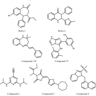
known. ...14 Figure 9: Chemical structures of inhibitors of SLTs for which the cellular target is still
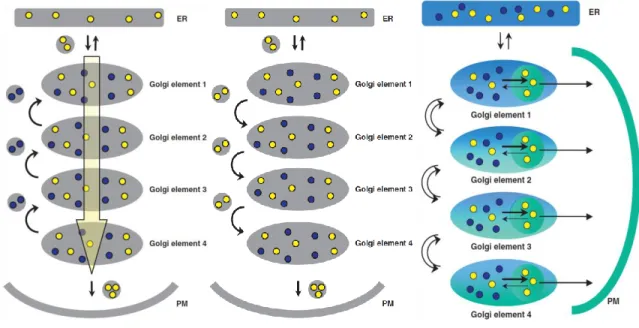
unknown, including the Retro compounds. ...17 Figure 10: Models for intra-Golgi vesicular transport. Cargo synthesized in the ER and
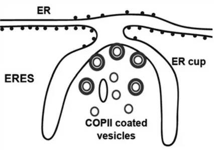
transported through the secretory pathway is shown in yellow; Golgi processing enzymes are shown in blue. Arrows indicate the direction of trafficking: From left to right: The model of cisternae maturation – the vesicular transport model – the rapid-partitioning model (Jackson, 2009) – Blue dots are Golgi localized enzymes; yellow dots are cargo; blue areas are glycero-phospholipid-enriched membranes; green areas are enriched membranes; green circles within each Golgi stack are sphingolipid-enriched export domains ...19 Figure 11: A schematic representation of an ER exit site (ERES) showing the ER cup lacking
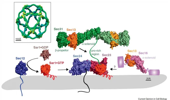
Figure 12: Model for assembly of the COPII coat complex, describing Sec16 as a scaffolding protein. The common model for assembly of the COPII coat complex is modified to include the role of Sec16. For simplicity, cargo molecules are omitted. (1) The Sec13–Sec16 tetramer is stably associated with the ER membrane and binds the integral membrane protein Sed4 or its homologue Sec12. Sar1 becomes associated with the membrane, when it is converted from the GDP- to GTP-bound state. Concentration of membrane-associated proteins begins to bend membrane. (2) Sec13–Sec16 and Sar1 collaborate to recruit the cargo adaptor Sec23–Sec24 dimer. (3) A precoat self-associates into higher-order oligomers. (4) Sec13–Sec16 and Sec23–Sec24–Sar1 form independent interactions with Sec13–Sec31, causing it to assemble near and/or in place of Sec16. (5) The forming coat contains progressively more Sec13–Sec31 and less Sec13–Sec16. Hand-off of Sec23–Sec24–Sar1 from Sec16 to Sec31 sets the stage for GTP hydrolysis by Sar1. (6) A final COPII coat is formed, and vesicle budding is complete. Sec13–Sec16 remains mostly associated with the ER. - (Whittle and Schwartz, 2010) ... 25 Figure 13: Structure and assembly of the COPII coat. The guanine nucleotide exchange
factor, Sec12 (McMahon et al., 2012) catalyzes GTP loading on Sar1, which switches from a cytosolic GDP-bound form (Huang et al., 2001) to a membrane-associated GTP-bound form (Bi et al., 2002) through exposure of an N-terminal amphipathic a-helix. Membrane-associated Sar1 recruits Sec23/Sec24 (Bi et al., 2002). Sec24 provides cargo-binding function by directly interacting with sorting signals on transmembrane clients. The Sar1/Sec23/Sec24 ‘pre-budding’ complex in turn recruits Sec13/Sec31 (Fath et al., 2007). Sec13/Sec31 self-assembles into a polyhedral cage (Stagg et al., 2006) that at least in part drives membrane curvature and contributes to vesicle scission. Sec23 is the GTPase-activating protein for Sar1, with Sec31 further contributing to hydrolysis via a proline-rich domain that extends across the surface of Sec23/Sar1. Sec16 is a peripheral component that binds to Sec13 (Whittle and Schwartz, 2010), modulates GTPase activity by preventing
Sec31 action and otherwise contributes to vesicle formation in poorly understood ways. - (Miller and Schekman, 2013) ...26 Figure 14: Pathways of entry into cells. Large particles can be taken up by phagocytosis,
whereas fluid uptake occurs by macro-pinocytosis. Both processes appear to be triggered by and are dependent on actin-mediated re-modelling of the plasma membrane at a large scale. Compared with the other endocytic pathways, the size of the vesicles formed by phagocytosis and macro-pinocytosis is much larger. Numerous cargoes can be endocytosed by mechanisms that are independent of the coat protein clathrin and the fission GTPase, dynamin. Some of these clathrin-independent pathways are also dynamin independent. Most internalized cargoes are delivered to the early endosome via vesicular (clathrin- or caveolin-coated vesicles) or tubular intermediates (known as clathrin-independent carriers (CLICs)) that are derived from the plasma membrane. Some pathways may first traffic to
intermediate compartments, such as the caveosome or
glycosylphosphatidylinositol-anchored protein enriched early endosomal compartments (GEEC), en route to the early endosome. - (Mayor and Pagano, 2007) ...27 Figure 15: Model for the assembly and disassembly of a clathrin coat. The drawing indicates
the direction of the rotational movement (counterclockwise) that is required to lock a relatively rigid clathrin triskelion into the lattice. The reverse process would be required to disassemble the coat. - (Kirchhausen, 2000) ...28 Figure 16: Clathrin-mediated endocytosis. (a) Stages of clathrin-mediated endocytosis.
Step 1: FCHo protein mediated nucleation. Step 2: Cargo recruitment by AP-2. Step 3: Coat assembly. Step 4: Dynamin-mediated scission. Step 5: Un-coating. (b) Network of interacting partners involved in CME. (c) Depletion of FCHo proteins, AP-2, clathrin, and dynamin and their effect on CCP formation (McMahon and Boucrot, 2011). ...30
Figure 17: (a) STxB-driven membrane invagination in interaction with the GSL Gb3. – adopted from (Johannes et al., 2016). ... 31 Figure 18: Model of actin-driven scission. After SLT binding on to Gb3, membrane curvature
is induced, leading to the clathrin independent formation of PM invaginations. Actin polymerization causes scission through membrane reorganization (Römer et al., 2010) ... 32 Figure 19: Gal3-driven membrane bending. (Johannes et al., 2015) ... 33 Figure 20: A schematic representation of the ‘tubular endosomal network’ (TEN). Endocytic
vesicles are delivered to clathrin-coated early endosomes, which start to mature involving progressive acidification of their lumen. The TEN contains various domains (labeled in different colors) and the necessary machinery to sort cargoes to their various destinations. - (Bonifacino and Rojas, 2006) ... 34 Figure 21: The generation of multi-vesicular bodies, leading to lysosomes. The degradation
pathway is shown by solid arrows. Membrane invaginations and internal vesicles are shown in red, highlighting multi-vesicular regions in EE and LE. The recycling pathways (slow and fast) are shown by dashed arrows. - (Gruenberg and Stenmark, 2004) ... 36 Figure 22: Schematic drawing of retrograde trafficking entry points. The retrograde
transport step from endosomes to the TGN can originate from several points: recycling (blue), early (green) or late (red) endosomes. - (Johannes and Wunder, 2011b) ... 37 Figure 23: Schematic presentation of a speculative model of the retromer coat on a
membrane tubule. SNX dimer in purple. - (Hierro et al., 2007) ... 39 Figure 24: SNARE proteins form a four-helical bundle complex that drives membrane
fusion. (a) VAMP (blue) on the vesicle interacts with syntaxin (red) and SNAP-25 (green) on the plasma membrane to form a four-helix bundle that zips up concomitant with bilayer fusion. (b) The backbone of the SNARE complex is
shown on the left, with the central ionic layer (red) and 15 hydrophobic layers (black) that mediate the core interactions. Top-down views of side-chain interactions are shown on the right, with the four SNARE helices that are represented as ribbons. The ball-and-stick structures represent the indicated amino acids; the dotted lines represent hydrogen bonds or salt bridges that stabilize interactions between SNAREs. Q-SNAREs and R-SNAREs are characterized by a glutamine (Q) or arginine (R) residue, respectively, in the central layer of the SNARE complex. (SNARE; soluble NSF attachment protein receptor, where NSF stands for N-ethyl-maleimide-sensitive fusion protein; SNAP-25, 25 kDa synaptosome-associated protein; VAMP, vesicle-associated membrane protein). - (Chen and Scheller, 2001) ...40 Figure 25: Model of SNARE-mediated lipid fusion. (a) The two membranes are in the vicinity
of each other but the SNAREs are not yet in contact. (b) SNARE complexes start zipping from the amino-terminal end, which draws the two membranes further towards each other. (c) Zipping proceeds, causing increased curvature and lateral tension of the membranes, exposing the bilayer interior. Spontaneous hemi fusion occurs as the separation is sufficiently reduced. (d) The highly unfavorable void space at the membrane junction in (c) causes the establishment of contacts between the distal membrane leaflets. (e) The lateral tension in the trans-bilayer contact area induces membrane breakdown, yielding a fusion pore. (f) The fusion pore expands and the membrane relaxes. - (Chen and Scheller, 2001) ...41 Figure 26: Schematic summary of known mammalian SNARE complexes and their site(s) of
action in the exocytic and/or endocytic pathways. The potential v-SNAREs are indicated in red. - (Hong, 2005) ...42 Figure 27: Proposed model of regulation of receptor-mediated endocytosis by dynamin
and miR-199a/b. Sense strands of the dynamin genes are transcribed and translated to synthetize dynamin proteins that are involved in endosome trafficking. miR-199a- 5p is transcribed in the nucleus from the antisense strand of introns in the DNM2 and DNM3 genes and regulates
receptor-mediated endocytosis and intracellular cholesterol levels by balancing the post-transcriptional levels of genes involved in endocytosis such as LDLR, CLTC, Cav-1, Rab5A and Rab21. - (Aranda et al., 2015) ... 44 Figure 28: Design and synthesis of Cu-free click chemistry reagents. (A) The
copper-catalyzed azide–alkyne cycloaddition. (B) The Cu-free click reaction of azides and DIFOs. (C) Single step synthesis of DIFO. (D) Derivatives of DIFO and a linear alkyne (alk) containing Alexa Fluor 488, Alexa Fluor 568, or biotin. - (Baskin et al., 2007) ... 50 Figure 29: Detection of protein-protein proximity with the proximity ligation assay (PLA).
(a) Schematic presentation of proximity probe-templated DNA circularization and subsequent rolling circle amplification (RCA) and detection. If two proximity probes bind close to each other, such as by binding two proteins present in the same complex, then subsequently added linear connector oligonucleotides are guided to form a circular structure covalently joined by enzymatic DNA ligation. After ligation, RCA is initiated using one of the proximity probes as a primer. The RCA product is detected through hybridization of fluorescence-labeled oligonucleotides complementary to a tag sequence in the RCA product. The green line in the circle that forms the proximity ligation reaction giving rise to multiple copies of complementary sequence in the RCA product (blue). This motif is detected by hybridizing fluorescence-labeled detection oligonucleotides (green).- (Söderberg et al., 2006)... 52 Figure 30: The RUSH system. (a) A schematic of the principle illustrates that the reporter is
retained in the donor compartment via its interaction with the hook. This interaction is mediated by the core streptavidin and the SBP. Release is induced by addition of biotin to allow trafficking of the reporter to its acceptor compartment. A fluorescent protein is fused to the reporter. (b) Schematics of hooks containing STIM1-NN, Ii or KDEL for ER retention, or Golgin-84 for Golgi retention fused to streptavidin, and of reporters containing SBP fusions with Golgi proteins ST, ManII, GalT or Golgin-84, plasma membrane proteins
VSVGwt, E-cadherin, TNFα or EGFP-GPI and secreted protein SBP-ssEGFP. HA, hemagglutinin tag; FP, fluorescent protein. (c) Schematics of genes coding for the hook and the reporter, expressed under the same CMV promoter (pCMV), separated by a synthetic intron (IVS, intervening sequence) and an internal ribosome entry site (IRES). - (Boncompain et al., 2012) ...54 Figure 31: Sec16A is involved in Retro-2 binding on cells. (A) Scheme of biorthogonal Click
chemistry adapted to Retro-2. The clickable Retro-2 probe, based on Retro-2.1 (Gupta et al., 2014) was coupled via a DIBO moiety to biotin, or a fluorophore. (B) In two independent pull-down experiments with the clickable Retro-2-biotin probe, Sec16A was identified as the top hit. When indicated, non-clickable Retro-2 was used in excess to compete with non-clickable Retro-2. DMSO without clickable Retro-2 was used as a control condition. (C) anti-Sec16A Western blots of a representative 2 pull-down (with the clickable Retro-2-probe). Shown are bands on the level of the 250 kDa marker. (D) Confocal acquisitions of Click-staining of a Retro-2-fluorophore-probe on mock-siRNA treated cells (=control), or Sec16A-depleted cells (=Sec16A). DNA was stained with DAPI. (E) Quantification (~100 cells per condition) of the fluorescence intensity of the Retro-2-fluorophore-probe (normalized to 100 ± 9.118 %). The intensity of the fluorescence-probe only (=probe) was set to 0 % intensity. The intensity of Retro-2-fluorophore-probe upon Sec16A depletion resulted in 21.5 ± 6.248 % intensity. *** = <0.0001. ...62 Figure 32: Depletion of Sec16A affects Shiga toxin trafficking similar to Retro-2 treatment.
(A) HeLa cells were transfected for 72 hours with the indicated siRNAs (scrambled or against Sec16A). After 30 min of pre-incubation with Retro-2 (or DMSO for control), cells were incubated for another 45 minutes at 37°C with STxB-Cy3 (green). The Golgi was immuno-labeled with an anti-rabbit-Giantin antibody (red); DNA was marked with DAPI (blue). The scale bar represents 10 µm. (B) Quantifications (~100 cells per condition of two independent experiments) of STxB-Cy3 intensity in the Golgi in the region indicated conditions. Control = 91.43 ± 0.9767 %, Retro-2 = 71.17 ± 2.289 %, siSec16A = 60.54 ± 2.309 %, siSec16A + Retro-2 = 46.17 ± 1.683 %. P value of T tests = ***
= <0.0001. (C) Intoxication of Hela cells with STx1 in the indicated conditions.
EC50siControl = 0.06178 ng/ml, EC50siSec16A = 0.3733 ng/ml, protection factor =
6.042-fold. ... 64 Figure 33: Depletion of Sec16A affects STX5 localization similar to Retro-2 treatment. (A)
HeLa cells were incubated for 30 min at 37°C with Retro-2 (or DMSO as control). STX5 was immuno-labeled with a rabbit antibody (green); the Golgi was immuno-labeled with an anti-goat-TGN antibody (red); DNA was marked with DAPI (blue). The scale bar represents 10 µm. (B) Quantifications (~50 cells per condition of two independent experiments) of STX5 intensity in the Golgi. Control = 69.4 ± 1.543 %, Retro-2 = 33.11 ± 0.9788 %, siSec16A =: 44.06 ± 2.739 %, siSec16A + Retro-2 = 31.49 ± 1.27 %. P value of T tests = *** = <0.0001. ... 65 Figure 34: Retro-2 treatment slows the anterograde transport of STX5. (A) Confocal
acquisitions of HeLa cells that expressed the STX5-RUSH construct. The cells were pre-treated for 60 min at 37°C with Retro-2, or DMSO as control (steady state). Trafficking was initiated upon addition of biotin, followed by incubation for 20 min at 37°C. STX5-GFP is shown in green; the Golgi (giantin) is shown in red; the scale bar represents 10 µm. (B) Quantification (four independent experiments, 60 cells per experiment) of STX5-GFP intensity in the Golgi area. Steady state: 17.93 ± 2.494 %, 20 min release in control conditions: 100 %, 20 min release in the presence of Retro-2: 68.15 ± 5.298 %, P value of T tests = *** = 0.0010. (C) Trafficking of the ManII-RUSH construct (green) was analyzed as in (B). (D) Quantifications (~50 cells per condition) of ManII-GFP intensity in the Golgi. Steady state: 17.15 ± 1.531 %, 20 min trafficking: 94.12 ± 1.967 %, 20 min trafficking + Retro-2: 91.18 ± 4.609 %, P value of T tests = NS = not significant. ... 67 Figure 35 (previous page): STX5-SNARE complexes are not affected upon Retro-2
treatment. (A) Representative confocal acquisitions of the cellular distribution of STX5, GS27, and GS28 in either control (DMSO) or Retro-2 treated cells. (B) Quantification (~110 cells per condition) of Golgi-localized fluorescent signal
of STX5, GS27, and GS28. TGN46 immuno-labeling was used as a Golgi mask. STX5: 56 ± 1 %, STX5 + Retro-2: 28.32 ± 1.0 %, P value of T tests = *** = <0.0001. GS27 = 64.47 ± 1.0 %, GS27 + Retro-2 = 61.18 ± 1.7 %, P value of T tests = NS = 0.09. GS28: 82.58 ± 0.9 %, GS28 + Retro-2: 81.14 ± 0.9 %, P value of T tests = NS = 0.2774. (C) Table for STX5 interacting proteins that are not competed for by Retro-2 treatment of cells. #-cis-Golgi STX5 SNARE complex proteins, *-trans-Golgi STX5 SNARE complex proteins. (D) anti-GFP, GS27 and GS28 Western blots of a representative eGFP-STX5 pull-down via GFP-trap beads. Controls were un-transfected cells or eGFP transfected cells. eGFP-STX5 cells were treated either with DMSO (control) or Retro-2. (D) anti-GFP, GS27 and GS28 Western blots of a representative eGFP-STX5 pull-down via GFP-trap beads. Controls were un-transfected cells or eGFP transfected cells. eGFP-STX5 cells were treated either with DMSO (control) or Retro-2. (E) Representative confocal acquisitions of STX5-PLA with either GS27 or GS28 upon DMSO (control) or Retro-2 treatment. One cell per picture is shown. (F) Quantification of PLA between STX5 and GS27. Number of dots were normalized by µm². P value of T tests = NS = 0.4766 (G) Quantification of PLA between STX5 and GS28. Number of dots were normalized by µm². P value of T tests = NS = 0.9543. ...70 Figure 36: The cytosolic domain of GPP130 interacts with STX5. (A) Table of
Retro-2-competed interacting proteins of STX5. (B) anti-GFP and GPP130 Western blots of a representative eGFP-STX5 pull-down via GFP-trap beads. Controls were un-transfected cells or eGFP transfected cells. eGFP-STX5 cells were treated either with DMSO (control), Retro-2, siRNA against STX5, siRNA against GPP130, or manganese (leading to GPP130 degradation). (C) SDS-Page analysis of purified STX5 (residues 202-355). The indicated amount (Load) of purified STX5 was incubated with GST, GST-GPP1301-108 (WT), GST- GPP1301-108 with a
substituted cytosolic domain from DPPIV (DGG), or GST- GPP1301-108 with
KR11,12AA alanine substitution in the cytosolic domain. Anti-GST-beads were
was determined by Coomassie staining of SDS-PAGE gels and (D) quantified (n=6±SD). ... 72 Figure 37 (previous page): STX5 binding site in GPP130 is required for its Golgi retrieval and
Shiga toxin trafficking. (A) Protein neo-biosynthesis was measured via the incorporation of S35-radiolabeled methionine in function of increasing toxin concentration after one hour of STx1 intoxication. Cells were pretreated either with scrambled siRNA or siRNA against GPP130 72 hours before intoxication. EC50siControl = 2.851 ng/ml, EC50siGPP130 = 27.47 ng/ml, protection factor = 9.6352. (B) STxB-Cy3 trafficking. Quantifications (~50 cells per condition of two independent experiments) of STxB-Cy3 intensity in the Golgi region after 45 min. Control = scrambled siRNA: 86.39 ± 1.488 %, siGPP130: 63.23 ± 2.475 %, WT rescue after GPP130 depletion: 83.23 ± 1.796 %, KR-AA mutant retransfection after GPP130 depletion: 57.08 ± 3.95 %. P value of T tests = *** = <0.0001, NS = non-significant. (C) Representative acquisitions of (D). (D) Gene-edited cells lacking GPP130 were transfected with either HA-GPP130 (WT) or an identical construct with the KR11,12AA alanine substitution (KR-AA)
that blocks binding to STX5. The cells were then untreated, treated with monensin for 1 hour to redistribute GPP130 to endosomes, or monensin-treated and then subjected to a 3-hour washout incubation. Only the GPP130 staining is shown to localize the GPP130 constructs, but giantin staining of the same cells indicated the position of the Golgi. Quantification of the washout was carried out by counting cells with primarily Golgi-localized, a mix of Golgi- and endosome-localized, or primarily endosome-localized GPP130 (n=9±SEM, about 50 cells counted per experiment).. Shown is the cellular distribution of the re-transfected GPP130 constructs (WT or KR-AA) upon control = untreated (left column), monensin treatment (middle column), or monensin washout (right column). The scale bar represents 10 µm. ... 75 Figure 38: Western blot against VPS26. Loading from left to right: molecular weight marker
(top to bottom in kDa: 100, 75, 55, 35, 25, 15), scrambled miR transfected cells (miRCM), miR199 transfected cells, cell lysate (input). Expected size: 38 kDa. . 80
Figure 39: Quantification STxB-Cy3 trafficking in mock or miR199-treated cells. Quantifications (~25 cells per condition) of STxB-Cy3 intensity in the Golgi region after 45 min of incubation. miRCM = scrambled miR: 52.46 ± 3.489, and miR199 treatment: 33.92 ± 2.713. Shown is the mean and the SD. P value of T tests = *** = <0.0001. ...81 Figure 40: Representative intoxication curves upon STx1 treatment. Points = control =
scrambled miR, triangles = miR199, cubes = siRNAVPS26, protection factors are shown in Figure 39. ...82 Figure 41: Quantifications of 3 independent STx1 intoxication experiments. Shown are
protection factors. Control = scrambled miR: 1-fold protection, miR199: 3.68 ± 0.8648 fold protection and siRNAVPS26 treatment: 4.507 ± 1.329 fold protection. The difference between miR199 and siRNAVPS26 was not significant (P = 0.6181) ...82
7
A
BBREVIATIONS
A
AP
Adaptor protein
AP180
Adaptor protein 180
Arf
ADP-ribosylation Factor GTPase
B
BFA
Brefeldin A
C
CALM
Clathrin assembly lymphoid myeloid leukaemia
CCP
Clathrin-coated pit
CCV
Clathrin-coated vesicle
CDC
Centers for Disease Control and Prevention
CD-MPR
Cation-dependent mannose-6-phosphate receptor
CMV
Human cytomegalovirus
COP I
Coatomer protein complex I
COP II
Coatomer protein complex II
CTx
Cholera toxin
CTxB
B-subunit of cholera toxin
D
E
EC50
50 % effective toxin concentration
EE
Early endosomes
EE-TGN
Early endosomes-Trans Golgi Network interface
EGF
Epidermal growth factor
eGFP
enhanced green fluorescent protein
EGFR
Epidermal growth factor receptor
EpsinR
Epsin related
ER
Endoplasmic reticulum
ERAD
ER-associated protein degradation
ERES
ER exit site(s)
ERGIC
ER-Golgi intermediate compartment
ESCRT
Endosomal sorting complex required for transport
F
G
GAP
GTPase activating factor
Gb3
Globotriaosyl ceramide
GCA
Golgicide A
GEEC
GPI-anchored protein enriched early endosomal compartment
GEF
Guanine-nucleotide exchange factor
GFP
Green fluorescent protein
GP73
Golgi membrane protein GP73 (Golgi membrane protein 1)
GPI-AP
Glycosylphosphatidylinositol-anchored proteins
GPP130
Golgi-localized phosphoprotein of 130 kDa (Golgi integral membrane protein 4)
GS15
Golgi SNARE of 15 kDa
GS28
Golgi SNARE of 28 kDa
H
HSC70
Heat shock cognate 70
HTS
High throughput screening
HUS
Hemolytic uremic syndrome
I
IL2R
Interleukin-2 receptor
J
K
L
LDL
Low-density lipoprotein
LDLR
Low-density lipoprotein receptor
M
MHC
Major histocompatibility complex
miR
micro interfering RNA
MPR
Mannose 6-phosphate receptor
MVB
Multivesicular body
N
O
P
PLA
Proximity ligation assay
PM
Plasma membrane
Q
R
Rab
Ras-like protein in brain
Rho
Ras homologous
RIP
Ribosome inactivating protein
RME
receptor-mediated endocytosis
RME-8
Receptor-mediated endocytosis-8
RNA
Ribonucleic acid
rRNA
Ribosomal RNA
RT
Room temperature
RTx
Ricin holotoxin
RUSH
Retention using selective hooks
S
siRNA
Small interfering RNA
SLT
Shiga-like toxins (Escherichia coli)
SNAP
Soluble N-ethylmaleimid sensitive factor (NSF) attachment protein
SNAP-25
Synaptosome-associated protein of 25 kDa
SNARE
Soluble N-ethylmaleimid sensitive factor attachment protein (SNAP) receptor
SNX
Sorting nexin
SNX-PX
Sorting nexin-phox homology domain
STEC
Shiga-like-toxin producing Escherichia coli
STx
Shiga toxin; holotoxin (Shigella dysenteriae)
STxA
A-subunit of Shiga toxin (Shigella dysenteriae)
STxB
B-subunit of Shiga toxin (Shigella dysenteriae); identical to Stx1B
Stx1
Shiga-like toxin 1; holotoxin (Escherichia coli)
Stx2
Shiga-like toxin 2; holotoxin (Escherichia coli)
STX5
Syntaxin-5
SV40
Simian virus 40
T
TEN
Tubular endosomal network
Tf
Transferrin
TfR
Transferrin receptor
TGN
Trans-Golgi network
TGN46
Trans-Golgi network protein 46
t-SNARE
targetSoluble N-ethylmaleimid sensitive factor attachment protein (SNAP)
receptor
U
V
8
S
UMMARY
The introduction of my PhD manuscript starts with describing plant and bacterial toxins (chapter 9.1), in particular Shiga toxin and Shiga-like toxins (SLTs) (chapter 9.1.2). Small molecule inhibitors of these toxins are summarized afterwards in chapter 9.1.3, notably the Retro-2 compound. Since these toxins rely on intracellular trafficking to reach their molecular targets, a general overview of endocytosis and endosomal trafficking is provided (chapter 9.2). Next, the retrograde route entry is presented (chapter 9.2.5), with focus on clathrin, the retromer and GPP130, a protein that constantly cycles between Golgi, plasma membrane, and endosomes. SNARE proteins, particularly syntaxin-5 and syntaxin-16, are then introduced (chapter 9.2.6). After a brief section of the micro RNA family 199 (chapter 9.3), the introduction finishes with the description of some salient techniques that were used in my work, such as - bio-orthogonal Click-Chemistry, anterograde trafficking synchronization with the retention using selective hooks (RUSH) assay, and the antibody-based proximity ligation assay (chapter 10.6.1, 0, 10.11.1).
Herein, my submitted publication opens the results part (chapter 11.1), in which I present the utility of biorthogonal click chemistry for the search of the cellular targets of Retro-2, a small molecule inhibitor that was previously shown to protect cells and animals against Shiga toxin and ricin. I describe that Sec16A is a likely cellular target candidate, and illustrate using the RUSH approach how interfering with Sec16A functions leads to the partial relocalization of syntaxin-5 to the endoplasmic reticulum (ER) by slowing-down its anterograde transport. The second part of the paper describes how syntaxin-5 relocalization causes the inhibition of Shiga toxin trafficking from endosomes to the TGN. I present a novel interaction between syntaxin-5 and the Golgi protein GPP130, which both have been already described in relation to Shiga toxin trafficking. My work connects both trafficking factors in retrograde trafficking at the endosomes-TGN interface. Strikingly, I demonstrate that this interaction is most probably based on a non-SNARE function of syntaxin-5.
In collaboration with Juan Francisco Aranda and Carlos Fernandez in the US, we put micro RNAs into an endogenous regulation context of Shiga toxin retrograde trafficking (chapter 11.2). An extended discussion will be given in chapter 12.
Last, a general outlook of ongoing projects is given in the perspectives section (chapter 13), in which further collaborations are highlighted.
Keywords: Retrograde transport, Shiga toxin, Shiga-like toxin (SLT), STxB, syntaxin-5, Sec16A, GPP130, Retro-2, Retro-2.1, azide-functionalized Retro-2, copper-free click chemistry, small molecule target identification, mass spectrometry, non-SNARE function, anterograde trafficking inhibition, miRNA, miR199, retromer, VPS26
9.1
P
LANT AND BACTERIAL RIBOSOMAL INACTIVATING PROTEIN-
TOXINSThe description of ribosome-inactivating proteins (RIPs) has been applied to plant proteins that enzymatically damage ribosomes in a catalytic manner, thus inhibiting protein biosynthesis (Desmyter et al., 2003; Peumans et al., 2001; Nielsen and Boston, 2001; Barbieri et al., 1993). The first identified RIPs were two potent toxins, known for more than a century: ricin, from the seeds of Ricinus communis, and abrin, from the seeds of Abrus precatorius.
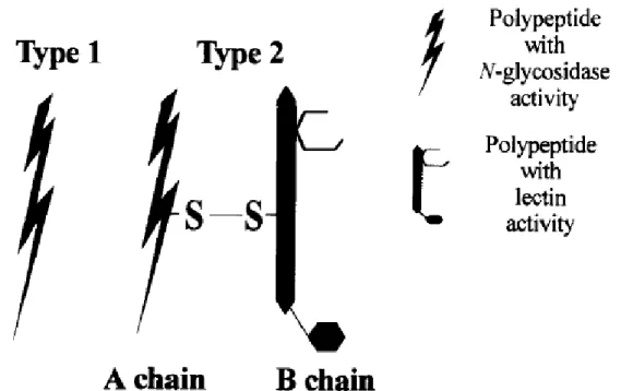
Later, further RIPs were discovered and classified into 3 types. Type 1 RIPs are single-chain proteins of around 30 kDa. Type 2 RIPs are built of two subunits: an A-subunit of about 30 kDa that provides the enzymatic activity, and a B-subunit with lectin activity, able to bind to oligosaccharides containing galactose. The third type of RIPs has been attributed to a maize b-32 RIP, regrouping RIPs with a proenzyme stage that get activated after cleavage of a short internal peptide, creating two fragments of 16.5 and 8.5 kDa (Walsh et al., 1991). For RIP JIP60, one of the two pieces is analog to a type 1 RIP, whereas the functions of the other part remains unknown (Reinbothe et al., 1994). Arguably, type 1 and 2 RIPs represent the majority of RIPs. Structural schemes of both classes are shown in Figure 1.
Figure 1: Structure of ribosome-inactivating proteins (RIP) type 1 and 2 (Barbieri et al., 1993). The number of binding sites per B-subunit of Type 2 RIPs can vary. The B-subunit of Shiga toxin provides in total 15 binding sites (3 binding sites per monomer) (Ling et al., 1998).
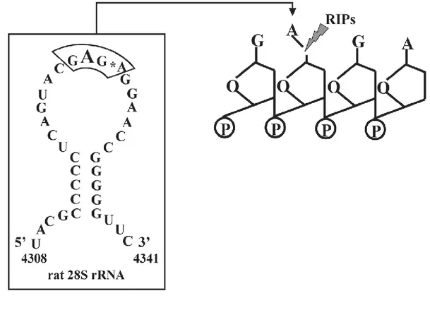
The unique characteristic of type 2 RIPs is the binding capacity of the B-subunit, which interacts with carbohydrate residues on cell membranes. Moreover, this binding results in the uptake of the toxins. A prominent member of the type 2 RIPs is ricin, which binds to mannose type glycans (Simmons et al., 1986). The endocytosis and intracellular trafficking of type 2 RIPs has been well studied. Characteristically, type 2 RIPs, such as ricin and Shiga toxin, undergo retrograde trafficking. The key step is the arrival to the Golgi and further the endoplasmic reticulum (ER) (Mallard et al., 1998; Sandvig et al., 1992; Sandvig and van Deurs, 2000; Johannes and Goud, 1998), from where their A-subunits are retrotranslocated into the cytosol to reach their molecular targets: the ribosomes. In the cytosol, the A-subunits cleave an adenine base on position 4,324 within the 28S-subunit of ribosomes (Endo et al., 1988; Saxena et al., 1989) (Figure 2). Through the catalytic cleavage, elongation factors are not recruited anymore, disabling ribosomes, inhibiting protein biosynthesis, and leading to cell death.
Figure 2: Schematic presentation of the biochemical action of ribosome-inactivating proteins (RIPs), such as ricin and Shiga toxin. The A-subunit targets the α-sarcin site on the large (28S) subunit of ribosomes, which results in an inhibition of protein biosynthesis. (Stirpe, 2005)
9.1.1 Bioterrorism, bio threat agents and biodefense
For the last decades, health emergencies due to infectious diseases have increased (E. coli O104:H4, Ebola virus, H1N1 influenza virus, etc.) (Morens et al., 2004; Jones et al., 2008). A recent outbreak that happened back in 2011 in central Europe, mainly Germany, demonstrated the overwhelming potential of toxins as bio-threats (King et al., 2012; Nr et al., 2011). The relative ease with which toxins are produced has enabled bioterroristic assaults (anthrax letters in 2001, or ricin letters to former US president Barak Obama), which kept bioterrorism on the political agenda throughout the years (Bekerman and Einav, 2015; Gottron and Shea, 2010; Gonzales et al., 2006; Council, 2007). As defined by the U.S. Centers for Disease Control and Prevention (CDC), the intended release of toxins, bacteria, viruses or other harmful biological agents to damage or kill people, animals, or plants is considered as a bioterrorist attack (Sciences et al., 2016). Biodefense is defined as the means or methods of preventing, detecting, or managing an attack involving biological weapons.
The discovery of effective and direct medical agents against biological threats is clearly important. In the past decade, high-throughput screenings (HTS) have been performed by the French Commission for Alternative Energies and Atomic Energy (CEA - Commissariat à l'énergie atomique et aux énergies alternatives) and the Curie institute in Paris. This will be discussed in chapter 9.1.3.
9.1.2 Shiga toxin and its Shiga-like toxins
In the following text, I will mainly focus on Shiga toxin and its isoforms. Shiga toxin was characterized by and named after Kiyoshi Shiga (Trofa et al., 1999; Konowalchuk et al., 1977). Shiga toxins are members of a family that includes Shiga toxin, produced by Shigella dysenteriae and Shiga-like toxins (SLTs), produced by enterohemorrhagic strains (EHEC) of Escherichia coli (or Shiga-like toxin producing (STEC) strains) (
Table 1) (Johannes and Römer, 2010; O’Brien et al., 1984). The production of these toxins by E. coli has been directly linked to the development of hemolytic uremic syndrome (HUS), which can have deadly outcome (Karmali et al., 1983). The enterohemorrhagic E. coli strains with the serotypes O157:H7 and O104:H4 are most common sources for SLTs. No direct treatments exist against EHEC-induced HUS, and the application of the anti-C5 complement component antibody eculizumab® remains questionable (Karpman, 2012). Furthermore, a treatment with conventional antibiotics seems to increase toxin release from bacteria and worsens the outcome (Agger et al., 2015).
Figure 3: Shiga toxin structures. (A) Schematic drawing of the Shiga holotoxin, catalytic A-subunit (STxA); five B-fragment monomers that form the homopentameric B-subunit (STxB). (B) A ribbon diagram of Shiga toxin, illustrating the binding sites on STxB for globotriaosylceramide (Gb3). Gb3 is shown in a ball-and-stick representation. (C) A zoom into the furin cleavage (Arg248-Val-Ala-Met251) site of STxA; the disulfide bond (between Cys242 and Cys261) is shown in yellow. (D) A ribbon diagram of a STxB from below, pointing out the three Gb3-binding sites. Gb3 is shown as a ball-and-stick representation. (Johannes and Römer, 2010)
Structurally and functionally, SLTs share many characteristics with other type 2 RIPs. Figure 3 schematically shows the molecular set up of SLTs (Johannes and Römer, 2010). SLTs are built of a catalytic A-subunit (STxA) and a homo-pentameric B-subunit (STxB). STxA inhibits protein biosynthesis through ribosomal RNA N-glycosidase activity, as mentioned above in chapter 9.1.
In the process of cellular trafficking, STxA can be cleaved into two fragments by the trans-Golgi-network-(TGN)-localized enzyme furin, which specifically recognizes an Arg-X-X-Arg sequence (Molloy et al., 1992) (Figure 3 C). Specifically, the cleavage occurs in the positions Arg248-Val-Ala-Met251 and
requires low pH for cell intoxication (Sandvig, 2001; Garred et al., 1995). This leads to the production of the STxA1 (28 kDa) and STxA2 (4 kDa) fragments. Both fragments remain connected via a disulfide bond between Cys242 and Cys261 until they reach the ER.
Table 1: Shiga toxin and isoforms (Johannes and Römer, 2010) Organism Toxin Sequence similarity to Shiga toxin [%] Characteristics Synonyms Cellular receptors A-subunit B-subunit* Shigella dysemterae Shiga
toxin 100 100 N/A N/A Gb3
STEC STx1 97 98 N/A SLTI & VT1 Gb3
STx1c 97 98 N/A SLTIc and VT1c Gb3
STx2 53 64 Associated with serve disease in humans SLTII and VT2 Gb3
STx2c 53 61 N/A SLTIIc and VT2c Gb3
STx2d 54 61 N/A SLTIId and VT2d Gb3
STx2e 53 61 Associated with the piglet edema disease SLTIIe and VT2e Gb3 & Gb4
STx2f 54 60 N/A SLTIIf and VT2f Gb3
Gb3, Globotriaosylceramide; N/A, not applicable; SLT, Shiga-like toxin; STEC, Shigella toxin-producing Escherichia coli; Stx1, Shiga toxin 1; VT: Vero toxin. *This is the sequence similarity for mature B-fragments, without signal sequences.
Strikingly, STxB (except STxB2e, Table 1) binds specifically the glycosphingolipid globotriaosylceramide (Gb3 or CD77) (Jacewicz et al., 1986; Waddell et al., 1990; Lindberg et al., 1987). Crystal structures revealed three binding sites per monomer, resulting in 15 binding sites per STxB (Ling et al., 1998). After binding to Gb3, SLTs were found in clathrin coated pits (Sandvig et al., 1989). Still, the depletion of clathrin only partially affects the uptake of Shiga toxin (maximally 35 % inhibition), suggesting that these toxins mostly enter cells through clathrin-independent endocytosis (CIE) (Lauvrak, 2004; Saint-Pol et al., 2004; Nichols et al., 2001). After endocytosis, SLTs traffic through the retrograde route, from early endosomes (EE) to the Golgi and the ER (Sandvig et al., 1992) (Figure 6, page 9). Chapter 9.2 is going to introduce this intracellular trafficking in further detail.
9.1.3 Small molecule inhibitors of the retrograde route
One major strategy in developing a direct treatment against plant and bacterial toxins, such as ricin and Shiga toxin is small molecule HTS of chemical libraries. Since the beginning of the 21st century, several
inhibitory small molecule compounds have been found that protect cells and animals against toxins through perturbation of their intracellular trafficking (Barbier et al., 2012). A recent review describes the latest development of new compounds that target the intracellular retrograde transport process (Gupta et al., 2017). In the upcoming chapter I am going to dissect their characteristics in further detail.
9.1.3.1 Retro compounds
In prior to 2010, my host laboratory collaborated with the CEA that had screened more than 16,000 small molecules for inhibitory effects against ricin, and found two protecting small molecules, called Retro1 and Retro-2 (structures are shown in Figure 4).
Figure 4: Structures of Retro compounds.
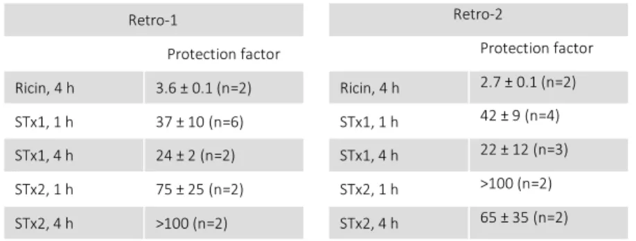
Ricin and SLT share the retrograde trafficking route. Therefore, those two compounds were also tested for their protective effect against intoxication of cells by SLTs, revealing an even higher protection factors (shown in Table 2), Moreover, it seemed that Retro-1 and Retro-2 affected only one out of 26 tested trafficking factors that were known to be involved in endosomes-to-Golgi trafficking: Syntaxin-5 (STXSyntaxin-5) was relocalized out of the Golgi without dispersing the Golgi itself (Stechmann et al., 2010), pointing to the possibility that the Retro compounds might be of interest for pharmaceutical developments. A proper introduction into intracellular trafficking is given in chapter 9.2, on page 18.
Table 2: Retro-1 and Retro-2 protection factors on HeLa cells against Ricin, Stx1, and Stx2. Protection factors calculated over the indicated number of experiments. Means ± SEM are shown. (Stechmann et al., 2010)
Retro-1 Retro-2
Protection factor Protection factor
Ricin, 4 h 3.6 ± 0.1 (n=2) Ricin, 4 h 2.7 ± 0.1 (n=2)
STx1, 1 h 37 ± 10 (n=6) STx1, 1 h 42 ± 9 (n=4)
STx1, 4 h 24 ± 2 (n=2) STx1, 4 h 22 ± 12 (n=3)
STx2, 1 h 75 ± 25 (n=2) STx2, 1 h >100 (n=2)
STx2, 4 h >100 (n=2) STx2, 4 h 65 ± 35 (n=2)
Remarkably, even in mice Retro-2 was effective against ricin, shown in Figure 5. Furthermore, Retro-2 was also effective in a more clinically relevant scenario in mice, an E. coli O104:H4 infection (Secher et al., 2015). On the cellular level, the Retro compounds blocked Shiga toxin trafficking in endosomal structures and delayed its entry into the Golgi, as illustrated in Figure 6 (Stechmann et al., 2010)
Figure 5: Retro-2 protects mice against ricin challenge. Comparison of survival curves from mice that were treated with single intraperitoneal dose of Retro-2 with indicated concentrations one hour before toxin exposure. Ricin was administered to mice via the nasal route (2 mg/kg). The control group received only the vehicle in prior to ricin exposure. The curves for treated animals are statistically different from control as measured by the log rank test (p < 0.0001 for 2 µg/kg of Retro-2, orange; p = 0.015 for 10 mg/kg, brown; p = 0.031 for 20 mg/kg, purple; p = 0.0007 for 200 mg/kg, red). (Stechmann et al., 2010)
Figure 6: Illustration of the retrograde trafficking of Shiga toxin and site of action of Retro-2 (Gupta et al., 2017). Toxins (e.g. ricin and SLTs) traffic via the retrograde route, starting from the plasma membrane through endosomes and Golgi to the ER (Johannes and Popoff, 2008). Retro-2 inhibits the toxin trafficking step from endosomes to the Golgi
Moreover, very recent studies have shown that Retro-2 protected against Leishmania, poxviruses, filoviruses and Chlamydiales (Nonnenmacher et al., 2015; Gupta et al., 2017). Together with ricin and
STEC, poxviruses, filoviruses and Chlamydiales are listed on the NIAID emerging disease list (National Institute of Allergy and Infectious Disease, 2017) and the CDC bioterrorism bio threat agent list (CDC, 2017). While Leishmania are not mentioned on those lists, a study from 2009 to 2012 showed that
Leishmania was reemerging in Madrid, Spain, thus representing an emerging health risk (Arce et al.,
2013).
9.1.3.1.1 Structural evolution of Retro-2
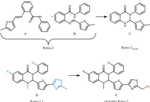
The original Retro-2 molecule was found to inhibit toxin trafficking (Stechmann et al., 2010). In 2012, it was shown that the actual active configuration of Retro-2 is a cyclic compound that forms spontaneously (Park et al., 2012). The groups of D. Gillet and J.-C. Cintrat optimized Retro-2 by performing a structure-activity relationship (SAR) study (Noel et al., 2013), which yielded a molecule that was termed Retro-2.1. After thereby improving the protection potency against SLTs by a factor of 500 when compared to the original hit compound (Noel et al., 2013), Retro-2.1 has been chemically functionalized with an azide group, herein referred to as “clickable Retro-2” (Figure 31) with the goal of identifying the cellular target of the molecule. As part of a target pull down strategy, this azide derivate enables biorthogonal click chemistry, which will be introduced in further detail in chapter 10.6.1 on page 49. A summary of the structural evolution of Retro-2 is illustrated in Figure 7.
Figure 7: Structural evolution of Retro-2. Structures A and B show the original hit compound Retro-2 (Stechmann et al., 2010). Structure B illustrates how the spontaneous cyclisation to molecule C (Retro-2cyclic) may take place (Park et al., 2012). Structure D shows the optimized molecule of Retro-2 based on C, called Retro2.1 (Noel et al., 2013). Additional structural groups are highlighted in blue. Molecule E is the azide-functionalized Retro-2.1, called “clickable Retro-2”, due to its potential to be used in a biorthogonal click chemistry approach (see chapter 0). The azide is marked in red.
Dihydro-quinazolinone analogs of Retro-2cycl showed a protective effect in monkey and human
polyoma- and papillomavirus infection in vitro (Carney et al., 2014; Nelson et al., 2013). Hence, the demonstrated protective effects of Retro-2 in vitro and in vivo turn the optimized derivatives into promising antidotes against many bio threats, including plant and bacterial toxins, viruses, intracellular parasites and bacteria.
9.1.3.1.2 Retro-2 effect on viral infections
Viruses are known to enter their host cells through different routes (Harper et al., 2013). Enveloped viruses dock and fuse directly with the plasma membrane (PM) and translocate their nucleo-capsid directly into the cytosol. Yet, non-enveloped viruses and others have to be taken up first, and then traffic through the host cell before the release into the cytosol. Some viruses even undergo retrograde trafficking (Grove and Marsh, 2011) which led to the idea that Retro-2 might protect against those viruses as well.
Adeno-associated viruses (AAV) have shown promising biomedical potential in gene therapy, and their evaluation for the treatment of various diseases is still ongoing. The retrograde trafficking of AAV is essential to reach the nucleus. Thus, its TGN arrival is a critical step, which has been shown to be syntaxin-5 (STX5) dependent and could be inhibited by Retro-2 (Nonnenmacher et al., 2015). Hence, Retro-2 could be exploited in anti-viral disease treatments.
As members of non-enveloped DNA viruses, polyomaviruses (PyV) (including human papillomaviruses (HPVs)) cause severe diseases in immunocompromised patients. BKPyV is the causative agent of polyomavirus-induced nephropathy and hemorrhagic cystitis, and JCPyV is the causative agent of the fatal demyelinating disease progressive multifocal leukoencephalopathy. Thus far, no vaccine or antiviral therapy for these viruses has been found (De Gascun and Carr, 2013). HPVs have been put in context with cancer development in the uterine cervix and oropharynx. Although vaccination showed success against some types of HPVs, many HPVs infections remain present and still are of public health concern (Carney et al., 2014). It has been shown that Retro-2 (c = 100µM) inhibited JCPyV, BKPyV and simian virus 40 (also a PyV) infections on tissue cells in average by around 30 %. Retro-2 blocked the PyVs’ arrival to the ER, which is strictly required for infection (Nelson et al., 2013). These results were confirmed in cell culture for PyVs. Additionally, Retro-2 protected cells also against HPVs (Carney et al., 2014).
As filamentous enveloped viruses, Ebola and Marburg filoviruses (FV) are members of the family Filoviridae, which cause viral hemorrhagic fevers in humans resulting in a high mortality rate of up to
90 % (Bausch et al., 2006; Feldmann and Geisbert, 2011). Thus far, only few drugs (favipiravir) have been tried in animals or humans, without promising results. Furthermore, no vaccination strategy is yet available (Sissoko et al., 2016), resulting in a biosafety level-4 classification for FVs. Recently, the growth of a new Ebola virus variant in West Africa has alerted health authorities. Favipiravir is the only small molecule drug that was tested in mice, showing an IC50 of 67 µM (Oestereich et al., 2014). Unpublished data of our collaborators at the CEA have shown that Retro-2 protected in vitro against an Ebola virus and Marburg virus infection (article in revision).
Two independent high throughput siRNA screens of Vaccinia virus (VACV) identified proteins of the endosomes-to-Golgi retrograde transport step to be pro-viral host factors (Sivan et al., 2013; Beard et al., 2014). Recently, further studies demonstrated that Retro-2 reduced spreading of VACV and Monkey pox viruses in cell cultures by interfering with their replication (Sivan et al., 2016; Harrison et al., 2016). The Retro-2 effect relied on a membrane wrapping process during late stages of virion maturation (Smith et al., 2002). Two viral proteins, involved in the maturation process, rely on retrograde trafficking from endosomes to the TGN. Retro-2 miss-localizes these proteins, and thus, blocks the maturation process.
In summary, Retro-2 shows significant potential in antiviral treatment, relying on its capacity to block retrograde trafficking.
9.1.3.1.3 Retro-2 effect on intracellular parasites
Leishmania is an intracellular parasite and causes leishmaniosis, affecting about twelve million people
with two million new cases per year worldwide. Although Leishmania is not classified as a bioterrorism agent, the Leishmania outbreak from 2009 to 2012 in Madrid, Spain, affected 446 individuals (Arce et al., 2013). Current treatments against Leishmania are either toxic or lead to the emergence of drug resistant strains, resulting in a strong need for new treatments against Leishmania (No, 2016; Sundar et al., 2014; Mansueto et al., 2014). Leishmania are internalized by macrophages into intracellular compartments called Leishmania parasitophorous vacuoles (LPVs), similar to phagosomes. Previous studies suggested that STX5 is involved in the creation of those LPVs. Consistently with the effect of Retro-2 on STX5 localization (see chapter 11.1.4.2, page 63), Retro-2 inhibited the development of LPVs in Leishmania amazonensis infected cells. Moreover, Retro-2 protected mice form L. amazonensis infections, without showing any toxicity by itself (Canton and Kima, 2012). Retro-2 also protected against L. donovani in vitro and in vivo. Thus, Retro-2 affects parasites inside and outside their host.
![Table 1: Shiga toxin and isoforms (Johannes and Römer, 2010) Organism Toxin Sequence similarity to Shiga toxin [%] Characteristics Synonyms Cellular receptors A-subunit B-subunit* Shigella dysemterae Shiga](https://thumb-eu.123doks.com/thumbv2/123doknet/2317460.28287/38.918.137.814.145.326/johannes-organism-sequence-similarity-characteristics-receptors-shigella-dysemterae.webp)