e-Polymers 2002, no. 035. http://www.e-polymers.org
pH Dependence of the morphology of aqueous micelles
formed by
polystyrene-block-poly(2-vinylpyridine)-block-poly(ethylene oxide) copolymers
Jean-François Gohy 1, Nicolas Willet 1, Sunil K. Varshney 2, Jian-Xin Zhang 2,
Robert Jérôme 1 *
1 Centre for Education and Research on Macromolecules (CERM) - Institute of
Chemistry B6, University of Liège, Sart-Tilman, 4000 Liège, Belgium; Fax +32 4 / 3663497; rjerome@ulg.ac.be
2 Polymer Source, 771 Lajoie Street, Dorval, Québec, H9P 1G7, Canada;
Fax +1 514 / 4229843; contact@polymersource.com
(Received: July 11, 2002 ; published: August 13, 2002)
Abstract: The morphology of micelles formed by two polystyrene-block-poly(2-vinylpyridine)-block-poly(ethylene oxide) (PS-b-P2VP-b-PEO) copolymers was studied in water by dynamic light scattering and transmission electron microscopy. Spherical micelles were observed that consist of a PS core, a P2VP shell and a PEO corona. The characteristic size of core, shell and corona depends on the copolymer composition. An important increase in micellar size occurs at pH < 5 as a result of P2VP block protonation. The reversibility of this pH effect depends on copolymer composition, too. The conformation of the PEO block plays an important role in this pH driven morphological transition.
Introduction
Aggregation and microphase separation of block copolymers yield a variety of well-defined supramolecular structures both in the bulk and in a selective solvent for one of the blocks [1]. Although ABC triblocks are precursors of very complex self-assembled structures in the bulk [2], little is known about their self-association in selective solvents of one (or two) of the constitutive blocks. In addition to ‘three-layer’ micelles in water [3], formation of asymmetrical micelles was reported in organic solvents [4]. These ‘Janus’ micelles consist of a crosslinked polybutadiene core and a corona with ‘southern’ polystyrene and ‘northern’ poly(methyl methacrylate) hemi-spheres. ‘Janus’ micelles actually result from supramolecular organization in the bulk which is transferred to solution. They have to be distinguished from micelles prepared by direct solubilization of ABC triblock copolymers in a selective solvent for one block.
An important characteristic feature of these self-assembled systems is their capability to respond to external stimuli, such as temperature and/or pH [5]. In this respect, pH sensitive ‘three-layer’ aqueous micelles were prepared by co-precipitation of polystyrene-block-poly(2-vinylpyridine) and poly(2-vinylpyridine)-block-poly(ethylene oxide) diblocks. However, ‘three-layer’ micelles are not completely formed by co-precipitation. They actually coexist with micelles formed by the individual diblocks [6].
Very recently, pH sensitive core-shell-corona (CSC) aqueous micelles of
polystyrene-block-poly(2-vinylpyridine)-block-poly(ethylene oxide) triblocks (PS-b-P2VP-b-PEO)
were prepared in water and characterized [7]. The pH sensitivity of P2VP allowed the hydrodynamic diameter to be tuned from 75 nm at pH > 5 to 135 nm at pH < 5, as a result of electrostatic repulsion of the charged P2VP blocks at low pH.
In this investigation, composition of the triblock copolymer has been changed, which completes the preliminary characterization of the pH sensitive CSC micelles of PS-b-P2VP-b-PEO copolymers.
Results and discussion
PS-b-P2VP-b-PEO copolymers of two compositions (Tab. 1) have been investigated. Although they contain a major PEO hydrophilic block, they are not readily soluble in water. Micelles have thus been prepared by the method reported by Eisenberg et al. for making ‘crew-cut’ micelles available [8]. So, the triblock copolymer was dissolved in a common solvent for the blocks (N,N-dimethylformamide, DMF) followed by addition of water until the hydrophobic blocks (PS and P2VP) are insoluble. The micelles were then frozen by dialysis of the organic solvent against water [8]. More-over, the triblock copolymers were also directly dissolved in a mixture of water and DMF (see Exptl. part) and then dialyzed against pure water. Regardless of the method, the same micelles were formed.
Tab. 1. Molecular weights of the investigated copolymers
Sample Mn
(PS) (P2VP) Mn (PEO) Mn (copolymer) Mw/Mn PS200-b-P2VP140-b-PEO590 20000 14000 26000 1.1
PS140-b-P2VP120-b-PEO795 14000 12000 35000 1.1
Self-association of the two copolymers in water was analyzed by dynamic light scattering (DLS), with the purpose to measure the mean hydrodynamic diameter (Dh)
of the micelles and to probe the effect of pH on the micellar characteristic features. DLS data in Tab. 2 agree with the formation of monodispersed micelles. The apparent diffusion coefficient of these micelles is independent of concentration, at least in the dilute regime, which is in agreement with ‘kinetically frozen’ micelles of an extremely low critical micellar concentration [9]. Moreover, the angular dependence of the apparent diffusion coefficient confirms that the micelles are spherical [10]. Analysis of the DLS data by the CONTIN method shows one population of CSC micelles with a narrow size distribution (Fig. 1).
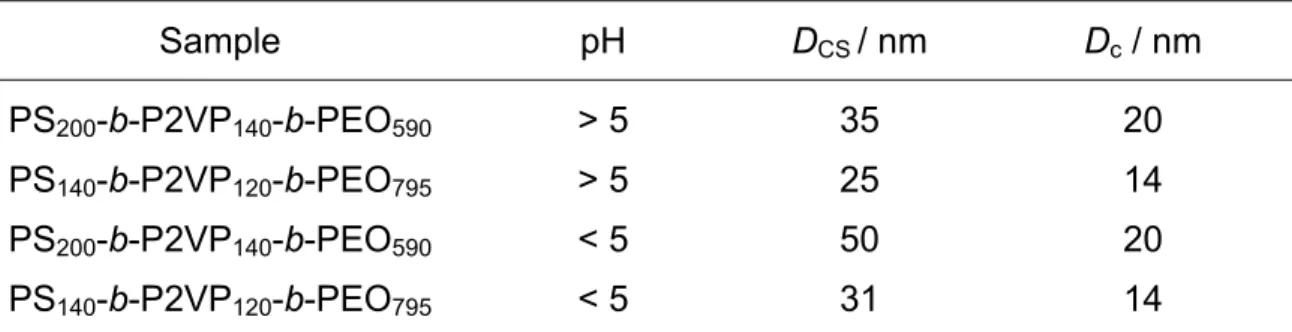
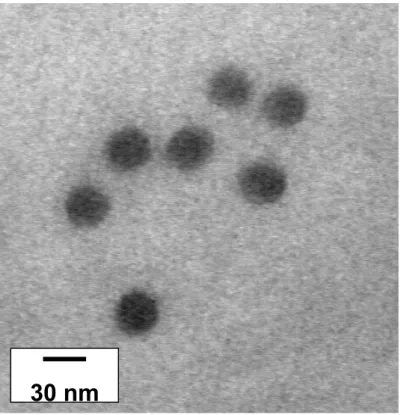
The micelles formed by the two copolymers have been directly observed by trans-mission electron microscopy (TEM), P2VP and PS blocks being selectively stained by RuO4. As shown in Fig. 2 for the PS200-b-P2VP140-b-PEO590 sample and in Fig. 3
for the PS140-b-P2VP120-b-PEO795 one, monodisperse spherical micelles are formed
at pH > 5, in agreement with DLS data. Because collapsed P2VP shell cannot be distinguished from the PS core in Fig. 2 and Fig. 3, the diameter of the core plus shell (DCS) has been extracted from the micrographs (Tab. 3).
Dh
1 10 100 1000I
N
T
E
N
S
I
T
Y
(%)
0 20 40 60 80 100Tab. 2. DLS data at pH higher and lower than 5 (PDI = Mw/Mn - 1)
Sample pH Dh / nm PDI
PS200-b-P2VP140-b-PEO590 > 5 67.9 (±2.7) 0.102 (±0.033)
PS140-b-P2VP120-b-PEO795 > 5 75.4 (±0.5) 0.087 (±0.017)
PS200-b-P2VP140-b-PEO590 < 5 89.8 (±3.5) 0.068 (±0.030)
PS140-b-P2VP120-b-PEO795 < 5 135.2 (±1.2) 0.146 (±0.021)
Fig. 1. CONTIN size distribution of CSC micelles formed by the PS200-b-P2VP140
-b-PEO590 sample at pH > 5
Tab. 3. Micellar dimensions measured from TEM pictures
Sample pH DCS / nm Dc / nm
PS200-b-P2VP140-b-PEO590 > 5 35 20
PS140-b-P2VP120-b-PEO795 > 5 25 14
PS200-b-P2VP140-b-PEO590 < 5 50 20
PS140-b-P2VP120-b-PEO795 < 5 31 14
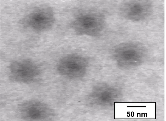
Nevertheless, an effort has been made to contrast PS and P2VP as selectively as possible. Fig. 4 illustrates CSC micelles at pH < 5 that have been stained by RuO4
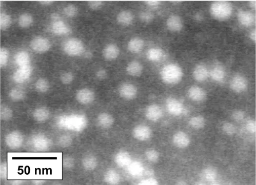
vapor, in such a way that the more compact PS core is more contrasted than the stretched P2VP shell, so giving rise to a ‘frog egg’ structure. The second method was based on specific interactions between the P2VP shell and phosphotungstic acid as shown in Fig. 5 for the PS140-b-P2VP120-b-PEO795 sample. Actually, phosphotungstic
30 nm
100 nm
acid is used as a negative contrasting agent for micelles. In Fig. 5, the concentration of phosphotungstic acid was kept low enough to contrast selectively the P2VP shell, which is observed as a dark ring. At higher phosphotungstic acid concentration (Fig. 6) the background is contrasted and the PS cores are observed as white spheres. These additional micrographs give access to the diameter of the PS core (Dc) as
reported in Tab. 3.
Fig. 2. TEM picture of micelles formed by the PS200-b-P2VP140-b-PEO590 copolymer
at pH > 5
Fig. 3. TEM picture of micelles formed by the PS140-b-P2VP120-b-PEO795 copolymer
50 nm
Fig. 4. TEM picture of micelles formed by the PS200-b-P2VP140-b-PEO590 copolymer
at pH < 5 (contrasted with RuO4)
Fig. 5. TEM picture of micelles formed by the PS140-b-P2VP120-b-PEO795 copolymer
at pH < 5. The P2VP shell is contrasted with H3PO4·12 WO3 (low concentration)
The water-solubility of the P2VP block strongly depends on pH [11], the block being hydrophobic and non-soluble in water at pH > 5 and ionized and water-soluble at pH < 5. Although the ionization/deprotonation of P2VP is a complex process [12], it allows for the reversible micellization of P2VP-b-PEO diblocks. Indeed, free chains are formed at pH < 5, whereas micelles consisting of a P2VP core and a PEO corona
50 nm
are observed at pH > 5 [11,13]. In the case of CSC micelles formed by PS-b-P2VP-b-PEO copolymers, a change in pH is expected to modify deeply conformation and water-solubility of the P2VP blocks. This block should be hydrophobic and collapsed on the PS core at pH > 5, and a highly stretched water-soluble protonated chain attached to the PS core at pH < 5, as illustrated in Scheme 1.
Fig. 6. TEM picture of micelles formed by the PS140-b-P2VP120-b-PEO795 copolymer
at pH < 5. The PS cores appear as bright spheres (high concentration of H3PO4·12
WO3)
Scheme 1. Effect of pH on CSC micelles (PS core as a sphere, P2VP chains in blue, PEO chains in black)
pH > 5
pH < 5
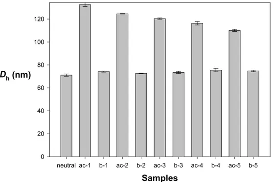
Samples
neutral ac-1 b-1 ac-2 b-2 ac-3 b-3 ac-4 b-4 ac-5 b-5
Dh (nm) 0 20 40 60 80 100 120
This pH effect and reversibility have been probed by DLS experiments, as reported in Fig. 7 for the PS200-b-P2VP140-b-PEO590 sample and in Fig. 8 for the PS140
-b-P2VP120-b-PEO795 one. A very important increase in Dh (from 75 to 135 nm) is noted
when an excess of HCl is added to micelles such that the P2VP block is completely protonated (Fig. 7, ac-1). Upon addition of NaOH and complete neutralization of the acid (Fig. 7, b-1), the initial Dh is recovered. Complete reversibility of the pH effect on
the size of the P2VP shell has been confirmed by pH cycling in the same vial (Fig. 7).
Dh of the ‘protonated’ CSC micelles, however, decreases with the number of cycles,
as a result of electrostatic screening by the increasing amount of salt formed by repeated neutralization of HCl by NaOH. Elimination of the screening salt and reset of pH below 5 restores the original data.
Fig. 7. pH Cycling of Dh of CSC micelles formed by the PS200-b-P2VP140-b-PEO590
copolymer (ac stands for the addition of 1.5 ·10-5 mol of HCl, b for the addition of 1.5·10-5 mol of NaOH)
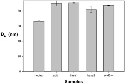
In sharp contrast, pH cycling of the PS140-b-P2VP120-b-PEO795 micelles is not
revers-ible (Fig. 8). Although Dh increases from 68 to 90 nm upon addition of an excess of
HCl (acid1, Fig. 8), the initial Dh is not restored when an equimolar amount of NaOH
is added (base1, Fig. 8). A small decrease in Dh (base2, Fig. 3) is only observed
when an excess of NaOH is used. This minor modification is however recovered upon neutralization of NaOH and acidification of the micellar solution (acid3+4, Fig. 8). So, the P2VP blocks of the PS140-b-P2VP120-b-PEO795 sample respond to pH but
the effect is not reversible upon pH cycling. Nevertheless, the initial Dh of the PS140
-b-P2VP120-b-PEO795 micelles can be restored by elimination of NaCl by dialysis and
heating at 50°C.
A tentative explanation of the non-reversible pH cycling of the PS140-b-P2VP120
-b-PEO795 copolymer compared to the PS200-b-P2VP140-b-PEO590 one might be found in
the chemical composition of these copolymers, particularly in the length of the PEO block which is much longer in the PS140-b-P2VP120-b-PEO795 copolymer. When pH is
Samples
neutral acid1 base1 base2 acid3+4
Dh (nm) 0 20 40 60 80
increased. This very important increase in Dh cannot be explained only by referring to
the fully extended conformation of the short P2VP block. More likely, conformation of the PEO block is influenced by stretching of the P2VP chains. Because the PEO blocks are no longer confined in the close neighborhood of the PS core when the P2VP blocks are protonated, there is a gain in conformational entropy. When the pH is increased, there is competition between the gain in energy by hydrophobic inter-actions of the deprotonated P2VP chains and loss of entropy associated to the possible collapse of the PEO chains which are again confined at the surface of the PS/P2VP core. This loss of entropy appears to be counterbalanced by the hydro-phobic interactions in case of the PS200-b-P2VP140-b-PEO590 copolymer but not when
PS140-b-P2VP120-b-PEO795 is concerned that contains a very long PEO block.
Finally, these experiments suggest that a change of pH only affects the P2VP shell of the micelles which otherwise remain unchanged in terms of shape and number of aggregation consistent with the frozen-in PS core [14].
Fig. 8. pH Cycling of Dh of CSC micelles formed by the PS140-b-P2VP120-b-PEO795
sample (acid stands for the addition of 1.5·10–5 mol of HCl, base for the addition of 1.5·10–5 mol of NaOH)
Data in Tab. 3 show that Dc is directly proportional to the number of styrenic units.
The number of aggregation has been calculated for both copolymers and was found to be 113 for the PS200-b-P2VP140-b-PEO590 micelles and 55 for thePS140-b-P2VP120
-b-PEO795 ones. The thickness of the P2VP shell has been also estimated for the
PS200-b-P2VP140-b-PEO590 micelles, viz., 7.5 nm at pH > 5 and 15 nm at pH < 5. As
far as PS140-b-P2VP120-b-PEO795 micelles are concerned, the thickness of the P2VP
shell is increased from 5.5 to 8.5 nm as the pH is decreased below 5. These charac-teristic dimensions have been compared to the fully extended length of the P2VP block (30 nm for the PS140-b-P2VP120-b-PEO795 sample and 35 nm for the PS200
-b-P2VP140-b-PEO590 one). The extension of the P2VP block can accordingly be
calcu-lated as 50% for the PS200-b-P2VP140-b-PEO590 sample and 23% for the PS140
-b-P2VP120-b-PEO795 one, thus being in relation to the length of the PEO block.
Copoly-mers with additional compositions are however needed before firm conclusions can be drawn.
Conclusions
This paper has demonstrated that PS-b-P2VP-b-PEO copolymers self-organize in water with formation of spherical micelles consisting of a PS core, a P2VP shell and a PEO corona. Combination of DLS and TEM assisted by selective contrasting techniques has made the sizes of the PS core and P2VP shell available. These results will be compared in the near future to data collected by SAXS and SANS. In the same experiments, composition of the triblocks will be systematically varied in order to establish the characteristic scaling laws. The pH sensitivity of the P2VP shell has been also investigated and found to be entirely reversible for one of the copoly-mers. This feature makes the investigated system particularly attracting for appli-cations including encapsulation and release of active species. Moreover, the P2VP shell can be used as a templating medium for the synthesis of, e.g., metal nanoshells [7] whose size and thickness can be tuned by the composition of the PS-b-P2VP-b-PEO copolymer.
Experimental part
The two PS-b-P2VP-b-PEO copolymers were synthesized by sequential anionic polymerization of the comonomers as reported elsewhere [7]. The micelles were prepared either by dissolving 0.1 g of the copolymer in a mixture of 0.5 g of bidistilled water and 4.4 g of N,N-dimethylformamide (DMF) or by dissolving 0.1 g of the copolymer in 4.4 g of DMF to which 0.1 g of water was added dropwise. Both methods led to the same results. DMF was then eliminated by dialysis against regularly replaced bidistilled water.
Dynamic light scattering (DLS) measurements were performed with a Brookhaven Instruments Corp. BI-200 apparatus equipped with a BI-2030 digital correlator and an Ion Laser Technology argon laser with a wavelength of 488 nm. The scattering angle was 90°. A refractive index matching bath of filtered water surrounded the scattering cell, and the temperature was controlled at 25°C. The experimental intensity corre-lation function was measured and analysed by using a cumulant expansion, as described elsewhere [15]. The mean hydrodynamic diameter (Dh), the angular
dependence of the diffusion coefficient and the polydispersity of the micelles/ aggregates were calculated accordingly. DLS data were also analyzed by the CONTIN routine, a method which is based on a constraint inverse Laplace transfor-mation of the data and which gives access to a size distribution histogram for the micellar object.
Transmission electron microscopy (TEM) observations were carried out with a Philips CM 100 apparatus operating at 100 kV. TEM photomicrographs were directly recorded with a Gatan 673 CCD camera and data were transferred to a computer equipped with the Kontron KS 100 software. Samples were prepared by casting one drop of a 0.2 wt.-% aqueous micellar solution onto a Formvar-coated copper grid. Micelles were stained by RuO4 vapor. In some experiments, H3PO4 · 12 WO3 was
used as a contrasting agent. In this case, a drop of micellar solution was placed on a Formvar-coated grid. The excess fluid was removed from the grid with a filter paper (Whatman). A drop of 1% phosphotungstic acid in water was immediately added to the surface. After one minute, excess fluid was removed and dried in air.
Acknowledgement: JFG is "Chargé de Recherche" by the Belgian National Foundation for Scientific Research (F.N.R.S.), and he thanks the European Science
Foundation SUPERNET program. JFG, NW and RJ are very much indebted to the "Services Fédéraux des Affaires Scientifiques, Techniques et Culturelles" for financial support in the frame of the "Pôles d’attraction Interuniversitaires 5/03: Supramolec-ular Chemistry and SupramolecSupramolec-ular Catalysis".
[1] Hamley, I. W.; “The Physics of Block Copolymers”; Oxford University Press, Oxford 1998.
[2] see, e.g.: (a) Goldacker, T.; Abetz, V.; Stadler, R.; Erukhimovich, I.; Leibler, L.;
Nature 1999, 398, 137. (b) Stupp, S. I.; LeBonheur, V.; Walker, K.; Li, L.S.; Huggins, K. E.; Keser, M.; Amstutz, A.; Science 1997, 276, 384.
[3] (a) Kriz, J.; Masar, B.; Plestil, J.; Tuzar, Z.; Pospisil, H.; Doskocilova, D.;
Macromolecules 1998, 31, 41. (b) Patrickios, C. S.; Hertler, W. R.; Abbott, N. L.; Hatton, T. A.; Macromolecules 1994, 27, 930. (c) Yu, G.; Eisenberg, A.;
Macromolecules 1998, 31, 5546.
[4] Erhardt, R.; Böker, A.;Zettl, H.; Kaya, H.; Pyckout-Hintzen, W.; Krausch, G.; Abetz, V.; Müller, A. H. E.; Macromolecules 2001, 34, 1069.
[5] (a) Chécot, F.; Lecommandoux, S.; Gnagnou, Y. ; Klok, H. A.; Angew. Chem. Int.
Ed. 2002, 41, 1340. (b) Gohy, J. F.; Varshney, S. K.; Jérôme, R.; Macromolecules 2001, 34, 3361. (c) Gohy, J.F.; Mores, S.; Varshney, S.K.; Zhang, J.X.; Jérôme, R.;
e-Polymers 2002, no. 21.
[6] Talingting, M. R.; Munk, P.; Webber, S. E.; Tuzar, Z.; Macromolecules 1999, 32, 1593.
[7] Gohy, J. F.; Willet, N.; Varshney, S.; Zhang, J. X.; Jérôme, R.; Angew. Chem. Int.
Ed. 2001, 40, 3214.
[8] Zhang, L.; Eisenberg, A.; Science 1995, 268, 1728.
[9] Zhang, L.; Eisenberg, A.; J. Am. Chem. Soc. 1996, 118, 3168. [10] Pecora, R. J.; J. Chem. Phys. 1968, 48, 4126.
[11] Martin, T. J.; Prochazka, K.; Munk, P.; Webber, S. E.; Macromolecules 1996,
29, 6071.
[12] (a) Puterman, M.; Koenig, J. L.; Lando, J. B.; J. Macromol. Sci. - Phys. 1979,
B16, 89. (b) Puterman, M.; Garcia, E.; Lando, J. B.; J. Macromol. Sci. - Phys. 1979,
B16, 117.
[13] Gohy, J. F.; Varshney, S. K.; Jérôme, R.; Macromolecules 2001, 34, 3361. [14] Wang, Y.; Balaji, R.; Quirk, R. P.; Mattice, W. L.; Polym. Bull. 1992, 28, 333. [15] Gohy, J. F.; Varshney, S. K.; Antoun, S.; Jérôme, R.; Macromolecules 2000, 33, 9298.