Université de Montréal
Comparative Mitochondrial Genomics Toward Understanding
Genetics and Evolution of Arbuscular Mycorrhizal Fungi
By Maryam Nadimi
Département de sciences biologiques Faculté des Arts et des sciences
Thèse présentée à la Faculté des arts et des sciences en vue de l’obtention du grade de docteur
en sciences biologiques
November, 2014
Résumé
Les champignons mycorhiziens arbusculaires (CMA) sont très répandus dans le sol où ils forment des associations symbiotiques avec la majorité des plantes appelées mycorhizes arbusculaires. Le développement des CMA dépend fortement de la plante hôte, de telle sorte qu'ils ne peuvent vivre à l'état saprotrophique, par conséquent ils sont considérés comme des biotrophes obligatoires. Les CMA forment une lignée évolutive basale des champignons et ils appartiennent au phylum Glomeromycota. Leurs mycélia sont formés d’un réseau d’hyphes cénocytiques dans lesquelles les noyaux et les organites cellulaires peuvent se déplacer librement d’un compartiment à l’autre. Les CMA permettent à la plante hôte de bénéficier d'une meilleure nutrition minérale, grâce au réseau d'hyphes extraradiculaires, qui s'étend au-delà de la zone du sol explorée par les racines. Ces hyphes possèdent une grande capacité d'absorption d’éléments nutritifs qui vont être transportés par ceux-ci jusqu’aux racines. De ce fait, les CMA améliorent la croissance des plantes tout en les protégeant des stresses biotiques et abiotiques. Malgré l’importance des CMA, leurs génétique et évolution demeurent peu connues. Leurs études sont ardues à cause de leur mode de vie qui empêche leur culture en absence des plantes hôtes. En plus leur diversité génétique intra-isolat des génomes nucléaires, complique d’avantage ces études, en particulier le développement des marqueurs moléculaires pour des études biologiques, écologiques ainsi que les fonctions des CMA. C’est pour ces raisons que les génomes mitochondriaux offrent des opportunités et alternatives intéressantes pour étudier les CMA. En effet, les génomes mitochondriaux (mt) publiés à date, ne montrent pas de polymorphismes génétique intra-isolats. Cependant, des exceptions peuvent exister. Pour aller de l’avant avec la génomique mitochondriale, nous avons besoin de générer beaucoup de données de séquençages de l’ADN mitochondrial (ADNmt) afin d’étudier les méchanismes évolutifs, la génétique des
population, l’écologie des communautés et la fonction des CMA. Dans ce contexte, l’objectif de mon projet de doctorat consiste à: 1) étudier l’évolution des génomes mt en utilisant l’approche de la génomique comparative au niveau des espèces proches, des isolats ainsi que des espèces phylogénétiquement éloignées chez les CMA; 2) étudier l’hérédité génétique des génomes mt au sein des isolats de l’espèce modèle Rhizophagus irregularis par le biais des anastomoses ; 3) étudier l’organisation des ADNmt et les gènes mt pour le développement des marqueurs moléculaires pour des études phylogénétiques.
Nous avons utilisé l’approche dite ‘whole genome shotgun’ en pyroséquençage 454 et Illumina HiSeq pour séquencer plusieurs taxons de CMA sélectionnés selon leur importance et leur disponibilité. Les assemblages de novo, le séquençage conventionnel Sanger, l’annotation et la génomique comparative ont été réalisés pour caractériser des ADNmt complets. Nous avons découvert plusieurs mécanismes évolutifs intéressant chez l’espèce Gigaspora rosea dans laquelle le génome mt est complètement remanié en comparaison avec Rhizophagus irregularis isolat DAOM 197198. En plus nous avons mis en évidence que deux gènes cox1 et rns sont fragmentés en deux morceaux. Nous avons démontré que les ARN transcrits les deux fragments de cox1 se relient entre eux par épissage en trans ‘Trans-splicing’ à l’aide de l’ARN du gene nad5 I3 qui met ensemble les deux ARN cox1.1 et cox1.2 en formant un ARN complet et fonctionnel. Nous avons aussi trouvé une organisation de l’ADNmt très particulière chez l’espèce Rhizophagus sp. Isolat DAOM 213198 dont le génome mt est constitué par deux chromosomes circulaires. En plus nous avons trouvé une quantité considérable des séquences apparentées aux plasmides ‘plasmid-related sequences’ chez les Glomeraceae par rapport aux Gigasporaceae, contribuant ainsi à une évolution rapide des ADNmt chez les Glomeromycota. Nous avons aussi séquencé plusieurs isolats de l’espèces R. irregularis et Rhizophagus sp. pour décortiquer leur position phylogénéque et inférer des relations évolutives entre celles-ci. La comparaison
génomique mt nous montré l’existence de plusieurs éléments mobiles comme : des cadres de lecture ‘open reading frames (mORFs)’, des séquences courtes inversées ‘short inverted repeats (SIRs)’, et des séquences apparentées aux plasimdes ‘plasmid-related sequences (dpo)’ qui impactent l’ordre des gènes mt et permettent le remaniement chromosomiques des ADNmt. Tous ces divers mécanismes évolutifs observés au niveau des isolats, nous permettent de développer des marqueurs moléculaires spécifiques à chaque isolat ou espèce de CMA.
Les données générées dans mon projet de doctorat ont permis d’avancer les connaissances fondamentales des génomes mitochondriaux non seulement chez les Glomeromycètes, mais aussi de chez le règne des Fungi et les eucaryotes en général. Les trousses moléculaires développées dans ce projet peuvent servir à des études de la génétique des populations, des échanges génétiques et l’écologie des CMA ce qui va contribuer à la compréhension du rôle primorial des CMA en agriculture et environnement.
Mots clés : champignons mycorhiziens arbusculaires (CMA), génome mitochondrial, mitogénomique comparative, marqueur moléculaire, homoplasmie, anastomose, analyses phylogénétique, remaniement génomique, élément génétique mobile, recombinaison.
Abstract
Arbuscular mycorrhizal fungi (AMF) are the most widespread eukaryotic symbionts that form mutualistic association with majority of plant-roots known as Arbuscular Mycorrhizae. AMF are obligate biotrophs belonging to an ancient fungal lineage of phylum Glomeromycota. Their mycelia are formed by a complex network made by coenocytic hyphae, where nuclei and cell organelles can freely move from a compartment to another. AMF are commonly acknowledged to improve plant growth by enhancing mineral nutrient uptake, in particular phosphate and nitrate and they confer tolerance to abiotic and biotic stresses for plants. Despite their significant roles in ecosystems, their genetics and evolution are not well understood. Studying AMF is challenging due to their obligate biotrophy, their slow growth and limited morphological treats. In addition, intra-isolate genetic polymorphism of nuclear DNA brings another level of complexity for investigation biology, ecology and function of AMF. Genetic polymorphism of nuclear DNA within a single isolate limits the development of efficient molecular markers mainly at lower taxonomic level (i.e. inter-isolate level). Thus, mitochondrial (mt) genomics have been used as an attractive alternative to study AMF. mt genomes have been shown to be homogeneous or much less polymorphic than nuclear DNA in AMF. However, we need to generate large mt sequence datasets in order to investigate its efficiency and usefulness in developing molecular marker toolkits in order to study its dynamic and evolutionary mechanisms as well as population genetics, community ecology and functions of Glomeromycota. In line with these challenges, the objectives of my Ph.D. project were therefore: 1) To investigate mitochondrial genome evolution using comparative mitogenomic analyses of closely related species and isolates as well as phylogenetically distant taxa of AMF; 2) To explore mt genomes inheritance among compatible isolates of the model AMF Rhizophagus irregularis through
anastomosis formation; and 3) To assess mtDNA and mt genes for marker development and phylogenetic analyses.
We used whole genome shotgun, 454 pyrosequencing and HiSeq Illimina to sequence some selected AMF taxa according to their importance and availability in our lab collections. De novo assemblies, Sanger sequencing, annotation and comparative genomics were then performed to characterize completed mtDNAs. We discovered interesting evolutionary mechanisms in Gigaspora rosea in which we found that its mtDNA revealed the fully reshuffled genome synteny compared to Rhizaphagus irregularis DAOM 197198 and presence of two fragmented cox1 and rns genes. We demonstrated that two cox1 transcripts are joined by trans-splicing. We also reported an unusual mtDNA organization in Rhizophagus sp. DAOM 213198 whose mt genome consisted of two circular mtDNAs. In addition, we observed a considerable amount of mt plasmid-related sequences in Glomeraceae compared with Gigasporaceae contributing to fast evolution of mtDNA in Glomeromycota. We also sequenced other isolates of R. irregularis and Rhizophagus sp. in order to unravel their evolutionary relationship and to develop molecular toolkits for their discrimination. Comparative mitogenomic analyses of these mtDNAs revealed the occurrence of many mobile elements such as mobile open reading frames (mORFs), short inverted repeats (SIRs), plasmid-related sequences (dpo) that impact mt genome synteny and mtDNA alteration. All together, these evolutionary mechanisms among closely related AMF isolates give us clues to design reliable an efficient intra- and inter-specific markers that are to discriminate closely related AMF taxa and isolates.
Data generated in my Ph.D. project advanced our knowledge on mitochondrial genomes evolution not only in Glomeromycota, but also in Fungal kingdom and in Eukaryotes in general. Molecular toolkits developed in this project will offer new opportunities to study population genetics, genetic exchanges and ecology of AMF, which will contribute to understand the role of
these fungi in nature with potential application in agriculture and environment protection.
Key words: arbuscular mycorrhizal fungi (AMF), mitochondrial genome, comparative mitogenomics, molecular marker, homoplasmy, anastomosis, phylogenetic analyses, genome rearrangement, mobile genetic elements, recombination.
Table of Contents
Résumé ... i
Abstract ... iv
Table of Contents ...vii
List of tables ...xii
List of figures ... xiv
List of abbreviations ...xvii
This thesis is dedicated to my mother and father, who had the patience to be far from me to complete this research in past five years. Thank you and I love you. ... xix
Acknowledgments ... xx
Chapter 1 - General introduction ... 1
1.1 Mycorrhiza ... 1
1.2 Arbuscular mycorrhizal fungi ... 2
1.3 AMF biodiversity and interaction with plants ... 4
1.4 AMF identification ... 5
1.5 Anastomosis in AMF ... 7
1.6 Mitochondrial DNA and its evolution ... 9
1.7 Mitochondrial inheritance in AMF ... 10
1.7 Mitochondrial inheritance in AMF ... 11
1.9 Research objectives, hypotheses and thesis presentation ... 12
Presentation of article 1 ... 16
Chapter 2 - Group I intron–mediated trans-splicing in mitochondria of Gigaspora rosea and a robust phylogenetic affiliation of arbuscular mycorrhizal fungi with Mortierellales . 17 2.1 Abstract ... 18
2.2 Key words ... 18
2.3 Introduction ... 18
2.4 Materials and Methods ... 22
2.4.2 DNA Purification ... 23
2.4.3 RNA Purification ... 23
2.4.4 Reverse Transcriptase–Polymerase Chain Reaction ... 24
2.4.5 Sequencing, Assembly, and Gene Annotation ... 25
2.4.6 Phylogenetic Analysis ... 27
2.5 Results and Discussion ... 27
2.5.1 Comparison of G. irregulare and G. rosea Mitochondrial Genomes ... 27
2.5.2 Mitochondrial Plasmid Insertions ... 31
2.5.3 Evolution of the Mitochondrial Genetic Code in Glomeromycota ... 32
2.5.4 Robust Phylogenetic Association of Glomeromycota with Mortierellales ... 34
2.5.5 Group I Intron–Mediated Trans-splicing of G. rosea cox1 ... 37
2.5.6 Gene for the Small Subunit rRNA in Two Pieces ... 41
2.5.7 How Many True Introns in rns and rnl? ... 43
2.6 Conclusion ... 43
2.7 Supplementary Material ... 44
2.8 Acknowledgments ... 44
Presentation of article 2 ... 45
Chapter 3 - Rapid Mitochondrial Genome Evolution through Invasion of Mobile Elements in Two Closely Related Species of Arbuscular Mycorrhizal Fungi ... 46
3.1 Abstract ... 47
2.2 Keywords ... 47
3.2 Introduction ... 48
3.3 Materials and Methods ... 51
3.3.2 DNA extraction ... 52
3.3.3 RNA extraction ... 52
3.3.4 cDNA synthesis ... 52
3.3.5 Polymerase chain reaction (PCR) ... 53
3.3.6 Reverse transcriptase – polymerase chain reaction (RT-PCR) ... 53
3.3.7 Cloning ... 55
3.3.8 Sequencing, assembly and gene annotation ... 56
3.4 Results and Discussion ... 57
3.4.1 Glomus sp. genome organization and structure ... 57
3.4.2 Comparative view of three Glomus mtDNAs ... 57
3.4.3 Rapid expansion of plasmid-like DNA polymerase sequences in Glomus ... 64
3.4.4 Mobile ORF elements (mORFs) in Glomus ... 65
2.5.5 Evidences of horizontal gene transfer between Glomus spp. ... 69
3.5 Conclusion ... 72
3.6 Acknowledgments ... 73
Presentation of article 3 ... 74
Chapter 4 - The mitochondrial genome of the glomeromycete Rhizophagus sp. DAOM 213198 reveals an unusual organization consisting of two circular chromosomes ... 75
4.1 Abstract ... 76
4.3 Introduction ... 77
4.4 Materials and methods ... 80
4.4.1 Fungal material and DNA extraction ... 80
4.4.4 Quantitative real-time PCR ... 83
4.5 Results and Discussion ... 84
4.5.1 Description of Rhizophagus sp. mtDNA ... 84
4.5.3 Short inverted repeats (SIRs) mediate recombination in mtDNA ... 91
4.5.4 Relative quantification of mtDNA chromosomes ... 94
4.5.5 Mitochondrial inheritance ... 94
4.6 Conclusions ... 95
4.7 Acknowledgments ... 96
Presentation of article 4 ... 97
Chapter 5 - Mitochondrial comparative genomics and phylogenetic signal assessment of mtDNA among arbuscular mycorrhizal fungal taxa ... 98
5.1. Highlights ... 99
5.2. Abstract ... 99
5.3. Introduction ... 100
5.4.1. Fungal material and DNA extraction ... 103
5.4.2. Sequencing, assembly and gene annotation ... 104
5.4.3. Polymerase Chain reactions (PCR) ... 104
5.4.4. Datasets and sequence alignments ... 105
5.4.5. Phylogenetic analyses ... 105
5.4.6. Statistical analysis of phylogenies ... 106
5.4.7. Distance matrix, principal coordinate analysis, and partitioning analysis ... 106
5.5. Results and Discussion ... 107
5.5.1. Overview of mtDNA diversity and evolution in the Glomeromycota ... 107
5.5.2. Assessment of the phylogenetic signal of single mt genes and subsets of genes ... 111
5.5.3. Assessment of the relationship between isolates ... 115
5.5.4. Unraveling the Rhizophagus irregularis complex ... 118
5.6. Conclusion ... 121
5.7. Acknowledgment ... 122
Presentation of article 5 ... 123
Chapter 6 - Detection of a transient mitochondrial DNA heteroplasmy in the progeny of crossed genetically divergent isolates of arbuscular mycorrhizal fungi ... 124
6.1 Summary ... 125
6.2 Keywords ... 126
6.3 Introduction ... 126
6.4 Materials and Methods ... 129
6.4.1 Growth conditions and maintenance of fungal cultures and roots ... 129
6.4.2 DNA extraction and sequencing ... 129
6.4.3 de novo assembly and sequence analysis ... 130
6.4.4 Molecular marker development ... 130
6.4.5 Experimental set up ... 132
6.4.6 Pre-symbiotic experiment ... 133
6.4.7 Symbiotic experiment ... 134
6.4.8 Microscopy, data collection, harvest and statistical analysis ... 136
6.4.9 PCR genotyping and sequencing of progeny spores ... 137
6.5.1 Mitochondrial genome comparison and marker development ... 138
6.5.2 Pre-symbiotic interactions ... 139
6.5.3 Symbiotic interactions ... 140
6.5.4 mtDNA genotyping and sequencing in spore progeny ... 144
6.5.5 Monosporal cultures with progeny from crossed experiments ... 145
6.5.6 Colonization detection ... 146
6.6 Discussion ... 146
6.7 Conclusions ... 150
6.8 Acknowledgements ... 151
Chapter 7 - General discussion, Conclusion and Perspectives ... 152
References ... 157
Annex 1 : Supplementary Information (Chapter 2) ... 171
Annex 2 : Supplementary Information (Chapter 3) ... 175
Annex 3 : Supplementary Information (Chapter 4) ... 180
List of tables
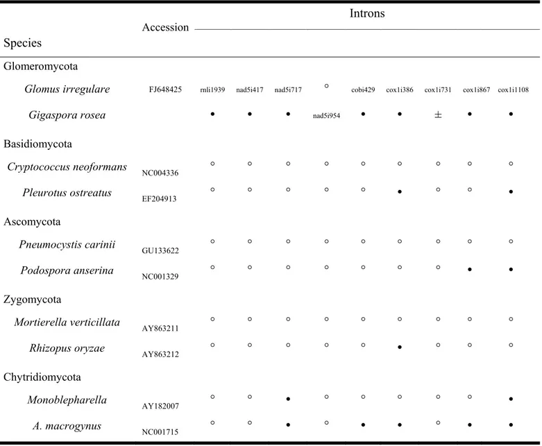
Table 2.1. Gene and Intron Content in Selected Fungal mtDNAs. ... 29 Table 2.2. Presence () or Absence (°) at Cognate Insertion Site of the Shared Introns between
Gigaspora and Glomus in Representatives of Other Fungi. ... 38 Table 3.2. Gene and intron content in AMF and selected fungal mtDNAs. ... 60 Table 3.3. Description of the gene hybrids found in Glomus sp. 229456 mtDNA. ... 68 Table 4.1. Primers and probes designed for the real-time qPCR assays and long range PCR
primers used to validate the circularity of the two mtDNAs in Rhizophagus sp. DAOM 213198. Primer direction (F, forward and R, reverse), sequence, PCR product size in base pair (bp) and melting temperature in degree Celsius (Tm) are indicated. ... 83 Table 4.2. Distribution of small inverted repeats (SIRs) found in R. irregularis DAOM 234179
and Rhizophagus sp. DAOM 213198 depending on categorized types and genome localization. ... 93 Table 6.1. Isolate-specific primers used to discriminate the three Rhizophagus irregularis isolates ... 131 Table 6.2. Anastomosis1,2 frequency from the interaction of Rhizophagus irregularis isolates in
the symbiotic experiment ... 141 Table S3.2. Sequence identity matrix of the atp9 native C-terminals along with the Glomus .... 176 sp. 229456 putative foreign inserted C*-terminal. ... 176 Table S3.3. Sequence identity matrix of the cox2 native C-terminals along with the Glomus .... 177 Table S3.4. Sequence identity matrix of the nad3 native C-terminals along with the Glomus sp.
229456 putative foreign inserted C*-terminal. ... 178 Table S4.1. Absolute quantitative real-time PCR assays performed on DAOM 213198 and
DAOM 197198 using cox1 and rnl for the large and small mtDNAs, respectively. PCR efficiencies were 98.33% and 99.86% for rnl and cox1, respectively. ... 186 Table S5.1. Number and percentage of anastomosis1 between germlings from spore clusters
Table S5.2. Total number of spores1 of R. irregularis (full lipids, empty and aborted
like-structures) produced in the interaction zone and at both sides in the crossing experiments and controls. ... 190 Table S5.3. Number and percentage of germinated and non-germinated spores of R. irregularis,
List of figures
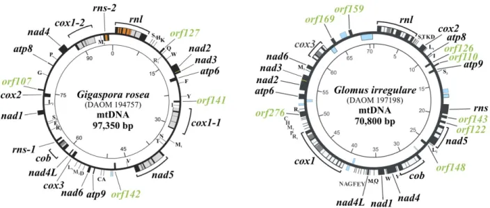
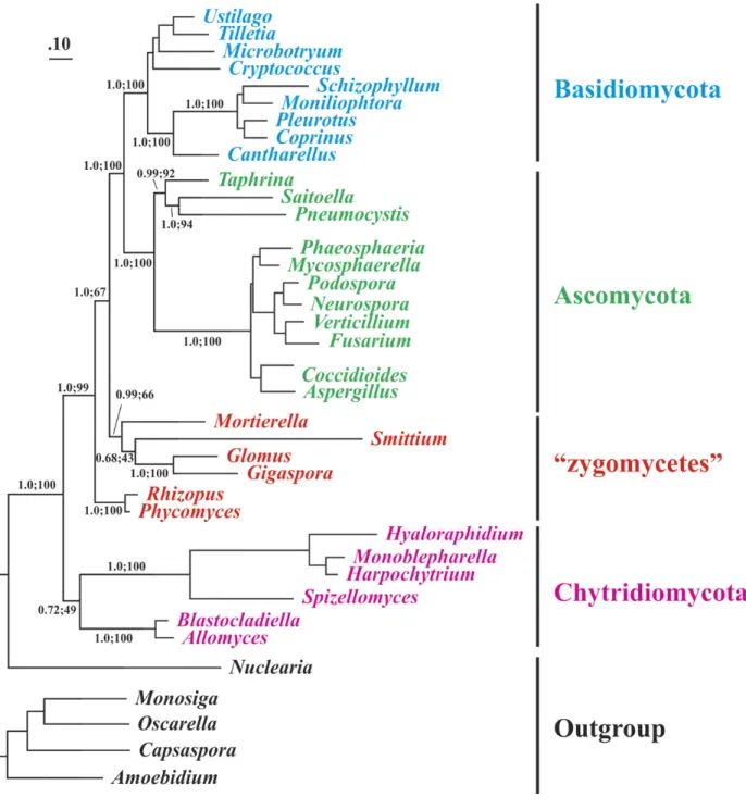
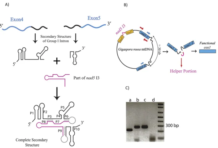
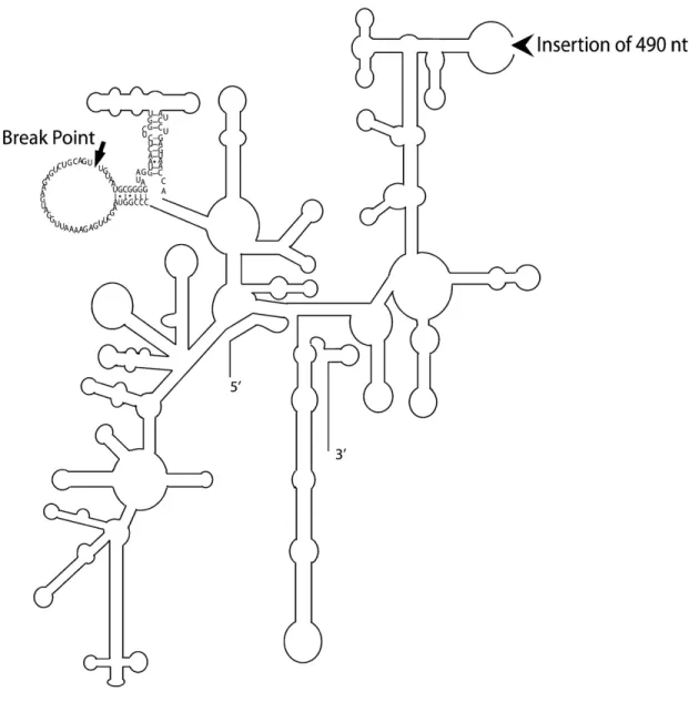
Figure 2.1. Comparison of Gigaspora rosea and Glomus irregulare mitochondrial genomes. .... 30 Figure 2.2. Phylogenetic positioning of Glomeromycota with mitochondrial protein data. ... 36 Figure 2.3. Model of group I intron–mediated trans-splicing in G. rosea and demonstration of
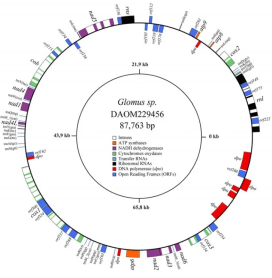
mRNA trans-splicing by RT-PCR. ... 40 Figure 2.4. Secondary structure model of the fragmented G. rosea small subunit rRNA. ... 42 Figure 3.1. The Glomus sp. 229456 mitochondrial genome circular-map was opened upstream of
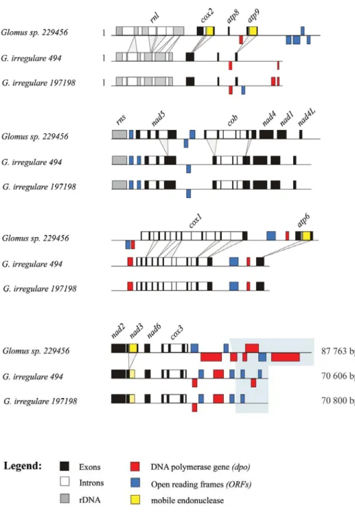
rnl. ... 58 Figure 3.2. Comparative view of the three mitochondrial genomes linear map where the exons
(black), introns (white), rDNA (gray), dpo plasmid insertions (red), ORFs (blue) and mobile endonuclease (yellow) are represented. ... 59 Figure 3.3. Schematic alignment representation of two mitochondrial intergenic regions (rnl-cox2
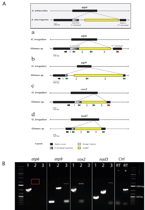
and cox3-nad6) showing the presence of numerous insertions and deletions (indels) ... 62 Figure 3.4. Comparison of gene hybrids found in atp6, atp9, cox2 and nad3. ... 67 Figure 3.5. Native and inserted C-terminals unrooted maximum likelihood phylogenetic trees. .. 71 Figure 4.1. A, Schematic representation of the two mtDNA contigs obtained after 454 reads
assembly of Rhizophagus sp. DAOM 213198. ... 86 Figure 4.2. mtDNA circular maps of Rhizophagus sp. DAOM 213198. ... 87 Figure 4.3. Hypothetical pathway of mtDNA conversion in Rhizophagus sp. DAOM 213198. ... 89 Figure 4.4. Linear representation of mtDNAs of R. irregularis DAOM 234179, DAOM 229456
and Rhizophagus sp. DAOM 213198 showing their genome synteny. ... 90 Figure 4.5. Multiple sequence alignment of the most common identified short inverted repeats
(SIRs). ... 92 Figure 5.1. Comparison of the mitogenomes of the Glomus aggregatum isolate (DAOM240163)
and the Rhizophagus irregularis isolate (DAOM240159). ... 108 Table 5.1. Arbuscular mycorrhizal fungal isolates used in our mitogenomic analyses. ... 109 Table 5.2. Sequence comparison of intergenic regions between R. irregularis 197198 and G
Figure 5.2. Difference in log-likelihood measured via SH test by comparing phylogenetic topologies inferred from single mt genes relative to that of the ‘‘supergene’’ set. ... 111 Figure 5.3. Topologies inferred from supergene set and concatenated set of rnl and cox1
revealing their high concordance in addition to topologies inferred from nad6 and cob as an example of individual genes with significant inconcordance to supergene topology. ... 113 Figure 5.4. PCoA plot based on the distance matrix obtained from mitochondrial genomic
alignments. ... 117 Figure 5.5. Complete mtDNA alignment of R. irreglaris isolates and G. aggregatum showing
sequence variations in intergenic regions, while coding genes are almost identical. ... 119 Figure 5.6. Linear genome representations of some mt genes, harbouring the relative
recombinations within the Rhizophagus sp. complex to compare with R. irregularis. ... 121 Figure 6.1. Isolate-specific mtDNA markers. ... 131 Figure 6.2. Self-fusion between spore clusters belonging to Rhizophagus irregularis DAOM-4240415. ... 134 Figure 6.3. Diagram presentation of the experimental set up for the study of anastomosis between
geographically distinct isolates of Rhizophagus irregularis in the symbiotic mycelium. ... 135 Figure 6.4. Variability in spore morphology of Rhizophagus irregularis at the interaction zone in
the combination DAOM197198/DAOM240415. ... 143 Figure 6.5. Aborted-like structures produced by the Rhizophagus irregularis isolates at the
interaction zone. ... 144 Figure 6.6. Gel electrophoresis showing patterns of mtDNA genotyping of ten progeny spores
(S1–S10) of three combinations using three Rhizophagus irregularis isolates DAOM-197198 (A) DAOM-240415 (B) and DAOM-234328 (C). ... 145 Supplementary Figure S2.1. Comparison of the Helicosporidium sp. (Hs), Trichoplax adhaerens
(Ta), Gigaspora rosea (Gr), Isoetes engelmannii (Ie) and Selaginella moellendorffii (Sm) cox1 group I intron trans-splicing positions represented on the Gigaspora rosea cox1 protein sequence. ... 171 Supplementary Figure S2.2. Predicted UUG translation initiation in Gigaspora and Mortierella. ... 172 Figure S3.1. Multiple DNA sequence alignment of numerous AMF representatives of the ... 175 atp6 native C-terminals along with the Glomus sp. 229456 putative foreign inserted C*- ... 175
terminal. ... 175 Figure S3.2. Multiple DNA sequence alignment of numerous AMF representatives of the atp9
native C-terminals along with the Glomus sp. 229456 putative foreign inserted C*- terminal. ... 176 Figure S3.3. Multiple DNA sequence alignment of numerous AMF representatives of the cox2
native C-terminals along with the Glomus sp. 229456 putative foreign inserted C*- terminal. ... 177 Figure S3.4. Multiple DNA sequence alignment of numerous AMF representatives of the nad3
native C-terminals along with the Glomus sp. 229456 putative foreign inserted C*- terminal ... 178 Fig. S4.1. Morphological description of spores of Rhizophagus sp. DAOM 213198 spores under
in vivo (a. b and c) and in vitro (e. f. g and h) culture conditions. ... 180 Fig. S4.2. mtDNA comparative analysis between R. irregularis DAOM 234179 and Rhizophagus
sp. DAOM 213198 isolates. ... 182 Fig. S4.3. Alignment of potentially conserved dpo-like translated amino acid sequences from R.
irregularis DAOM 234179 and Rhizophagus sp. DAOM 213198. ... 183 Fig. S4.4. Multiple sequence alignment of less frequent short inverted repeats. ... 184 Fig. S4.5. Hypothetical pathway of mtDNA inheritance and dynamics in Rhizophagus sp. DAOM
213198. ... 185 Figure S5.1. Spore germination of R. irregularis through the subtending hyphae. ... 187 Figure S5.2. Polymerase chain reaction banding patterns of the progenies coming from different
List of abbreviations
~: approximatelyAM: arbuscular mycorrhiza
AMF: arbuscular mycorrhizal fungi
RNA: acide ribonucléique mRNA: ARN messager ATP: adenosine triphosphate
atp: ATP synthetase
BiP: binding protein
BLAST: basic local alignmentBLAST: basic local alignment tool Bp: base pair(s)
cDNA: complementary deoxyribonucleic acid cob: apocytochrome b
cox: cytochrome oxidase C*-terminal: carboxy-terminal
DAOM: agriculture and agri-Food Canada national mycological herbarium DGGE: denaturing gradient gel electrophoresis
DNA: deoxyribonucleic acid
dNTP: deoxyribonucleoside triphosphate dpo: DNA polymerase
e.g.: Latin: exempli gratia (English: for example) HMM: Hidden Markov Model
i.e.: Latin: id est (English: that is) in vitro: Latin: in glass
in vivo: Latin: within the living organism ITS: internal transcribed spacer
Kb: kilo base pair(s) LSU: large subunit μl: micro liter μM: micro molar mM: mili molar
ML: Maximum likelihood
mRNA: messenger RNA
mt: mitochondrial MM: minimal medium
MUCL: mycothèque de l'universite catholique de Louvain nad: NADH dehydrogenase
NGS: next generation sequencing
NCBI: the national center for biotechnology information
N*-terminal: amino-terminal
NSERC: natural sciences and engineering research council (Canada) Numts: nuclear mitochondrial DNA
Orf: open reading frames
PCR: polymerase chain reaction
PCG: protein coding gene
RAPD: Randomly Amplified Polymorphic rDNA: ribosomal deoxyribonucleic acid
RFLP: restriction fragment length polymorphism
Ri T-DNA: transfer DNA transmission from Agrobacterium root-inducing plasmid into the genome of Dacus carota
rnl: large ribosomal RNA rns: small ribosomal RNA rpm: revolutions per minute rpo: RNA polymerase gene rps: ribosomal protein subunit Rpo: RNA polymerase
rRNA: ribosomal ribonucleic acid RNA: ribonucleic acid
ROC: root organ culture
RT-PCR: real-time polymerase chain reaction SSU: small subunit
Spp.: species
Taq: Thermus aquaticus
TGGE: temperature gradient gel electrophoresis Tm: melting temperature
tRNA: transfer ribonucleic acid
TRFLP: terminal restriction fragment length polymorphism UV: ultraviolet
v/v: volume/volume w/v: weight/volume
This thesis is dedicated to my mother and father, who had the patience to be far from me to complete this research in past five years. Thank you and I love you.
Acknowledgments
“And still, after all this time, the Sun has never said to the Earth, "You owe me."
Look what happens with love like that. It lights up the sky.”
― Hafez
It is my pleasure to thank the many people who made this research possible, first and foremost my supervisor Dr. Mohamed Hijri. I wish to express my most sincere appreciation to his nice attitude and kindness. His smiley face always full me up with energy for continueing my research. I was not only learned scientific knowledge from him but also how patience and calmness can be as effective as anything else. I would like to express very special gratitude to Dr. Franz Lang for transferring his worthy knowledge to me in bioinformatic analyses. Wiothout his assistance I would not be able to persue my research in this field. My appreciation also goes to my husband, Amir Yadghar, my beloved parents, Hamid and Ensieh, my sister, Fatima and my dear parents-in-law, Ali and Elaheh for their love, understanding, patience, and calmness that are the most effective support.
I wish to express my sincere appreciation to knowledgeable postdoctoral fellows Dr. Franck Stefani and Cristina Micali for their support and valuable advices. The work could also not have been completed without the aid of my colleague Dr. Eva Boon, Dr. Denis Beaudet, Dr. Sebastien Halary, Dr. Ivan de la Providencia, Laurence Daubois, Alice Roy-Bolduc, Guillaume Bourdel, dear Stephanie Berthiaume, Iffis Bachir, and Rim Ben Haj Sassi. I would like to thank Dr. Firas Bou Daher for his special supports. I would also acknowledge Dr. Anne Bruneau and Dr. Simon Joly and Dr. Sebastien Renaut and Edeline Gagnon for their assistance in phylogenetic analyses and Dr. Marc St. Arnaud and Dr. Mario Cappadocia for their beneficial advises in my predoctoral
exam. I wish to express my last, but not least, wholehearted thanks to my lovely friends for their motivation, encouragement and support. Finally, I am the most grateful person in the world by having the sweetest son, Ali, during my Ph.D. I am sorry that I did not have enough time for him and I wish he would excuse me.
Chapter 1
- General introduction
_________________________________
1.1 Mycorrhiza
Exploring the plants' rhizosphere reveals that it acts such as a factory and microorganisms and plants activities produce the outcome of this natural factory which is essential to soil, plant and ecosystem health. ‘Mycorrhiza' (Greek word means ‘fungus-root') used for the first time by Franck (1885) to describe the symbiotic association between fungi and root of trees. This association was dated back 400 million years ago based on the fossils records (Remy et al. 1994). Mycorrhizas are the most important and widespread symbiosis between plants and fungi on earth. This mutualistic association provides the fungus with carbohydrates, in a form that they can easily translocate and absorb. In return, fungal mycelia extend out of the roots into the soil where they uptake mineral nutrients and water that will be delivered to plants' root. As a consequence, mycorrhizal fungi play a crucial role in plant nutrient uptake, water relations, ecosystem establishment, plant productivity and diversity. Up to date, seven major important groups of mycorrhizae have been characterized and described, (Peterson et al. 2004) as; (1) arbuscular mycorrhiza, (2) ectomycorrhiza, (3) ectendomycorrhiza, (4) arbutoid mycorrhiza, (5) monotropoid mycorrhiza, (6) ericoid mycorrhiza and (7) orchid mycorrhiza. Different groups of mycorrhizas not only diverge in structural features such as intracellular structure (coil in Arbuscular, ericoid and arbutoid mycorrhizas; peloton in orchid mycorrhiza, fungal peg in monotropoid mycorrhiza and arbuscule in arbuscular mycorrhiza) but also in their preferred host plants and ecosystems which outcome their distribution (Smith and Read 2008). Arbuscular
mycorrhiza is the most important group of mycorrhizae, which is widespread that is found in all ecosystems and concerns more than 80% of plants. Arbuscular mycorrhizal fungi (AMF), is an early divergent fungal lineage that can form mycorrhiza with most important crops in which they improve growth and productivity. Therefore, they were largely used as an alternative to hazardous mechanisms of agriculture and chemical fertilizers (biofertilizers), substituting the development and maintenance of healthy ecosystems. Nowadays, many commercial inoculants of AMF with a wide range of formulations are available in the market.
1.2 Arbuscular mycorrhizal fungi
Arbuscular mycorrhizal fungi (AMF) are a group of high potential microorganisms for agriculture that belong to the ancient phylum Glomeromycota (Schüβler et al. 2001). They are considered as living fossils since they are found to be 460 million years old (Redecker et al. 2000). AMF contributed directly to the evolution and survival of plant species, to the expansion of biodiversity in the earth and consequently to the equilibrium of ecosystems. These microorganisms are ubiquitous putative mutualistic fungi that can form symbiotic associations with the majority of vascular plants (Parniske 2008). However, non-mutualistic or exploitive (i.e. an association in which only the plant profits from the nutrient exchange, (Taylor and Bruns 1999) is a challenging term for mycorrhizal fungi in association with myco-heterotrophic plants that are completely supported by a fungus (Imhof 1999 and Brundrett 2004). The mycelium of these coenocytic (i.e. hyphae that are lacking septa) root symbionts acts as root extension for plants improving the use of soil water and soil minerals from a much larger volume of soil. Improvement in nutrient uptake enhances plant growth and health (Smith 1997) lead to resistance
and controlling the environmental stresses (e.g. drought, salinity and pollution) and plant pathogens (Barea et al. 2002). Moreover, AMF stimulate the fitness of plants in polluted environments (Hildebrandt et al. 1999) and affect soil structure by improving soil quality (Barea et al. 2002). AMF also raises resistance to biotic and abiotic stresses, representing a relevant alternative for sustainable agriculture (Subramanian and Charest 1999, Aliasgharzad et al. 2006). These fungi are declared to be obligate biotrophs, which means they are not able to complete their life cycle and cultivate without a host plant. However, Hildebrandt and colleagues questioned this putative virtue of AMF by finding G. intraradices (G. irregulare) capable of completing its life cycle in the presence of Paenibacillus validus in the absence of a plant root (Hildebrandt et al. 2006). AMF grow a hyphal network both within the root cortex (linear and coiling; (Smith 1997)) and the adjacent soil. The physical properties of the mycelium (a high surface-to-volume ratio), enhance uptake of nutrients more than the plant's root system by exploring a larger volume of soil (Bolan 1991, Tuomi et al. 2001). AMF were previously considered as asexual (clonal) organisms (Gandolfi et al. 2003). However, increasing number of evidences such as detection of genetic recombination, the presence of ‘core meiotic genes' and retrotransposons (Halary et al. 2013), questioned this phenomenon. These fungi form spores containing hundreds of nuclei that are most likely haploid (i.e., one set of chromosome in a single nucleus) (Hijri and Sanders 2004). Analyses of Kuhn et al. (2001) by fluorescence in situ hybridization (FISH) revealed two highly variable variants of internal transcribed spacer 2 (ITS2) in different nuclei of Scutellospora castanea, suggesting AMF heterokaryosis. Pawlowska and Taylor (2004) believed this fungus to be homokaryotic because of the analysis of POL-like sequences from Glomus etunicatum, which show that all sequence variants were present in offsprings. However, Sanders and Croll (2010) explained the results of Pawlowska and Taylor (2004) in a review, and concluded that AMF are most likely heterokaryotic. Hijri & Sanders
(2005) and Boon et al. (2015) support the heterokaryosis hypothesis in AMF by reanalyzing POL-like genetic sequence variations in G. etunicatum (and R. irregularis DAOM 197198). It is important to note that AMF do not seem to go through a bottleneck of genetic variation (Marleau et al. 2011); in other words, no observations of a single nucleus stage have been reported in AMF life history. The lack of a genetic bottleneck is considered as a potential source for AMF extreme intra-isolate genetic diversity (Kuhn et al. 2001). However, Lin et al. (2015) sequenced the genomes of individual nuclei of R. irregularis DAOM 197198 and they found a low level of polymorphism among nuclei consistent with homokaryotic hypothesis. Likewise, homokaryosis has been again challenged by Beaudet et al. (2015) who demonstrated that Next Generation Sequencing underestimates intra-isolate polymorphism that is a strong bias. Overall, some recent publications support the hetrokaryosis hypothesis (Boon et al. 2015; Beaudet et al. 2015).
1.3 AMF biodiversity and interaction with plants
Primarily, observation of about 225,000 terrestrial plant species colonized by approximately 160 species of AMF (Smith 1997) suggested AMF low host specificity. In contrary, some researchers have claimed a high rate of host specificity and local diversity of certain taxa (Yang et al. 2012). If AMF possesses low host specificity, the researchers speculate, the fungi would distribute widely although most AMF have a restricted geographical distribution, which suggests host specificity (Yang et al. 2012). This speculation would indicate that the effects of ecosystems (biogeographical territories and climatic zones) on AMF distribution might be mediated by plants through host selectivity and preference. It has been reported by Alkan et al. (2006) that the interface among several AMF and host plant roots would offer multiple benefits to the host plant
in comparison to a host plant colonized by a single AMF species. Concisely, different AMF species might affect plant growth and biodiversity in different ways (Ravnskov and Jakobsen 1995) and possess a defined role in the community structure (Alkan et al. 2006). Thus, appropriate management of mycorrhizae in agriculture should result in a considerable drop in harmful chemical usage and also production expenses. The first step toward AMF application in sustainable agriculture and industrial treatment is a development of precise identification and quantification procedures.
1.4 AMF identification
Arbuscular mycorrhizal fungi (AMF) have traditionally been identified by various taxonomic characters including spore size, colour, sporocarp structure (spore ontogeny, Franke and Morton 1994) and hyphal attachment morphology. Furthermore, the intraradical structures allow identification to the family level the absence of spores (Clapp et al. 1995). In other words in the absence of sexual reproduction, AMF were characterised mainly by morphological criteria. However, there are several limitations for morphological identification and characterisation. For example, AMF sporulation is influenced by ecological conditions and physiological parameters. Several characteristics such as immaturity, degradation, or infection by parasites and chemical reactions might influence spore shapes (Redecker 2002). Meanwhile, similarity of AMF intraradical structures, obscuring usage of the light microscopy method for simultaneously detecting of more than one AMF species in a common root fragment (Alkan et al. 2006). Thus, neither morphology nor ultrastructure characters is sufficient to statistically support phylogenetic inferences for identification and quantification of AMF. Therefore, molecular techniques are
powerful approaches that complement and overcome limitations of morphological-based identification, detection and quantification of AMF providing information on the biodiversity, community structure, and function of AMF in an ecosystem. Many investigations have attempted to develop molecular markers based on nuclear genes in AMF. The high sequence variation within the nuclear genome of AMF (heterogeneity) with high intra-isolate genetic diversity (Kuhn et al. 2001) encountered researchers to some errors and complexities in identification. Existence of multiple sequences within a single AMF isolates or even single spores (Clapp et al. 1995, Sanders et al. 1995), causes overestimation of the number of species making an ambiguity in population analyses interpretation. An ideal molecular marker should be present in a broad range of organisms from the target taxon and easily amplifiable by PCR. The marker should also comprise highly conserved as well as variable regions so that it can be used for phylum to species or even to isolate identification. In AMF, nuclear ribosomal DNA such as the internal transcribed spacer (ITS) of rDNA region including the 5.8S rRNA gene (White et al. 1990, Sanders et al. 1995, Wubet et al. 2003, Hempel et al. 2007), small subunit (SSU) rRNA gene (Wubet et al. 2003, Helgason et al. 1999, Lee et al. 2008) and the large subunit (LSU) rRNA gene (Gollotte et al. 2004, Pivato et al. 2007, Rosendahl et al. 2009, Stockinger et al. 2009) are the most commonly regions used for marker development. Some attempts have been made to develop markers in functional genes such as actin and elongation factor 1-α (Helgason et al. 2003, Sokolski et al. 2010), β-tubulin (Msiska and Morton 2009), phosphate transporter genes (Sokolski et al. 2011), H+-ATPases (Requena et al. 2003). Although the molecular data have changed the systematic by adding several new genera and families (Walker et al. 2004, Walker et al. 2007) there is still a long way to make these approaches trustworthy. For example, although the SSU rDNA region provides the largest taxon sampling for AMF, allowing phylogenetic resolution no more than genus level (Walker et al. 2007). Stockinger et al. (2009) used a combination of SSU-ITS-LSU
rDNA amplicon and resolved AMF phylogenetic down to species' level. Single-copy genes are another type of sequences that have been studied so far to elucidate evolutionary history of AMF (Helgason et al. 2003, Stukenbrock and Rosendahl 2005, Msiska and Morton 2009, Sokolski et al. 2010). Single-copy genes have the advantage that any sequence variation within a single spore can be recognized as a variation among nuclei (Helgason et al. 2003). However, a weak point of analyzing a single gene for evolutionary analyses is that a single-gene tree may not reflect the entire organisms' evolutionary history. In fact, the evolutionary history and divergence time of different segments of the genome may differ from each other and thus, their separate histories do not reflect the entire organisms' evolutionary history (species tree) (Aguilera et al. 2008).
1.5 Anastomosis in AMF
Hyphal fusion (anastomosis) is a phenomenon that results in the cytoplasmic and genetic connection between the same or genetically divergent individuals and is ubiquitous in many filamentous fungi (reviewed in (Saupe 2000, Esser 2006). This phenomenon has crucial functions specifically in the absence of the assumed sexual AMF mode of reproduction. Anastomosis has a major effect in extending the sphere of the underground network among different host plants. It is believed that fitness of AMF and consequently host plants can be enhanced via migration of nutrient, cytoplasm, organelles and even nuclei especially under stressed conditions. Although the host-plant communities influence the frequency of AMF hyphal fusion, their genome similarity is another important prerequisite for anastomosis occurrence (Giovannetti et al. 2001). It is still unknown how the process of hyphal fusion is regulated in AMF. Another important issue is to comprehend the genetic distance required prohibiting the formation of an anastomosis
between two individual AMF, which might lead to a definition for AMF species. The self-recognition and non-self self-recognition during hyphal fusion were first reported by Giovannetti et al. (1999) in six different species of AMF. Hyphal fusion was observed within the same germline (i.e. a single germinated spore) and between different germlines belonging to the same Glomus species (Giovannetti et al. 1999). The authors also detected the exchange of nuclei in the middle of hyphal bridges connecting mycelia (anastomosis) from either the same or different plant species. They introduced anastomosis as a mechanism for maintaining the genetic diversity in the absence of sexual recombination (Giovannetti et al. 2001). The authors performed another test for vegetative compatibility to study the hyphal interactions between six isolates of G. mosseae sampled from various worldwide ecosystems and they observed incompatibility responses among them (Giovannetti et al. 2003). However, based on a new report from Croll et al. (2009a) hyphal fusion (anastomosis) can occur even between genetically distinct isolates of Glomus irregulare but with lower rates in comparison with genetically similar isolates. This event causes the creation of more genetically diverse networks via nuclei exchange. The authors analyzed the progenies by genetic markers and reported the evidence for biparental inheritance. Sets of experiments have been also conducted by Angelard et al. (2010), which declared both genetic exchange and random distribution (segregation) of nuclei in offspring affecting their phenotypes, plant symbiosis and productivity. The most important consequence of non-self fusion is the formation of heterokaryon, which is in agreement with the hypothesis of heterokaryosis vs. homokaryosis. It seems that in contrast to what is prominent about the fitness of organisms in favour of homokaryosis (Ballard and James 2004) (with a good match between nucleus and mitochondria), mito-nuclear interaction in AMF is under different mechanisms that needs to be explored.
1.6 Mitochondrial DNA and its evolution
Mitochondrial DNA (mtDNA) as it stands from the term, is the DNA located in organelle called mitochondria. mtDNA contains set of genes essential for normal mitochondrial function that is conversion of food chemical energy into a form that cells can consume (adenosine triphosphate, ATP) via oxidative phosphorylation pathway. The current most popular theory about the origin and evolution of mitochondria (endosymbiont theory) suggests that the origin of nuclear genome of eukaryotic cells was occurred in parallel to the origin of mitochondrial genome (Gray et al. 1999, Lang et al. 1999). This is one of the reasons why mtDNA has been studied extensively in many diverse eukaryotic lineages and evolutionary analysis. Mitochondrial genomes are present in multiple copies within a cell that results in better yield of amplification-based methods (e.g., PCR) compared to single nuclear genes. mtDNA was thought to be fully inherited from maternal lines (clonal), and so there is usually no or minor change in mtDNA from parent to offspring with low chance of recombination (Birky 2001). Uniparental inheritance of mitochondria is the main support of the homoplasmy (i.e. all the mitochondrial genomes in the cell are essentially identical), which drastically decreases the chance of recombination (Lynch 1996). However, clonal and uniparental inheritance have been questioned and rejected in some organisms such as mussel from bivalve family (Breton et al. 2007). Beside this phenomenon, mtDNA higher rate of mutation to compare with nuclear DNA (because of biochemical processes of mitochondrion itself, lack of histones in DNA structure, and low efficiency of the DNA repair system (Rand 2001)), suggested this genomic pool as a potential candidate tool for evolutionary analysis. mtDNA elevated mutation rate made it highly variable in natural population offering a good tracer for population history over a short period of time. It has also been thought that the
mutation rate in mtDNA was constant, and mtDNA is almost neutral which both have been questioned by collection of recent data (Galtier et al. 2009). Therefore, caution should be taken, and mtDNA should be studied in a variety of organisms to upgrade the understanding of its evolutionary constraints.
1.7 Mitochondrial inheritance in AMF
Mechanisms of mitochondrial inheritance in AMF remains to be demonstrated. So far, the putative homoplasmy reported for the mitochondrial genome of Rhizophagus irregularis could be explained by four hypotheses: (1) Vegetative incompatibility among genetically different individuals prevents co-inheritance of divergent mtDNA haplotypes in the progeny. (2) The exchange of both mitochondrial haplotypes occurs, but homoplasmy is reestablished in subsequent generations. (3) Homoplasmy existed only in AMF in vitro cultures because of a homogenized condition, and it does not exist in the nature. (4) Based on a hint by Barr et al. (2005), heteroplasmy might rarely or only transiently occur, leading to recombination and removal of some mutations. After all, in case of production of mitochondrion harboring incomplete genome, mitochondrial autophagy (mitophagy), results in a selective degradation of damaged mitochondria in cells. Therefore, studying the inheritance pattern, maintenance, and stability of mtDNA in AMF help to address these important biological questions and facilitate the development of mitochondrial markers as a promising choice for identification purposes.
1.7 Mitochondrial inheritance in AMF
In order to circumvent depletions and the probable complexity of nuclear genes for AMF identification, studies of an independent genomic pool of mtDNA have noted several advantages (Moritz et al. 1988, Raab et al. 2005, Borstler et al. 2008, Lang and Hijri 2009, Lee and Young 2009). Conversely, the use of such genomic information is not without pitfalls because we have little understanding of selective forces that cause mitochondrial gene loss and reshuffling. Bruns et al. (1989) found that the gene for the large subunit of the mitochondrial ribosomal RNA (rnl gene) is useful for molecular identification of the ectomycorrhizal fungi. It has been reported that the rnl gene has a very low degree of intra-isolate genetic variation in the AMF G. intraradices (G. irregulare) and G. proliferum (Raab et al. 2005). However, sequences of this region were found to be polymorphic among isolates of these species (Raab et al. 2005), and sequence variation is more substantial (Borstler et al. 2008). It has also been reported that both exons and introns of the rnl offered opportunities to develop markers for discriminating haplotypes of G. intraradices (G. irregulare). Subsequently, investigating and mapping of several mitochondrial genomes from R. irregularis species proposed homoplasmy in AMF offering mitochondrial genome as a promising choice for species identification (Lee and Young 2009, Formey et al. 2012). Rapid genetic segregation (i.e. random distribution of mtDNA in the offsprings) (Lee and Young 2009) and/or an active process of transmitting homogenous mtDNA to descendants (Ling and Shibata 2004) are suggested to be the main reasons for lack of polymorphism in AMF mtDNA. Thus, the mitochondrial genome of AMF has a yet-to-be-explored potential for understanding the evolution of this important fungal group and designing molecular tools for further characterization in natural populations.
1.9 Research objectives, hypotheses and thesis presentation
When I started my Ph.D. project on AMF, little was known about their mtDNA structure, characteristics, evolutionary constraints and signals. There were only one published mitogenome (Glomus intraradices isolate#494, Lee and Young, 2009) paper and few studies based on ribosomal large subunit (mtLSU) (Raab et al. 2005; Borstler et al. 2008)! Considering the difficulties and complexities of AMF identification required for their application, many questions were raised regarding the sue of mtDNA for the development of molecular toolkits for identification, quantification in order to investigate evolution, function, population genetics and ecology of AMF. In this context, the general objective of my Ph.D. project was to investigate mitochondrial genome diversity and their evolutionary mechanisms among representative glomeromycotan taxa using next-generation sequencing, bioinformatics and molecular biology approaches.
The specific objectives are:
1. To investigate mitochondrial genome evolution using comparative mitogenomic analyses of closely related species and isolates as well as phylogenetically distant taxa of AMF 2. To assess mitogenomes and mt genes for marker development and phylogenetic analyses. 3. To explore mitogenomes, inheritance among compatible isolates of the model AMF
Rhizophagus irregularis through anastomosis formation
I have addressed the following scientific questions:
i. What is the mt genome structure, gene synteny in Gigaspora rosea, a phylogentetically distant AMF taxon compared to R. irregularis DAOM 197198?
iii. Is mt gene synteny conserved among taxa of the genus Rhizophagus?
iv. Can mtDNA be used for development of molecular toolkits and for phylogenetic analysis in AMF?
v. Do the mt genes possess the same evolutionary signal?
vi. Are mtDNA haplotypes from two crossed isolates of R. irregularis inherited to the progeny after anastomosis?
vii. Does heteroplasmy as an outcome of anastomosis make drawbacks in utilization of mtDNA for phylogenetic and identification analyses?
I tested the following hypotheses in my Ph.D. project: a. mtDNA evolves rapidly in AMF.
b. mtDNA is highly polymorphic among isolates of a given species. c. mtDNA is suitable for developing isolate specific markers.
d. mt genes are not possessing the same evolutionary signal as it was thought and so some mt genes should be targeted for facilitating phylogenetic analyses.
e. mtDNAs are exchanged via anastomosis but there are some unknown mechanisms (e.g., segregation and selection) which act in favor of homoplasmy.
Through the first investigation (2nd chapter), mtDNA of Gigaspora rosea DAOM 194757 has been sequenced revealing relatively large genome size (97,349 bp) comparing other sequenced Glomus irregulare who's name has been changed to Rhizophagus irregularis, during my Ph.D. project. We also annotated unorthodox fragmented genes (rns and cox1) in the genome of Gigaspora rosea.
The 3rd chapter of my thesis (second article) consisted of sequencing, assembling and analyzing mtDNA of Rhizophagus sp. DAOM 229456. Comparative mitogenomics on the
mitochondrial genome of Rhizophagus sp. DAOM 229456 revealed the first evidence of AMF interspecific exchange of mitochondrial coding sequences resulting in formation of hybrid genes in atp6, atp9 (coding for the subunit 6 and 9 of the ATP synthase complex), cox2 (cytochrome C oxidase subunit 2) and nad3 (NADH dehydrogenase subunit 3) genes.
MtDNA sequencing and mapping of Rhizophagus sp. DAOM 213198 also reveal an a novel mtDNA structure that has never been reported in fungi. This peculiar organization has been characterized in the 4th chapter (third article). Further quantification approaches have been conducted in order to measure mitochondrial genomes copy number. Comparative mitogenomics analyses were also applied to trace the probable origins and causes of this novel mtDNA structure formation in Rhizophagus sp. DAOM 213198.
Finally in 5th chapter (fourth article), I evaluated the power of individual mt genes in phylogenetic analyses of AMF and revealing their evolutionary relationship. Mitochondrial genes evolutionary signals have been compared to phylogenetic signal of "supergene" set signal in order to explore the best-performing genes. Mitogenomics comparative analysis also has been implemented to identify uncertain position isolate of AMF and to reveal the evolutionary history among closely related isolates.
The 6th chapter (fifth article) of my thesis represents the investigation of heteroplasmy status within genetically divergent isolates of R. irregularis in case of anastomosis occurrence. We performed three crossing combinations both in pre-symbiotic and symbiotic phases. Progeny spores per each crossing combination were genotyped using isolate-specific markers that have been developed in the mitochondrial genome. Genotyping patterns of individual spores from the progenies revealed the fate of the two parental mtDNA haplotypes. Further germination of some progenies and the genotyping pattern of them also gave us a speculation about the fate of mtDNA in next generation and persistence of our result.
My thesis is presented under the scientific article form for the Doctorate in Biological Science Program at the Université de Montreal. Chapter 1 of the thesis (Introduction and Literature review) introduces the current knowledge about the arbuscular mycorrhizal fungal biology, genetics and genomics as well as mitochondrial genome evolution. The personal experiments, methods and results are then introduced in the chapters 2, 3, 4, 5 and 6 in four published articles and one submitted manuscript. Chapter 7, the last section of the thesis, serves as a general discussion and conclusion of all the obtained results following by the perspectives of this project.
Presentation of article 1
In 1990, arbuscular mycorrhizal fungi were organized in order "Glomales" containing three families (Acaulosporaceae, Gigasporaceae, and Glomeraceae) and six genera (Acaulospora, Entrophospora, Gigaspora, Glomus, Sclerocystis, and Scutellospora) within phylum Zygomycota (Morton 1990). However, the authors have not implemented molecular aspects and evidences such as their symbiotic habit, lack of zygospores that suggest AMF form a monophyletic group distinct from other Zygomycotan lineages (Schüβler et al. 2001). Based on these evidences, the phylum Glomeromycota has been proposed by Schüßler et al. (2001). Phylogenetic analyses based on 18S rDNA revealed that Glomeromycota are the sister group to Ascomycota and Basidiomycota (Schüßler et al. 2001). Advances made in fungal genome sequencing last decade, allowed generating a huge datasets in fungal mtDNAs such as many mtDNA of the Dikarya (Basidiomycota and Ascomycota) as well as basal fungal lineages Chitridiomycota, Zygomycota and Glomeromycota. Publication of the first mitochondrial genome of AMF species by Lee and young in 2009 opens the avenue through mitochondrial assessment of AMF (Lee and Young 2009). The increasing number of sequenced mtDNAs in fungal kingdom offers opportunities to infer robust phylogenies using phylogenomics approaches. Sequencing of an AMF member relatively distinct from the available sequenced mtDNAs (Rhizophagus and Glomus members) would enhance our knowledge of AMF evolutionary history.
In this article, we report the complete mtDNA sequence of Gigaspora rosea, which encodes two fragmented genes, transcripts of one of which undergo group I intron–mediated trans-splicing. We further report the results of a phylogenetic analysis of mitochondrial proteins that allows more confident positioning of Glomeromycota within Fungi.
This article was published in Molecular Biology and Evolution. I have contributed significantly to data analyses using bioinformatics in particular analyses of detailed fragmentation annotation (via generating secondary structure) for G. rosea. I have also contributed together with D. Beaudet and L. Forget to RNA analyses using molecular biology methods. I have written some sections of this article.
Chapter 2
- Group I intron–mediated trans-splicing in
mitochondria of Gigaspora rosea and a robust phylogenetic
affiliation of arbuscular mycorrhizal fungi with
Mortierellales
Maryam Nadimi
*1, Denis Beaudet
*1, Lise Forget
2, Mohamed Hijri
1, and B.
Franz Lang
+21 Université de Montréal, Département de sciences biologiques, Institut de recherche en
biologie végétale (IRBV), 4101 rue Sherbrooke Est, Montréal, QC, H1X 2B2, Canada.
2 Département de Biochimie, Centre Robert-Cedergren, Université de Montréal, Québec,
Canada
*
These authors contributed equally to this work.Published in : Molecular Biology and Evolution (MBE), March 12, 2012. 29(9): 2199-210, doi: 10.1093/molbev/mss088.
2.1 Abstract
Gigaspora rosea is a member of the arbuscular mycorrhizal fungi (AMF; Glomeromycota) and a distant relative of Glomus species that are beneficial to plant growth. To allow for a better understanding of Glomeromycota, we have sequenced the mitochondrial DNA of G. rosea. A comparison with Glomus mitochondrial genomes reveals that Glomeromycota undergo insertion and loss of mitochondrial plasmid-related sequences and exhibit considerable variation in introns. The gene order between the two species is almost completely reshuffled. Furthermore, Gigaspora has fragmented cox1 and rns genes, and an unorthodox initiator tRNA that is tailored to decoding frequent UUG initiation codons. For the fragmented cox1 gene, we provide evidence that its RNA is joined via group I-mediated trans-splicing, whereas rns RNA remains in pieces. According to our model, the two cox1 precursor RNA pieces are brought together by flanking cox1 exon sequences that form a group I intron structure, potentially in conjunction with the nad5 intron 3 sequence. Finally, we present analyses that address the controversial phylogenetic association of Glomeromycota within fungi. According to our results, Glomeromycota are not a separate group of paraphyletic zygomycetes but branch together with Mortierellales, potentially also Harpellales.
2.2 Key words
Arbuscular mycorrhizal fungi (AMF), mitochondrial genome, intron evolution, phylogeny, tRNA structure, genetic code.
2.3 Introduction
Arbuscular mycorrhizal fungi (AMF) is a group of ubiquitous soil-borne fungi that form symbiotic associations with the majority of vascular plants (Parniske 2008). AMF are obligate
biotrophs, that is, they are unable to grow without a host plant that provides them with carbohydrates; in turn, AMF transfer nutrients such as phosphate to the plant (reviewed in (Strack et al. 2003)). At the cellular level, AMF are characterized by the formation of large, multinucleate hyphae, and asexual spores (e.g., (Marleau et al. 2011)). Apparently, the genetic segregation of the hundreds of distinct nuclei that are present in these species does not follow canonical but rather population rules, and recent analyses demonstrate substantial sequence variation in certain nuclear genes (Hijri and Sanders 2005, Croll et al. 2009, Croll and Sanders 2009, Boon et al. 2010). It is therefore no surprise that the Glomus irregulare nuclear genome project has turned into a sequence assembly nightmare (Martin et al. 2008). Yet in stark contrast, the first complete Glomus mitochondrial DNAs (mtDNAs) that have been deciphered recently by 454 sequencing are homogeneous in sequence (Lee and Young 2009) and GenBank ′FJ648425; Bullerwell CE, Forget L, Lang BF, unpublished data), that is, genetic segregation of mtDNA is as effective in Glomus as in other fungi. In these two cases, long homopolymer stretches that introduce systematic pyrosequencing error are surprisingly absent. In other, more A+T-rich mtDNAs, however, we have observed intolerable levels of 454 sequence error (close to one per 1 kbp sequence on average in a heterolobosean amoeba; Bullerwell CE, Forget L, Lang BF, unpublished data) causing frameshifts in several protein-coding genes. It therefore remains advisable to carefully examine homopolymer-rich sequences for potential error, for instance by resequencing with Sanger technology.
The taxonomic and phylogenetic identity of AMF have been, and continues to be, controversial. Initially assigned to zygomycetes, a fungal taxon that is strongly suspected to be paraphyletic (e.g., (Schwarzott et al. 2001, Seif et al. 2006, Hibbett et al. 2007, Liu et al. 2009), AMF have been recently moved into a separate fungal phylum, Glomeromycota (Hibbett et al. 2007). Yet the underlying published phylogenies are controversial and often lack significant
statistical support, either due to a limited amount of sequence data (based on one or only few gene sequences), poor taxon sampling, or a combination of both. In many instances, analyses further suffer from potential phylogenetic artifacts such as long-branch attraction (e.g., Felsenstein 1978).
A phylogenetic data set of complete mtDNA sequences is currently restricted to two G. irregulare isolates (Lee and Young 2009 and GenBank ′FJ648425) having identical coding sequences. Likewise, a previous phylogenomic analysis with a large number of nuclear genes (Liu et al. 2009) had only limited taxon sampling. Accordingly, phylogenetic analyses with both mitochondrial and nuclear genome data have provided only a tentative answer to the question of where AMF belong within Fungi. In some cases, they both show a weak affinity of Mortierellales with Glomeromycota (Lee and Young 2009, Liu et al. 2009), unsupported however by strict statistical analysis (such as the AU test, Shimodaira 2002). An updated, comprehensive fungal phylogenomic analysis with nuclear sequence data published in 2011 (Ebersberger et al. 2012) nicely summarizes the confusing state of the art, commenting that ‘‘at the moment, available data do not allow to confidently attach glomeromycetes to the phylogenetic backbone of the fungi.’’ In the latter phylogenomic analysis, Mortierellales are shown separate from Mucorales, that is, excluding Mortierellales from a monophyletic taxon Mucoromycotina favored by others and in contradiction to conclusions reached in a previous phylogenomic analysis (Liu et al. 2009). Evidently, better taxon sampling of genomic data sets is required to resolve these questions, in particular by adding to both mitochondrial and nuclear gene data sets members of Mortierellales, and AMF lineages that are distant from Glomeraceae.
Our rationale for sequencing the Gigaspora rosea mtDNA is that Gigasporaceae are at a large evolutionary distance to Glomus species, with clearly distinct morphological characteristics. Gigasporaceae form auxiliary cells in the extraradical mycelium, and giant spores that are usually