Development and Characterization of Polymeric Nanoparticles
(NPs) made from Functionalized Poly (D,L- lactide) (PLA)
Polymers
par
Sherief Essa
Faculté de Pharmacie
Thèse présentée à la Faculté des études supérieures
en vue de l’obtention du grade de Philosophiae Doctor (Ph. D.)
en Sciences Pharmaceutiques
Option: Technologie Pharmaceutique
May 2011
© Sherief Essa, 2011
Université de Montréal Faculté des études supérieures
Cette thèse intitulée:
Development and Characterization of Polymeric Nanoparticles (NPs) made from Functionalized Poly (D,L-lactide) (PLA) Polymers
présentée par:
Sherief Essa
a été évaluée par un jury composé des personnes suivantes :
Dr. Suzanne Giasson, président-rapporteur Dr. Patrice Hildgen, directeur de recherche
Dr. François-Xavier Lacasse, membre du jury Dr. Pascal Janvier, examinateur externe
Résumé
Les nanoparticules polymériques biodégradable (NPs) sont apparues ces dernières années comme des systèmes prometteurs pour le ciblage et la libération contrôlée de médicaments. La première partie de cette étude visait à développer des NPs biodégradables préparées à partir de copolymères fonctionnalisés de l’acide lactique (poly (D,L)lactide ou PLA). Les polymères ont été étudiés comme systèmes de libération de médicaments dans le but d'améliorer les performances des NPs de PLA conventionnelles. L'effet de la fonctionnalisation du PLA par insertion de groupements chimiques dans la chaîne du polymère sur les propriétés physico-chimiques des NPs a été étudié. En outre, l'effet de l'architecture du polymère (mode d'organisation des chaînes de polymère dans le copolymère obtenu) sur divers aspects de l’administration de médicament a également été étudié. Pour atteindre ces objectifs, divers copolymères à base de PLA ont été synthétisés. Plus précisément il s’agit de 1) copolymères du poly (éthylène glycol) (PEG) greffées sur la chaîne de PLA à 2.5% et 7% mol. / mol. de monomères d'acide lactique (PEG2.5%-g-PLA et PEG7%-g-PLA, respectivement), 2) des groupements d’acide palmitique greffés sur le squelette de PLA à une densité de greffage de 2,5% (palmitique acid2.5%-g-PLA), 3) de copolymère « multibloc » de PLA et de PEG, (PLA-PEG-PLA)n. Dans la deuxième partie, l'effet des différentes densités de greffage sur les propriétés des NPs de PEG-g-PLA (propriétés physico-chimiques et biologiques) a été étudié pour déterminer la densité optimale de greffage PEG nécessaire pour développer la furtivité (« long circulating NPs »). Enfin, les copolymères de PLA fonctionnalisé avec du PEG ayant montré les résultats les plus satisfaisants en regard des divers aspects d’administration de médicaments, (tels que taille et de distribution de taille, charge de surface, chargement de drogue, libération contrôlée de médicaments) ont été sélectionnés pour l'encapsulation de l'itraconazole (ITZ). Le but est dans ce cas d’améliorer sa solubilité dans l'eau, sa biodisponibilité et donc son activité antifongique. Les NPs ont d'abord été préparées à partir de copolymères fonctionnalisés de PLA, puis ensuite analysés pour leurs paramètres physico-chimiques majeurs tels que l'efficacité d'encapsulation, la taille et distribution de taille, la charge de surface, les propriétés thermiques, la chimie de surface, le pourcentage de poly (alcool vinylique) (PVA) adsorbé à la surface, et le profil de libération de médicament. L'analyse de la chimie de surface par la spectroscopie de photoélectrons rayon X (XPS) et la microscopie à force
atomique (AFM) ont été utilisés pour étudier l'organisation des chaînes de copolymère dans la formulation des NPs. De manière générale, les copolymères de PLA fonctionnalisés avec le PEG ont montré une amélioration du comportement de libération de médicaments en termes de taille et distribution de taille étroite, d’amélioration de l'efficacité de chargement, de diminution de l'adsorption des protéines plasmatiques sur leurs surfaces, de diminution de l’internalisation par les cellules de type macrophages, et enfin une meilleure activité antifongique des NPs chargées avec ITZ. En ce qui concerne l'analyse de la chimie de surface, l'imagerie de phase en AFM et les résultats de l’XPS ont montré la possibilité de la présence de davantage de chaînes de PEG à la surface des NPs faites de PEG-g-PLA que de NPS faites à partie de (PLA-PEG-PLA)n. Nos résultats démontrent que les propriétés des NPs peuvent être modifiées à la fois par le choix approprié de la composition en polymère mais aussi par l'architecture de ceux-ci. Les résultats suggèrent également que les copolymères de PEG-g-PLA pourraient être utilisés efficacement pour préparer des transporteurs nanométriques améliorant les propriétés de certains médicaments, notamment la solubilité, la stabilité et la biodisponibilité.
Mots-clés: Acide poly(lactique), Architecture, PEG-PLA, Multi-bloc, Organisation de la chaîne, Spectroscopie de photoélectrons Rayon X (XPS), Microscopie à force atomique (AFM), Imagerie de phase, Faible solubilité.
Abstract
Biodegradable polymeric nanoparticles (NPs) have emerged as promising drug delivery carriers for the controlled drug release and targeting. The first part of this study aimed to develop biodegradable NPs from functionalized copolymers of poly (D,L-Lactide) (PLA). Those copolymers were explored as drug delivery systems in attempt to improve the drug delivery performance of conventional PLA NPs. The effect of PLA functionalization (insertion of chemical substituents onto PLA backbone) on the physicochemical properties of the obtained NPs was investigated. Moreover, the effect of polymer architecture (mode of organization of polymer chains in the resultant copolymer) on various drug delivery aspects was also studied. To reach those goals, various PLA based copolymers namely poly(ethylene glycol) (PEG) grafted on PLA backbone at 2.5% & 7% mol/mol of lactic acid monomers (PEG2.5%-g-PLA and PEG7%-g-PLA, respectively), palmitic acid grafted on PLA backbone at 2.5% grafting density (palmitic acid2.5%-g-PLA), and multiblock copolymer of PLA and PEG, (PLA-PEG-PLA)n were synthesized. In the second part, the effect of different PEG grafting densities over PLA backbone on the properties of PEG-g-PLA NPs either physicochemical or biological properties was investigated to reveal the optimal PEG grafting density required to develop stealth particles (long circulating NPs). Finally, functionalized PEG/PLA copolymers that showed the most satisfactory results in terms of various drug delivery aspects, such as size and size distribution, surface charge, drug loading, and controlled drug release were selected for encapsulation of itraconazole (ITZ) to improve its aqueous solubility, bioavailability and hence its antifungal activity. NPs were first prepared from functionalized PLA copolymers then analyzed for their major physicochemical parameters such as encapsulation efficiency, size and size distribution, surface charge, thermal properties, surface chemistry, % poly(vinyl alcohol) (PVA) adsorbed at the surface of NPs, and drug release pattern. Surface chemistry analysis using x-ray photoelectron spectroscopy (XPS), and atomic force microscopy (AFM) phase imaging were used to study the chain organization behavior of each functionalized copolymer during NPs formulation. Generally speaking, functionalized PEG/PLA copolymers showed improved drug delivery behavior in terms of narrow size and size distribution, enhanced loading efficiency, less plasma protein adsorption onto their surfaces and less macrophage uptake, and finally better antifungal activity for ITZ loaded NPs. For the surface chemistry analysis, AFM phase imaging and XPS studies revealed the possibility of existence of more PEG chains at the surface of
PEG-g-PLA NPs than (PLA-PEG-PLA)n during NPs formation. Our results demonstrate that properties of PLA-based NPs can be tuned by proper selection of both polymer composition and polymer architecture. Resultsalsosuggest that PEG-g-PLA copolymers could be used efficiently as a nanocarrier to improve various drug properties e.g. solubility, stability, and bioavailability.
Keywords: Poly (D,L lactide), Architecture, PEG-PLA, Multiblock, Chain organization, X-ray photon electroscopy (XPS), Atomic force microscopy (AFM) phase imaging, Poor solubility.
Table of Contents
Résumé...I Abstract…………...III Table of Contents…………...V List of tables...XIV List of figures...XV List of abbreviations...XX Acknowledgements...XXIV CHAPTER ONE...1 INTRODUCTION... 1Polymeric Nanoparticles (NPs) as Drug Delivery Systems...1
1.1. A Brief Overview of Nanotechnology...2
1.2. Nanotechnology and Drug Delivery... 2
1.3. Design of Nanotechnology–Polymeric based Drug Delivery Systems...3
1.4. Review of Polymeric Nanoparticles as Delivery Systems...4
1.5. Factors affecting Performance of Polymeric Nanoparticles………...5
1.6. Applications of Polymeric Nanoparticles...7
1.7. Preparation of Polymeric Nanoparticles...11
1.7.1. Classification of Copolymers used for Nanoparticles Formation...12
1.7.2. Polyesters ...13
1.7.3. Poly(lactic acid) (PLA) ...15
1.7.4. Functionalized Polyesters for Nanoparticles Preparation...16
1.7.5. Emulsification Solvent Evaporation...17
1.8. NPs Characterization...18
1.8.1. Size and Morphology...18
1.8.2. Surface Characteristics and Stability…...20
1.8.4. Drug Loading...22
1.8.5. Drug Release...23
1.9. Thesis rationale and research objectives...24
1.9.1. Rationale...24
1.9.2. Research objectives...26
1.10. References...29
CHAPTER TWO...48
RESEARCH PAPER...48
Effect of Aqueous Solubility of Grafted Moiety on The Physicochemical Properties of Poly(D,L-lactide) (PLA) based Nanoparticles1...48
2.1. Abstract...49
2.2. Keywords...50
2.3. Introduction ...50
2.4. Materials and methods...52
2.4.1. Materials ...52
2.4.2. Synthesis of Polymers...53
2.4.3. Preparation of nanoparticles (NPs) ...54
2.4.4. Characterization of NPs...55
2.4.4.1. Particle size and zeta ( ) potential measurements...55
2.4.4.2. Determination of residual PVA...55
2.4.4.3. NPs surface morphology and phase image analysis...56
2.4.4.4. Encapsulation efficiency (EE) ...56
2.4.4.5. Differential Scanning Calorimetry (DSC) ...57
2.4.4.7.1H NMR spectroscopy...58
2.4.4.8. Erosion study...58
2.4.4.9. In vitro drug release study...59
2.5. Results and discussion: ...59
2.5.1. Characterization of Polymers...59
2.5.2. Particle size and size distribution...60
2.5.3. Zeta ( ) potential measurements...63
2.5.4. Residual PVA...63
2.5.5. Surface morphology and phase analysis...64
2.5.6. Encapsulation efficiency (EE) ...66
2.5.7. DSC...67
2.5.8. XPS analysis...70
2.5.9. 1H-NMR of NPs in D2O...75
2.5.10. Erosion study...78
2.5.11. In Vitro Drug Release. ...79
2.6. Conclusion...82
2.7. Acknowledgment ...83
2.8. References...84
CHAPTER THREE...88
Effect of Polyethylene Glycol (PEG) Chain Organization on the Physicochemical Properties of
Poly(D,L-lactide) (PLA) based Nanoparticles1. ...88
3.1. Abstract...89
3.2. Keywords...90
3.3. Introduction ...90
3.4. Materials and methods...92
3.4.1. Materials ...92
3.4.2. Synthesis of Polymers..…...92
3.4.3. Preparation of nanoparticles (NPs)...93
3.4.4. Characterization of NPs...94
3.4.4.1. Particle size and size distribution...94
3.4.4.2. NPs surface morphology and phase image analysis...94
3.4.4.3. Zeta ( ) potential measurements...95
3.4.4.4. Encapsulation efficiency (EE) ...95
3.4.4.5. Determination of residual PVA...96
3.4.4.6. Differential Scanning Calorimetry (DSC) ...97
3.4.4.7. XPS analysis...97
3.4.4.8.1H NMR spectroscopy...98
3.4.4.9. Erosion study...98
3.4.4.10. In vitro drug release study...98
3.5.1. Characterization of Polymers. ...99
3.5.2. Particle size and size distribution...100
3.5.3. Surface morphology and phase analysis...103
3.5.4. Encapsulation efficiency (EE) ...104
3.5.5. Zeta ( ) potential measurements...105
3.5.6. Residual PVA...106
3.5.7. DSC...107
3.5.8. XPS analysis...112
3.5.9. 1H-NMR of NPs in D2O...116
3.5.10. Erosion study...118
3.5.11. In Vitro Drug Release...119
3.6. Conclusion...124
3.7. Appendix A. Supplementary material...125
3.8. Acknowledgment ...125
3.9. References...126
CHAPTER FOUR...132
RESEARCH PAPER...132
Characterization of Rhodamine Loaded PEG-g-PLA Nanoparticles (NPs): Effect of Poly(ethylene glycol) Grafting Density1...132
4.1. Abstract...133
4.3. Introduction ...134
4.4. Materials and methods...135
4.4.1. Materials ...135
4.4.2. Synthesis of Polymers...136
4.4.3. Preparation of nanoparticles (NPs)...137
4.4.4. Characterization of NPs...138
4.4.5. Encapsulation efficiency (EE) ...139
4.4.6. In vitro drug release study…...139
4.4.7. Evaluation of protein adsorption to NPs surface...140
4.4.8. Cell culture...140
4.4.8.1. Evaluation of cellular toxicity of PEG-g-PLA NPs...140
4.4.8.2. Cellular interaction with RAW 264.7: CLSM study...140
4.4.8.3. Cellular interaction with RAW 264.7: Fluorimetry analysis...141
4.4.9. Statistical analysis...141
4.5. Results and discussion...142
4.5.1. Characterization of Polymers...142
4.5.2. NPs characterization...143
4.5.3. Encapsulation efficiency (EE) ...147
4.5.4. In Vitro Drug Release...149
4.5.5. Plasma protein adsorption...151
4.6. Conclusions...154
4.7. Appendix A. Supplementary material...157
4.8. Acknowledgment...157
4.9. References...158
CHAPTER FIVE...164
RESEARCH PAPER...164
Improved Antifungal Activity of Itraconazole Loaded PEG/PLA Nanoparticles2...164
5.1. Abstract...165
5.2. Keywords...166
5.3. Introduction ...166
5.4. Materials and methods...168
5.4.1. Materials ...168
5.4.2. Synthesis of Polymers. ...168
5.4.3. Preparation of nanoparticles (NPs) ...169
5.4.4. Characterization of NPs...170
5.4.5. Loading efficiency (% LE) ...171
5.4.6. Stability studies...171
5.4.7. Differential scanning calorimetry (DSC) ...172
5.4.8. Powder X-ray diffractometry (PXRD) ...172
5.4.9. Fourier transform infrared spectroscopy (FT-IR) study...172
5.4.11. Hemolysis test...173
5.4.12. Fungal Growth Inhibition Studies...173
5.4.13. Strains and growth media...174
5.4.14. Petri Plate Culture...174
5.4.14.1. C. albicans...174
5.4.14.2. A. fumigatus...174
5.4.15. Statistical analysis...174
5.5. Results and discussion...175
5.5.1. Characterization of Polymers. ...175
5.5.2. NPs characterization. ...175
5.5.3. Stability studies...178
5.5.4. Differential scanning calorimetry (DSC) ...180
5.5.5. Powder X-ray diffractometry (PXRD) ...181
5.5.6. Fourier transform infrared spectroscopy (FT-IR) study...185
5.5.7. In Vitro Drug Release...185
5.5.8. Hemolysis analysis: ...188
5.5.9. Fungal growth inhibition in Petri Plate Culture...189
5.6. Conclusions...192
5.7. Acknowledgment...193
5.8. References...194
GENERAL DISCUSSION...199
6.1. Synthesis and characterization of functionalized (PLA) polymers……….…………....201
6.2. Preparation and physicochemical characterization of NPs...203
6.3. 1H-NMR and XPS analysis of NPs ...206
6.4. In vitro release...207
6.5. Plasma protein adsorption...208
6.6. Cellular toxicity and uptake studies...209
6.7. Hemolysis analysis...210
6.8. Fungal growth inhibition in Petri Plate Culture...210
6.9. References...212
CHAPTER SEVEN...215
CONCLUSION AND PERSPECTIVES...215
7.1. Conclusion………...……….……216
ist of tables
Table 1.1. Different applications of polymeric nanoparticles in pharmaceutical field………… 10 Table 1.2. Polymers and methods widely used in nanoparticle preparation……… 14 Table 2.1. Polymers characterization by 1H NMR, DSC, and Gel Permeation Chromatography (GPC)……….61
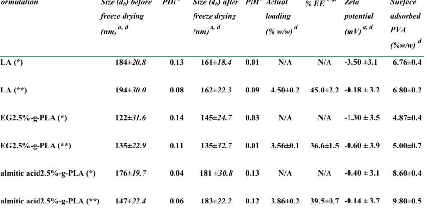
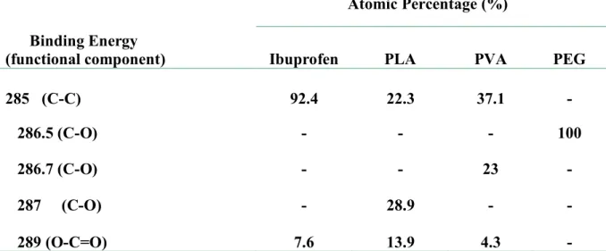
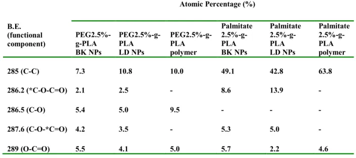
Table 2.2. Characteristics of different NPs formulation……….. 68 Table 2.3. Relative atomic percentages calculated from XPS Surface Analysis of pure materials used in NPs preparation……… 75
Table 2.4. Relative atomic percentages calculated from XPS Surface Analysis of synthesized polymers and formulated NPs using those polymers………76
Table 3.1. Polymers characterization by 1H NMR, DSC, and Gel Permeation Chromatography (GPC)……….. 101
Table 3.2. Characteristics of different NPs formulation……… 108
Table 3.3. Relative atomic percentages calculated from XPS Surface Analysis of pure materials used in NPs formulation……….. 113
Table 3.4. Relative atomic percentages calculated from XPS Surface Analysis of synthesized polymers and formulated NPs using those polymers……….. 117
Table 4.1. Polymer characterization by 1H NMR and Gel Permeation Chromatography (GPC)……….. 145
Table 4.2. Size distribution characteristics for different NPs after different stages of preparation……….. 147
Table 4.3. Other physicochemical characteristics for different NPs formulation………...148 Table 5.1. Characteristics of different NPs formulation……….176 Table 5.2. Diameter of growth inhibition zone measured with c. albicans fungal strains……. 191
List of figures
Fig.1.1. Schematics of polymeric nanoprticles. A. Matrix type nanosphere, drug molecules are uniformly dispersed in the polymer matrix. B. Core shell nanocapsules, drug molecules are presented in an oily or aqueous core covered with polymeric shell. C. matrix type nanosphere where drug crystals are embedded in a polymer matrix………. 5 Fig.1.2. Factors affecting the drug delivery performance of polymeric nanoparticles………...…7 Fig.1.3. Architectures of different copolymers used in the preparation of polymeric nanoparticles………..13 Fig. 1.4. Chemical structure of poly(lactic acid)………...15 Fig.1.5. Schematic representation of the emulsification-evaporation technique……….. 18 Figure 2.1. 1H NMR spectra and chemical structures of PEG2.5%-g-PLA (1), and palmitic acid2.5%-g-PLA (2)……….. 62 Figure 2.2. Tapping mode AFM images of NPs, left panel shows topography (T) and right panel shows corresponding phase images (P); all images are acquired in air. Scan size [400 nm × 400 nm]; PLA (a), PEG2.5%-g-PLA (b), palmitic acid2.5%-g-PLA (c)……… 69
Figure 2.3.a. DSC curves of ibuprofen, physical mixture of PEG2.5%-g-PLA with ibuprofen, PEG2.5%-g-PLA polymer, and ibuprofen loaded NPs. Inset inside the figure shows clear thermograms for both physical mixtures and NPs, Ibuprofen melting peak is encircled………. 71
Figure 2.3.b. DSC curves of ibuprofen, physical mixture of palmitic acid2.5%-g-PLA with ibuprofen, palmitic acid2.5%-g-PLA polymer, and ibuprofen loaded NPs. Inset inside the figure shows clear thermograms for both pure polymer and NPs. Ibuprofen melting peak is encircled……….72
Figure 2.4. 1H-NMR of blank NPs of PLA (1), palmitic acid2.5%g-PLA (2), and PEG2.5%g-PLA (3) in D2O. PEG peaks at 3.6 ppm are encircled………. 77
Figure 2.5. Schematic representation of polymer chain organization inside the NPs: PLA (a), Palmitic acid2.5%-g-PLA (b), PEG2.5%-g-PLA (c)……… 78
Figure 2.6. Erosion of different ibuprofen loaded NPs in phosphate buffer saline (PBS, pH 7.4) at 37 ºC………. 80
Figure 2.7. Effect of PLA grafting on the in vitro release behavior of ibuprofen loaded NPs; values are represented as mean ±S.D. of three independent experiments……… 82
Figure 3.1. 1H NMR spectra and chemical structures of PEG7%-g-PLA, and multiblock copolymer, (PLA-PEG-PLA)n………... 102
Figure 3.2. Tapping mode AFM images of NPs, left panel shows topography (T) and right panel shows corresponding phase images (P); all images are acquired in air. PLA (a) [Scan size: 1 µm×1 µm], PEG7%-g-PLA (b) [Scan size: 2 µm×2 µm], and (PLA-PEG-PLA)n (c) [Scan size: 750 nm×750 nm]………. 109
Figure 3.3.a. DSC curves of ibuprofen, physical mixture of PEG7%-g-PLA with ibuprofen, PEG7%-g-PLA polymer, and ibuprofen loaded NPs. Inset inside the figure shows clear thermogram for NPs……… 111
Figure 3.3.b. DSC curves of ibuprofen, physical mixture of (PLA-PEG-PLA)n with ibuprofen, (PLA-PEG-PLA)n polymer, and ibuprofen loaded NPs. Inset inside the figure shows clear thermograms for both physical mixtures and NPs……….. 112
Figure 3.4. 1H-NMR of blank NPs of PLA (1), PEG7%-g-PLA (2), and (PLA-PEG-PLA)n (3) in D2O. PEG peaks at 3.6 ppm are encircled……….. 118
Figure 3.5. Erosion of different ibuprofen loaded NPs in phosphate buffer saline (PBS, pH 7.4) at 37 ºC……… 120
Figure 3.6. Effect of PEG chain organization on the in vitro release behavior of ibuprofen loaded NPs; values are represented as mean ±S.D. of three independent experiments………. 121
Figure 3.7. Schematic representation of polymer chain organization inside the NPs: PLA (a), PEG7%-g-PLA (b), and multiblock copolymers (PLA-PEG-PLA)n (c)……… 124
Figure 3.S (supporting information): XRD spectra of ibuprofen, physical mixture of PEG7%-g-PLA with ibuprofen, PEG7%-PEG7%-g-PLA polymer, and ibuprofen loaded NPs……… 131
Figure 4.1. 1H NMR spectra and chemical structure of PEG-g-PLA copolymers of different PEG grafting densities over PLA backbone……… 144
Figure 4.2. AFM images of PEG7%-g-PLA NPs encapsulating rhodamine B (RHO), before (a) and after (b) lyophilization; surface morphology (left, S) and phase image (right, P)………... 149
Figure 4.3. In vitro release behavior of RHO from different PEG-g-PLA NPs in comparison to PLA NPs and RHO solution; values are represented as mean ±S.D. of three independent experiments………. 150
Figure 4.4. a): DLS size distribution data (nm) of different NPs upon incubation at 37 °C for 24 h with 5% FBS. b): DLS size data (nm) of NPs upon incubation at 37 °C for 24 h with 2% BSA………. 152
Figure 4.5. Cytotoxicity of pegylated NPs of different PEG grafting densities over PLA backbone (1, 7, and 20% mol/mol) in RAW 264.7 cells by MTT assay……… 154
Figure 4.6. Fluorescence images (right panels) and their corresponding phase contrast images (left panels) of RAW 264.7 cells after incubation with a) RHO, b) RHO loaded PLA NPs, c) RHO loaded PEG1%-g-PLA NPs, d) RHO loaded PEG7%-g-PLA NPs, and d) RHO loaded PEG20%-g-PLA NPs. red images show RHO. Scale bar=50 µm……….. 156
Figure 4.7. RAW 264.7 cellular uptake of RHO encapsulated NPs made from PEG-g-PLA copolymer of different PEG grafting densities in comparison to PLA NPs. RAW 264.7 cells were incubated with NPs at 37°C for 24 h………..… 157
Figure 4.S1. a): Polydispersity index values of different NPs upon incubation at 37 °C for 24 h with 5% FBS. b): Polydispersity index values of NPs upon incubation at 37 °C for 24 h with 2% BSA………..163
Figure 5.1. AFM image of lyophilized ITZ loaded PEG7%-g-PLA NPs………..177 Figure 5.2. The stability index of ITZ-NPs in the particle size shown as diameter [D] (a), polydispersity index [PDI] (b), and loading efficiency [% LE] (c) during storage for 30 days [d] at room temperature. Data are expressed as the mean±S.D. (n = 3)………179, 180
Figure 5.3a. DSC curves of ITZ, physical mixture of PLA)n with ITZ, (PLA-PEG-PLA)n copolymer, and ITZ loaded (PLA-PEG-(PLA-PEG-PLA)n NPs………...181
Figure 5.3b. DSC curves of ITZ, physical mixture of PEG7%-g-PLA with ITZ, PEG7%-g-PLA copolymer, and ITZ loaded PEG7%-g-PLA NPs………182
Figure 5.4a. XRD spectra of ITZ, physical mixture of PLA)n with ITZ, (PLA-PEG-PLA)n copolymer, and ITZ loaded (PLA-PEG-(PLA-PEG-PLA)n NPs………...183
Figure 5.4b. XRD spectra of ITZ, physical mixture of PEG7%-g-PLA with ITZ, PEG7%-g-PLA copolymer, and ITZ loaded PEG7%-g-PLA NPs………184
Figure 5.5a. FT-IR spectra of ITZ, physical mixture of PLA)n with ITZ, (PLA-PEG-PLA)n copolymer, and ITZ loaded (PLA-PEG-(PLA-PEG-PLA)n NPs [ITZ main characteristic peaks are marked with arrows]………186
Figure 5.5b. FT-IR spectra of ITZ, physical mixture of PLA with ITZ, PEG7%-g-PLA copolymer, and ITZ loaded PEG7%-g-PEG7%-g-PLA NPs [ITZ main characteristic peaks are marked with arrows]……….187
Figure 5.6. In vitro release behavior of ITZ from PEG/PLA NPs either from PEG7%-g-PLA or (PLA-PEG-PLA)n in comparison to PLA NPs and ITZ solution; values are represented as mean ±S.D. of three independent experiments..………188
Figure 5.7. Erythrocyte lysis caused by ITZ loaded PEG/PLA NPs preparations compared to free ITZ and ITZ-PLA NPs. Values are expressed as mean of % lysis of three separate experiments ±S.D……….…189
Figure 5.8a. Plates inoculated with C. albicans fungal cells and incubated at 30 ºC, treated with 12.5 µl of (a) ITZ-Water, (b) ITZ-DMSO, (c) ITZ-PLA NPs, (d) ITZ-PEG7%-g-PLA NPs, and (e) ITZ-(PLA-PEG-PLA)n NPs. Lower plates (a-e) represent control experiments corresponding to each upper plate either using only the solvent in case of ITZ-Water, and ITZ-DMSO or only blank NPs ( no ITZ) in case of NPs formulations………....191
Figure 5.8b. Plates inoculated with A. fumigatus fungal cells and incubated at 30 ºC, treated with (a) Water, (b) DMSO, (c) PLA NPs, (d) PEG7%-g-PLA NPs, and (e)
ITZ-(PLA-PEG-PLA)n NPs. Lower plates (a-e) represent control experiments corresponding to each upper plate either using only the solvent in case of ITZ-Water, and ITZ-DMSO or only blank NPs ( no ITZ) in case of NPs formulations………..192
List of abbreviations
AFM Atomic Force microscope
ANOVA Analysis of variance
BSA Bovine serum albumin
ºC Degree Celsius
CDCl3 Deuteriated chloroform
DCM Dichloromethane
DLS Dynamic light scattering
DL Drug loading
DMEM Dulbecco’s modified eagl’s medium
DMSO Dimethyl sulfoxide
D2O Deuterium oxide
DSC Differential scanning calorimetry
EPR Enhanced permeability and retention FBS Fetal bovine serum
FTIR Fourier transform infrared spectroscopy GPC Gel permeation chromatography
h Hour
HCl Hydrochloric acid
1
H NMR Proton nuclear magnetic resonance
HPLC High performance liquid chromatography ITZ Itraconazole
IV Intravenous
kDa Kilo Dalton
LE Loading efficiency
Mn Number average molecular weight Mw Weight average molecular weight
MPS Mononuclear phagocytic system
MTT 3-(4,5-dimethylthiazol-2-yl)-2,5-diphenyltetrazolium bromide
MWCO Molecular weight cut off
Mwt Molecular weight
NPs Nanoparticles
NaCl Sodium chloride
NaOH Sodium hydroxide
O/W oil in water
PBS Phosphate buffered saline
PCL Poly(ε-caprolactone)
PDI Polydispersity index
PEG Poly(ethylene glycol)
PCS Photon correlation spectroscopy
PEG-g-PLA Poly(ethylene glycol)-grafted-poly(D,L-lactide)
PEG2.5%-g-PLA Poly(ethylene glycol)2.5%-grafted-poly(D,L-lactide)
PEG20%-g-PLA Poly(ethylene glycol)20%-grafted-poly(D,L-lactide)
(PLA-PEG-PLA)n (poly(D,L-lactide)-block- poly(ethylene glycol) block-poly(D,L-
lactide))n PLA Poly(lactic acid) PGA Poly(glycolic acid)
PLGA Poly(lactic-co-glycolic acid)
PVA Polyvinyl alcohol
PXRD Powder x-ray diffraction pattern
dh Hydrodynamic diameter
RHO Rhodamine
RPMI Royal Park Memorial Institute (culture medium) SD Standard deviation
SEM Scanning electron microscopy
Tg Glass transition temperature
TEM Transmission electron microscopy
TM-AFM Tapping mode atomic force microscope
UV Ultraviolet wt Weight
λmax Wavelength of maximum absorbance
λex Excitation wavelength
λem Emission wavelength
To my mother, my kids, my wife and the souls of of Egyptian Revolution martyrs.
Acknowledgements
This work was carried out at the Division of Pharmaceutics, Faculty of Pharmacy, University of Montreal. I wish to express my gratitude to my supervisor, Dr. Patrice Hildgen for his continuous support and guidance into the world of science. I would also like to thank him for always being available and willing to help me in the challenges of this work. During my studies, he has given me the freedom and reliable basis necessary to advance this project and to develop as an independent researcher. Also, I really value his deep involvement and valuable criticism throughout the course of my doctoral studies without which it would have been difficult to finish this thesis.
I am sincerely grateful to all my co-authors, Jean Michel Rabanel, Fatiha Louhichi, and Dr. Martine Raymond, for their remarkable contribution to this work. Special thanks are due to Jean Michel Rabanel for his technical assistance, valuable discussion and suggestions during my study. I would like to express my warmest thanks to all my lab colleagues for discussions, friendship and for creating a pleasant atmosphere during these years. I also express my gratitude for Dr. Grégoire Leclair, evaluator of my progress reports for his valuable time and thoughtful disccussion. I also acknowledge Mme. Suzie Poulin, research associate, Ecole polytechnique, Montreal University for her unlimited help and patience in interpretation of XPS data.
Finally, I would like to express my gratitude to my mother, my wife, and my little kids for their endless support and love. It is their sacrifice and understanding that allowed me to work hard to finish this thesis.
The financial support by the missions department, Ministry of Higher Education, Egypt is gratefully acknowledged.
CHAPTER ONE
__________________________________________________________________
INTRODUCTION
Polymeric Nanoparticles (NPs) as Drug
1.1. A Brief Overview of Nanotechnology
In recent years, significant effort has been devoted to research and applications in the area of nanotechnology. Nanotechnology is the design, characterization, synthesis and application of materials, structures, devices and systems by controlling shape and size at nanometer scale [1]. Nanotechnology is widely applied nowadays to many areas of life as; fiber and textiles [2], agriculture [3], electronics [4], forensic science [5], space [6] and many other industrial applications. Medicine is one of the areas likely benefited from advances in nanotechnology [7-10]. Many applications of nanotechnology in medicine have been investigated so far including drug delivery, both in vitro and in vivo diagnostics, and production of high quality biocompatible materials [11, 12]. It is reported that the global market of nanotechnology is expanding very fast with many expectations that it will reach $1.5 trillion by year 2012 [13].
1.2. Nanotechnology and Drug Delivery
Development of nanotechnology for drug delivery has attracted a great deal of attention in recent years. Nanotechnology for drug delivery aims at formulating medicinal agents in biocompatible nanocarries (nanoparticles) for improving their clinical outcomes. The development of nanotechnology products for currently existing drugs may optimize their therapeutic performance, and minimize the need for developing new drugs with better properties. Formulating drugs into nanocarriers has proven successful to achieve all the following tasks [14]. To enhance the solubility and hence; absorption of poorly water-soluble drugs; efficiently target drugs into distant areas in the body; enhance cellular uptake of drugs across tight epithelial and endothelial barriers; efficiently deliver large macromolecular drugs (e.g. proteins, peptides and nucleic acids) to intracellular sites; deliver two or more drugs using the same carrier [15, 16]; and follow drug delivery journey in the body using imaging tools combined with the drug in the same carrier [17].
Nowadays, nanotechnology is widely used for encapsulating many drugs/bioactive moieties onto nanocarriers to improve either their pharmaceutical performance (e.g. bioavailability, stability, and controlled release) or their pharmacological performance (e.g. therapeutic activity, reducing side effects, and effective targeting) [18-20]. Several drugs used for treating incurable diseases like cancer [21], AIDS [22], diabetes [23], malaria [24], prion disease [25] and tuberculosis [26] are in the clinical testing phase and some of them are available in the market
[27, 28]. As a result of the last mentioned applications, nanotechnology holds enormous promise for drug delivery and it is widely expected that nanotechnology will be able to change the landscape of pharmaceutical and biotechnology industries for the foreseeable future [14].
1.3. Design of Nanotechnology–Polymeric based Drug Delivery Systems
There are a number of parameters that need to be considered for the development and manufacturing of successful drug delivery vehicles [29]. These include (a) the use of well- characterized, biodegradable, biocompatible materials; (b) the ability to tune the biophysicochemical properties of the used materials (e.g. hydrophilicity/ hydrophobicity balance, charge, molecular weight) to develop nanocarriers with adjustable properties (e.g. size, surface charge, release behavior); and (c) developing small unit operations amenable to scaling-up (manufacturing large quantities of drug delivery systems) for clinical applications. Numerous classes of drug delivery nanocarriers composed of different materials including lipids, polymers and inorganic materials have been developed, resulting in different delivery systems that vary in their physicochemical properties and thus their pharmaceutical uses.
Growing interest in formulating polymeric nanoparticles is rapidly expanding for the following reasons. Advances in the scientific fields of polymer chemistry and polymer colloid physicochemistry have resulted in the availability of many tailor-made polymers for drug delivery. Polymeric drug delivery systems are often biodegradable, and biocompatible with potential of controlled drug release. They often have a superior drug delivery performance (e.g. size, surface charge, drug release, and cellular uptake properties) relative to other non-polymeric carriers. Moreover, they can be designed with hydrophilic coats, such as poly(ethylene glycol) (PEG), which creates a steric barrier decreasing the adsorption of opsonin proteins onto particle surface. This helps nanoparticles avoid recognition by the mononuclear phagocytic system (MPS) and circulate for prolonged period of time in the blood [30, 31]. Nanoparticles assembled from water soluble polymers usually have a molecular weight above glomerular filtration threshold (42-50 kDa), which is another cofactor for their prolonged circulation in the body [32]. An interesting feature of polymeric carriers is their ability to control the release of a therapeutic agent to achieve desired therapeutic level in certain pathological areas in the body. This could be achieved by the use of stimuli responsive polymers which release their payload under the effect of external stimuli (i.e. change in pH, temperature or ionic strength) [33, 34].
This allows maximum drug release at the target site, achieving optimum therapeutic efficacy [35]. Polymeric nanoparticles can be delivered to distant target sites either by localized delivery using a catheter-based approach [36] or they can be attached to a recognition marker or/ ligand which could direct them to the target area [37]. They usually achieve high drug loading, which maximizes drug/excipients ratio. This results into reducing the frequency of administration and hence, patient expenses, and risks of toxicity could also be reduced.
One of the most distinct advantages of polymeric carriers for drug delivery is their adjustable nanometer size range. Due to their sub-cellular and sub-micron size, polymeric nanoparticles can homogenously penetrate into tissues, cross the fenestration of the epithelial lining (e.g., liver), and efficiently taken up by the cells [38]. In addition, their nanometer size range allows them to be efficiently captured by some tumor cells particularly solid tumors, infarcts and inflamed tissues. This phenomenon is called enhanced permeability and retention effect (EPR) or passive accumulation of nanocarriers into the tumor tissues [39]. EPR mainly occurs because of the pathophysiological features of tumors vessels that are mainly distinct from normal vessels. Tumor cells are characterized by hypervascularity, incomplete vascular architecture, poorly aligned endothelial cells and wide fenestrations (20 nm-1.2 µm) [40, 41]. These features make the vasculature of pathological tissues more “leaky” or more “permeable” than that of normal tissue. This increased permeability or leaky vasculature together with compromised lymphatic drainage facilitates accumulation of macromolecules and nanoparticles in pathological tissues. Based on the EPR effect, drug concentration in the tumor tissues was found to be 10-30 times higher than that in the blood and other normal tissues [42]. To sum up, nanoencapsulation of medicinal drugs using polymeric carriers (nanomedicines) was shown to enhance drug efficacy, specificity, tolerability and therapeutic index of many drugs [43-45].
1.4. Review of Polymeric Nanoparticles as Delivery Systems
Polymeric nanoparticles are solid or semisolid colloidal particles that vary in size from 10 nm to 1000 nm [46]. They are mainly consisting of macromolecular substances and can be used as drug carrier. The drug is adsorbed, dissolved, entrapped, or encapsulated into the nanoparticles matrix. Either nanospheres or nanocapsules can be obtained from the same polymer depending on the used method of preparation [47, 48]. Nanospheres are matrix systems in which the drug is uniformly dispersed whereas; nanocapsules are vesicular systems in which
the drug is confined to a cavity surrounded by a polymer shell (Fig. 1.1). After preparation, nanoparticles are usually dispersed in aqueous solution. The resultant drug nanoparticle dispersion can be administered to humans by many routes as injection, oral route, or applied topically either to the skin or the eye. Nanoparticles can be also used for pulmonary delivery after being dried to a powder, or can be compressed into tablets or capsules.
A
B
C
Fig.1.1. Schematics of polymeric nanoprticles. A. Matrix type nanosphere, drug molecules are uniformly dispersed in the polymer matrix. B. Core shell nanocapsules, drug molecules are presented in an oily or aqueous core covered with polymeric shell. C. matrix type nanosphere where drug crystals are embedded in a polymer matrix.
1.5. Factors affecting Performance of Polymeric Nanoparticles as Delivery Vehicles Formulation of nanoparticles as delivery vehicles depends on the choice of suitable polymeric substance having acceptable biocompatibility and biodegradability, higher incorporation efficiency, and an extraordinary ability to enhance the drug retention time in vivo. Nanoparticles are superior to conventional drug carriers (e.g. tablets, capsules….etc.) with respect to control release, targeted delivery and therapeutic potential.
Polymer chains
Oily or aqueous core Drug crystals Drug molecules
Many factors affect the drug delivery performance of polymeric nanoparticles as particle size, surface charge, surface modification, and hydrophobicity (Fig.1.2). Size and size distributions of nanoparticles play an important role in determining their release kinetics as well as their interaction with the cell membrane and their penetration across the biological barriers. The ability of nanoparticles to penetrate different biological barriers is mainly dependent on the particle size, the target tissue biophysiological characteristics e.g. tissue size, tissue thickness, and blood circulation [49]. It has been shown before that large size particles are rapidly cleared by mononuclear phagocytic system (MPS) than smaller size particles [50]. The size of the carrier might have a role in complement activation [51]. For long circulating colloidal nanoparticles, optimum size is favored to be 150-200 nm [52, 53].
Surface charge is also important in determining the in vivo stability as well as cellular interaction of nanoparticles. Surface charge measurement (zeta potential) evaluates whether nanoparticles would aggregate in blood flow or would adhere to, or interact with oppositely charged cells membrane [54]. As a biological rule, to enhance the rate and extent of cellular uptake, positively charged surfaces or cationic surface charges are required as it promotes the interaction of nanoparticles with the negative charges (phospholipids) of the cell membrane [10]. However, cationic charges could enhance the non-specific cell sticking or uptake of the particles. As a result, it is recommended that colloidal carriers should have neutral or near neutral surface charge to avoid both the non specific cell uptake and uncontrolled plasma protein adsorption onto their surfaces [50, 55].
Intravenously (IV) administered nanoparticles are easily recognized by the body immune system, and then massively cleared by the macrophages (MPS) rich organs such as liver, spleen, lungs and bone marrow [56]. The NPs surface hydrophobicity determines the amount of adsorbed blood components, mainly proteins (opsonins) which initiate the phagocytosis process [57]. Hence, to prolong the circulation time of NPs in the blood, it is necessary to minimize the opsonization process (plasma protein adsorption onto NPs surface). This can be achieved by surface modification of hydrophobic particles by coating their surfaces with different hydrophilic molecules such as polyethylene glycol (PEG), polyethylene oxide, polyoxamer, poloxamine and polysorbate 80 (Tween 80) [46, 58].
Finally, the in vivo performance of nanoparticles is affected by morphological characteristics, surface chemistry, and molecular weight. Molecular weight of the carrier can
play a role in modulating the release behavior. For a slow drug release from the particles, high molecular weight of the starting polymer should be used [59]. Careful formulation of polymeric nanoparticles with respect to target and route of administration may solve many pharmaceutical problems of either marketed drugs or newly discovered drugs.
Fig.1.2. Factors affecting the drug delivery performance of polymeric nanoparticles
1.6. Applications of Polymeric Nanoparticles
Nanoparticles in pharmaceutical applications have gained plenty of research attention during recent decades taking advantage of their small size, and their biodegradable nature [13]. Nanoparticles can be administered orally as a reconstituted aqueous dispersion. NPs dispersion is easily uptaken and absorbed into the systemic circulation across the mucosal epithelium by enterocytes [60]. They could protect the encapsulated active ingredient against the
gastro-Different factors affecting polymeric Nanoparticles
Surface charge Size and size
distribution Surface modifications Hydrophobicity Molecular weight (Mwt)
intestinal (GI) tract harsh conditions and/or prolong the contact time of its payload on the mucous membrane by efficiently adhering to mucosal surfaces (bioadhesion). Many examples of the oral applications of NPs are reported in the literature particularly for delicate macromolecules e.g. oral delivery of peptides such as salmon calcitonin [61] and elcatonin [62]. NPs were also used as an oral carrier for the delivery of hormones as mifepristone [63] and estradiol [64]. Oral delivery of vaccines by NPs has achieved considerable success through the targeted uptake of the particles in the M cells of peyer’s patches. Many examples for the oral immunization using PLGA nanoparticles are also reported such as tetanus toxoid [65], ovalbumin [66] and bovine serum albumin (BSA) [67].
Several applications of stealth (long circulating) nanoparticles can be found in the area of tumor/cancer therapy. The mean residence time, effective concentration, half life of some anticancer drugs loaded into polymeric nanoparticles have been enhanced compared to pure drug e.g. camptothecin [68], and paclitaxel [69].
PLA and PLGA NPs have also been applied for the sustained delivery of many drugs into an intracellular target [70-72]. NPs could enhance the cytoplasmic delivery of many drugs through their marked ability to rapidly escape the endo-lysosomal compartment to the cytoplasmic compartment e.g. PCL nanoparticles [73]. Dexamethasone-loaded nanoparticles has achieved higher antiproliferative activity in vascular smooth muscle cells compared to free dexamethasone due to the enhance uptake of NPs by the glucocorticoid receptors which are cytoplasmic [74].
Another interesting application of nanoparticles is their ability to deliver drugs across the Blood–Brain Barrier (BBB) [75]. Nanoparticles has been shown to sustain the delivery of therapeutic agents to brain tumours following the opening of tight junctions of the BBB by hyperosmotic mannitol [76]. Poly(alkylcyanoacrylate) nanoparticles have been widely investigated as a drug carrier to the brain [77]. Many types of polymeric nanoparticles have also been used for the brain delivery e.g. PEG nanoparticles functionalized by lectin [78], PLA-PEG nanoparticles bearing cationic BSA coating [79], PLA-polysorbate 80 nanoparticles [80], and PLGA peptide nanoparticles [81]. Loperamide has been loaded into peptide derivatized PLGA NPs then delivered successfully to the central nervous system of rats [82]. Till now, the mechanisms of particle uptake across the BBB are not clearly identified; other processes may be also involved [83].
Nanoparticles with mucoadhesive properties have also been investigated as an ocular drug delivery carrier. They mainly act as an ocular carrier based on their ability to prolong the mean residence time of the drug in the cornea, control its release and reduce irritation after topical application. Flurbiprofen loaded into PLGA nanoparticles exhibited higher anti-inflammatory action upon instillation into the eye than free drug [84]. Similar enhancement of the ocular absorption of acyclovir-loaded PEG-PLA nanoparticles has been observed [85].
In gene delivery studies, non-viral vectors are assembled through the interaction between the negative charge of nucleic acids and the positive charge of the particles. Non viral vectors have been demonstrated a marked ability to encapsulate the entrapped gene, protect it against stressful body conditions, sustain its release, and facilitate its delivery and interaction with the cell membrane. For example, PLA-PEG nanoparticles administered orally have achieved higher transfection efficiency for the encapsulated DNA [86]. PLGA nanoparticles have been used as non viral vector for the sustained release of DNA [87]. Many challenges in nanoparticulate gene delivery applications are faced particularly the key challenge of enhancing the coupling potential between nucleic acid and the polymer while maintain the size of the carrier in the nanometer scale.
Despite the extensive research in the area of nanotechnology and drug delivery, few polymeric nanoparticulate products have marketed so far. A commercialized product, Abraxane™, obtained from the protein albumin bound with paclitaxel, is administerd intravenously [88]. Another product, Doxorubicin Transdrug®, consisting of doxorubicin-loaded poly(isohexylcyanoacrylate) nanoparticles is currently investigated clinically [89].
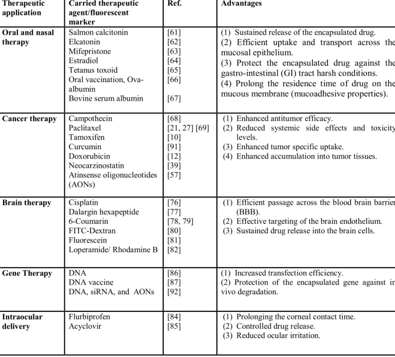
As a general summary of the investigated applications of nanoparticles in the pharmaceutical field, nanoparticles could protect the encapsulated active ingredient, enhance its action, reduce its toxicity and side effects, sustain its release and target it into the desired area. For clinical translation, most of the research is now focused on developing processes for the scaling up of nanoparticles [90]. A range of different types of nanoparticles and their applications are outlined in Table 1.1.
Therapeutic application Carried therapeutic agent/fluorescent marker Ref. Advantages
Oral and nasal therapy Salmon calcitonin Elcatonin Mifepristone Estradiol Tetanus toxoid Oral vaccination, Ova-albumin
Bovine serum albumin
[61] [62] [63] [64] [65] [66] [67]
(1) Sustained release of the encapsulated drug.
(2) Efficient uptake and transport across the mucosal epithelium.
(3) Protect the encapsulated drug against the gastro-intestinal (GI) tract harsh conditions. (4) Prolong the residence time of drug on the mucous membrane (mucoadhesive properties).
Cancer therapy Campothecin Paclitaxel Tamoxifen Curcumin Doxorubicin Neocarzinostatin Atinsense oligonucleotides (AONs) [68] [21, 27] [69] [10] [91] [12] [39] [57]
(1) Enhanced antitumor efficacy.
(2) Reduced systemic side effects and toxicity levels.
(3) Enhanced tumor specific uptake.
(4) Enhanced accumulation into tumor tissues.
Brain therapy Cisplatin
Dalargin hexapeptide 6-Coumarin FITC-Dextran Fluorescein Loperamide/ Rhodamine B [76] [77] [78, 79] [80] [81] [82]
(1) Efficient passage across the blood brain barrier (BBB).
(2) Effective targeting of the brain endothelium. (3) Sustained drug release into the brain cells.
Gene Therapy DNA
DNA vaccine
DNA, siRNA, and AONs
[86] [87] [92]
(1) Increased transfection efficiency.
(2) Protection of the encapsulated gene against in vivo degradation. Intraocular delivery Flurbiprofen Acyclovir [84] [85]
(1) Prolonging the corneal contact time. (2) Controlled drug release.
(3) Reduced ocular irritation. Table 1.1. Different applications of polymeric nanoparticles in pharmaceutical field.
1.7. Preparation of Polymeric Nanoparticles
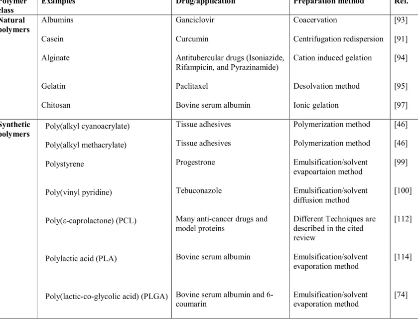
Polymeric nanoparticles for pharmaceutical applications are prepared from a class of synthetic and natural polymers. Natural polymers are considered safer and more biocompatible than synthetic ones. Synthetic polymers became more acceptable than before in the preparation of NPs due to the greater control over their biophysicochemical properties. Natural polymers include e.g. albumin [93], casein [91], alginate [94], gelatin [95, 96], and chitosan which is considered the most widely used natural polymer for nanoparticle preparation [97, 98]. Synthetic polymers include aliphatic polyesters, polyanhydrides, polyorthoesters, and polycyanoacrylates [46]. Other investigated synthetic polymers include polystyrene [99] and poyl(vinyl pyridine) [100].
Two techniques are widely used for nanoparticles preparation; (i) polymerization of monomers and (ii) deposition of an already synthesized or preformed polymer [58, 101]. Nanoparticle preparation from preformed polymers is more common because of the ease of the preparation, more control of the manufacturing process, and reproducibility of the used method. Polymer deposition method mainly depends on dissolving a preformed polymer in a convenient solvent followed by precipitation in a liquid medium leading to nanoparticle formation. The drug intended to be encapsulated in the particles is usually incorporated in the process during the polymer solvation process. Nanoprecipitation, salting-out, emulsification/solvent diffusion, and emulsification/solvent evaporation methods are widely applied techniques used for nanoparticles preparation by polymer deposition method [46, 102-104].
Nanoprecipitation method was introduced and patented by Fessi and co-workers [105]. In this method, particles are formed spontaneously by precipitation and subsequent solidification of the polymer upon rapid solvent diffusion. The polymer is dissolved in a water miscible organic solvent (or solvent mixture) and poured under magnetic stirring into a non solvent (usually water containing surfactant), in which the organic solvent diffuses. The mechanism of formation of NPs by this technique has been explained by the interfacial turbulence generated at the interface of the solvent and non-solvent. Thus, the process is often called solvent displacement or interfacial deposition. In the salting-out method, nanoparticle formation is based on the separation of a water-miscible solvent containing the polymer from aqueous solutions via a salting-out effect. Acetone is commonly used as the water-miscible solvent because of its
solubilizing efficiency and rapid separation from aqueous solutions by salting-out effect with electrolytes. In emulsification/solvent diffusion method, nanoparticle formation starts when the saturation limit of a partially water-miscible solvent (e.g. benzyl alcohol) is exceeded by addition of water. In both techniques, vigorous stirring is a prerequisite for inducing the phase separation. The emulsification solvent evaporation method is presented in section 1.7.5. Another widely used technique nowadays for nanoparticles preparation is supercritical fluid technology [106, 107]. Nanoparticles can also be prepared directly from monomers using different polymerization techniques as described in those reviews [46, 102]. A list of some used polymers and the commonly used methods to prepare nanoparticles are summarized in Table 1.2.
Nanoparticles are usually prepared as an aqueous dispersion. If nanoparticles are stored in an aqueous medium, polymer degradation, drug leakage, and/ or drug degradation may occur [108]. In addition, handling (storage, transportation) of a liquid particle system is inconvenient. To improve the physicochemical stability of nanoparticles, freeze-drying in is usually performed to conserve the structure of particles, facilitate handling and transport, and help the further processing of particles into other dosage forms (e.g. tablets, capsules, and powder).
1.7.1. Classification of Copolymers used for Nanoparticles Formation
Nanoparticles have been formulated using a wide range of polymers. Copolymers are widely used than homopolymers in NPs preparation due to their tunable properties. Copolymers can be divided into 4 main types based on their architecture (how the building blocks are connected together) (Fig.1.3):
1. Block copolymers: linear copolymers where the end group of one chain is covalently attached to the head of another chain giving diblock or triblock architectures [109].
2. Graft copolymers: branched copolymers with a comb-like architecture where different branches emanate from one main chain.
3. Random copolymers: linear copolymers with the building blocks arranged randomly [110]. 4. Alternating copolymers: linear copolymers with perfectly alternating arrangement of their building blocks.
1.7.2. Polyesters
Biodegradable polyesters (e.g. PLA, PGA, PLGA and PCL) are, so far, the most extensively used biomaterials for biomedical applications. They are the preferred synthetic polymers for nanoparticle preparation for the following reasons. They are mainly characterized by their biocompatibility and their controlled degradation to biocompatible monomers [111]. They are degraded by bulk hydrolysis of their ester bonds [112]. Their degradation products (e.g. lactic acid or glycolic acid) are eliminated from the body by citric acid cycle [113], hence, they do not require any kind of surgery to be removed from the body. They are easily synthesized through ring opening polymerization of cyclic lactones. They could efficiently protect the entrapped drug against degradation and control its site specific delivery. They are more efficient for encapsulation of many drugs particularly hydrophobic drugs. Encapsulated drugs are slowly released from polyester based matrices upon the controlled degradation of the polymeric materials.
Diblock copolymer Mutliblock copolymer
Graft copolymer
Random copolymer Alternating copolymer
Fig.1.3. Architectures of different copolymers used in the preparation of polymeric nanoparticles.
Polymer class
Examples Drug/application Preparation method Ref.
Natural polymers Albumins Casein Alginate Gelatin Chitosan Ganciclovir Curcumin
Antitubercular drugs (Isoniazide, Rifampicin, and Pyrazinamide) Paclitaxel
Bovine serum albumin
Coacervation
Centrifugation redispersion Cation induced gelation
Desolvation method Ionic gelation [93] [91] [94] [95] [97] Synthetic polymers Poly(alkyl cyanoacrylate) Poly(alkyl methacrylate) Polystyrene Poly(vinyl pyridine) Poly(ε-caprolactone) (PCL)
Polylactic acid (PLA)
Poly(lactic-co-glycolic acid) (PLGA)
Tissue adhesives Tissue adhesives Progestrone
Tebuconazole
Many anti-cancer drugs and model proteins
Bovine serum albumin
Bovine serum albumin and 6-coumarin Polymerization method Polymerization method Emulsification/solvent evapoartaion method Emulsification/solvent diffusion method
Different Techniques are described in the cited review Emulsification/solvent evaporation method Emulsification/solvent evaporation method [46] [46] [99] [100] [112] [114] [74] Table 1.2. Polymers and methods widely used in nanoparticle preparation.
1.7.3. Poly(lactic acid) (PLA)
Poly(lactic acid) (PLA) belongs to the family of linear aliphatic polyesters commonly made from α-hydroxy acids. PLA mainly obtained from lactic acid monomers (Fig. 1.4.). Being biodegradable polyester, PLA is widely used nowadays for nanoparticle preparation. PLA is also used for the manufacture of resorbable sutures, bone implants, artificial organs, tissue screws, and contraceptive implants [115, 116].
PLA is the simplest hydroxy acid with an asymmetric carbon atom and exists in two optically active configurations. Polymers of L(+) lactic acid and D(-) lactic acid are partially crystalline, while the racemic poly(D,L-lactic acid) is amorphous. The self-condensation of lactic acid results in low molecular weight (Mwt) PLAs, whereas ring opening polymerization of lactide usually results into higher Mwt PLA polymers [117]. PLAs are insoluble in water and ethanol, but they are soluble in organic solvents such as dichloromethane and chloroform [117-119]. PLA undergoes hydrolytic degradation of its ester bond in an aqueous medium [120]. The degradation products are biocompatible and metabolizable; they are removed from the body by the citric acid cycle. PLA degradation rate is mainly controlled by these factors; (i) Mwt (high Mwt PLAs degrade slower); (ii) crystallinity (amorphous PLA degrades faster); (iii) environmental conditions (pH, ionic strength, temperature); (iv) particle morphology (porous particles degrade faster); (v) size of the particles (small size particles degrade faster) [121]. Drugs encapsulated into PLA particles also could affect its degradation rate. Both acidic and basic drugs may catalyze the degradation of PLA polymeric carriers either microparticles or nanoparticles.
1.7.4. Functionalized Polyesters for Nanoparticles Preparation
Extensive work has been recently devoted towards functionalizing polyesters in order to enhance the drug delivery behavior of the obtained nanoparticles, increase the number of drugs that can be encapsulated into the polymeric particles and finally to efficiently control the drug release pattern. Most polyesters do not have significant number of functional groups that could enhance their potential applications. Such applications are greatly widened when functional pendant groups are incorporated into the polymer backbone.
Introduction of functional groups that can be easily substituted or conjugated with the compound of interest is a challenge. Many synthetic protocols were tried in the past for the synthesis of functionalized polyesters with better characteristics [122-124]. Most of these trials were complex including various tedious steps and the process itself was not reproducible and out of control in many cases. For example, functionalized malolactonate copolymers were synthesized with low yields (12–45%) and polymerization reactions were very slow (over 4–30 days) [122]. PLA grafted polysaccharide copolymers were also synthesized with a multistep method that required protection/deprotection step [125]. Moreover, most of the polyesters developed had molecular weights that are usually not high enough to be used for nanoparticles preparation [126]. Thus, there are very few methods that could be regarded as easy and efficient for the synthesis of functionalized polyesters. An efficient method for the synthesis of functionalized PCL and PLLA with controlled molecular weight and low polydispersity was previously reported [124].
In our study, nanoparticles were prepared from novel functionalized PLA copolymers. Those copolymers were developed by grafting different functional substituent as poly(ethylene glycol), PEG onto PLA backbone according to a previously reported method by our group [127]. This method simply depends on the development of versatile copolymers of PLA and allyl glycidyl ether. The grafted PLA with allyl group is undergoing subsequent steps to convert the allyl group into hydroxyl and then carboxyl groups to which various functional groups could be grafted easily to the polymer backbone. Various bioactive molecules like salens [128] or ligands for E-selectin and for P glycoprotein were successfully grafted onto PLA Backbone. Also, methoxy PEG-g-PLA has been previously synthesized by our group using the same principle and successfully used to prepare colloidal nanoparticles for sustained drug release [129].
1.7.5. Emulsification Solvent Evaporation
The emulsification solvent evaporation method was early described by Niwa et al and has been widely used to prepare NPs from a range of polymeric materials, particularly PLA and PLGA [130]. In this method, the polymer is first dissolved in a water immiscible, volatile organic solvent (e.g. chloroform, methylene chloride or ethyl acetate) then emulsified into an aqueous phase as shown in Fig.1.5. Emulsification can be achieved with the help of mechanical stirring, sonication or a high-energy source such as ultrasound or homogenization [Fig. 1.5]. The aqueous phase usually containing a surfactant like poly(vinyl alcohol), PVA to stabilize the system and help the formation of relatively small sized particles with uniform size distribution [131]. Nanoparticles tend to precipitate into the aqueous phase in the form of solid particles after the organic solvent has been removed under reduced pressure. This technique has been widely used for encapsulating hydrophobic drugs. The procedure has been modified to help encapsulating hydrophilic compounds and large macromolecules as peptides and proteins. The modified protocol depends on the use of the double or multiple emulsion technique (w/o/w) instead of the single emulsion (o/w). Simply, a water soluble drug and a surfactant are dissolved in water. The primary emulsion is prepared by dispersing the aqueous phase into an organic solvent containing a dissolved polymer. This is then re-emulsified in an outer aqueous phase also containing surfactant [132-134]. From here, the procedure for obtaining the nanoparticles is similar to the single emulsion technique (o/w) for solvent removal.
Several factors can influence the physicochemical properties of the obtained particles, these include; type and molecular weight of the starting polymer, concentration of polymer in the organic phase, type of organic phase, volume ratio of organic: aqueous phase, nature and concentration of surfactant, and stirring speed.
The main advantages of using this technique is the use of water as the nonsolvent; this is reduces the cost of production, facilitates the washing step of the particles, facilitates the further processing of particles [102]. However, there are some major disadvantages associated with the use of this method to prepare NPs. First of all, residual chlorinated solvents have serious toxicity potential. Second, the excess surfactant used is difficult to remove. Poly(vinyl alcohol), PVA is the most widely used stabilizer to prepare nanoparticles. However, PVA remains adsorbed at the surface of the nanoparticles and is difficult to be removed by multiple washing steps. It is
reported that PVA adsorbed at the surface of nanoparticles could change biodegradability, biodistribution, NPs cellular uptake, and drug-release pattern [135]. Third, this procedure is good for a laboratory-scale, but not for a large-scale pilot production.
Fig.1.5. Schematic representation of the emulsification-evaporation technique (taken from ref. [102]).
1.8. NPs Characterization
Nanoparticles as colloidal dispersions differ remarkably from other macroscopic dispersions. Nanoparticles have some unique physicochemical properties related to their sub-micron size as high surface area and surface free energy, and movement of particles by random motion (Brownian diffusion). Different physicochemical methods are being used specifically for NPs characterization to help understand their performance in vivo.
1.8.1. Size and Morphology
Particle size affects release kinetics, biodistribution potential and cellular uptake properties of NPs [46, 136]. Therefore, it is crucial to measure the size of the particles before conducting any further characterization. A detailed knowledge of the NPs size could help identify the