Université de Montréal
Decoding protein networks during porcine epidemic
diarrhea virus (PEDV) infection through proteomics
Par Camila Andrea Valle Tejada
Département de pathologie et microbiologie Faculté de médecine vétérinaire
Mémoire présenté à la Faculté de médecine vétérinaire en vue de l’obtention du grade de Maîtrise ès sciences (M.Sc.)
En sciences vétérinaires option microbiologie
Avril, 2019
Université de Montréal
Faculté de médecine vétérinaire
Decoding protein networks during porcine epidemic
diarrhea virus (PEDV) infection through proteomics
Présenté par
Camila Andrea Valle Tejada
A été évalué par un jury composé des personnes suivantes :
Dr. Marcio Carvalho Costa, président-rapporteur Dr. Levon Abrahamyan, directeur de recherche
Dr. Carl A. Gagnon, co-directeur de recherche Dr. Francis Beaudry, co-directeur de recherche
Résumé
Le virus de la diarrhée épidémique porcine (VDEP) est responsable de graves pertes économiques. Les épidémies de VDEP ont détruit plus de 10% de la population porcine américaine au cours des 3 dernières années. Malheureusement, la compréhension insuffisante des interactions hôte-virus empêche la mise au point d'un vaccin efficace contre le VDEP. Les interactions hôte-virus sont très dynamiques et peuvent impliquer des complexes multiprotéiques. De plus en plus de preuves indiquent que les microvésicules extracellulaires (MVE) et la composition des particules virales jouent un rôle important dans la pathogenèse virale et la modulation de la réponse immunitaire de l'hôte à l'infection. De plus, on pourrait s’attendre à ce que la composition des virions de la diarrhée épidémique porcine (DEP) soit dépendante du type cellulaire, en raison de l'incorporation ou de l'association de protéines de cellules hôtes dans ou avec des virions. Par conséquent, la caractérisation des profils protéomiques des MVEs produits par les cellules infectées par le VDEP, et l'identification des protéines hôtes spécifiquement encapsidées dans les virions sont importantes pour notre compréhension plus approfondie des interactions virus-hôte. Pour atteindre cet objectif, nous avons produit et purifié des virions et des MVE de VDEP et analysé leur composition en protéines en utilisant une approche protéomique. Afin d'étudier la régulation spatio-temporelle de l'infection virale, une certaine optimisation de l'infection par le VDEP était nécessaire. Pour cela, nous avons synchronisé et augmenté l'entrée de virus dans les cellules et étudié les schémas protéomiques des cellules infectées par le VDEP selon un mode de résolution temporelle.
Nous avons constaté que l'infection par le VDEP affectait l'abondance de diverses protéines de l'hôte associées aux microvésicules produites par les cellules infectées. Plus précisément, nos données protéomiques ont révélé que les protéines impliquées dans la liaison aux acides nucléiques, les processus métaboliques et les voies de la réponse immunitaire étaient parmi les plus touchées par l'infection. Fait intéressant, les protéines de l'hôte impliquées dans la régulation du cycle cellulaire et du système cytosquelettique ont également été touchées en abondance, ce qui n'est pas étonnant, car plusieurs chercheurs ont rapporté que les protéines cytosquelettiques participent activement au déplacement des composants viraux vers le site d'assemblage et que de nombreux virus manipulent la réparation de l'ADN, ainsi que le cycle cellulaire. La présente étude a démontré
l’incorporation de nombreuses protéines cellulaires dans les virions de la DEP. De plus, nous avons démontré que les polycations (molécules à charge positive) eu augmente 9-fois l'efficacité de l'entrée et de l'infection du VDEP. Ainsi, les polycations peuvent être utilisés pour optimiser l’infection par le VDEP, et améliorer la production de vaccins.
À notre connaissance, il s'agit de la première étude de la composition des virions et des microvésicules de DEP produits par une infection par le VDEP.
Mots-clés : Le virus de la diarrhée épidémique porcine, protéomiques, polycation,
Abstract
Porcine epidemic diarrhea virus (PEDV) is responsible for severe economic losses. The PEDV epidemics have destroyed more than 10% of the US swine population in the past 3 years. Unfortunately, the insufficient understanding of virus-host interactions impedes the development of an effective vaccine against PEDV. Virus-host interactions are highly dynamic and may involve multiprotein complexes. Growing evidence indicates that extracellular microvesicles (EMV) and composition of the viral particles play an important role in viral pathogenesis and modulation of host immune responses to infection. Additionally, it could be expected that the composition of porcine epidemic diarrhea (PED) virions is cell type dependent, due to the differential incorporation or association of host cell proteins into or with virions. Consequently, the characterization of the proteomic profiles of the EMV, produced by the PEDV-infected cells, and identification of the host proteins that are specifically encapsidated into the virions are important for our further understanding of virus-host interactions. To accomplish this objective, we produced and purified PEDV virions and EMV and analyzed their protein composition using a proteomic approach. In order to investigate the spatial-temporal regulation of viral infection and due to the low overall infectivity of the virus, a certain optimization of the PEDV infection was needed. To this end, we synchronized and increased virus entry into the cells. This allowed us to study the proteomic patterns of the PEDV-infected cells in a time-resolved mode.
We found that PEDV infection affected the abundance of various host proteins associated with microvesicles produced by the infected cells. More precisely, our proteomic data revealed that proteins involved in nucleic acids binding, metabolic processes and immune response pathways were among the most affected by the PEDV infection. Interestingly, host proteins involved in cell cycle regulation and cytoskeletal system also were affected in abundance, which is not surprising since several investigators have reported that cytoskeletal proteins are actively participating in moving the viral components to the assembly site, and that many viruses manipulate DNA repair and cell cycle. The present study has demonstrated the incorporation of numerous cellular proteins into the PED virions. Additionally, we demonstrated that treatment of PEDV virions with polycations (positively charged molecules) induced a nine-fold increase in the efficiency of viral entry and infection.
Thus, polycations can be used for the optimization of PEDV infection and improved vaccine production.
To the best of our knowledge, this is the first study of the composition of PED virions and microvesicles produced by PEDV infection.
Keywords: Porcine epidemic diarrhea virus, proteomics, polycation, polybrene,
Table of content
Résumé……….. ... III Abstract…………. ... V Table of content ... VII List of tables ... X List of figures ... XI List of acronyms and abbreviations ... XIII Acknowledgments ... XVIII
I. Introduction ... 1
II. Review of the literature ... 5
1. Introducing porcine epidemic diarrhea virus (PEDV) ... 6
1.1. Porcine epidemic diarrhea ... 6
1.2. Biology of PEDV ... 7 1.2.1. Genome organization ... 7 1.2.2. Non-structural proteins ... 8 1.2.3. Structural proteins ... 8 1.2.3.1. Spike protein (S) ... 9 1.2.3.2. Membrane protein (M) ... 10 1.2.3.3. Nucleocapsid protein (N) ... 10
1.2.3.4. Envelope protein (E) ... 11
1.2.4. Viral life cycle ... 11
1.2.4.1. Cellular receptors ... 12
1.2.4.2. Viral entry ... 14
1.2.4.3. Genome replication ... 14
1.2.4.4. Assembly and viral spread ... 14
1.2.4.5. Microvesicles and exosomes ... 15
1.3. Global distribution ... 16
1.4.1. Diagnostics ... 17
1.4.2. Treatment ... 18
1.4.3. Vaccination ... 19
1.5. PEDV-host interactions ... 22
1.5.1. Immunogenic interactions between host cells and PEDV ... 22
2. Proteomic analyses ... 25
2.1. Key steps in proteomic analysis ... 25
2.2. Types of mass spectrometry analyses ... 28
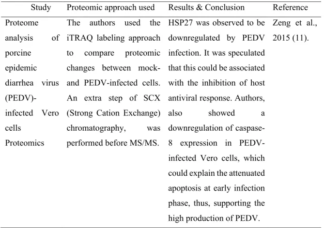
2.3. Proteomic studies of PEDV-infected cells ... 30
3. Methods to enhance PEDV viral infection ... 34
3.1. Polycations ... 35
III. Hypotheses and objectives ... 38
IV. Methodology ... 40
Cell lines and viral strains ... 41
Viral stocks production ... 41
Viral titration ... 41
Cell viability assay ... 42
PEDV proliferation assay to evaluate the effects of polycations ... 43
Indirect immunofluorescence assay (IFA) ... 43
Statistical analysis ... 45
Multistep purification of the PEDV for proteomic assay ... 45
Evaluation of the expression of viral RNA ... 46
Proteomic analysis ... 46
V. Results ... 49
1. Changes in the intracellular levels of host proteins during PEDV infection. .... 50
1.1. Optimization of PEDV infection by polycations ... 50
2. Identification of host cell proteins associated with or encapsidated into PEDV and EMV during viral infection. ... 59
2.1. Production of PEDV using simian cell lines that are routinely used for PEDV studies. ... 59
2.2. Analysis of the composition of virions and microvesicles/exosomes through proteomics approach. ... 59 VI. Discussion ... 79 1. Optimization of PEDV infection by polycations ... 80 2. Identification of host cell proteins associated with or encapsidated into PEDV and EMV induced by PEDV infection. ... 83 VII. Conclusions and perspectives ... 90 VIII. References ... 92 IX. Communications and founding ... XIX
List of tables
Table I. Strategies for PEDV-infection treatment ... 18
Table II. Available vaccines for PEDV prevention in Asia and North America ... 19
Table III. Variety of mass spectrometer configurations commonly used for quantitative proteomic analysis ... 26
Table IV. Reported proteomic studies of PEDV infected-cells ... 30
Table V. Proteins of the semipurified viral preparation affected by the PEDV infection ... 63
Table VI. Proteins affected by PEDV infection identified in the purified viral preparation ... 74
List of figures
Figure 1. Schematic representation of the structure of porcine epidemic diarrhea viral
particle. ... 6
Figure 2. Schematic representation of porcine epidemic diarrhea viral genome. ... 8
Figure 3. Overview of porcine epidemic diarrhea viral life cycle. ... 12
Figure 4. Inflammatory response to cytosolic or endosomal nucleic acid sensors. ... 23
Figure 5. Labeled and label-free quantitative proteomics. ... 29
Figure 6. Biophysical model of electrostatic interactions between the virus, target cell, and charged polymer. ... 35
Figure 7. Schematic chart of the experimental procedure, for evaluation of polycations effect on PEDV infectivity, at different times p.i. ... 44
Figure 8. Effect of DEAE-dextran at different concentrations, on viability of Vero-76 cells after different times post-treatment. ... 50
Figure 9. Effect of polybrene at different concentration on viability of Vero-76 cells after different times post-treatment. ... 51
Figure 10. Cytotoxic effect of DEAE-dextran at different concentration on Vero-76 cells measured after 24- and 48-hours post-treatment. ... 52
Figure 11. Cytotoxic effect polybrene at different concentration on Vero-76 cells measured after 24- and 48-hours post-treatment. ... 53
Figure 12. Synchronization of infection effect on PEDV infectivity in presence of polycations. ... 54
Figure 13. Effect of pre-incubation of Vero-76 cells with polycations before viral adsorption and infection. ... 55
Figure 14. Effect of pre-incubation of virus with polycations before viral infection. ... 56
Figure 16. Dose effect of DEAE-dextran treatment on PEDV infectivity. ... 58
Figure 17. Dose effect of polybrene treatment on PEDV infectivity. ... 59
Figure 18. Comprehensive quality control assessed by Spearmen-Karber method (viral titer), RT qPCR (presence of viral RNA) and Bradford assay (protein concentration). ... 60
Figure 19. Volcano plot of dysregulated tryptic peptides in the semipurified preparation containing viruses and EMV after PEDV infection. ... 61
Figure 20. Functional categories of cellular proteins of the semipurified viral preparation. ... 62
Figure 21. Subcellular localization of dysregulated host proteins identified in semipurified viral preparation. ... 63
Figure 22. Network of specifically the significantly dysregulated by PEDV infection proteins present in the semipurified viral preparation. ... 71
Figure 23. Functional categories of cellular proteins identified in the purified viral preparation. ... 72
Figure 24. Subcellular localization of dysregulated host proteins identified in the purified viral preparation. ... 73
Figure 25. Network of specifically the significantly dysregulated by PEDV infection proteins present in the purified fraction. ... 78
List of acronyms and abbreviations
3C1: Poliovirus 3C-like proteinaseABPP: Activity-based protein profiling ANOVA: One-way Analysis of variance ASFV: African swine fever virus
B2M: b-actin
Bcl-2: B-cell lymphoma 2 BtCo: Bat coronavirus
cAMP: Cyclic adenosine monophosphate CDV: Canine distemper virus
CHIKV: Chikungunya virus CPE: Cytopathic effect
CREB: cAMP responsive element binding
CRIPA : Le centre de recherche en infectiologie porcine et avicole CsCl: Cesium chloride
DENV: Dengue virus
DEP : Diarrhée épidémique porcine DHBV: Duck hepatitis B virus
DMEM: Dulbecco Modified Eagle Medium DTT: DL-dithiothreitol
EBOV: Ebola virus, EBV: Epstein-Barr virus
EDTA: Ethylene-diamine-tetra acetic acid EGF: Epidermal growth factor
ELISA: Enzyme-linked immunosorbent assay EMCV: Encephalomyocarditis virus
EMV: Extracellular microvesicles ER: Endoplasmic Reticulum
ERGIC: Endoplasmic reticulum Golgi intermediate compartment ESI: Electrospray ionization
EV: Extracellular vesicles
FACS: Fluorescence-activated cell sorter FBS: Fetal bovine serum
FDR: False discovery rate
FRQNT : Fonds de recherche du Québec – Nature et technologies FT-ICR: Fourier-transform ion cyclotron resonance
FWHM: Full width at half maximum
GAPDH: Glyceraldehyde-3-phosphatase dehydrogenase Gfl: One Growth factor-like motif
GREMIP : Le Groupe de recherche sur les maladies infectieuses en production animale h.p.i: Hours post infection
HBV: Hepatitis B virus
hCMV: Human cytomegalovirus HCoV: Human coronavirus HCV: Hepatitis C virus Hel: Helicase motif
HIV: Human immunodeficiency virus
HPLC: High Performance Liquid Chromatography
HPLC-MS/MS: High Performance Liquid Chromatography tandem-mass spectrometry HPV: Human papilloma virus
HSV: Herpes simplex virus
HTLV: Human T-cell leukemia virus IAA: Iodoacetamide
IFA: Immunofluorescent assay IFN: Interferon
IgY: Immunoglobulin Y IKK: IκB kinase
IL: Interleukin
IPEC: Intestinal porcine epithelial cells IRFs: Interferon regulatory factors
JAK-STAT: Janus kinase- signal transducer and activator of transcription JEV: Japanese encephalitis virus
kDa: Kilo Daltons
KSHV: Kaposi's sarcoma-associated herpesvirus LC: Liquid chromatography
LFP: Label-free proteomics LFQ: Label free quantification LIT: Linear ion trap
LTQ: Linear trap quadrupole
MALDI: Matrix-assisted laser desorption /ionization MARV: Marburg virus
Mb: Metal ion binding domain MeV: Measles virus
MOI: Multiplicity of infection mRNA: Messenger RNA MS: Mass spectrometry
mTOR: Mammalian (or mechanistic) target of rapamycin, a serine/threonine kinase MVBs: Multivesicular bodies
MVE : Microvésicules extracellulaires MW: Molecular weight
NDV: Newcastle disease virus
NF-κB: Nuclear factor kappa-light-chain-enhancer of activated B cells NK: Natural killer cell
NSERC: Natural Sciences and Engineering Research Council of Canada Nsp: Non-structural proteins
ORF: Open reading frame p.i.: Post-infection
pAPN: N aminopeptidase PBS: Phosphate-buffered saline PCR: Polymerase chain reaction PED: Porcine epidemic diarrhea
PEDV: Porcine epidemic diarrhea virus PFA: Paraformaldehyde
PI3K-AKT: Phosphoinositide 3-kinases- Protein kinase B PIN: Protein interaction networks
Plp: Papain-like proteinase PMF: Peptide mass fingerprinting PML: Promyelocytic leukemia PPIA: Peptidylprolyl isomerase PPIs: Protein–protein interactions
PPRV: Peste des petits ruminants’ viruses PRCoV: Porcine respiratory coronavirus PRD: Positive regulatory domain PRRs: Pattern recognition receptors
PRRSV: Porcine reproductive and respiratory syndrome virus PVRL4: Poliovirus like receptor 4
PVX: Potato virus X
RdRp: RNA-dependent RNA polymerase domain RIPA: Radioimmunoprecipitation assay buffer RLRs: RIG-I-like receptors
RNA: Ribonucleic acid
RNP: Ribonucleic protein complexes RSV: Respiratory syncytial virus
RT-qPCR: Real-time quantitative reverse transcription polymerase chain reaction RTC: Replication and transcription complex
SARS-Co: Severe acute respiratory syndrome-related coronavirus SCX: Strong cation exchange chromatography
SFV: Simian foamy virus sgRNA: Subgenomic RNA
SILAC: Stable isotope labeling by amino acids SINV: Sindbis virus
SV40: Polyomavirus simian virus 40
TBK1: TANK-binding kinase 1 Serine/threonine-protein kinase TCID50: 50% Tissue culture infective dose
TFA: Trifluoroacetic Acid
TGEV: Transmissible gastroenteritis coronavirus TLR: Toll-like receptor
TMEV: Theiler's encephalomyelitis virus TMT: Tandem mass tags
TMV: Tobacco mosaic virus TNE: Tris-NaCl-EDTA buffer TNF: Tumor necrosis factor TOF: Time of flight
TRIS-HCL: tris-hydrochloric acid TTV: Transfusion-transmitted virus UPR: Unfolded protein response
VDEP : Virus de la diarrhée épidémique porcine VEEV: Venezuelan equine encephalitis virus VSV: Vesicular stomatitis virus
VZV: Varicella-zoster virus WNV: West Nile virus WT: Wilde-type
YFV: Yellow fever virus ZIKV: Zika Virus
Acknowledgments
First, I will like to start with acknowledging my director, Dr. Levon Abrahamyan, for giving me the opportunity to work on this project initially as an intern and then as a master student. I will also like to thank my co-directors, Dr. Carl A. Gagnon and Dr. Francis Beaudry.
I would like to thank the member of both virology labs for the help in the laboratory as well as for suggestions regarding my oral presentations. Special thanks to Dr. Chantale Provost and to Yaima Burgher, for giving me tips for some techniques.
Thanks to all the members of my «comité-conseil» and jury: Dr. Levon Abrahamyan, Dr. Carl A. Gagnon, Dr. Francis Beaudry, Dr. Christopher Fernandez-Prada, Dr. Mariela Segura, Dr. Marcio Costa, and Dr. Neda Barjesteh.
I would like to acknowledge all institutions for their financial support: FRQNT, GREMIP, CRIPA, NSERC, and the University of Montreal.
Particular thanks to my professors at the Pontificia Universidad Javeriana, for making me fell in love with research, and, more specifically, with research in virology field.
I am forever thankful to my family in Colombia, Mom, Poff, Susan, Sebas. Thank you for making the path easier for me, as well as for all the long-distance emotional support.
I thank the Cuban team for making the lunch-time funnier and more interesting. And finally, I thank my closest friends in St- Hyacinthe: Catarina, Agustina, and Vincent, for the advices, the laughs, love, and support. Without you this journey wouldn’t have been the same.
Porcine epidemic diarrhea virus (PEDV) is considered as an emerging pathogen of swine. It is the causative agent of an enteric disease characterized by severe diarrhea, vomiting, dehydration, anorexia, and death on newborn piglets (1). It was first reported in England in 1971, then virus has spread to different European and Asian countries. Nowadays, PEDV circulates on the Asian, American and European continents and causes outbreaks in Asia and North America, resulting in a tremendous impact on the swine industry (1). PEDV-caused diarrhea is clinically indistinguishable from other diarrhoeal diseases such as the transmissible gastritis-enteritis virus infection. Therefore, to diagnose PEDV, several sensitive and specific laboratory-based techniques have been developed (2). Despite significant efforts to develop safe vaccines for controlling the epidemics of PEDV, the development of an effective vaccine remains elusive. Thus, a better understanding of the molecular interactions between PEDV and host cells and the evidence-based improvements of vaccine technological platform are indispensable for a cost-effective anti-PEDV vaccine. PEDV is a member of the order Nidovirales, Coronaviridae family, genus Alphacoronavirus, and belongs to the group IV, according to the Baltimore classification. It is an enveloped virus with a positive sense ssRNA of 28 kb. PEDV genome consists of 7 open reading frames (ORFs) encoding 3 non-structural proteins: replicases 1a and 1b, and ORF 3; and four structural ones: spike protein (S), the envelope protein (E), membrane protein (M), and the nucleocapsid protein (N) (1, 3). Recent studies have shown close similarities between PEDV and bat coronavirus (BtCo), suggesting that PEDV might have originated from coronavirus present in bats, a natural reservoir for coronaviruses (4).
Despite that just one serotype of PEDV has been reported, studies of the S protein (also known as the Spike protein) gene have proposed that, PEDV could be classified in two groups G1 (classical strains) and G2 (epidemic or pandemic strains). Among G1, strains containing insertions or deletions in the sequence of the S gene have been described, which could have implications on the levels of PEDV virulence (2). The M (membrane) protein is the most abundant protein on the PEDV virions membrane. It not only serves as a structural protein for the virions, but it was reported that M protein can induce the production of antibodies (5). Another component of the viral membrane is the E (envelope) protein, which also can induce immune response (6). The N (nucleocapsid) protein is known to form a complex with the genomic RNA and provides a helical shape to the viral capsid (7). Finally,
the ORF 3, an accessory protein, is believed to function as an ion channel. It was shown that ORF 3 gene is dispensable for the PEDV replications in cell cultures, but it is tightly related to cell adaptation and virulence (1).
PEDV infection disrupts the absorption capacity of the villus of the small intestine, by damaging the integrity of the cells (8). Both PEDV M and E proteins can arrest the host cell in the S phase of its growing cycle through the cyclin A pathway. This probably provides a more favorable intracellular environment for viral replication, and virus takes advantage of the replication machinery of the cell, available at this cellular step. Furthermore, it was reported that N protein suppresses the IFN (interferon) type I and III response (9), while S protein is known for promoting cell apoptosis by interacting with PARP9 (10). Thus, evidence from numerous studies suggests that, similar to other viruses, PEDV infection is mediated by multiple protein–protein interactions (PPIs), which globally can be represented as molecular networks (protein interaction networks, PIN). Understanding the complex dynamics of the virus-host cell interaction will provide the necessary knowledge for the design of effective strategies against this enteric swine coronavirus. This is fundamental to our understanding of the PEDV epidemiology and pathogenesis. Proteomic-based approaches are used at increasing rates to characterize the dynamic virus–host molecular interplay. However, only a very few studies used proteomics tools to characterize the PEDV-host molecular interactions (11–16). Furthermore, for some viruses, it has been reported the incorporation or association of host cell proteins into or with virions, which could have an implications in viral life cycle and pathogenicity (17). Importantly, incorporation or association of viral proteins into or with exosomes/microvesicles has been largely studied, and it was shown that they have an important impact on viral assemble, antigenicity, viral spread, cell signaling, etc. (18).
At cellular level, PEDV viral growth kinetics have shown a peak of viral production at 15 hours post infection (h.p.i.), reaching a titer of 105,5 virus/mL (19, 20). Routinely, PEDV
production in simian cells yields up to 105,5 to 106,5 virus/mL, which could be considered low
and not ideal for variety of downstream applications.
Based on the aforementioned findings, we hypothesized that: a) PEDV can change the intracellular levels of host proteins in order to modify the intracellular environment, to escape host defenses and facilitate their own replication and spread, and b) that host-virus
interactions are highly dynamic and may involve viral-host-protein complexes. Thus, it could be expected that the compositions of PED virions are cell-type dependent.
Accordingly, the main objectives of this work are to investigate the changes in the intracellular levels of host proteins during PEDV infection, and to identify the host cell proteins associated with or encapsidated into PEDV and microvesicles/exosomes of infected cells.
To this end, the next specific aims were proposed: 1. To optimize PEDV infection, using polycations;
2. To produce and purify PEDV progeny virions using simian cell lines that are routinely used for PEDV production and studies;
3. To analyze the composition of virions and microvesicles/exosomes through proteomics approach.
1. Introducing porcine epidemic diarrhea virus (PEDV)
Porcine epidemic diarrhea virus is a member of the Alphacoronavirus genus in the
Coronaviridae family of the Nidovirales order (categorize in the group I). It is an enveloped
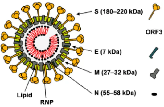
virus, with a positive sense non-segmented ssRNA of 28 kb. Its genome contains 7 open reading frames (ORFs) that codify 16 non-structural and 4 structural proteins. The structural proteins are the spike protein (S), the envelope protein (E), the membrane protein (M), and the nucleocapsid protein (N) (Figure 1) (1, 3).
Figure 1. Schematic representation of the structure of porcine epidemic diarrhea viral
particle. Structural proteins forming the virion are indicated, with their respective molecular weights (MW) (2).
1.1. Porcine epidemic diarrhea
PEDV is the etiological agent of porcine epidemic diarrhea (PED), a re-emergent virus, of enormous impact on the porcine industry. This virus was reported for the first time in England, and it was mistaken with transmissible gastroenteritis virus (TGEV) due to the similar symptoms produced by both viruses (21).
The primary sign of infection with PEDV is a watery diarrhea. It can affect pigs of all ages. Following the major symptom, the vomiting accompanied by anorexia and depression are common. Depending on the pigs' age, severity and morbidity could vary and reach 100% in piglets, but for sows it can have a lower impact (2). The incubation period is from 1 to 8 days, and viral particles can be detected during the first 48h of infection in a fecal sample.
Symptoms can last between 3 and 4 weeks. In adult animals, the disease is self-limiting, and recovery is within 7-10 days. Nevertheless, PED can have a significant impact on the growth of weanling piglets and on the reproductive performance of gilts and sows (reproductive failure) (22).
At the intestine level, PEDV completes its cycle in the cytoplasm of the villous epithelial cells, disrupts the lamina propria, and affects the intake of nutrients and electrolytes, which results in the characteristic diarrhea and deadly dehydration. Severe consequences on piglets could be due to the low rate of regeneration of the intestinal epithelial cells (23).
1.2. Biology of PEDV
1.2.1. Genome organization
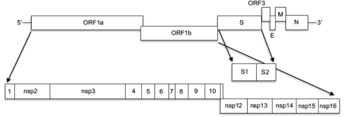
PEDV has a positive sense non-segmented ssRNA genome of about 28 kb. Viral genome contains 7 ORFs that are organized in the following way: 5’ ORF 1a/1b-(S)-ORF3-(E) -(M) -(N) 3’. The extreme 5’ is capped, and the 3’ tail is polyadenylated. 70% of the PEDV genome is occupied by the ORF 1a/1b, which codifies the non-structural polyproteins pp1a and pp1ab. These polyproteins are translationally processed into the 16 non-structural proteins (nsp) that play a key role in viral RNA replication, sub-genomic (sg) mRNA transcription and translation, besides, having an important function in the mechanisms of viral evasion of the host immune response. On the other hand, the genes encoding the structural proteins are a nested set of subgenomic RNAs (sgRNA) that perform a discontinuous transcription (Figure 2) (2).
Figure 2. Schematic representation of porcine epidemic diarrhea viral genome.PEDV
genome contains 7 ORFs. ORF1ab codifies for 16 nsp. ORF S codifies for the spike proteins; ORF3 results in an accessory protein. ORF E, M, N results in the expression of envelope, membrane and nucleocapsid proteins, respectively (6).
1.2.2. Non-structural proteins
The polyproteins pp1a and pp1ab encoded by the ORF1ab and the accessory protein encoded by ORF3 are the non-structural proteins found in the PED virions (2). Pp1a and pp1ab are cleaved internally by proteases into the 16 other proteins: poliovirus 3C-like proteinase (3C1), papain-like proteinase (Plp), one growth factor-like motif (Gfl), X domain (X), metal ion binding domain (Mb), an RNA-dependent RNA polymerase domain (RdRp) and a helicase motif (Hel), among others, which are highly conserved among coronaviruses (2). Interestingly, the accessory protein ORF3 has a high level of genetic diversity. Even though this protein is not essential for the PEDV replication, it has been associated with cell culture adaptation and strain pathogenicity (24).
1.2.3. Structural proteins
Similar to coronaviruses, PED virion is composed of the structural spike (S), membrane (M), envelope (E), and nucleocapsid (N) proteins. They are essential for viral life cycle and stimulation of antiviral host response. These genes and the corresponding proteins are important antiviral targets for viral diagnostics and for the vaccine’s development.
1.2.3.1. Spike protein (S)
The spike protein plays an essential role in PEDV life cycle (2). The S protein is a key factor in the host receptor-virus interaction and virus-cell fusion. It is a class I fusion glycoprotein and has an apparent molecular mass of about 180 to 200 kDa. In order to activate the fusogenic properties of the S protein, it should be cleaved by trypsin-like proteases, after virus attachment (3). The cleavage results in two subunits S1 and S2. The S1, also called the N-terminal, binds to the cellular receptor. The S2, also called the C-terminus, contains the fusion peptide, allowing the virus to enter into the cell by membrane fusion. Successful entry of PEDV depends on this step (3). Additionally, the cell-surface associated S proteins, cleaved by exogenous proteases, can mediate cell-cell fusion and produce multinuclear cells (syncytium), inducing an obvious cytopathic effect (CPE) (25).
Aside from the attachment and fusion, the spike protein of the PEDV is implicated in other steps of viral replication. Wicht et al., in 2014 (26) showed that S protein of PEDV is an essential factor for viral progeny release. Authors infected Vero cells with PEDV classical strain CV777 (wild type WT) and mutant strain (cell cultured adapted and trypsin independent strain), in presence of trypsin or not. After 16 h.p.i they collected the supernatant and measured the amount of virion released. They found that WT strains’ progeny was released in higher quantities, due to its dependency to trypsin (26).
The S protein is also a key factor of the cell adaptation and PEDV virulence. Sato et al., in 2011 (27) described that PEDV adapts to the Vero cell line by acquisition of several mutations in the S protein encoding gene. On the contrary, other structural proteins of the virus remained conserved over time. Interestingly, strains with mutations in the S protein showed attenuated phenotype during in vivo experiments. Thus, authors concluded that S protein is important for PEDV pathogenesis and virulence. The less was the amount of mutations in the S gene the more virulent the strain was. However, after long passage history PEDV S-mutants may revert in virulence and show a milder virulence (27). Insertion and deletion of nucleotides in the S gene are implicated in the PEDV pathogenic variability and facilitate the PEDV vaccine evasion. These strains were designated as S-Indel. As it was discussed earlier, deletions and insertions in S gene can attenuate the stain or enhance its
virulence and cause high mortality in suckling piglets. Thus, these strains are becoming a serious problem for the swine industry. Similar to what was observed for classical strains, severity of clinical signs of S-Indel strains also depends on the age of the animals (28).
The PEDV tropism is defined by the viral S protein. The N-terminal of S protein confers PEDV tropism to respiratory and intestinal tracts of the pigs. Virus with the deletions in this domain is able to replicate only in the enterocytes or in the respiratory tract, but not in both tissues. Similar phenotype has been observed for porcine respiratory coronavirus (PRCoV) and for the natural deletion variant of the TGEV (respiratory tract tropism) (15).
1.2.3.2. Membrane protein (M)
The membrane protein (M) is a type III glycoprotein with a molecular mass of 27-36 kDa. The M protein is the major component of the PED virions. Hence, the M protein is highly conserved among all strains, it is an excellent antiviral target. This feature of the M has important implications in the diagnostics (5). Antibodies produced against M protein of PEDV have been reported to be specific to the PEDV, when they were compared to other coronaviruses M protein (5). This protein plays an important role in PEDV viral life cycle, particularly, in viral assembly through its interaction with the viral E protein of virus (24).
1.2.3.3. Nucleocapsid protein (N)
The nucleocapsid protein (N) is a 58 kDa phosphoprotein. N protein plays a fundamental role in viral genome management (24). For instance, together with the viral RNA it forms the nucleocapsid of PEDV and provides a stable helical shape to the genome. This complex binds to the M protein, in this way protecting the viral genome (29). The N protein is produced in abundance during the early stages of infection and along the viral life cycle. It can be readily detected at the early time of infection (i.e. 6 h.p.i) (30). It has been shown that N protein mainly localizes in the endoplasmic reticulum, interacts with the several molecules involved in the cell cycle, and arrest host cells in S phase (29). Additionally, it has been reported that PEDV inhibits the host immune response by blocking the interferon (IFN)
signal signaling through its nucleocapsid protein. These strategies will be further explained in the section describing the PEDV-host interactions (13).
Recently (in 2019), a group of researchers from the National Science and Technology Development Agency (NSTDA) of Thailand, demonstrated that PEDV N protein can accelerate the growth ratio of a slow-growing PEDV strain. Additionally, authors observed a slight enhancement of infection of porcine reproductive and respiratory syndrome virus (PRRSV), on stable protein PEDV N protein-expressing Vero cells. On the contrary, they didn’t observed any positive effect of PEDV N protein on Influenza virus replication (31).
1.2.3.4. Envelope protein (E)
The envelope protein (E; 7 kDa), a small transmembrane protein of PEDV, is a key component of the viral membrane. It has ion-channel properties and plays an important role in virion morphogenesis and maturation (24). In the infected cells, the E protein is located in the nucleolus or endoplasmic reticulum (ER). The ER localization of E protein induces ER stress, which can lead to unfolded protein response (UPR) and stimulation of inflammatory antiviral responses (14).
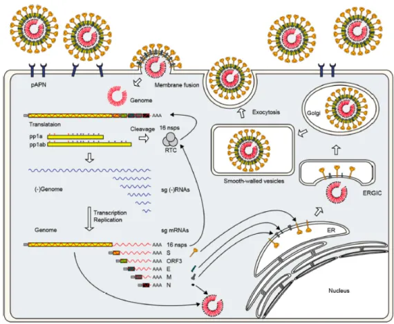
Figure 3. Overview of porcine epidemic diarrhea viral life cycle. PEDV binds its host
cell using he spike protein. Translation of replicases pp1a and pp1ab starts immediately. Then, polyproteins are proteolytically cleaved into 16 non-structural proteins (nsp), which are part of the replicase-transcriptase complex (RTC). Transcription and replication of the genome takes place. Next, the envelope proteins are inserted in the endoplasmic reticulum (ER) and fixed in the Golgi apparatus. Finally, the progeny virus is assembled at the ER-Golgi intermediate compartment (ERGIC) and virions are released by the exocytosis-like fusion (2).
1.2.4.1. Cellular receptors
Until 2016, the N aminopeptidase (pAPN), a receptor abundantly expressed on the epithelial cells (particularly, in the small-intestinal microvillar membrane), was believed to be a principal host receptor for PEDV. Several studies showed that the presence of N aminopeptidase on the surface of permissive cell lines is essential for the PEDV biding and
cell entry. Moreover, in 2010, Nam and Lee reported that overexpression of pAPN in the non-expressing and non-permissive cell lines conferred them a susceptibility to PEDV infection (33). Earlier, Li and Li in 2007, demonstrated the blockage of PEDV infectivity when pAPN was masked by antibodies (34). Additionally, biochemical interactions between the S1 domain of the S protein with the pAPN has been also reported (35). But contrary to what was described for other permissive cell lines, Vero cells, which are widely used for the PEDV propagation, don’t express pAPN. Nonetheless, Vero cells are permissive to PEDV (cell-adapted strains) infection, a fact that suggests the existence of different receptor for the PEDV. For example, PEDV was reported to bind to the sialic acid (15). Shirato et al. in 2017 (36) showed that pAPN was not a cellular receptor for the PEDV, but it could act as a promotor of the infectivity. In order to prove this, authors created a HeLa cell line stably expressing pAPN and infected them with PEDV or TEGV, known to use pAPN as main cellular receptor. Their experiments revealed that recombinant cell line was resistant to the PEDV infectivity but was susceptible to the TEGV. On the contrary, cell transfected with PEDV genome were able to produce infectious PEDV particles. Interestingly, overexpression of pAPN in porcine cells had a positive effect on PEDV infectivity, but it was attributed to the enzymatic activity of the receptor (36).
In 2016, Li et al. (37) showed that overexpression of porcine APN in non-susceptible cells didn’t change their susceptibility towards the PEDV infectivity. They also didn’t find any interaction between PEDV S1 and pAPN using fluorescence-activated cell sorter (FACS)-based assays. All their experiments were performed with multiple PEDV strains to exclude strain-specific artifacts (37), thus, there are no robust evidences that PEDV infects pAPN-expressing cells.
The ability to bind to specific cellular receptors is an important factor in determining the host range and tropism of viruses. It is well-documented that PEDV has tropism for small and large intestinal epithelial cells. However, additional studies are critical for better understanding the cellular tropism and evolution of PEDV. Nevertheless, in the process to discover the main cellular receptor for PEDV, co-receptor molecules or entry enhancement proteins have been found; such as occludin protein, member of the tight junctions of the small intestine (38).
1.2.4.2. Viral entry
After viral attachment via S protein to the cellular receptors, the spike protein undergoes conformational changes, which expose its trypsin cleavage site. Next, the trypsin cleaves the S protein at two sites: first, at the borderline between the subunits S1 and S2, and, second, at the S2, activating the exposure of the fusion peptide and positioning it in a close proximity to the host cell membrane. Then, cellular and viral membrane merge and PEDV genome is delivered into the host cells (Figure 3). This step might result in formation of syncytia, which is a characteristic CPE of the PEDV (25).
Occludin is a protein present in the tight junctions of the epithelial barrier, located in the intestinal epithelium cells (39). Luo et al., in 2017 (38), suggested that overexpression of the occludin in target cells enhanced susceptibility to the PEDV infection. Additionally, authors showed that reduction of occluding expression in target cells through RNAi assay, decreased significantly their susceptibility to PEDV infection. It has been observed that macropinocytosis inhibitors impeded occludin internalization and virus entry, indicating that virus entry and occludin internalization are tightly linked. Yet, the macropinocytosis inhibitors didn’t impede virus replication, once the virus was inside the cells. This finding suggested that occludin internalization by macropinocytosis or a macropinocytosis-like activity is implicated in PEDV entry, but occludin is not involved in the initial attachment of virus to the cell (38).
1.2.4.3. Genome replication
Immediately after membrane fusion, the viral genome is released into the cytoplasm. It is translated into the first 2 replicases pp1a and pp1ab, which are proteolytically cleaved into the 16 non-structural proteins (nsps) that are part of the replication and transcription complex (RTC). Then, the RTC synthetizes the negative-strand RNA using genomic RNA and produces full-length genomic RNA and subgenomic mRNAs. Each subgenomic mRNA is translated into a structural protein (Figure 3) (2).
During viral replication, the viral envelope proteins, S, E, and M are inserted into the endoplasmic reticulum and attached to the Golgi apparatus. The N protein interacts with the genomic RNA to form helical ribonucleic protein complexes (RNP). The progeny virus is assembled after maturation of the RNP in the endoplasmic reticulum Golgi intermediate compartment (ERGIC), and then freed by the exocytosis-like fusion of smooth-walled vesicles with the host cell plasma membrane (Figure 3) (2).
1.2.4.5. Microvesicles and exosomes
Extracellular vesicles (EV) are vesicles release by the cells into the media. They are classified in three mayor groups according their size; exosomes, microvesicles and multivesicular bodies (MVBs) (40). Microvesicles are cell-derived membrane vesicles that mediate the cellular signaling and transport of various molecules. Their main objective is to communicate by delivering molecules to the cells. Exosomes are the most studied and well-characterized EVs. They are derived from the MVB and they play an important role in intercellular communication via RNAs and proteins between neighbor cells (41). Different cellular and extracellular processes and signals can trigger production of the exosomes, such as cell differentiation, activation, stress, cell death, and viral infection have been reported (40). The function of each type of EVs varies among them, besides cells communication, exosomes contain RNases, trypsin, or any degradative substance, due the composition of its bilipid membrane (42). Microvesicles on the other hand, are strictly related to the cell communication processes (40).
Coronaviruses replicate their genomes in the cytoplasm, in the specific replicative structures associated with cellular membranes. Viral infection induces formation of the cell-derived organelle-like membranous structures, where the viral replication-transcription complexes (RTCs) localize. Initially, the intracellular rearrange results in two types of organelle-like replicative structures: the double membrane vesicles (DMV) and convoluted membranes (CMs). Later, highly organized cubic membrane structures, the large virion-containing vesicles (LVCVs) and condensed tubular bodies are formed (43). These microvesicles have been recently described for PEDV infection. More specifically, PEDV
non-structural proteins trigger the synthesis of these microvesicles, appearing after 24 h.p.i, with a peak after 60h post infection. It was suggested that the endoplasmic reticulum, which plays a key role in late viral assembly, is the mostly likely source of DMVs (44).
Porcine reproductive and respiratory syndrome virus (PRRSV) is a nidovirus, part of the Coronaviridae family. Recently in a study, exosomes purified form PRRSV-infected cells were analyzed through proteomics, showing that these particles contained genomic viral RNA and viral proteins. Interestingly, infection of PRRSV-susceptible and -non susceptible cells with the purified exosomes, performed successful infection. Authors conclude that exosomes can be a mechanism of PRRSV to evade host immune response (45). Importantly, exosomes have been proven as a vaccine mechanism against PRRSV. Authors have report that exosomes isolated form non-viremic animals (animals previously exposed with PRRSV but free of virus at the moment of isolation) contained antigenic viral proteins. Moreover, the serum of non-viremic pigs reacted against the purified exosomes. Authors conclude that exosomes could be a new approach to control PRRSV infection (46).
Little is known about PEDV infection stimulating the use of microvesicles and exosomes. Demonstrating the utility of these molecules for PEDV viral life cycle, could help us to identify a new antiviral therapy strategy, as well as to fill the knowledge gap of PEDV-host molecular interactions.
1.3. Global distribution
Diarrheal disease resulted from PEDV infection in pigs was first reported in England in 1971. Initially, due to the typical symptoms shown by sick pigs, it was proposed that the causative agent is the transmissible gastroenteritis coronavirus (TGEV), due to the typical symptoms shown by sick pigs. Five years later, an outbreak of PEDV was reported in Belgium, and subsequently, in the 1980’s and 1990’s, PEDV was identified throughout Europe.
With the time, the outbreaks decreased on this continent and PEDV crossed borders to Asia, where it became endemic. Since 2010, several Asian countries such as Korea, Vietnam, China and Japan, large outbreaks causing major impact on the porcine industry have been observed (47). Significant outbreaks of PEDV with 50 to 90% mortality and 80 to
100% morbidity in suckling piglets emerged (48). In 2013, PEDV was reported for the first time in United States, and within a year it extended all over the country and neighbors (Mexico and Canada). It was shown that the strains isolated in the US are genetically related to the strains from China, with a distinguished U insertion. One year later, the PEDV strains isolated in the US showed different deletions and insertions in the S gene, suggesting a possible recombination event between the Chinese and US strains (2). Thus, taking in the consideration the phylogenetic studies of the S gene of PEDV, it was proposed that PEDV strains can be classified into 2 genotypic groups: genogroup 1 (G1; the classical) and genogroup 2 (G2; the field epidemic or pandemic). Each genogroup was additionally divided into the subgroups 1a and 1b, and 2a and 2b, respectively. G1a includes the classical PEDV strain CV777, vaccine strains, and viral strains adapted to the cell cultures. G1b contains new variants identified initially in China and then in the US and South Korea. The G2 genogroup contains global field isolates, which are grouped into 2a and 2b subgroups, responsible for previous epidemic outbreaks in Asia and current pandemic outbreaks in North America (2).
1.4. Strategies for the control of PEDV
1.4.1. Diagnostics
Because of PEDV similarities to any pathogen causing diarrhea, vomiting and anorexia; diagnostic of porcine epidemic diarrhea virus can’t be based on symptoms presented by the animal. Diagnostic approaches such as direct and indirect immunofluorescence, ELISA are routinely performed, however, the RT-qPCR is the standard technique to diagnose PEDV-infected pigs. Immunohistochemical assay and direct electron microscopy are other additional techniques, which help determining the presence of PEDV in the samples. Depending on the type of samples, one or other type of technique could be more suitable. ELISA is used generally to study the presence of IgG or IgA in the sera of PEDV-infected piglets (49, 50). For the diagnostics purposes, an indirect ELISA, based on the PEDV structural protein M, has been designed as well, showing no cross-reactivity with the M proteins of other Coronaviruses (5).
RT-qPCR is the most commonly used technique, because of its sensitivity, specificity, and rapid time to results. Primers for the conserved regions of PEDV genome such as M gene, N gene, and ORF 1 are utilized to analyze samples for the PEDV presence (49). Since the S gene is the least conserved and undergoes through insertions and deletions processes, it is not used for diagnostics purposes (28).
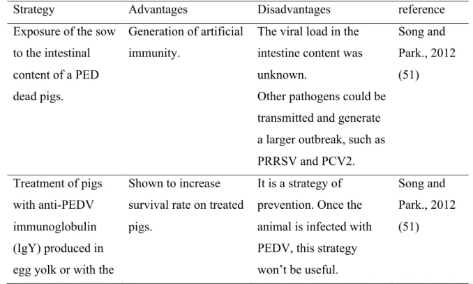
1.4.2. Treatment
Four strategies have been described to treat PEDV outbreaks (Table I). Although, most of these options could work, majority of them have important disadvantages, that can’t be ignored. The best approach to overcome and avoid any PEDV outbreak seems to be safe and effective vaccines.
On the conventional side, farmers treat suckling piglets with oral electrolyte solutions, to overcome dehydration. For adult pigs, it is recommended drop the intake of dry food upon 12–24 h and then, water should freely available for the pigs (48).
Table I. Strategies for PEDV-infection treatment
Strategy Advantages Disadvantages reference
Exposure of the sow to the intestinal content of a PED dead pigs.
Generation of artificial immunity.
The viral load in the intestine content was unknown.
Other pathogens could be transmitted and generate a larger outbreak, such as PRRSV and PCV2. Song and Park., 2012 (51) Treatment of pigs with anti-PEDV immunoglobulin (IgY) produced in egg yolk or with the
Shown to increase survival rate on treated pigs.
It is a strategy of prevention. Once the animal is infected with PEDV, this strategy won’t be useful.
Song and Park., 2012 (51)
colostrum of immunized cow. Expression of neutralizing anti-PEDV antibodies in E. coli to block or
treat viral infection
in vivo.
Demonstrated to neutralize PEDV in
vitro.
Performed only in vitro.
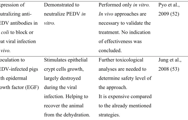
In vivo approaches are
necessary to validate the treatment. No indication of effectiveness was concluded. Pyo et al., 2009 (52) Inoculation to PEDV-infected pigs with epidermal growth factor (EGF)
Stimulates epithelial crypt cells growth, largely destroyed during the viral infection. Helping to recover the animal from the dehydration.
Further toxicological analyses are needed to determine safety level of the approach.
It is expensive compared to the already mentioned strategies.
Jung et al., 2008 (53)
1.4.3. Vaccination
Vaccine development against PEDV began earlier in Asia compared to Europe and North America. PEDV is endemic in several Asian countries; therefore, it was a constant demand for effective vaccines against the PEDV. In Europe, the frequency of PEDV outbreaks decreased by 2007, however, mild symptoms in pigs of all ages have been reported in positive farms. In North America, PEDV appeared in 2013 in the United States and has been around since then (54).
Table II. Available vaccines for PEDV prevention in Asia and North America
Location Type of vaccine
Administratio n
North America iPED: Inactivat e particle Two intramuscular doses within three weeks, in young pigs. Three oral inoculation to sows Truncated in the S gene, produced in SirraVax℠ RNA Particle. Viral shedding reduced in young pigs.
Mortality drop from 91% to 63% in sows Fredrickso n et al., 2014 (55) Porcine Epidemi c Diarrhea Vaccine (Zoetis): Inactivat ed whole virus Two oral doses within three weeks for pregnant sows Liquid from with an adjuvant that claims to increase the immune response Increase titer of neutralizing antibody in sows compared to the control group Schwartz et al., 2016 (56) Vaccine develop ed by InterVac : Inactivat ed virus Two intramuscular doses within two weeks for pregnant sows (pre-farrowing) Liquid from with an adjuvant that claims to increase the immune response High levels of neutralizing antibodies were found in the milk and serum of piglets born to vaccinated sows Makadiya et al., 2016 (57) Asia Trivalen t vaccine (PEDV, TGEV and porcine rotavirus One-time intramuscular inoculation to piglets Based on the classical CV777 (G1-a) strain, produced on Vero cells Resulted in partially protected piglets against PEDV Chen et al., 2010 (58)
): Attenuat ed vaccine Bivalent (PEDV and TGEV): Attenuat ed Vaccine One-time intramuscular inoculation Contained the PEDV strain ZJ08 (G1-b) or AJ1102 strain (G2-b). Still under evaluation Song et al., 2015 (1) P-5V vaccine: live Vaccine Two-time intramuscular inoculation PEDV strain 83P-5 (G1-a) attenuated through several passages on Vero cells The vaccinated sows displayed PEDV specific antibody responses, containing neutralizing antibody in the colostrum. Reduction of clinical signs and mortality on piglets fed with the immunized sows-colostrum was observed. Sato et al., 2018 (59) South Korean G2-b vaccine: Inactivat Two-time intramuscular inoculation of Pregnant sows based on G2-b strain KOR/KNU-141112/2014 piglets born to vaccinated sows had reduced morbidity, mortality after
Song et al., 2015 (1)
ed vaccine (pre-farrowing) challenge with PEDV
The most common approaches are to inoculate the sows before farrowing, in order to generate lactic immunogenicity, or direct inoculation to the young pigs. In general, the reduction of mortality, the morbidity and virus shedding, in orally inoculated young pigs were higher than in new born piglets. In comparison, sows inoculated with either viral genome or attenuated virus, produce a higher titer of neutralizing antibodies in the milk and colostrum. New generation of vaccines, directed against the PEDV strains G2, showed increased significantly pig’s survival (54).
1.5. PEDV-host interactions
1.5.1. Immunogenic interactions between host cells and PEDV
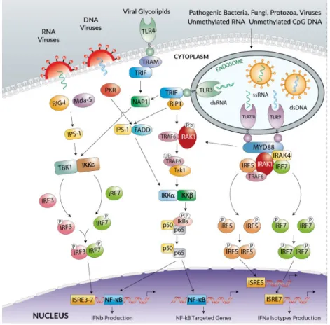
To any pathogen invasion, cells will display several strategies such as cytokines and chemokines production to eradicate or control infection. One of the most important cytokines to restrict viral replication is the interferon (IFN) production (60). However, viruses have developed strategies to supress IFN production through their viral proteins (61–63). The activation of IFN response can be mediated by toll-like receptors (TLRs) and RIG-I-like Receptors (RLRs), which recognize viral RNA or DNA in the endosomes or cytosol. Then, activation of Serine/threonine-protein kinase (TBK1)-mediated phosphorylation of the IRFs (interferon regulatory factors) take place, activating the IFN transcription (Figure 4) (64). Serine/threonine-protein kinase (TBK1), a member of the IKK protein kinase family, is one of the many molecules that play key roles in the IFN regulation. TBK1 is a member of the IKK protein kinase family.
Figure 4. Inflammatory response to cytosolic or endosomal nucleic acid
sensors.Recognition of nucleic acids through endosomal or cytosolic sensors, will activate a cascade of transcription of different molecules (IFRs, NF-kB). This process promotes cell activation as well as expression of different genes, resulting in TNF, IFN and IL responses (65).
Since TBK1 plays an important role in the IFN signaling pathway, viruses evolved mechanisms aimed at inhibition of the IFN production (66). This immune response evasion has been shown also for the PEDV. Its structural protein N impairs the IFN production by interacting with TBK1, sequestering this molecule and avoiding vital interaction between TBK1 and IRF3. The precise mechanism of this interaction is yet to be investigated, but several theories are proposed (9). Moreover, PEDV can interfere with type I INF production not only through its structural proteins but also through its non-structural proteins like nsp1(6). As it was mentioned earlier, the interaction between TBK1 and IRF3 is important for regulation of IFN expression. Upon PEDV infection the IRF3 displays a signal to form a
complex with the transcription co-activator CREB (cAMP responsive element binding)-binding protein (CBP)/p300. The IRF3-CBP/p300 complex then binds to the positive regulatory domain (PRD) regions of the IFN-β promoter, assembling together with NF-κB and other factors, to stimulate the transcription of type I IFN genes. The IRF3–CBP/p300 interaction is vital for IFN transcription. The nsp1 of PEDV causes the CBP degradation by the proteasome-dependent pathway (6).
Moreover, little is known about the pro-inflammatory response (chemokines) against PEDV. It has been shown that PEDV down-regulates different chemokines (IL-1α, IL-1β, CXCL8) to promote its own replication (67). Yu el al., in 2019 (67) further showed that PEDV nsp4 contributed to the up-regulation of IL-1α, IL-1β, TNF-α, and CXCL8, inhibiting PEDV viral life cycle in vitro (67). Additionally, Xu et al. in 2013 (68) demonstrated that cells overexpressing PEDV E protein were significantly up regulating IL-8. Authors related the up regulation of the IL-8 with the fact that E protein is normally and mainly localized on the ER, where it causes ER stress and IL-8 activation. At the same time, overexpression of E protein causes high expression of B-cell lymphoma 2 (Bcl-2) protein, a cell survival anti-apoptotic factor. Additional findings will be essential to elucidate the exact role of E protein in the antiviral host response against PEDV (68).
Likewise, it has been described that N protein up-regulates IL-8, causes ER stress and prolongs cell cycle phases, which are beneficial for viral infection. The S phase of cell cycle provides an optimal cellular environment for viral replication. Interestingly, the N protein of PEDV is able to inhibit the cell proliferation and prolongs the S-phase cell cycle (29) . Cyclin A is an important molecule for cells to pass from the S phase to G2/M phase. It has been shown that in PEDV N protein-expressing cell lines, cyclin A is significantly lower than in control cell lines (29). Also, it was found that PEDV N protein significantly inhibits the transcription of cyclin A. Since the PEDV N protein is mainly localizes in the ER and up regulates the chaperon GRP78, the ER stress response during PEDV infection (at least partially) is attribute to this protein. Finally, because PEDV N protein induces ER stress, it significantly activates NF-kB, which leads to induction of IL-8 transcription (29). Further studies are needed for understanding the roles of pro-inflammatory response in PEDV replication and host immune response.
2. Proteomic analyses
During the past three decades, mass spectrometry (MS) based proteomics has become one of the preferred methods for identifying protein-protein interactions and gaining insights into the complex networks of molecular interactions between the host and pathogen (69–72). There are different types of proteomic approaches: structural, functional, quantitative, and comparative expression profiles. These approaches can be performed through labeling proteomics, or label-free proteomics (Figure 6) (73). In general, the proteomic strategies involve following common steps: production and extraction of the proteins of interest from the sample, preparation of the protein samples for chemical or enzymatic digestion, digestion of proteins followed by the cleanup or desalting of the final peptide mixture prior to MS, analysis of the produced peptides by different types of mass spectrometers (Table III). The final step of proteomics workflow is the performing a database search to identify the proteins based on the peptides discovered in the sample.
Proteomics approaches proved to be effective at characterizing the composition of viral composition, studying viral life cycle, and changes in the virally-infected cells. Furthermore, proteomic tools are widely employed for searching new targets for antiviral strategies (74, 75).
2.1. Key steps in proteomic analysis
Once proteins have been produced, they can be separated or not prior to MS analysis. Separation before the MS analysis is most commonly done through one-dimensional or two-dimensional gels. Depending on the complexity of the sample, separation of proteins can be reasonable or not (76).
Prior to MS, proteins are enzymatically or chemically digested into peptides. There are two ways digest proteins; the first one is directly with proteases (in-solution), and the second one is in gel digestion, if gel electrophoresis separation was performed before (77). Then, the resulted peptides are ionized and desalted through a mass spectrometer. The mass spectrometers are typically composed of four elements: an ionization source, mass analyzers, an ion mirror, and a detector. Variety of mass spectrometer configurations are used, either
simple or hybrid (Table III). Recently, a new generation of mass spectrometers has been developed combining segmented quadrupole and Orbitrap mass analyzer, called the Q-Exactive. It designed to make easier the measurement and coupled with a higher sensitivity, compared to older generation of spectrometers (78). Among the new features of the Q-Exactive instrument are the high ion currents, fast high-energy collision-induced dissociation peptide fragmentation, double mass spectrometric resolution, 1 s for a top10 higher energy collisional dissociation (79).
Table III. Variety of mass spectrometer configurations commonly used for quantitative
proteomic analysis
Mass spectrometer Specifications Reference
Electron spay ion source (ESI)
Involves 3 phases: a dispersal of charge droplets in a delicate spray, then a solvent evaporation, and, finally, an ion ejection of the very charged droplets, resulting in the foundation of desolvated ions
Ho et al., 2008 (80)
Matrix Assisted Laser Desorption/Ionization (MALDI)
The technique involves the following three steps: first, a low organic compound matrix is added to the digested sample, and the mixture is applied to a metal plate and dried; then, the sample plate is subjected to a laser irradiation for a short time, forming molecular ions; third, the resulting ionized peptides are analyzed by a mass analyzer to reveal characteristic information about the composition of the sample based on their mass-to-charge ratios.
Clark et al., 2013 (81)
Triple-quadrupole mass spectrometers
Often used to obtain amino acid sequences. This system performs the tandem mass spectrometry (MS/MS). Called that way
Graves and
Hystead 2002 (77).
because it involves two stages of mass analysis by two different mass analyzers
Quadrupole-time-of-flight (QqTOF)
This is a combination of a quadrupole mass spectrometer with a TOF analyzer. The principal application of a Qq-TOF mass spectrometer is the protein identification by amino acid sequencing, including any potential post-translation modifications that those amino acids might have undergone.
Graves and
Hystead 2002 (77).
Matrix Assisted Laser Desorption/Ionization-time-of-flight
(MALDI-TOF)
Is a combination of matrix-assisted laser desorption/ionization and time-of-flight mass analyzers. Its main application is mass fingerprinting of peptides. It is completely automatic, which makes it easier to work with a large scale of samples.
Graves and
Hystead 2002 (77).
Fourier transform ion cyclotron resonance (FT-ICR)
Is a Fourier transform ion cyclotron resonance mass spectrometer. It reaches a high mass resolution and mass accuracy. Similar to others, this technique identifies the amino acid sequences and protein fingerprints
Perry et al., 2008 (82)
Linear trap quadrupole (LTQ)-Orbitrap
Can pull up a mass resolution up to 150,000 ions. It has a high mass accuracy, and a larger capacity of ion trapping compared to FT-ICR, among other characteristics. This system is less expensive compare to others, smaller and easier to manage.
Perry et al., 2008 (82)
The last step in a proteomic analysis is the data analysis. After the processing the sample by a mass analyzer, the peptides are identified through peptide mass fingerprinting (PMF). This technique uses the masses of peptides derived from the analyte’s spectra as to
check against the database of predicted peptide masses from databases of known proteins. If they overlap, the identification and changes can be assessed. There are different data searching programs such as MASCOT, SONAR, SEQUEST that are available for this task. MASCOT seems to be the most complete database. The main disadvantage of these programs is their vagueness, when identifying proteins due to peptide redundancy. In other words, similar amino acid sequences with small differences in the post-translational changes will have comparable peptide masses (77, 83).
2.2. Types of mass spectrometry analyses
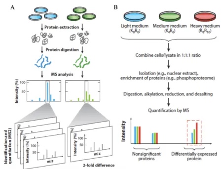
As it was discussed before, there are two types of quantitative proteomics: label-free or label-based proteomics (Figure 6). For the labeling techniques, peptides are tagged metabolically, chemically, or enzymatically. The label-free quantitation technique determines ion quantity or peak intensity (73).
Among the metabolic labeling based techniques, the stable isotope labeling in cell culture (SILAC) is one of the best developed approaches. This technique relies on growing cells in culture media containing “light” and “heavy” isotopically labeled amino acids, which are going to be incorporated into the cell proteins by metabolic processes of protein synthesis. Heavy-labeled proteins are going to be distinguished from the pool of the proteins, and the difference between the peak’s intensities will reflect the relative abundance of proteins labeled with the same amino acid (Figure 6. B) (84).
Along chemical labeling, isobaric tags for relative and absolute quantitation (iTRAQ) and the tandem mass tags (TMT), are the most used techniques. These tags are distinguished by their abundance and scores in the mass spectrometers. There are 12 available isobaric tags, which mean that at least 12 conditions can be compared (85). Additionally, trypsinization of samples can’t be performed, because trypsin is unable to cleave modified lysine, so protein digestion step becomes complex (73).
Labeled proteomics has major advantages when studies are targeting a known group of proteins. Nevertheless, when performing a discovery proteomics approach, labeling is limited to a certain number of labels. Economically, these types of studies are high-priced, so limitation on number of samples would be a concern (84).
Figure 5. Labeled and label-free quantitative proteomics. (A) Spectral counting–based
label free quantification (LFQ) techniques for identification and quantitation using MS/MS spectra. Quantitation is based on the number of spectra identified for each peptide. (B) Amino acid tagging and targeted proteomics strategies. Cells are grown in media containing light, medium, or heavy amino acids with stable isotopes, and lysates are combined for processing (73).
Label-free proteomics (LFP) is a simple and cost-effective application in quantitative proteomics. This approach has been used to either replace or to enhance labeling techniques. LFP can be divided in two types, ion counting, and intensity based. Ion counting determines the number of peptides of a protein in a sample and divides it by the theorical number of peptides of the identify protein. One of the disadvantages of this approach is that the number of peptides generated by proteolytic digestion (with trypsin) depends on the length of the protein. Therefore, quantitation of lighter proteins (< 20 kDa) won’t be as precise as for larger proteins (73).The second LFP approach is intensity based, where the MS-signal intensity is measure, in the area under the chromatographic peak of the precursor peptide ion, while it is eluted in the liquid-chromatography (LC) column (73).