Article Number: 9EC92A663380 ISSN 1996-0875
Copyright © 2020
Author(s) retain the copyright of this article http://www.academicjournals.org/JMPR
Journal of Medicinal Plants Research
Full Length Research Paper
Antimycobacterial activities, cytotoxicity and
phytochemical screening of extracts for three medicinal
plants growing in Kenya
François Nimbeshaho
1,2, Charity Ngina Mwangi
1, Fred Orina
3, Meryl Chacha
3,
Nicholas Adipo
3, Jones O. Moody
1and Elizabeth M. Kigondu
31
Pan African University Life and Earth Sciences Institute (Including Health and Agriculture)-University of Ibadan, Ibadan, Oyo State, Nigeria.
2
Centre de Recherche en Sciences Naturelles et Envionnementales-Université du Burundi, Burundi. 3
Kenya Medical Research Institute-Nairobi, Kenya.
Received 3 January, 2020; Accepted 24 February, 2020
Tuberculosis (TB), an airborne disease, is among the ten leading deadly diseases worldwide. Despite the efforts of WHO and its partners to eradicate it, it is still a public health issue especially with the rise of multi-drug resistant tuberculosis (MDR-TB) and extensively drug- resistant tuberculosis (XDR-TB).
Commiphora species (Burseraceae family) are known in the Kenyan traditional medicine to treat
respiratory diseases including TB. In the search of new anti-TB alternative drugs, plant materials from
Commiphora mildbraedii Engl. (root bark and stem bark), Commiphora edulis (Klotzsch) Engl. (stem
bark and leaves) and C. ellenbeckii Engl. (Stem bark and leaves) were tested for antimycobacterial activity, cytotoxicity and phytochemistry. 100 g of the powdered plant materials were macerated using the serial method with solvents of increasing polarity. Aqueous extraction was carried out by decoction. The microbroth dilution method was used to determine the antimycobacterial activity (MIC) against a model Mycobacterium smegmatis ATCC607 while the cytotoxicity evaluation (CC50) was carried out using the MTT assay. The most active extract was fractionated using preparative TLC and fractions were analysed by GC-MS. Thirty extracts were obtained from the 6 different plant materials and eleven of them exhibited the antimycobacterial activity with the methanolic extracts of the stem and root bark of C. mildbraedii, and the aqueous extract of the C. ellenbeckii leaves exhibiting high activities (MIC= 0.39, 0.78 and 0.78 mg/L respectively). The MTT assay showed no or low cytotoxicity. The GC-MS analysis of the preparative TLC fractions from the methanolic extract of C. mildbraedii revealed the presence of 42 compounds belonging to 10 different classes of phytochemicals. Lup-20(29)-en-3-one and o-xylene were the most abundant. Except o-xylene and α-terpineol, all the compounds were detected for the first time in the Commiphora genus. These findings justify the ethnomedicinal uses of
Commiphora species in TB treatment.
Key words: C. mildbraedii, C. ellenbeckii, C. edulis, antimycobacterial activity. INTRODUCTION
Tuberculosis (TB) is an airborne contagious disease caused by Mycobacterium tuberculosis. Despite all the efforts of the World Health Organization (WHO) and its partners to eradicate this disease, TB continues to be a
public health concern. TB is one of the top 10 causes of death worldwide. In 2017, there were an estimated 10 million new TB infections worldwide. People co-infected with HIV and TB accounted for 9% of the total
130 J. Med. Plants Res.
and the majorities were adults up to 90% (aged ≥15 years). TB is also the main cause of deaths related to antimicrobial resistance and the leading killer of people with HIV. Globally, the TB mortality rate has widely decreased from 23% in 2000 to 16% in 2017 and the TB incidence rate is generally reducing to about 2% yearly worldwide (WHO, 2018). This reduction of the incidence is contrasted with the continuous rise of drug resisting TB strains, hence the need for developing new intervention methods. In 2017, an estimation of 558 000 new cases with resistance to rifampicin (RR-TB) was reported and eighty-two percent (82%) of those cases had multi-drug resistance-tuberculosis (TB). Moreover, an estimated 8.5% of people with MDR-TB had extensively drug-resistant MDR-TB (XDR- MDR-TB). About 23% of the world’s population has latent TB infection and is therefore prone to develop active TB disease during their lifetime (WHO, 2018). It is for these major reasons that researches carried out these days, focus more on developing new alternatives to counteract this hectic problem of drug resistance in bacterial infections and particularly in TB.
Synthetic drugs are not the only focal point, nature provides also a great reservoir of medicinally active ingredients that could alleviate or suppress this phenomenon of drug resistance (Zumla et al., 2013).
Commiphora plants (myrrh plants) are well known in
various cultures to have medicinal virtues including treating infectious diseases. The myrrh plants (Commiphora sp.) belonging to the Burseraceae family are among the most popular plants that have been in use since time immemorial.
The myrrh genus (Burseraceae family) is the richest in terms of species among the flowering plants of the Burseraceae family. The plant list reports that it comprises approximately 208 species (https://www.plantlist.com). They are shrubs and trees mostly distributed throughout the sub-tropical regions of Africa, the western Indian Ocean islands, the Arabian Peninsula, India and Vietnam (Daly et al., 2011). The genus Commiphora has been exploited worldwide as a natural drug to treat pain, skin infections, inflammatory conditions, diarrhea, periodontal diseases, and wounds (Nomicos, 2007; Abdul-Ghani et al., 2009). Its ethnomedicinal uses, pharmacology and phytochemistry have been thoroughly reviewed by Hanuš et al. (2005) and Shen et al. (2012).
Egyptians used C. myrrha early 4000 years ago, in the process of embalming bodies and its perfume was used in spiritual rites (Abdul-Ghani et al., 2009). In the Islamic inspired culture, it was utilized for the treatment of intestinal parasites, diarrhea, wound treatment,
persistent chest ailments (Ghazanfar, 1994). Chinese medicineexploitsC.myrrha forthetreatment of wounds, dysmenorrhea, abnormal blood clotting (thrombosis) (Hanuš et al., 2005), for toothache, oral ulcer, tumor, inflammatory diseases and for acroanesthesia (Shoemaker et al., 2005). Pharmacological studies carried on C. myrrha proved that it exhibits good antimicrobial activity. It was tested against Gram-positive and Gram-negative bacteria and some fungal strains, and it inhibited the growth of the tested strains (Omer et al., 2011, Alhussaini et al., 2015).
South Africans valued C. molmol (or C. mollis) for the treatment of fever (malaria and typhoid), wound healing, cancer, ulcer, rheumatic conditions, colds, nasal congestion and coughing (Van Wyk et al., 2002). It is reported that the methanolic extracts of C. molmol exhibited high antibacterial activity against Gram-positive bacteria than tested Gram-negative bacteria with MIC ranging from 31.25 to 500 µg/mL (Abdallah et al., 2009). In another study, the oil from C. molmol has strong activity against clinical S. aureus isolates including multi-drug resistant strains (Mohammed and Samy, 2013).
In Nigeria, the seed decoction of C. Africana is traditionally used for expelling tapeworms (McGuffin et al., 2000). An alkaloid isolated from C. africana presented antimicrobial activities against all the test microorganisms (Banso and Mann, 2006). On the other hand, the chloroform and methanolic extracts (leaves and stem) of ten South African Commiphora species including C. africana, C. schimperi, C. grandulosa, C.
marlothii, C. mollis, C. neglecta, C. pyracanthoides, C. tenuipetiolata, C.viminea weretestedforthe antimicrobial activity and were found to possess high bioactivity with MICs of 0.01-8.00, 0.25-8.00 and 1.00-8.00 mg/ml against positive bacteria, yeasts and Gram-negative bacteria respectively (Paraskeva et al., 2008).
In folk medicine, the bark infusion of C. edulis was used to treat malaria and its roots, leaves, and stem are used in the treatment of stomachache, menstrual problems and spirits related illnesses (Orwa et al., 2009). Paraskeva et al. (2008) tested C. edulis for the antimicrobial activity and found that it was able to inhibit the growth of yeasts and Gram-positive and Gram- negative bacteria tested with MIC ranging from 2-8 mg/mL.
The literature provides a number of reports where some Commiphora species were tested for antimycobacterial activity using different Mycobacteria model strains and the pathogenic M. tuberculosis, a pre-requisite step in TB drug discovery.
The essential oil from the fresh aerial parts of C. *Corresponding author. E-mail: nimbeshahof@gmail.com.
Author(s) agree that this article remain permanently open access under the terms of the Creative Commons Attribution License 4.0 International License
opobalsamum L., a plant used by Saudi Arabians to treat
infectious problems (Shen et al., 2007) was evaluated for the antimicrobial activity and exhibited a good activity with IC50 of 80, 90, 150 and 15 µg/mL against Candida
glabrata, C. krusei, Cryptococcus neoformans, and Mycobacteria intracellulare respectively (Al-Massarany et
al., 2008). The yellowish sap from the stem of C. mukul (guggul) is well known in Ayurvedic medicine over 2000 years for the treatment of arthritis inflammation, hyperlipidemia related diseases and to improve the hepatic antioxidant defense system (Al-Rejaie, 2012). It is also known to treat some infections and is reported to exhibit good antimycobacterial activity against two models of Mycobacteria, M. aurum and M. smegmatis with a MIC 62.5 µg/mL for both (Newton et al., 2002).
Tanzanian pastoralists use aromatic saps from the stem bark of C. eminii in the treatment of skin infections, gastrointestinal infections, wounds and as well as dry and blood cough (Erasto, 2012). Two sterols (4 -methyl-cholest-7-en-3-ol and -methyl-cholest-7-en-3-ol) isolated from the sap of C. eminii stem bark were tested for antimycobacterial activity against Mycobacterium madagascariense and Mycobacterium indicus pranii and
only the second sterol (cholest-7-en-3-ol) exhibited antimycobacterial activity with MIC values of 1.6 mg/mL against both Mycobacteria strains used.
Indigenous tribes in the Northeast of Brazil utilize C.
leptophloeos as an infusion, tea or syrup for the
treatment of their illnesses, such as infectious and inflam-matory ones (Silva et al., 2011). Hinokinin, a compound that was isolated from C. leptophloeos presented antimicrobial activity against a variety of microbes with MIC values ranging from 0.0485 to 3.125 mg/mL (against different S. aureus clinical isolates) and showed a bactericidal activity against Methicillin- resistant
Staphylococcus aureus (MRSA) isolated from blood with
of MMC= 0.40 mg/mL. Additionally, the C. leptophloeos extracts were tested for antimycobacterial and it exhibited moderate activity against M. smegmatis (MIC=12.5 mg/mL) and M. tuberculosis (MIC=52 mg/mL) (De Souza et al., 2017). The Maasai community of southern Kajiado District of Kenya utilizes Commiphora sp. for various medicinal uses including the treatment of killing chiggers, open wounds, as a skin moisturizer and for the treatment of respiratory diseases such as coughing and tuberculosis (Kiringe, 2006).
Kenyan biodiversity is so rich in Commiphora species but less has been subject to studies for proving the above mentioned ethnomedicinal uses. Therefore, this study was designed to establish the antimycobacterial, cytotoxicity and phytochemical properties of selected African Commiphora species growing in Kenya. These include Commiphora mildbraedii Engl., endemic in
regions ranging from Ethiopia to Kenya, and Commiphora
edulis (Klotzsch) Engl. And Commiphora ellenbeckii Engl.
found abundantly in Kenya, Ethiopia and Tanzania (Vollesen, 1989). This work intends then to verify the efficacy and safety of the three Commiphora plants used
in the treatment of TB by the Kenyan local communities; and to screen their bioactive compounds.
MATERIALS AND METHODS Plant collection
Plant materials consisting of C. mildbraedii stem and root bark, C.
edulis stem bark and leaves and C. ellenbeckii stem bark and
leaves were identified by a taxonomist and collected in various locations of Kenya. C. edulis was collected at Chekbor, Marakwet County and C. mildbraedii and C. ellenbeckii were collected at Kibwezi forest, Kenya. Voucher specimen were prepared and deposited at the University of Eldoret Herbarium, Eldoret, Kenya where they were assigned voucher numbers: CFW/31/1/19/003 for
C. edulis, CFW/3/2/19/008 for C. mildbraedii and CFW/3/2/19/010
for C. ellenbeckii.
Sample processing
Drying of the plant materials was carried out in open-air and well-ventilated room for about one month. Once the plant materials were dry, grinding was undertaken using an electric mill “Christy 8 MILL, No. 51474” to obtain a coarse powder.
Chemical reagents
Chemicals that were utilized in the various experiments were bought from Merck-Sigma Aldrich. All the reagents were of analytical grade.
Plant extracts preparation
Organic solvent extracts were prepared using the serial or successive extraction method with solvents of increasing polarity: n-hexane-dichloromethane (DCM)- ethyl acetate (EtOAc)- methanol (MeOH). 100 g ofthepowderedsamplesweresoaked successively. For each solvent, the maceration was carried twice before using the following solvent. The filtrates were concentrated under vacuum, dried on open-air for two weeks and kept in a dark place until needed. Aqueous extracts were prepared separately from the fresh powdered sample. A quantity of 50 g of the plant material was mixed with about 300 mL of water and placed in a warm water bath (60°C) for 2 h. After the filtration using normal Whatman filter paper (45 µm), the filtrate was placed in a round-bottomed flask, cooled and coated with dry-ice in acetone. The coated flasks were freeze-dried using the freeze-dryer machine Butchi, LYOVAPOR L-300 for about 24 h. The aqueous dried samples were kept in the dark and at 4°C to prevent re-humidification.
Antimycobacterial activity
Preparation of the broth 7H9 media
Powdered Middlebrook 7H9 broth was dissolved in distilled water and glycerol was added up to 0.2%. The mixture was autoclaved at 121°C for about 15 min and cooled down. A sterile pre- prepared tween 80 (20%) in distilled water was added on the broth media up to 0.25% to prevent clumping and the volume was topped up with the Middlebrook oleic acid-albumin-dextrose (OADC) enrichment up to 10%.
132 J. Med. Plants Res.
Preparation of plant extracts stock solution
A sufficient volume (10 mL) of plant extract stock solution at 100 mg/mL was prepared. The organic plant extract (100 mg) was first dissolved in 100 µL dimethyl sulfoxide (DMSO) and diluted with 9.9 mL of broth 7H9 media (supplemented with Middlebrook OADC and Tween 80 20%) which brought the DMSO to 1% in the whole solution (Dorin et al., 2001). Aqueous extract stock solutions were prepared with the broth 7H9 media. A volume of 2 to 4 mL of each plant extract was filter-sterilized and stored at 4°C until when needed for experimentation.
Inoculum preparation
A reference strain of Mycobacterium smegmatis ATCC607 was used to test the extracts for the antimycobacterial activity. The existing seed stock at the Tuberculosis laboratory, Centre for Respiratory Diseases Research-Kenya Medical Research Institute was diluted in the ratio 1:1000 (v/v) with filter-sterilized broth media in a 750 mL culture bottle and pre-cultured for 16 h at 37°C with no shaking. The pre-grown inoculum was again diluted in the ratio 1:1000 at the time of use (Kigondu, 2015).
Broth microdilution method
The method was carried out according to Collins and Franzblau (1997) with some slight modifications. A two-fold dilution in a 96-well plate was used to determine the minimum inhibitory concentration (MIC99). The broth media 7H9 (50 µL) was added in all the wells, except the first column where the initial concentration of plant extracts was added. Each plant extract was tested in duplicate (100 µL of plant extract solution 100 µg/mL in column 1), with two rows (1st and 8th) serving as negative control and positive control respectively. The two-fold serial dilution was carried out by transferring 50 µL of the content (plant extract + broth media) of the wells of the first column to the next wells until the wells of the 12th column are reached, where 50 µL was aspired off. An aliquot (50 µL) of the pre-grown inoculum (M. smegmatis ATCC607) was added in all wells except the row 1 wells serving as the control. The total volume in each well was 100 µL and the initial concentration was diluted to 50 µg/mL by adding the inoculums solution. After sealing the micro plates with parafilm paper, they were placed in a tight box and incubated for 48 h at 37°C.
Freshly prepared resazurin blue dye (20 µL) at 0.01% in distilled water was added and the plates were incubated for another 24 h. The change in color from blue to pink indicated the growth of microorganisms (Inactivity of the plant extract) and the non-color change indicated the inhibition of growth of microorganisms (the activity of the plant extract). Rifampicin was used as the reference standard drug in this study.
Cytotoxicity evaluation of the bioactive plant extracts
The cytotoxicity assessment of the active plant extracts against M.
smegmatis ATCC607 was carried out according to the MTT-based
assay, a method described by Mosmann (1983). Vero cells were grown to confluence. They were trypsinized and the required seeding density of 2.105 cells per mL (20,000 cells/ 100 µL) was
determined. The cells were seeded (100 µL of the cells suspension) into the 96-well plates and incubated at 37°C for 24 h at 5% CO2,
80% humidity for the cells to adhere conveniently into the wells. The maintenance media (35 μL) was added in the plates with cells that were incubated the day before, bringing the volume to 135 μL in each well.
An aliquot of 15 μL of the plant extract solution (at 10,000 μg/mL in PBS) was then added in all row H wells. The mixture was homogenized and this brought the total volume per well to 150 μL and the plant extract concentration to 1000 μg/mL. A three-fold serial dilution was performed from row H to row B, with row A serving as a positive control (non-drug-treated cells). The last 50 μL picked from row B was discarded. The micro plates were closed and incubated at 37°C for 48 h at 5% CO2, 80% humidity.
Rifampicin was used as a reference standard drug. For each test sample, two columns were used as a negative control (media and plant extract only) and the tests were carried out in quadruplicates. After 48 h of incubation, 10 μL MTT dye was added to all the wells and the plates incubated for another 4 h (at 37°C at 5% CO2, 80%
humidity) until a purple precipitate was clearly visible in the wells under the light of a microscope. The liquid content of the wells was aspirated off and 100 μL DMSO was added to dissolve the formazan crystals (attached into the wells) produced by viable cells. The absorbance was read using ELISA plate reader at 540 nm with a reference wavelength of 720 nm. The percentage cell viability at different extracts concentration was obtained using this formula:
Percentage cell viability =
Where, At is the absorbance value of the test compound, Ab the absorbance value of the negative control (blank) and Ac the absorbance value of the positive control. The cytotoxic concentration of plant extract which reduces at 50% the Vero cells (CC50 value) was estimated using a linear regression equation (Y=
aX + b) obtained after plotting the percentage cell viability against drug concentration.
Preliminary phytochemical screening
The phytochemical screening of the active plant extracts was carried out according to Harborne (1984) with minor modifications. Alkaloids were screened using the Dragendorff’s reagent, the flavonoids using the alkaline method, phenols by the ferric chloride method, the tannins by the ferric chloride method on a pre-heated sample, the terpenoids by the chloroform-sulphuric acid method and the saponins by the foaming method.
Preparative TLC fractionation
Preparative TLC was carried out on the most active plant extract using a glass plate pre-coated with silica gel 60 PF254, 2 mm of
thickness. The sample was loaded continuously from left to right 5 mm above the base of the plate and the best solvent system (Hexane: ethyl acetate in ratio 6: 4) obtained from the TLC experiment was applied. After development, the different bands were scraped off from the plates and dissolved into the most polar solvent of the system, that is, ethyl acetate. The fractions were analyzed further by a Gas Chromatography-Mass Spectrometry (GC- MS) machine.
Gas chromatography-mass spectrometry analysis (GC-MS)
The samples were diluted in ethyl acetate. They were filtered through PTFE 0.45 µm syringe filters and transferred into auto-sampler vials for MS analysis. A Shimadzu QP 2010-SE GC-MS with an auto-sampler was used for the analysis. Ultrapure Helium was used as the carrier gas at a flow rate of 1 mL/ minute. A BPX5 non-polar column, 30 m; 0.25 mm ID; 0.25 µm film thickness, was used for separation. The GC was programmed as follows: 50°C (1 min); 5°C/min to 250°C (4 min). The total run-time was 45
Table 1. Percentage yield of extracts obtained from the selected six plant materials of the 3 plants
Botanical name Part % Yield
n-hexane DCM EtOAc MeOH H2O
C. mildbraedii Stem bark 1.51 0.18 0.27 7.5 7
Root bark 2.59 1.65 0.32 3 3
C. edulis Stem bark 0.21 0.34 0.15 10 24
Leaves 1.7 1.06 0.42 10 20
C. ellenbeckii Stem bark 1.69 0.99 0.21 10 10
Leaves 0.25 0.82 0.57 8.33 18
DCM=Dichloromethane; EtOAc= Ethyl acetate; MeOH= Methanol; H2O = Water.
min. Only 1 µL of the sample was injected. The injection was done at 200°C in split mode, with a split ratio set to 10:1. The interface temperature was set at 250°C. The electron-ion (EI) source was set at 200°C. Mass analysis was done in full scan mode, 50 – 600 m/z. Detected peaks were matched against the National Institute of Standards and Technology (NIST 2014 MS library) for possible identification.
RESULTS AND DISCUSSION Extraction percentage yield
The organic solvent and aqueous extractions of the six well-dried plant materials (2 plant parts per plant) led to thirty (30) extracts. Table 1 reports the percentage yield of the extraction with the different solvents. The non-polar and moderate solvents (n-hexane, DCM and Ethyl acetate) extracted more in the root bark (0.32-2.59%) than in the stem bark (0.18-1.51%) for C. mildbraedii, while those solvents extracted more in the leaves (0.42-1.7%) than in the stem bark (0.15-0.34%) for C. edulis. Hexane and DCM extraction yielded more in the stem bark (1.69% and 0.99%) than in the leaves (0.25% and 0.82%) for the C. ellenbeckii. On the contrary, it was not the case for the ethyl acetate extraction.
Extractionusingmethanolyieldedgenerally comparable quantities of extracts for the different parts of plants under study, except for the C. mildbraedii where the yield was higher in the stem bark (7.5%) than in the roots (3%). Moreover, the methanolic extracts represented the major yields compared to others. A comparison between different solvents indicated that methanol is capable of extracting higher quantities and more types of phytochemicals (Truong et al., 2019; Gahlot et al., 2018; Dhawan and Gupta, 2017).
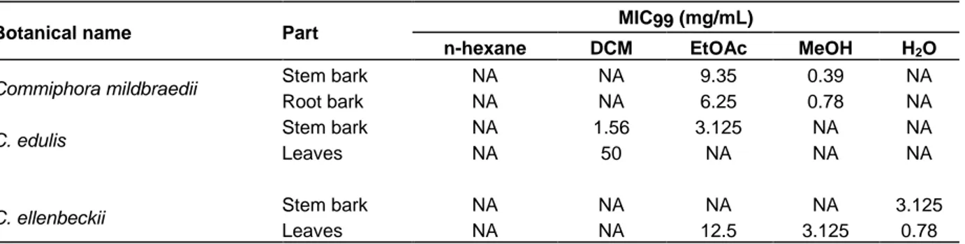
Antimycobacterial activity of Commiphora sp. plant materials
The antimycobacterial activities of the 30 plant extracts
from the three Commiphora species were evaluated using the broth microdilution method. The assay was performed against M.smegmatis ATCC607 and eleven
(11) out the thirty (30) extracts exhibited antimycobacterial activity (Table 2) with MICs ranging from 0.39 to 50 mg/mL. At least one of the plant parts of the 3 Commiphora species under study have showed antimycobacterial activity with C. mildbraedii proving to be more active than the other two species i.e. C. edulis and C. ellenbeckii. The methanolic extracts of C.
mildbraedii stem bark and root bark, and the aqueous
extract of C. ellenbeckii showed greater bioactivity with MICs less than 1000 µg/mL (0.39, 0.78, 0.78 mg/mL respectively). The highest MIC (50 mg/mL) was recorded for the DCM extract of C. edulis leaves, hence the less active among the tested plant extracts. All the tested plant extracts showed lower activity than the one of rifampicin, the reference drug (MIC= 0.015 mg/mL).
The best bioactivity of the methanolic and ethyl acetate extracts is supported by the qualitative phytochemical screening which proved that those extracts in general, contain phenols, flavonoids, terpenoids, saponins, tannins, and alkaloids (Table 4). Indeed, those bioactive molecules are well known to exhibit antimicrobial activity (Newton et al., 2002; Cushnie and Lamb, 2011, Gupta et al., 2012, Akiyama et al., 2001, Liu and Henkel, 2002; Alves et al., 2013).
Nevertheless, further work for purification of the most active extracts, isolation and testing of the purified compounds are worth to be carried out for a thorough understanding of the active principles behind this antimycobacterial property.
The Commiphora genus has been used since time immemorial as an antimicrobial agent (Hanuš et al., 2005). Researchers have assessed several species within this genus for the antimicrobial activity including antibacterial, antifungal and antimycobacterial activity.
The findings obtained from this study can therefore be compared to the findings from previous studies. For instance, C. edulis was investigated for the antibacterial
134 J. Med. Plants Res.
Table 2. Antimycobacterial activity (expressed as MIC99) of the different extracts obtained from the 3 Commiphora
species under study against M. smegmatis ATCC607.
Botanical name Part MIC99 (mg/mL)
n-hexane DCM EtOAc MeOH H2O
Commiphora mildbraedii Stem bark NA NA 9.35 0.39 NA
Root bark NA NA 6.25 0.78 NA
C. edulis Stem bark NA 1.56 3.125 NA NA
Leaves NA 50 NA NA NA
C. ellenbeckii Stem bark NA NA NA NA 3.125
Leaves NA NA 12.5 3.125 0.78
NA = Not active at highest tested concentration (50 mg/mL). MIC99 for the rifampicin (reference standard drug) = 0.015 mg/mL.
property Commiphora species have been evaluated for the antibacterial activity, including C. myrrha (Alhussaini et al., 2015), C. caudata and C. berryi (Latha et al., 2005), C. molmol (Abdallah et al., 2009, Kuete et al., 2011, Mohammed and Samy, 2013), C. gileadensis (Iluz et al., 2010; Al-Sieni, 2014; Al-Mahbashi et al., 2015), C.
swynertonii (Mkangara et al., 2014), C. africana, C. shimperi, C. grandulosa, C. marlothii, C. negleta, C. pyracanthoides, C. tenuipetiola, C. veminea (Paraskeva
et al., 2008), C. guidottii (Gebrehiwot et al., 2015), C.
pedunculata (Sallau et al., 2014), C. kerstingii (Ibrahim et
al., 2016). The antifungal activity was demonstrated in C.
wightii (Fatopea et al., 2013), C. kua (Berzinji et al.,
2014), C. wildii (Sheehama et al., 2019) and C. guidottii (Gebrehiwot et al., 2015).
Four Commiphora species so far have been subjected to antimycobacterial evaluation and some of the results obtained are comparable to the ones attained in this study. In a study carried out by De Souza et al. (2017), the chloroform extract of C. leptophloeos stem bark was tested against M. smegmatis and M. tuberculosis and proved to have antimycobacterial activity with MICs of 12.5 and 52 mg/Ml respectively, while the dichloromethane extract of C. eminii sap and the sterols isolated from it were tested against M. madagascariense and M. indicus pranii and showed same activity of MIC at 2.5 and 1.6 mg/mL respectively for the DCM extract and the isolated sterols (Erasto, 2012).
This is comparable to the MIC99 (1.56 mg/mL) of the DCM extract of C. edulis stem bark obtained in this study. In another study, the C. mukul gum was tested against M.
aurum and showed a MIC of 62.5 µg/ml (Newton et al.,
2002), while C. opobalsamum essential oils from the fresh aerial parts exhibited good activity (MIC = 15 µg/mL) against M. intracellulare (Al- Massarany et al., 2008). The reported results for C. mukul and C.
opobalsamum essential oils are better than the ones
obtained from this study. This implies that further studies using essential oils or gum from the Commiphora species under investigation here are needed for proper
comparison. Nevertheless, the present study demonstrated the antimycobacterial activity of C.
mildbraedii, C. edulis and C. ellenbeckii, which has not
been reported previously.
Cytotoxicity of the active extracts
The MTT-based assay and the Vero cells were used to assess the cytotoxicity of the eleven plant extracts. The CC50 values of the active plant extracts ranged from 339.65 ± 1.38 to 1734.05 ± µg/mL, as presented in Table 3. The active plant extracts tested showed low or no cytotoxicity compared to the reference standard drug rifampicin. The aqueous extract of C. ellenbeckii leaves and the ethyl acetate extract of C. edulis stem bark presented the lowest cytotoxicity, at CC50 of 1509.64 and 1734 µg/mL respectively, compared to rifampicin.
According to Zirihi et al. (2005) and Kigondu et al. (2009),theplantextractscanbeconsiderednon-cytotoxic when their CC50 values ˃ 20 µg/mL. However, the literature reports that South African C. edulis stem bark and leaves chloroform/methanolic extracts exhibited high cytotoxicity (CC50 = 194.0 µg/mL and CC50 = 99.5 µg/mL respectively) when tested against Graham cells (Paraskeva et al., 2008).
Based on the findings reported here, there is need to carry out further studies to investigate the in vivo antimycobacterial activity and toxicity of the Commiphora species used in this study.
Preliminary phytochemical screening
Phytochemical screening of various extracts of the plant materials used in this study as presented in Table 4 revealed the presence of alkaloids, phenols, terpenoids, saponins, flavonoids, tannins. The active extracts of C.
mildbraedii seemed contain similar phytochemicals either
Table 3. Cytotoxicity evaluation (expressed as CC50) of the bioactive plant extracts on Vero cells.
Botanical name Plant part Solvent used Mean CC50 (µg/mL)
C. mildbraedii
Stem bark EtOAc 432.65 ± 9.41
MeOH 452.80 ± 4.37
Root bark EtOAc 339.65 ± 1.38
MeOH 559.30 ± 35.37
C edulis Stem bark DCM 393.54 ± 36.64
EtOAc 1734.05± 186.04
C. ellenbeckii
Leaves DCM 506.41 ± 26.08
Stem bark Aqueous 448.62 ± 19.00
Leaves
EtOAc 420.15 ± 12.59
MeOH 608.53 ± 43.62
Aqueous 1509.647±67.47
Rifampicin 527.65 ± 48.30
Table 4. Preliminary phytochemical screening of the active plant extracts from the Commiphora sp. under studies. Botanical
name
Plant
part Solvent
Parameter
Alkaloids Saponins Phenols Tannins Flavonoids Terpenoids
C. mildbraedii
Stem bark EtOAc + - + - - +
MeOH - + + - - +
Root bark EtOAc + - + + + -
MeOH - + + + - -
C. edulis Stem bark DCM - - - - + +
EtOAc - - - - + -
Leaves DCM - - - -
C. ellenbeckii
Stem bark Aqueous - + + + - +
Leaves
EtOAc - - - - + -
MeOH + + + + + -
Aqueous - + + + + +
C. edulis was found to contain a small number of classes
of metabolites, at the same time only flavonoids and terpenoids were detected in its active extracts (DCM and ethyl acetate extracts).
The active extracts (aqueous extract of the stem bark and ethyl acetate, methanol and aqueous extracts of the leaves) of C. ellenbeckii generally manifested the presence of all the tested phytochemicals. Some or all the phytochemicals detected in the Commiphora plants used in this study have been reported present in other species of this genus. These include C. africana root bark (Okwute and Ochi, 2017) and stem bark (Nuhu et al., 2016), C. myrrha resins (Chandrasekharnath et al., 2013), C. opobalsamum aerial parts (Al-Howiriny et al., 2004), C. caudata and C. pubescensleaves (Deepa et al.,
2009), C. berryi stem bark (Selvamani et al., 2009), C.
gileadensis stem bark (Al-Mahbashi et al., 2015), C. mukul stem bark and seeds (Singh et al., 2016), C. kerstingii leaves (Ibrahim et al., 2016), C. pedunculata
stem bark (Sallau et al., 2014), C. guidottii (Gebrehiwot et al., 2015) and others.
All the detected phytochemicals have been investigated for their antimicrobial properties including antibacterial, antimycobacterial and antifungal activities. Reports from the literature show that alkaloids have antibacterial (Karou et al., 2006) and antimycobacterial (Newton et al., 2002) activities, while flavonoids (Cushnie and Lamb, 2011) and tannins have antibacterial activity (Akiyama et al., 2001; Lerato, 2017). Terpenoids (Gupta et al., 2011), saponins (Liu and Henkel, 2002) and phenols exhibit
136 J. Med. Plants Res.
antimicrobial activity against a wide range of bacteria and fungi (Alves et al., 2013).
However, it is recommendable to further investigate other phytochemicals that have not been included in this present work. Meanwhile, this is the first time that phytochemical screening of the C. mildbraedii, C. edulis and C. ellenbeckii is being reported to the best of my knowledge.
Preparative thin layer chromatography (p-TLC) and gas chromatography-mass spectrometry (GC-MS) analysis of the methanolic extract of C. mildbraedii stem bark
The most active plant extract (MIC99= 0.39 mg/mL), that is the methanolic extract of the stem bark of C.
mildbraedii, was further fractionated by p-TLC using
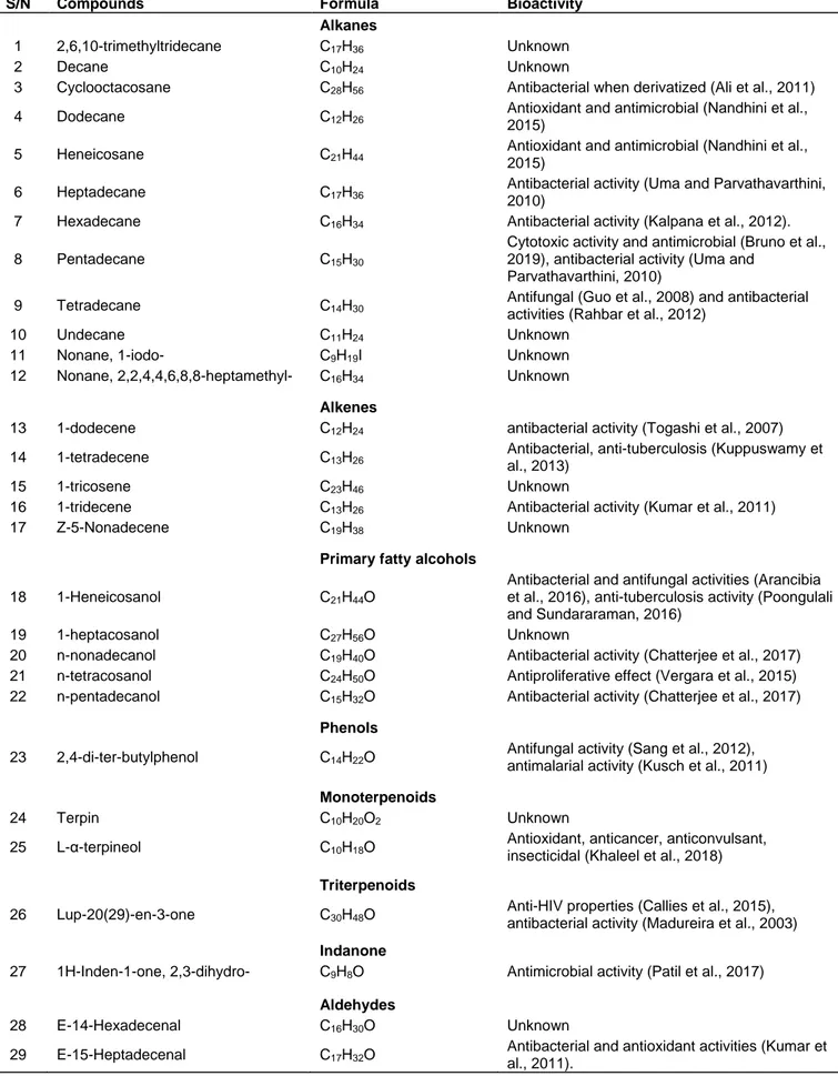
hexane-ethyl acetate (6:4) as the solvent system. Seven bands/layers (F1-F7) were obtained and their scraping from the plate led to five reconstituted fractions in ethyl acetate, FF1-FF5 (with F1 and F2 and F6 and F7 pulled together). A plethora of different bioactive compounds were detected in each fraction (Appendix 1; by the GC-MS analysis). In total, 42 different bioactive compounds belonging to various chemical classes were found to be present across the different fractions (Table 5). Two compounds were detected as the most abundant; triterpenoid- lup-20(29)-en-3-one (lupenone) present in FF3 (45.29% peak area) and FF4 (12.6% peak area) and an aromatic compound o- xylene present in FF1 and FF5 at 10% peak area.
The phytoconstituents revealed by GC-MS can be grouped into saturated hydrocarbons-alkanes (12), unsaturated hydrocarbons-alkenes (5), primary fatty alcohols (5), phenols (1), monoterpenoids (2) and triterpenoids (1), indanone (1), aldehydes (2), ethers (2), carboxylic acid (1), carbonate esters (3), fatty acids esters (6) and aromatic hydrocarbon (1). Even though the chemistry of Commiphora plants has been thoroughly studied (Hanuš et al., 2005; Shen et al., 2012), the phytoconstituents detected in C. mildbraedii stem bark (methanolic extract) are almost all newly reported in this genus, except three compounds including α-terpineol detected in the gum resin of C. mukul (Saxena and Sharma, 1998), C. leptophloeos essential oils (Da Silva et al., 2015), C. gileadensis essential oils (Dudai et al., 2017), C. wildii essential oils (Sheehama et al., 2019); tridecene in C. gileadensis essential oils (Dudai et al., 2017) and o-xylene in the C. quadricincta essential oils (Assad et al., 1997). Researchers have paid more attention and studied the most ethnomedicinally used plant part/product, that is, resinous exudates or gum resin and not the other plant parts i.e. leaves, roots or the entire stem bark (Shen et al., 2012). This is a great contribution so far in the study of Commiphora genus chemistry as nobody else has reported on the chemistry
of C. mildbraedii before, to the best of my knowledge. Twenty-three compounds out of the 42 have been reported to exhibit antibacterial, antifungal and antimycobacterial activities (Table 5). The antimycobacterial activity of the methanolic extract of C.
mildbraedii may be due to two of its bioactive compounds
which have been reported to possess anti-tuberculosis activity, tetradecene (Kuppuswamy et al., 2013) and 1-heneicosanol (Poongulali and Sundararaman, 2016). Synergism effect of all the antimicrobial phytochemicals may be responsible for the high antimycobacterial activity of the named plant extract under study (Doern, 2014) because the most abundant detected components, that is, lup- 20(29)-en-3-one (Madureira et al., 2003) and o-xylene (Tiwari et al., 2016) are reported to have good antibacterial activity. Thus, the C. mildbraedii methanolic extract is also presumably a potential antimicrobial agent against different bacteria and fungi strains based on the reports from the literature on the bioactivities of compounds detected by GC-MS in this study.
Conclusion
The methanolic extract of C. mildbraedii stem bark presented the highest antimycobacterial activity and the active plant extracts showed low cytotoxicity. The phytochemical screening revealed that the active extracts of C. mildbraedii contain all the tested bioactive phytochemicals except the flavonoids. GC-MS analysis of the p-TLC fractions identified 42 different compounds with 39 compounds being detected for the first time in
Commiphora genus. Twenty-three (23) of them are
reported in the literature to have antimicrobial activities with two particular compounds, tetradecene and 1-heneicosanol being reported to have anti-tuberculosis activity.
The results provided in this study therefore constitute an additional and reliable contribution to the study of the complex and interesting Commiphora genus. The findings justify the ethnomedicinal practices of different cultural societies which utilize the Commiphora species to treat respiratory diseases including TB.
ACKNOWLEDGEMENTS
The authors would like to thank the African Union through the Pan African University, and The World of Academic Science (TWAS) for the financial support. Our gratitude goes also to Mr. Bernard K. Wanjohi (University of Eldoret, Kenya) for his help in the identification and collection of the plant materials.
CONFLICT OF INTERESTS
Table 5. Summary of detected compounds by GC-MS and their reported bioactivities.
S/N Compounds Formula Bioactivity
Alkanes
1 2,6,10-trimethyltridecane C17H36 Unknown
2 Decane C10H24 Unknown
3 Cyclooctacosane C28H56 Antibacterial when derivatized (Ali et al., 2011)
4 Dodecane C12H26 Antioxidant and antimicrobial (Nandhini et al.,
2015)
5 Heneicosane C21H44 Antioxidant and antimicrobial (Nandhini et al.,
2015)
6 Heptadecane C17H36 Antibacterial activity (Uma and Parvathavarthini,
2010)
7 Hexadecane C16H34 Antibacterial activity (Kalpana et al., 2012).
8 Pentadecane C15H30
Cytotoxic activity and antimicrobial (Bruno et al., 2019), antibacterial activity (Uma and
Parvathavarthini, 2010)
9 Tetradecane C14H30 Antifungal (Guo et al., 2008) and antibacterial
activities (Rahbar et al., 2012)
10 Undecane C11H24 Unknown
11 Nonane, 1-iodo- C9H19I Unknown
12 Nonane, 2,2,4,4,6,8,8-heptamethyl- C16H34 Unknown
Alkenes
13 1-dodecene C12H24 antibacterial activity (Togashi et al., 2007)
14 1-tetradecene C13H26 Antibacterial, anti-tuberculosis (Kuppuswamy et al., 2013)
15 1-tricosene C23H46 Unknown
16 1-tridecene C13H26 Antibacterial activity (Kumar et al., 2011)
17 Z-5-Nonadecene C19H38 Unknown
Primary fatty alcohols
18 1-Heneicosanol C21H44O
Antibacterial and antifungal activities (Arancibia et al., 2016), anti-tuberculosis activity (Poongulali and Sundararaman, 2016)
19 1-heptacosanol C27H56O Unknown
20 n-nonadecanol C19H40O Antibacterial activity (Chatterjee et al., 2017) 21 n-tetracosanol C24H50O Antiproliferative effect (Vergara et al., 2015) 22 n-pentadecanol C15H32O Antibacterial activity (Chatterjee et al., 2017)
Phenols
23 2,4-di-ter-butylphenol C14H22O Antifungal activity (Sang et al., 2012), antimalarial activity (Kusch et al., 2011)
Monoterpenoids
24 Terpin C10H20O2 Unknown
25 L-α-terpineol C10H18O Antioxidant, anticancer, anticonvulsant,
insecticidal (Khaleel et al., 2018)
Triterpenoids
26 Lup-20(29)-en-3-one C30H48O Anti-HIV properties (Callies et al., 2015), antibacterial activity (Madureira et al., 2003)
Indanone
27 1H-Inden-1-one, 2,3-dihydro- C9H8O Antimicrobial activity (Patil et al., 2017)
Aldehydes
28 E-14-Hexadecenal C16H30O Unknown
29 E-15-Heptadecenal C17H32O Antibacterial and antioxidant activities (Kumar et al., 2011).
138 J. Med. Plants Res.
Table 5. Contd.
Ethers
30 Ethanol, 2-butoxy- C6H14O2 Mild antimicrobial activity ((Bailey and Neihof, 1976)
31 Pentane, 1-ethoxy- C7H16O Unknown
Carboxyilic acids
32 Pentanoic acid,
3-methyl-4-oxo- C6H10O3
Antimicrobial, antioxidant and anticancer activities (Madkour et al., 2017)
Carbonate esters
33 Carbonic acid, eicosyl
vinyl ester C23H44O3
Unknown
34 Carbonic acid,
decyl 2-ethylhexyl ester Unknown
35 Carbonic acid, 2-ethylhexyl octyl ester
C19H38O3
Unknown C17H34O3
Fatty acids esters
36 Isopropyl myristate C17H3402 antioxidant and antimicrobial activity
(Chandrasekar et al., 2016, Begum et al., 2016)
37 Isopropyl palmitate C19H38O2 hepatoprotective and mild anticancer properties (Saxena et al., 2007)
38 Eicosyl trifluoroacetate C22H41F3O2 Unknown
39 Heneicosyl
heptafluorobutyrate
C25H43F7O2
Unknown
40 Tetradecyl trifluoroacetate C16H29F3O2 Unknown
41 Trifluoroacetoxy hexadecane C18H33F3O2 Antifungal activity (Ibrahim et al., 2017)
Aromatic hydrocarbons
42 O-xylene C6H4(CH3)2 Antioxidant, antimicrobial and antifungal activities (Tiwari et al., 2016)
REFERENCES
Abdallah EM, Khalid AS, Ibrahim N (2009). Antibacterial activity of oleo-gum resins of Commiphora molmol and Boswellia papyrifera against Methicillin Resistant Staphylococcus aureus (MRSA). Scientific Research Essay 4(4):351-356.
Abdul-Ghani RA, Loutfy N, Hassan A (2009). Myrrh and trematodoses in Egypt: an overview of safety, efficacy and effectiveness profiles. Parasitology International 58(3):210- 214.
Akiyama H, Fujii K, Yamasaki O, Oono T, Iwatsuki K (2001). Antibacterial action of several tannins against Staphylococcus aureus. Journal of Antimicrobial Chemotherap 48(4):487-491. Al-Howiriny TA, Al-Sohaibani MO, Al-Said MS, Al-Yahya MA, El-Tahir
KH, Rafatullah S (2004). Hepatoprotective properties of Commiphora
opobalsamum ("BALESSAN"), a traditional medicinal plant of Saudi
Arabia. Drugs and Experimental Clinical Research 30(5-6):213-220. Alhussaini SM, Saadabi AM, Alghonaim IM, Ibrahim EK (2015). An
Evaluation of the Antimicrobial Activity of Commiphora myrrha Nees (Engl.) Oleo-gum Resins from Saudi Arabia. Journal of Medicinal Sciences 15(4):198-203.
Ali KA, Hosni HM, Ragab EA, El-Moez SIA (2011). Synthesis and Antimicrobial Evaluation of Some New Cyclooctanones and Cyclooctane-Based Heterocycles. Archiv der Pharmazie, 345(3), 231-239.
Al-Mahbashi MH, El-Shaibany A, Saad AF (2015). Evaluation of acute toxicity and antimicrobial effects of the bark extract of Bisham (Commiphora gileadensis L.). Journal of Chemical and Pharmaceutical Research 7(6): 810-814.
Massarany MS, Abbas AF, Demirci B, Baser HCK, Khan IS, Al-Rehaily JA, Jaber S, Mossa SJ, Abourashed AE (2008). Chemical Composition and Biological Evaluation of the Essential Oil of
Commiphora opobalsamum L. Journal of Herbs, Spices & Medicinal
Plants 13(4):111-121.
Al-Rejaie SS (2012). Effect of oleo-gum-resin on ethanol induced hepatotoxicity in rats. Journal of Medical Sciences 12(1):1-9. Al-Sieni IIA (2014). The antibacterial activity of traditionally used
Salvadora persica l. (miswak) and Commiphora gileadensis (palsam)
in Saudi Arabia. African Journal of Traditional, Complementary and Alternative Medicine 11(1): 23-27.
Alves MJ, Ferreira ICFR, Froufe HJC, Abreu RMV, Martins A, Pintado M (2013). Antimicrobial activity of phenolic compounds identified in wild mushrooms, SAR analysis and docking studies. Journal of Applied Microbiology 115(2):346-357.
Arancibia LA, Naspi VC, Pucci NG, Arce EM, Colloca BC (2016). Biological activity of 1-heneicosanol isolated from Senecio
coluhuapiensis, an endemic species from Patagonia, Argentina.
Pharmaceutical Chemistry Journal 3(4):73-77.
Bailey CA, Neihof RA (1976). Biocidal Properties of Anti-Icing Additives for Jet Aircraft Fuels. Naval Research Lab Washington DC,
ADA022839. Available online at
https://apps.dtic.mil/docs/citations/ADA022839
Banso A, Mann A (2006). Antimicrobial alkaloid fraction from
Commiphora africana (A. Rich). Journal of Pharmacy and
Bioresources 3(2):98-102.
Barzinji AKR, Nasher AK, Mothana RA, Al-Hamadi MMS (2014). In vitro Antimalarial activity of selected Yemeni plants used in traditional
medicine. International Journal of Medicinal Plants-Photon 107:526-535.
Begum FI, Mohankumar R, Jeevan M, Ramani K (2016). GC–MS Analysis of Bio- active Molecules Derived from Paracoccus
pantotrophus FMR19 and the Antimicrobial Activity against Bacterial
Pathogens and MDROs. Indian Journal of Microbiology 56(4):426-432.
Bruno F, Castelli G, Migliazzo A, Piazza M, Galante A, Verde VL, Calderone S, Nucatolo G, Vitale F (2019). Cytotoxic Screening and In Vitro Evaluation of Pentadecane against Leishmania infantum Promastigotes and Amastigotes. Journal of Parasitology 101(6):701-705.
Callies O, Bedoya LM, Beltrán M, Muñoz A, Calderón PO, Osorio AA, Jiménez AI, Alcamí J, Bazzocchi IL (2015). Isolation, Structural Modification, and HIV Inhibition of Pentacyclic Lupane-Type Triterpenoids from Cassine xylocarpa and Maytenus cuzcoina. Journal of Natural Products 78(5):1045-1055.
Chandrasekar T, Rao MRK, Kumar RV, Prabhu K, Nandha KV, Divya D (2016). GC–MS analysis, antimicrobial, antioxidant activity of an Ayurvedic medicine, Nimbapatradi Choornam. Journal of Chemical and Pharmaceutical Research 7(9):124-136.
Chatterjee S, Karmakar A, Azmi SA, Barik A (2017). Antibacterial Activity of Long- Chain Primary Alcohols from Solena amplexicaulis Leaves. Proceedings of the Zoological Society 71(4):313-319. Collins L, Franzblau SG (1997). Microplate Alamar Blue Assay versus
BACTEC 460 System for High-Throughput Screening of Compounds against Mycobacterium tuberculosis and Mycobacterium avium. Antimicrobial Agents and Chemotherapy, 41:1004-1009.
Cushnie TT, Lamb AJ (2011). Recent advances in understanding the antibacterial properties of flavonoids. International Journal of Antimicrobial Agents 38(2):99-107.
Daly DC, Harley MM, Martinez-Habile MC, Weeks A (2011). Burseraceae. In The Families and Genera of Vascular plants (ed. K.
Kubtzki), 10:76-104. Available at
https://www.springer.com/series/1306
De Souza PJJ, Pereira PCA, Jandú JJB, Da Paz JA, Crovella S, Dos Santos CMT, De Azevêdo SJ (2017). Commiphora leptophloeos: Phytochemical and Antimicrobial Characterization. Frontiers Microbiology 8:52.
Deepa SV, Kumar SP, Latha S, Selvamani P, Srinivasan S (2009). Antioxidant studies on the ethanolic extract of Commiphora spp. African Journal of Biotechnology 8(8):1630-1636.
Dhawan D, Gupta J (2017). Comparison of Different Solvents for Phytochemical Extraction Potential from Datura metel Plant leaves. International Journal of Biological Chemistry 11(1):17-22.
Dorin D, Le Roch K, Sallicandro P, Alano P, Parzy D, Poullet P, Meijer L, Doerig C (2001). Pfnek-1, a NIMA-related kinase from the human malaria parasite Plasmodium falciparum. European Journal of Biochemistry 268(9):2600-2608.
Dudai N, Shachter A, Satyal P, & Setzer NW (2017). Chemical composition and monoterpenoid enantiomeric distribution of the essential oils from Apharsemon (Commiphora gileadensis). Medicines 2017(4):66. doi:10.3390/medicines4030066.
Erasto P (2012). Antimycobacterial Sterols from Aromatic Stem Sap of
Commiphora eminii Engl. Journal of Advanced Scientific Research, 3(4), 27-31. Available online through http://www.sciensage.info/jasr
Evans CW (2009). Trease and Evans-Pharmacognosy (16th Edition).
Elsevier. Available at
https://www.academia.edu/38664516/Trease_and_Evans_Pharmaco gnosy_SIXTEENTH_EDITI ON
Fatopea MO, Burtomania SKS, Ocheib JO, Abdulnoura AO, Al-Kindya SMZ, Takeda Y (2003). Muscanone: α3O(1ʺ, 8 ʺ, 14 ʺ -trimethylhexadecanyl) naringenin from Commiphora wightii. Phytochemistry 62(8):1251-1255.
Gebrehiwot M, Asres K, Bisrat D, Mazumder A, Lindemann P, Bucar F (2015). Evaluation of the wound healing property of Commiphora
guidottii Chiov. ex. Guid. Complementary and Alternative Medicine
15:282.
Ghazanfar SA (1994). Handbook of Arabian Medicinal Plants. CRC Press, Boca Raton, FL, P 176. Available online through https://www.crcpress.com/Handbook-of-Arabian-Medicinal- Plants/Ghazanfar/p/book/9780849305399
Guo L, Wu J, Han T, Cao T, Rahman K, Qin L (2008). Chemical Composition, Antifungal and Antitumor Properties of Ether Extracts of
Scapania verrucosa Heeg. And its Endophytic Fungus Chaetomium
fusiforme. Molecules 13(9):2114-2125.
Gupta A, Kaul A, Tsolaki AG, Kishore U, Bhakta S (2012).
Mycobacterium tuberculosis: Immune evasion, latency and reactivation. Immunobiology 217(3):363-374.
Gupta N, Saxena G, Kalra SS (2011). Antimicrobial activity pattern of certain terpenoids. International Journal of Pharmacology and Biological Sciences 2(1):87-91.
Hanuš OL, Renzanka T, Dembitsky MV, Moussaieff A (2005). Myrrh- Commiphora Chemistry Biomedical Papers 149(1):3-28.
Harborne JB (1984). Phytochemical Methods. Springer Netherlands,
Dordrecht. Available online through
https://link.springer.com/book/10.1007/978-94-009-5570-7
Ibrahim T, Gidado Muluh K, Alexander A (2016). Phytochemical Screening, Antioxidant and Antibacterial Activities of Commiphora
kerstingii. International Biological and Biomedical Journal
2(3):127-133.
Iluz D, Hoffman M, Gilboa-Garber N, Amar Z (2010). Medicinal properties of Commiphora gileadensis. African Journal of Pharmacy and Pharmacology 4(8):516-520.
Kalpana DV, Shanmugasundaram R, Mohan VR (2012). GC–MS analysis of ethanol extracts of Entada pursaetha Dc. seed. Bioscience Discovery 3(1):30-33.
Karou D, Savadogo A, Canini A, Yameogo S, Montesano C, Simpore J, Colizzi V, Traore SA (2006). Antibacterial activity of alkaloids from
Sida acuta. African Journal of Biotechnology 5(2):195-200.
Khaleel C, Tabanca N, Buchbauer G (2018). α-Terpineol, a natural monoterpene: A review of its biological properties. Open Chemistry 16(1):349-361.
Kigondu EVM, Rukunga GM, Keriko JM, Tonui WK, Gathirwa JW, Kirira PG, Irungu B, Ingonga JM, Ndiege IO (2009). Anti-parasitic activity and cytotoxicity of selected medicinal plants from Kenya. Journal of Ethnopharmacology 123(3):504-509.
Kigondu ME (2015). Repurposing chlorpromazine and its metabolites for anti-tuberculosis drug discovery. PhD dissertation, University of
Cape Town. Available online through
https://open.uct.ac.za/handle/11427/16702
Kiringe WJ (2006). A Survey of Traditional Health Remedies Used by the Maasai of Southern Kaijiado District, Kenya. Ethnobotany Research and Applications 4: 061-073.
Kuete V, Wiench B, Hegazy M-E, Mohamed T, Fankam A, Shahat A, Efferth T (2011). Antibacterial Activity and Cytotoxicity of Selected Egyptian Medicinal Plants. Planta Medica 78(02):193-199.
Kumar V, Bhatnagar AK, Srivastava JN (2011). Antibacterial activity of crude extracts of Spirulina platensis and its structural elucidation of bioactive compound. Journal of Medicinal Plants Research 5(32):7043-7048.
Kuppuswamy MK, Jonnalagadda B, Arockiasamy S (2013). GC-MS AnalysisofChloroformextractofCroton bonplandianum. International
Journal of Pharmacology and Biological Sciences 4(4):613-617. Kusch P, Deininger S, Specht S, Maniako R, Haubrich S, Pommerening
T, Lin TKP, Hoerauf A, Kaiser A (2011). In Vitro and In Vivo Antimalarial Activity Assays of Seeds from Balanites aegyptiaca: Compounds of the Extract Show Growth Inhibition and Activity against Plasmodial Aminopeptidase. Journal of Parasitology Research ID 368692, pp 1-9.
Latha S, Selvamani P, Sen JD, Gupta KJ, Pal T, Ghosh LK (2005). Antibacterial activity of Commiphora caudata and Commiphora berryi leaves. Indian Drugs 42(10):696-698.
Liu J, Henkel T (2002). Traditional Chinese medicine (TCM): are polyphenols and saponins the key ingredients triggering biological activities? Current Medicinal Chemistry 9(15):1483-1485.
Madkour HMF, Ghareeb MA, Abdel-Aziz MS, Khalaf OM, Saad AM, El-Ziaty AK, Abdel-Mogib M (2017). Gas chromatography-mass spectrometry analysis, antimicrobial, anticancer and antioxidant activities of n-hexane and methylene chloride extracts of Senna
italica.JournalofAppliedPharmaceuticalSciences7(06):023-032. Madureira AM, Ascenso JR, Valdeira L, Duarte A, Frade JP, Freitas G,
Ferreira MJU (2003). Evaluation of the Antiviral and Antimicrobial Activities of Triterpenes Isolated from Euphorbia segetalis. Natural
140 J. Med. Plants Res.
Product Research 17(5):375-380.
McGuffin MJT, Kartesz A, Keung A, Tuker AO (2000). Herbs of Commerce. American Herbal Products Association (2nd edition).
Available online through
https://law.resource.org/pub/us/cfr/ibr/001/ahpa.herbs.1992.pdf Mkangara M, Chacha M, Erasto PK (2014). Antimicrobial and
Cytotoxicity Efficacy of Commiphora swynnertonii (Burtt) Extracts. International Journal of Science and Research, 3(7):2319-7064. Mohammed EA, Samy AS (2013). Antimicrobial activity of essential oil
and methanol extract from Commiphora molmol (Engl.) resin. International Journal of Current Microbiology and Applied Sciences 2(12):1-6.
Mosmann T (1983). Rapid Colorimetric Assay for Cellular Growth and Survival: Application to Proliferation and Cytotoxicity Assays. Journal of lmmunological Methods 65(1-2):55-63. Nandhini SU, Sangareshwari S, Lata K (2015). Gas chromatography-mass spectrometry analysis of bioactive constituents from the marine
Streptomyces. Asian Journal of Pharmacology and Clinical Research
8(2):244-246.
Newton SM, Lau C, Gurcha SS, Besra GS, Wright CW (2002). The evaluation of forty-three plant species for in vitro antimycobacterial activities; isolation of active constituents from Psoralea corylifolia and
Sanguinaria canadensis. Journal of Ethnopharmacology 79(1):57-67.
Nomicos NY (2007). Myrrh: medical marvel or myth of the Magi? Holistic Nursing Practice, 21(6):308-323.
Nuhu A, Danmalam UH, Ilyas N, Zakariya AM, Shehu S, Jajere UM (2016). Evaluation of Antiulcer Activity of Commiphora africana (A. Rich) Engl. (Burseraceae) Stem- bark Extracts Using Ethanol Induced Ulcer Model in Albino Rats. International Journal of Current Microbiology and Applied Sciences 5(3):15-25.
Okwute SK, Ochi IO (2017). Phytochemical analysis and cytotoxic activity of the root extract of Commiphora africana (Caesalpiniaceae). Journal of Pharmacognosy and Phytochemistry 6(6):451-454. Omer SA, Adam SEI, Mohammed OB (2011). Antimicrobial activity of
Commiphora myrrha against some bacteria and Candida albicans
isolated from gazelles at King Khalid Wildlife Research Centre. Research Journal of Medicinal Plant 5(1):65-71.
Orwa C, Mutua A, Kindt R, Jamnadass R, Anthony S (2009). Agroforestree Database: a tree reference and selection guide version
4.0. Retrieved from
[http://www.worldagroforestry.org/sites/treedbs/treedatabases.asp.] Paraskeva MP, Van Vuurena SF, Van Zyl RL, Davids H, Viljoen AM
(2008). The in vitro biological activity of selected South African
Commiphora species. Journal of Ethnopharmacology
119(3):673-679.
Patil SA, Patil R, Patil SA (2017). Recent developments in biological activities of indanones. European Journal of Medicinal Chemistry 138:182-198.
Poongulali S, Sundararaman M (2016). Antimycobacterial, anticandidal and antioxidant properties of Terminalia catappa and analysis of their bioactive chemicals. International Journal of Pharmacy and Biological Sciences 6(2):69-83.
Rahbar N, Shafagha A, Salimi F (2012). Antimicrobial activity and constituents of the hexane extracts from leaf and stem of Origanum
vulgare L. sp. Viride (Boiss.) Hayek. Growing wild in Northwest Iran.
Journal of Medicinal Plant Research 6(13):2681-2685.
Sallau MS, Tajuddeen N, Ndukwe GI, Musa AM, Dambatta BM, Sani YM (2014). Phytochemical and antimicrobial properties of
Commiphora pedunculata (engl) stem bark extracts. Bayero Journal
of Pure and Applied Sciences 7(1):101-104.
Sang MK, Kim KD (2012). The volatile-producing Flavobacterium
johnsoniae strain GSE09 shows biocontrol activity against Phytophthora capsici in pepper. Journal of Applied Microbiology
113(2):383-398.
Saxena M, Faridi U, Srivastava SK, Darokar MP, Mishra R, Pal A, Shisodia B, Khanuja S PS (2007). A Cytotoxic and Hepatoprotective Agent from Withania somnifera and Biological evaluation of its Ester Derivatives. Natural Product Communications 2(7).
Saxena VK, Sharma RN (1998). Constituents of the essential oil from Commiphora mukul gum resin. In: Hanuš OL, Renzanka T, Dembitsky MV, Moussaieff A. (2005). Myrrh- Commiphora Chemistry. Biomedical Papers 149(1):3-28.
Selvamani P, Sen JD, Gupta KJ (2009). Pharmacognostical standardization of Commiphora berryi (Arn) Engl and phytochemical studies on its crude extracts. African Journal of Pharmacy and Pharmacology 3(2):037-046.
Sheehama JT, Mukakalisa C, Amakali T, Uusiku LN, Hans RH, Nott K, Nott A, Louw S (2019). Chemical characterization and in vitro antioxidant and antimicrobial activities of essential oil from
Commiphora wildii Merxm. (omumbiri) resin. Flavour and Fragrance
Journal 34(4):241-251.
Shen T, Li G-H, Wang X-N, Lou H-X (2012). The genus Commiphora: A review of its traditional uses, phytochemistry and pharmacology. Journal of Ethnopharmacology 142(2):319-330.
Shen T, Wan W, Yuan H, Kong F, Guo H, Fan P, Lou H (2007). Secondary metabolites from Commiphora opobalsamum and their antiproliferative effect on human prostate cancer cells. Phytochemistry 68(9):1331-1337.
Shoemaker M, Hamilton B, Dairkee SH, Cohen I, Campbell MJ (2005).
In vitro anticancer activity of twelve Chinese medicinal herbs.
Phytotherapy Research 19(7):649-651.
Silva MIG, De Melo CTV, Vasconcelos LF, De Carvalho AMR, Sousa FCF (2011). Bioactivity and potential therapeutic benefits of some medicinal plants from the Caatinga (semi-arid) vegetation of Northeast Brazil: a review of the literature. Revista Brasileira de Farmacognosia 22(1):193-207.
Singh A, Chawhan SE, Tiwari A (2016). Phytochemical screening of
Commiphora Mukul Seeds and Bark Powder: A Comparative Studies.
International Journal for Innovative Research in Science and Technology 2(9):157-159.
Tiwari S, Mishra S, Misra RD, Upadhyay R (2016). Identification of New Bioactive Compounds from Fruit of Abutilon indicum through GC-MS Analysis. Biological Forum-An International Journal 8(1):548-554. Togashi N, Shiraishi A, Nishizaka M, Matsuoka K, Endo K, Hamashima
H, Inoue Y (2007). Antibacterial activity of long-chain fatty alcohols against Staphylococcus aureus. Molecule 12(2):139-148.
Truong D-H, Nguyen DH, Ta NTA, Bui AV, Do TH, Nguyen HC (2019). Evaluation of the Use of Different Solvents for Phytochemical Constituents, Antioxidants, and In Vitro Anti-Inflammatory Activities of
Severinia buxifolia. Journal of Food Quality ID 8178294, pp. 1-9.
https://doi.org/10.1155/2019/8178294
Uma B, Parvathavarthini R (2010). Antibacterial effect of hexane extract of sea Urchin, Temnopleurus alexandri (Bell, 1884). International Journal of PharmTech Research, 2(3):1677-1680.
Van Wyk BE, Van Heerden F, Van Oudtshoorn B (2002). Poisonous
Plants of South Africa. Briza Publications, South Africa. P 288.
Available at
https://www.researchgate.net/publication/280314250_Poisonous_Pla nts_of_South_Africa
Vergara M, Olivares A, Altamirano C (2015). Antiproliferative evaluation of tall-oil docosanol and tetracosanol over CHO-K1 and human melanoma cells. Electronic Journal of Biotechnology 18(4):291-294. Vollesen K (1989). In Burseraceae. Flora of Ethiopia. Hedberg I.,
Edwards S., Eds., Addis Ababa University Press, Addis Ababa, 3:442-478.
World Health Organization (WHO) (2018). Global Tuberculosis Report
2018. Available online through
https://www.aidsdatahub.org/sites/default/files/publication/WHO_Glob al_TB_Report_2018.pdf
Zirihi GN, Mambu L, Guédé-Guina F, Bodo B, Grellier P (2005). In vitro antiplasmodial activity and cytotoxicity of 33 West African plants used for treatment of malaria. Journal of Ethnopharmacology 98(3):281-285.
Zumla A, Raviglione M, Hafner R, von Reyn CF (2013). Tuberculosis. New England Journal of Medicine 368(8):745-755.
Appendix 1. Compounds detected by GC-MS analysis in different prep-TLC fractions FF1-FF5 of C. mildbraedii stem bark MeOH extract. Prep-TLC
fraction Rf value GC-MS detected constituents Retention time Area (%)
1 0.20-0.33 Pentanoic acid, 3-methyl-4-oxo- 3.05 1.62
Pentane, 1-ethoxy- 3.24 0.90
o-Xylene 5.29 9.37
1-Dodecene 10.07 0.91
Cyclohexanemethanol,4-hydroxy-α,α,4-trimethyl- or p-menthane-1,8-diol or Terpin 12.33 0.94
1-Tridecene 13.09 2.02 Tetradecane 13.18 1.48 Pentadecane 14.55 0.73 2,4-Di-tert-butylphenol 14.88 1.37 Tetradecyl trifluoroacetate 15.77 3.01 Hexadecane 15.84 1.73 Heptadecane 17.06 0.92 n-Nonadecanol-1 18.17 3.29 Heptadecane 18.22 1.97 Isopropyl myristate 18.52 1.59 Nonane, 1-iodo- 19.33 0.92 n-Nonadecanol-1 20.33 3.17 Heneicosane 20.37 1.34 Isopropyl palmitate 20.64 3.91 1-Tricosene 21.39 2.26 n-Tetracosanol-1 22.45 3.68 1-Heptacosanol 25.30 2.46
Carbonic acid, 2-ethylhexyl octyl ester 27.42 1.46
2 0.47 Pentane, 1-ethoxy- 3.24 1.02 Ethanol, 2-butoxy- 5.42 1.00 Undecane 6.86 0.81 1-Dodecene 10.07 1.01 Dodecane 10.18 0.65 1-Tridecene 13.09 1.99 Tetradecane 13.18 1.45 2,4-Di-tert-butylphenol 14.88 1.33 E-15-Heptadecenal 15.77 3.02 Hexadecane 15.84 1.78 Heptadecane 17.06 0.66
142 J. Med. Plants Res. Z-5-Nonadecene 18.17 3.16 Heptadecane 18.22 1.58 Isopropyl myristate 18.52 1.45 n-Pentadecanol 19.28 0.57 Heneicosane 19.33 0.27 n-Nonadecanol-1 20.33 3.06 Heneicosane 20.37 1.33 Isopropyl palmitate 20.64 3.83 Trifluoroacetoxy hexadecane 21.39 1.38 n-Tetracosanol-1 22.45 2.64 1-Heptacosanol 25.30 3.32
Carbonic acid, 2-ethylhexyl octyl ester 27.41 2.06
3 0.6 Pentane, 1-ethoxy- 3.24 0.52 Undecane 6.86 0.43 1-Dodecene 10.07 0.51 Dodecane 10.18 0.35 1-Tridecene 13.09 1.15 Tetradecane 13.18 0.83 Pentadecane 14.55 0.30 2,4-Di-tert-butylphenol 14.88 0.80 E-15-Heptadecenal 15.77 1.73 Hexadecane 15.84 1.02 Heptadecane 17.06 0.38 n-Nonadecanol-1 18.17 1.95 Heptadecane 18.22 0.96 Isopropyl myristate 18.53 0.94 n-Nonadecanol-1 20.33 1.79 Heneicosane 20.37 0.80 Isopropyl palmitate 20.64 2.32 Heneicosyl heptafluorobutyrate 21.39 2.08 1-Heneicosanol 22.45 1.39 2,6,10-Trimethyltridecane 22.49 0.33 Eicosyl trifluoroacetate 25.30 1.39 Lup-20(29)-en-3-one 30.91 45.29 4 0.73 Pentane, 1-ethoxy- 3.24 0.95 Undecane 6.86 0.74 1-Dodecene 10.07 0.87 Dodecane 10.18 0.73 L-alpha-Terpineol 10.44 0.73
1H-Inden-1-one, 2,3-dihydro- 12.01 0.48 Nonane, 2,2,4,4,6,8,8-heptamethyl- 12.08 0.33 1-Tetradecene 13.09 1.84 Tetradecane 13.18 1.26 Pentadecane 14.55 0.66 2,4-Di-tert-butylphenol 14.88 1.00 E-15-Heptadecenal 15.77 2.53 Hexadecane 15.84 1.45 Heptadecane 17.06 0.60 n-Nonadecanol-1 18.17 2.72 Heptadecane 18.22 1.37 Isopropyl myristate 18.52 1.28 n-Pentadecanol 19.27 0.57 Heptadecane 19.32 0.19 n-Nonadecanol-1 20.33 2.41 Heneicosane 20.37 1.16 Isopropyl palmitate 20.63 3.13 n-Tetracosanol-1 21.39 1.44 1-Heneicosanol 22.45 1.21
Carbonic acid, decyl 2-ethylhexyl ester 27.42 1.65
Lup-20(29)-en-3-one 30.81 12.60 5 0.8-0.87 Pentane, 1-ethoxy- 3.24 1.10 o-Xylene 5.29 10.42 Decane 6.86 0.89 1-Dodecene 10.07 1.02 1-Tridecene 13.09 1.67 Tetradecane 13.18 1.13 2,4-Di-tert-butylphenol 14.88 1.12 E-14-Hexadecenal 15.77 2.97 Hexadecane 15.84 1.82 n-Nonadecanol-1 18.16 3.00 Heptadecane 18.22 1.32 Isopropyl myristate 18.52 1.57
Carbonic acid, eicosyl vinyl ester 19.32 1.73
n-Nonadecanol-1 20.33 2.57
Heneicosane 20.37 1.28
Isopropyl palmitate 20.63 3.68
Cyclooctacosane 21.39 4.52