Brain imaging of the central executive component of working memory
Fabienne Collette
1& Martial Van der Linden
1,21
Neuropsychology Unit, University of Liège, Belgium
2
Cognitive Psychopathology Unit, University of Geneva
Corresponding author: F. Collette, Service de Neuropsychologie, Boulevard du Rectorat 3 (B33), 4000 Liège (Sart-Tilman) BELGIUM.
Tel: 32 4 366 23 99; Fax: 32 4 366 28 08; E-mail: [email protected]
M. Van der Linden, Service de Neuropsychologie, Boulevard du Rectorat 3 (B33), 4000 Liège (Sart-Tilman) BELGIUM.
ACKNOWLEDGEMENTS. F. Collette is a Post-doctoral Researcher at the Belgian National Fund for Scientific Research (FNRS)
Abstract:
COLLETTE, F. and Van der Linden, M. Brain imaging of the central executive component of working memory. NEUROSCI AND BIOBEHAV REV, Submitted, 2001.- This review presents neuroimaging studies which have explored the cerebral substrates of the central executive component of the working memory model proposed by Baddeley and Hitch [12, see also 7]. These studies have demonstrated that different executive functions (manipulating and updating of information, dual-task coordination, inhibition and shifting processes) not only recruit various frontal areas but also depend upon posterior (mainly parietal) regions. Such results are in agreement with the hypothesis that executive functions rely on a distributed cerebral network not restricted to anterior cerebral areas. Moreover, the intervention of similar prefrontal regions in a large number of executive tasks suggests that the central executive functioning must be understood in terms of different interactions between a network of regions rather than in terms of a specific association between one region and one higher-level cognitive process.
The ability to maintain temporarily information in memory is necessary for performing a wide range of cognitive skills including comprehension, learning and reasoning. The system whereby information is temporarily maintained in memory for use in ongoing mental operations is referred to as working memory.At present, the working memory model proposed by Baddeley and Hitch [12, see also 7] represents the most extensively investigated theoretical construct of working memory.
The majority of studies on working memory have been carried out using behavioural experiments in normal subjects and in brain-damaged patients [e.g., 10,121; for a review, see 122]. These studies were mainly interested in demonstrating functional dissociations supporting the multi-component view of working memory, with little focus on the specific brain regions involved in the processes. However, with the development of neuroimaging techniques (such as positron emission tomography, PET, and functional magnetic resonance imaging, fMRI), a new line of research has emerged that involves determining brain areas associated with the functioning of working memory.
In this paper, we present functional imaging studies in normal subjects which explored one of the components of the working memory model proposed by Baddeley [7], namely the central executive. The central executive is considered as a general attentional control system that has been attributed a large range of separate high-level cognitive functions, on the basis of cognitive and neuropsychological studies. In that context, the aim of this review is to determine whether the fractionation of the central executive revealed in these studies is also demonstrated with neuroimaging techniques.
The central executive of working memory
Working memory refers to a limited capacity system that is responsible for the temporary storage and processing of information while cognitive tasks are performed [7,8,9,12]. This model comprises a modality-free controlling central executive that is aided by a number of subsidiary slave systems which ensure temporary maintenance of information. Two such systems have been more deeply explored: the phonological loop and the visuospatial sketchpad. The visuospatial sketchpad system is assumed to be involved in setting up and maintaining visuospatial images. The phonological loop system is specialized for processing verbal material and is composed of two subsystems: a phonological store and an articulatory rehearsal process. Recently, Baddeley [6] proposed a supplementary component, the episodic buffer, to encompass a range of phenomena which are not readily explained by the actual model of working memory. The episodic buffer consists of a limited capacity system providing temporary storage of information held in a multi-modal code, capable of binding information from the subsidiary systems and from long-term memory into a unitary episodic representation by means of a common multi-dimensional code. This concept of episodic buffer resembles Tulving’s concept of episodic memory [120]. It differs, however, in being a short-term system that is linked to long-term memory but not dependent on it.
The core of the working memory model is the central executive. The central executive is assumed to be an attentional control system responsible for strategy selection and for control and coordination of the various processes involved in short-term storage and more general processing tasks. Baddeley [7] has suggested that the Supervisory Attentional System (SAS) component of the attentional control of action model proposed by Norman and Shallice [87] might be an adequate approximation of the functions of the central executive system. Norman and Shallice assume the existence of two attentional control processes: the Contention Scheduling Mechanism, which would be involved in making selections between conflicting routine actions, and the Supervisory Attentional System (SAS), which would intervene when the selection of routine actions is not sufficient (for instance, in tasks that require planning or decision making, or when coping with novel or dangerous situations).
According to Shallice [106], dysfunction of the SAS could plausibly account for the cognitive deficits following frontal-lobe lesions. Indeed, studies that explored cognitive functioning of patients with brain damage in frontal areas demonstrated impaired performance in a large range of tasks assessing executive functioning, such as planning abilities, inhibition processes and rules detection [21,23,90,108], although a normal executive performance was also reported in some cases. For example, studies using tasks requiring similar cognitive processes (such as dual task coordination or verbal fluency) have demonstrated either preserved (for example [2,11]) or impaired performance in frontal-lobe patients [33,94]. Moreover, executive deficits are found more frequently after diffuse
frontal lesions [33,112] than focalised frontal lesions [4,5,125]. On the other hand, there is also evidence suggesting that patients with nonfrontal lesions can show executive deficits similar to frontal patients (e.g., [3,83]). Taken as a whole, these data seem to indicate that the presence of frontal lesions does not necessarily involve executive dysfunction and that executive processes are not exclusively based upon a network of prefrontal regions.
Besides the question of the cerebral localisation of the central executive, another field of research concerned the identification of the cognitive processes really carried out by the system. Single-case analyses of brain-damaged patients demonstrated the existence of double dissociations between tasks assessing different executive functions, which is in agreement with the existence of independent functions [22,109]. For example, Shallice and Burgess [22,110] described frontal patients with a correct performance on a task assessing inhibitory processes (the Hayling task) but impaired performance on another task evaluating rules detection (the Brixton test), while other patients demonstrated the reverse profile. Another line of evidence for the nonunitary nature of the central executive comes from group studies which examined executive functions in several target populations, including normal young adults [71], normal elderly adults [74,101] and brain-damaged patients [19,20,42]. All these studies employed a large battery of widely used executive tasks and examined how well these tasks correlated with one another. A highly consistent pattern of results across these studies is that the intercorrelations among the different executive tasks are low and are often not statistically significant. Thus, results from these group studies are often used to argue that the functions of the central executive are not unitary and hence need to be fractionated.
In that context, Baddeley [9] proposed to fractionate the central executive into different functions: the capacity to allocate resources during the simultaneous execution of two tasks (dual-task coordination), the capacity to switch retrieval strategies (such as those used in a random generation task), the capacity to selectively attend to one stimulus and inhibit the disrupting effect of others, and a last process involving the capacity to hold and manipulate information stored in long-term memory. Another function classically attributed to the central executive is the updating process [80,111]. This process consists in continuously modifying the content of working memory according to newer external (sensory input) or internal (long-term memory retrieval) information.
More fine-grained dissociations between the different functions of the central executive have also been suggested. For example, Yee, Hunt and Pellegrino [129; see also 79] made a distinction between coordination tasks and dual tasks. More precisely, during the execution of dual tasks, the two tasks compete for attentional resources but are unrelated to each other. On the other hand, coordination tasks require the integration of information coming from different sources.
In a recent study, Miyake, Friedman, Emerson, Witzki and Howerter [78] used latent variable analysis to determine to what extent different executive functions can be considered to be unitary (in the sense that they are a reflection of the same underlying mechanism or ability) or nonunitary. The principle of latent variable analysis is to extract statistically what is common among several tasks selected to tap a putative executive function and use that purer latent variable factor to examine how different executive functions relate to one another. Thus, the authors administered a set of tasks, requiring executive processes, to a large group of young participants in order to examine the separability of three often-postulated executive functions (shifting, updating and inhibition). Confirmatory factor analysis indicated that these three executive functions are moderately correlated with one another, but are clearly separable, thus indicating both unity and diversity of executive functions. Moreover, structural equation modelling suggests that the tasks often used in cognitive and neuropsychological studies to explore executive functioning are not completely homogeneous in the sense that the three executive functions isolated contribute differentially to performance on complex executive tasks. For example, performance on the Wisconsin Card Sorting Test (computerised version developed by Kimberg, D’Esposito & Farah, [67] was related most strongly to the shifting function while performance on the Tower of Hanoi [58] depends on inhibition processes. The only complex executive task that did not relate clearly to the three target executive functions was the dual task (requiring the simultaneous performance of a spatial maze task and a verbal word generation task). This suggests the possibility that the simultaneous coordination of multiple tasks is an ability that is somewhat distinct from the three executive functions explored in this study. According to Miyake et al. [78], at least two explanations may be proposed to take into account the intercorrelations between
the three target executive functions. One is that all the tasks used in the study require the maintenance of goal and context information in working memory. In other words, they require “controlled attention” capacity, which is a domain-free attentional capacity to maintain or suppress actively working memory representations [45]. The other interpretation is that the three executive functions involve an inhibitory capacity, which is considered by certain authors as a basic unit of working memory or executive functioning [e.g., 37,130]. Taken as a whole, the results of this study suggest that executive functioning is characterized both by the unity and diversity of processes. Indeed, the three target executive functions are clearly distinguishable even if these functions are not completely independent and do seem to share some underlying commonality.
Despite these recent advances in the identification of executive processes, it must be noted that the exploration of the central executive is still confronted with major theoretical and methodological problems (for a review, see [19] and [99]). A first problem is that a wide range of various cognitive functions has been ascribed to this system and the exact nature and relationships of these functions are not yet completely understood. A second problem is that executive tasks are less pure measures than the non-executive ones and consequently the performance of participants on these tasks can be contaminated by the ability to perform the non-executive requirements of the task. From a neuroimaging point of view, this multicompound aspect of executive tasks leads to important difficulties in finding control tasks that enable the isolation of one specific executive process. A related problem is finding a task in which the role of the central executive can be clearly distinguished from that of the slave (storage) systems. A third problem is that each executive function can be assessed by several cognitive tasks. For example, inhibition function has been evaluated by the Stroop test [117], the Hayling task [23], the stop-signal paradigm [73], the antisaccade task [100], the negative priming [119] and the directed forgetting paradigm [130]. Finally, as Duncan [43; see also 19 and 99] has pointed out, tasks cease to be effective tests of executive function as soon as they are performed more than once and thus become automatic.
In conclusion, results obtained from cognitive and neuropsychological studies have not succeeded in resolving completely several important questions about the nature of executive processes, their relationships, and their cerebral substrates. In that context, functional neuroimaging techniques (PET and fMRI) appear to be a fruitful alternative to the study of brain-damaged patients and normal subjects. These techniques should allow a more precise identification of the cerebral areas involved in specific executive tasks and should also be useful to examine the unitary or nonunitary nature of the central executive.
We will present here a description of neuroimaging studies that explored the neural substrates of the central executive and whose methodology enabled the isolation of the cerebral areas involved in the running of specific executive functions. The first part of this review will be devoted to a brief presentation of the initial studies that demonstrated the key role of the prefrontal cortex in executive functioning. Next, studies that have clearly dissociated the storage requirement of the tasks from the executive processes will be discussed. However, one criticism that can be made about these studies is that they explored relatively large and ill-defined executive processes (i.e., manipulation function). Consequently, we will finally describe studies that explored more specific executive processes, namely updating of information, inhibition, shifting and dual task coordination. These processes were selected on the basis of Miyake et al’s study [78] that clearly demonstrated their separability at a cognitive level.
Functional imagery of the central executive of working memory
The first studies that explored the neural substrates of the central executive used multi-compound executive tasks such as the random generation and n-back tasks. For example, in a first study, Petrides, Alivisatos, Meyer and Evans [96] were interested by the cerebral areas associated with the monitoring of self- or externally-generated verbal stimuli (numbers). In the control condition, participants were required to count aloud from 1 to 10. In the experimental self-ordered condition, the participants were asked to say aloud, in a random order, the numbers from 1 to 10. In the experimental externally-ordered condition, participants were read the numbers from 1 to 10 in a random sequence
and had to detect the number which was omitted. Examination of the difference in activation revealed strong bilateral activation within the mid-dorsolateral prefrontal and frontopolar cortices (BA 9/46 and BA 10) as well as within posterior cortical areas (BA 40 and BA 7) during both experimental tasks. In a second study [95], these authors explored the self-ordered process for non-verbal information. The procedure consisted in the presentation of a series of cards, one card at a time. The same set of eight abstract designs was printed on each card, but the position of these designs varied randomly from card to card. In the control condition, the experimenter pointed to one of the designs and, during scanning, the participants were required to point to this design on each card presented. In the self-ordered condition, the same cards were used but now the participants were required to point to a different design on each card presented until all eight designs had been selected. The comparison of the two conditions again demonstrated strong bilateral activation in the mid-dorsolateral prefrontal cortex (BA 9/46), anterior cingulate and posterior parietal cortex.
Using fMRI, Cohen et al. ([27]; see also [77]) explored the neural substrates associated with the n-back task. In this task, letters were sequentially presented and participants had to decide if each letter was similar to the one presented two items previously. The control task consisted in detecting each appearance of the letter X in the sequence. Again, results indicated strong bilateral increases of activity in middle (BA 46, 9 and 10) and inferior (BA 45, 10, 44, 46 and 47) frontal regions.
Taken as a whole, these first studies demonstrated the key-role of the prefrontal cortex in executive functioning. However, these studies did not determine precisely which executive process is associated to the areas more activated in the experimental than in the control condition. Indeed, experimental tasks were multi-compound and probably required more than one executive component (i.e., for the self-ordered tasks: to select a strategy to point to a different design on each trial, to update the content of working memory, to check that a design had not been previously indicated,…). More importantly, the control tasks used in these studies were constructed to suppress motor and sensorial requirement but not an eventual intervention of the slave (storage) systems during the experimental task. Consequently, controlling the influence of the slave systems during the execution of executive tasks directly interested some of the next studies.
1) Dissociation of the storage requirement and processing function
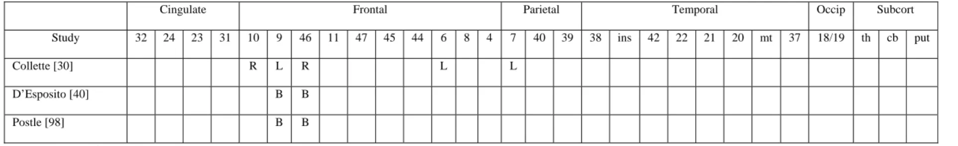
The objective of the studies described in this section was to explore the cerebral areas involved in the manipulation of information maintained in working memory, by controlling the contribution of the storage functions. A typical example of the procedures which were used to explore the manipulation processes is the alphabetisation span task (“alpha span task”). This task requires subjects to recall presented verbal information not in serial order (as in a classical span task) but according to the alphabetical order. The processes involved in this type of tasks which can be considered to be dependent upon the central executive include the inhibition of serial order recall, the extraction of alphabetical order from long-term memory and the checking of that order to rearrange the sequence of items before producing the response [14].In a recent study, we explored the cerebral substrates involved in the execution of the alpha span task, by using a procedure enabling to isolate the manipulation function from the storage processes [30]. The procedure involved the presentation of word lists whose length corresponded to the individual subject’s span minus one item. In the first condition, participants had to recall the presented words in serial order. In the second condition, the words had to be recalled in alphabetical order. With the storage requirement being equated between both conditions, the only difference consisted of a central executive intervention during alphabetical recall. Two supplementary conditions were added to compare manipulation with and without storage of information: a serial reading task and an alphabetical reading task, which did not implicate any storage of presented information. A factorial design was thus obtained, with two levels of memory (tasks with and without memory) and two levels of manipulation (tasks with and without manipulation), yielding four task conditions.
The regions involved in manipulation of information (comparison of the two alphabetical tasks to the two serial tasks) were a predominantly left-sided parietal region (BA 7), at the limit between superior and inferior parietal areas, and bilateral middle frontal areas (BA 10/46 and BA 9/6). Moreover, the interaction analysis between memory and manipulation conditions showed no
significant increase of activity, indicating that the cerebral areas involved in manipulation of information are similar in the presence or absence of a memory load.
As indicated above, the implication of the prefrontal cortex in tasks assessing the central executive of working memory has been frequently reported using various cognitive tasks [e.g., 27,95,96], with the prefrontal dorsolateral cortex (BA 9/46) being the most frequently activated region. However, in our study, the region most significantly activated by the manipulation of information was a left superior parietal region (BA 7). Increase of cerebral regional blood flow in this region has also been found in other working memory tasks [96,103,104,114]. However, since our experimental design was constructed to isolate precisely executive functioning from that of the storage systems, the predominant activation of BA 7 should be specifically related to the running of the central executive. These data are consistent with the hypothesis of a parieto-frontal network sustaining the executive functioning previously proposed by several authors [38,51,81,127]. Another interesting finding in this study is the lack of interaction between the variables “manipulation” and “memory”, indicating that the functioning of the central executive is not necessarily related to the presence of a memory load. These results are consistent with the view that the central executive of working memory is mainly devoted to executive processes, not necessarily in relation with the short-term storage of information.
The cerebral areas associated to the alphabetisation span task were also explored by D’Esposito and collaborators using event-related fMRI. In a first experiment [40], sequences of five letters were presented and changes in cerebral activity were compared in two conditions. In the first one (maintenance), participants had to retain a sequence of letters in their order of presentation during a delay period while in the second they had to reorder the sequence into alphabetical order during the delay period (manipulation). In order to determine prefrontal cerebral areas specifically involved in maintenance and manipulation of working memory, the authors created two regions of interest, one encompassing the dorsolateral prefrontal cortex (areas 9 and 46) and one containing the ventrolateral prefrontal cortex (areas 44, 45 and 47). These regions were determined on the basis of previous studies which suggested their major involvement in storage and manipulation processes in working memory. Activity during the delay period was found in both the dorsolateral and ventrolateral prefrontal cortex in both types of conditions. However, dorsolateral prefrontal activity was greater in the manipulation condition. These findings suggest that the dorsolateral prefrontal cortex may exhibit greater recruitment during behavioural conditions that require transformation of the information held in working memory.
In a second study, Postle, Berger, and D’Esposito [98] used the same task to test the hypothesis that the processes supporting strictly the storage demands of the task can be dissociated anatomically from the processes supporting the executive demands. They conceived an event-related fMRI experimental design isolating the variables of load and alphabetisation. These two variables index, respectively, mnemonic processes and executive control processes contributing to performance on the working memory task. Manipulation requirements were operationalized by asking participants to reorder into alphabetical order the five randomly ordered letters of a memory set. The evoked fMRI signal from this condition was contrasted with signals from trials in which no alphabetization of the five memoranda was required. The storage of information was operationalized by varying the number of items (five versus two) to be remembered during the delay. Again, regions of interest were defined in order to explore load and alphabetization sensitivity. These regions were located in the dorsolateral (BA 9/46) and ventrolateral (BA 44/45/47) prefrontal cortex and in posterior cortical regions (BA 22, 39 and 40). Results showed a functional neuroanatomical double dissociation of mnemonic and executive processes that contribute to the storage and manipulation of items in a working memory task: voxels in the left perisylvian cortex (BA 39 and 40) showed load sensitivity while voxels of dorsolateral prefrontal cortex (BA 9 and 46) showed manipulation (alphabetisation) sensitivity.
Taken as a whole (see Table 1), these three studies demonstrated the involvement of similar prefrontal areas when participants have to manipulate actively series of items, namely the dorsolateral prefrontal cortex BA 9/46. Moreover, one of these studies [30] indicates that manipulation of information would be also dependent upon posterior (mainly parietal) cerebral areas. Obviously, further studies with specific attention to cerebral metabolism in the superior parietal regions are necessary to confirm the role of this area in active manipulation of information stored in working
memory. Such a pattern of activity would be a strong argument in favour of the proposition that executive control requires the integration of information coming from different antero-posterior cerebral areas.
[Insert Table 1 near here]
2. The fractionation of the central executive
2.1. Updating function
Several authors [78,111] suggest that the updating process is one of the important executive functions. This function consists in continuously modifying the content of working memory according to newer incoming information. Memory updating intervenes in a large number of activities of everyday life, such as the learning and organization of recently acquired information. The updating function has been classically explored with the running memory paradigm which requires participants to watch strings of consonants of unknown length, and then to recall serially a specific number of recent items. The running span task requires considerable flexibility of information processing and a progressive shift of attention, i.e., discarding some items while new ones are registered. Morris and Jones [80] showed that the running memory task requires two independent mechanisms: the phonological loop (phonological store and articulatory rehearsal process) and the central executive. The updating process requires central executive resources but not the phonological loop. Conversely, the serial recall component of the task requires the phonological loop but not the central executive. Some studies demonstrated that this updating function is impaired in older healthy subjects although these subjects still evidence an intact phonological loop [123].
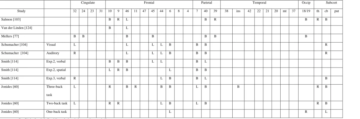
We conducted two PET studies using the running span task to explore the cerebral areas associated with the updating process. In the first study [103], changes in cerebral metabolism were compared during the performance on a phonological short-term memory and an updating working memory task. In the phonological task, participants were instructed to rehearse serially series of six consonants in order to detect whether a target consonant, presented after a delay, was present in the list. In the updating task, lists of 8, 9 and 10 consonants were presented. Participants were not informed of the length of the lists. They were asked to rehearse silently and to remember serially only the six last items in order to decide whether a target consonant, presented after a delay, was present in the six last consonants for this particular list. When working memory updating was compared to phonological short-term memory, increases of activity occurred in the right mid-dorsal prefrontal cortex (BA 9), in left middle frontal regions (BA 46 and possibly BA 10) and in the right frontal pole (BA 10). Increased rCBF was also found in a broad area of the right inferior parietal and angular gyri (BA 40/39), and in the left supramarginal gyrus (BA 40).
These data confirm that the dorsolateral prefrontal cortex is a key structure for the functioning of the central executive. The involvement of posterior (mainly parietal) areas during the updating condition also seems in agreement with the hypothesis that posterior areas underlie executive functioning. However, a problem with this study was that the Morris and Jones’ [80] original updating procedure was changed by converting the serial recall task into a short-term memory recognition task1. We recently obtained preliminary data suggesting that the recognition and the recall procedures promote the preferential utilization of visuospatial and phonological strategies, respectively. In addition, post hoc questioning of the participants examined in the initial PET study indicated that half of them used a phonological strategy in the updating memory experiment, while the other half used a visual imagery strategy or a combination of phonological and visuospatial strategies (although most of the participants used a phonological strategy in the control task). As it seems that the participants had frequently adopted a visuospatial strategy in our experimental updating condition, some of the observed activation (e.g., parietal and occipital activation) might be related to generation and short-term storage of visuospatial images rather than to central executive functioning. Another problem was that the participants were asked to remember the last six items, a memory load close to, or beyond their memory span. According to Baddeley [7; see also 121], span performance depends on both the phonological loop system and the central executive. The phonological loop system is able to store only
a limited number of items but the central executive may increase this number either by improving the working of the phonological loop (for example, by grouping items into higher level units) or even by using long-term memory information. In that perspective, it is plausible that holding a six-item memory load is very dependent upon central executive resources and consequently, in our updating experiment, the central executive system would have been involved not only in the updating process but also in the storage function.
Consequently, we carried out a second PET experiment [124] to re-examine the brain regions involved in working-memory updating by using a serial recall procedure instead of a recognition procedure and by using a sub-span (four-item) memory load which is presumably less dependent upon the central executive. This time, the most significant increase of rCBF occurred more specifically in the left frontopolar cortex (BA 10). Activation spread to the left middle frontal cortex (BA 46), and it was also observed in the right frontopolar cortex.
This second study demonstrated that the updating of information is mainly related to activity in the left frontopolar cortex (BA 10). Activity in that area had been previously found by Grasby et al. [55] who showed that activation in the frontopolar cortex increased in relation to the number of times that participants had to recall a single word. This task also involves a continuous updating process, i.e., to control how many times the word is repeated and has again to be repeated, and the updating process becomes more important as the number of repetitions increases. However, it remains to be determined whether the activation observed in the frontopolar cortex is related to a general updating process (independent of the type of updated information; i.e. semantic, visual or phonological) or to an updating operation specifically devoted to serial verbal recall. Furthermore, it could be argued that the attribution of an updating function to the frontopolar cortex is not related to the specific involvement of the central executive component but rather to a greater difficulty in the updating task than in the serial recall task. Accordingly, in our study, recall performance tended to be lower in the updating task than in the serial recall task. However, when the error score of the participants was taken as the covariate, similar activations were observed in the comparison of the updating and serial recall task.
The updating function can also be assessed by another kind of cognitive task: the n-back tasks. In the n-back tasks, items (letters, spatial positions, or patterns) are sequentially presented and subjects have to evaluate if each item is similar to the one presented n items previously. So, the task requires encoding, temporary maintenance and rehearsal, tracking of serial order, updating and comparison, and response processes. A great number of studies explored cerebral areas involved in n-back tasks. Most of them used as control condition a task in which subjects have to decide if each item sequentially presented is similar or not to a predetermined number of specific items, this number corresponding to the items being stored in working memory during the n-back condition [17,60,104,114]. The interest of such a control task is trying to remove the influence of the short-term maintenance of items on the updating process (contrary to the initial studies of Cohen et al. [27] and Mellers et al. [77]). Taken as a whole, these studies showed that the n-back task performance was associated to cerebral activity not only in the prefrontal dorsolateral cortex (BA 9/46), in the inferior frontal cortex (BA 44), in the anterior cingulate (BA 24) but also in posterior cerebral areas such as the superior and posterior parietal cortex (BA 40/7). Nevertheless, one criticism that could be made about these studies concerns the control tasks used. Indeed, memory load was equated between control and experimental conditions by requiring participants to detect, in the control task, a number of target items similar to that which had be maintained in working memory. However, a difference between the two conditions is that the n-back task requires to update continuously the letters to be maintained in working memory while in the control condition, the same items are maintained in working memory during the whole task. Thus, we cannot reject the hypothesis that the control task became more automatic or that participants used long-term memory to perform the task while the experimental task required a continuous activation of the phonological loop. Consequently, some increases of activity observed during the experimental task might not be due to the updating process per se but rather to a greater involvement of the phonological loop.
In conclusion (see Table 2), results of studies using the n-back paradigm and the running span task with a high memory load and a recognition procedure [103] demonstrated the intervention of a distributed antero-posterior cerebral network (involving mainly the dorsolatetal prefrontal cortex BA
9/46) when the tasks require updating processes. However, in a task based upon the same process (but with a lower memory load and in a recall procedure), it appears that the left frontopolar cortex (BA 10) is the area most involved in the task [124]. These data are indicative of interactions between the executive and non-executive components of a task, leading to slightly different patterns of cerebral activation according to the exact non-executive requirements of the task.
[Insert Table 2 near here]
2.2. Inhibition
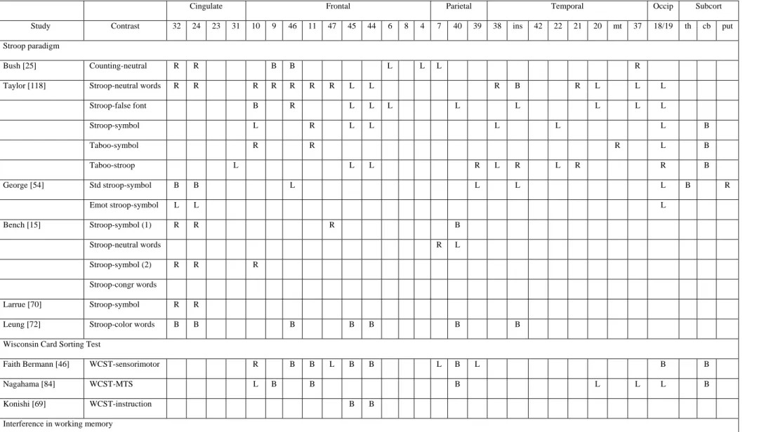
It is generally considered that inhibitory control constitutes an important executive function [9,87]. Different aspects of inhibitory control may be distinguished [56], especially those preventing access to goal-irrelevant information that may be partially activated, restraining access to strong but situationally inappropriate responses, and suppressing the activation of no longer relevant information. Most of the studies exploring inhibition processes have used different variants of the Stroop paradigm and showed increased activity during the interference condition (i.e., naming the font colour of letters that spell a colour word different than the colour-to-be-named) in the anterior cingulate gyrus and right orbitofrontal regions [15,70,91]. Moreover, increased activity has also been found in left inferior frontal regions [25,54,118] and in temporal and parietal areas [e.g., 25,118].
Semantic inhibition processes were explored by Nathaniel-James et al. [86] with the Hayling task [23]. In this study, participants were visually presented sentences in which the last word was missing. In one condition, they had to complete the sentence with the expected word (“initiation”) while in the other condition they had to provide a word unrelated to the sentence (“inhibition”). The modifications of metabolism in these two conditions were also compared with a condition in which participants had to read the last word of the sentence. In comparison to the reading condition, both the initiation and inhibition conditions led to increased rCBF in the left operculum (BA 45) and right cingulate area (BA 32). However, when the inhibition condition was compared to the initiation condition, no supplementary activation focus was found. A possible interpretation of that absence of supplementary activity relates to the paradigm used. Indeed, the procedure of Nathaniel-James et al. could be insensitive to inhibition processes since sentences were presented every 6 seconds for each condition. Thus, if participants gave responses faster than the time limit of 6 sec., they had some extra time in each condition during which cognitive processes were not controlled. This procedure could have made the task less sensitive to inhibition processes since these processes are not maximised in the contrast between the inhibition and initiation condition.
Consequently, we recently re-explored the cerebral areas involved in the initiation and inhibition conditions of the Hayling task [31]. The procedure used by Nathaniel-James et al. [86] was modified in order to maximise the involvement of inhibition processes: for all conditions, the next sentence was displayed as soon as the participants had produced a response. Moreover, participants were trained before the experimental session to use inhibition processes efficiently. Results indicated that inhibition processes are associated with left-sided increases of activity in the middle (BA 10 and BA 9) and inferior (BA 45) frontal gyrus. The involvement of several cerebral areas during the execution of the inhibition condition is not surprising since this condition involves, in comparison to the initiation condition, different types of processes, including not only the inhibition of a prevalent response but also lexical access, search strategies and working memory, at least. In agreement with previous studies, activity in the left inferior frontal area (BA 45) was attributed to the selection of a semantic response among numerous alternatives [i.e., 52], activity in the left middle frontal gyrus (BA 9) was attributed to the manipulation of information in order to build up an efficient search strategy [i.e., 36] and, finally, the middle frontal region (BA 10) was more specifically attributed to inhibition processes [i.e., 53].
Finally, it is important to note that increases of activity in several prefrontal cerebral areas are also found when the tasks require inhibitory processes that can be considered as more automatic. For example, D’Esposito et al. [41] used a working memory task in which participants had to decide as quickly as possible whether a probe letter matched a letter in a target set seen previously. Increase of activity in the left inferior frontal region (BA 45) was found when participants had to respond to trials in which the probe did not match any item in the target set of the present trial but did match an item
from the target set of the two previous trials, in comparison to trials in which the probe did not match items from either the current or the two previous target sets. The authors attributed the increase of activity in the left inferior frontal region to processes of interference resolution during retrieval of information in working memory. In another study, Chee et al. [26] characterized cerebral areas activated by a task, the Implicit Association Test, which assesses inhibitory processes engaged when participants have to process items with ingrained (as distinct from arbitrary) associations. This task examines the differential association of two object categories (e.g., flower and insect) with two attribute categories (e.g., pleasant and unpleasant). Participants are asked to make dichotomous choices in two distinct classifications on alternating trials. In the first classification, the participants have to decide if a word belongs to a given category (e.g., flower / insect) while in the second they have to judge an attribute of a word (pleasant / unpleasant). When items from congruent categories (e.g., flower / pleasant) share a response key, performance is faster and more accurate than when items from incongruent categories (e.g., insect / pleasant) share a key. In the congruent task, categories with similar valences are mapped to the same response key while in the incongruent one, categories with discordant valences are mapped to the same key. In order to complete the incongruent task successfully, the participants have to inhibit the prepotent tendency to assign items with implicitly linked attributes to the same response key. Results indicated a more extensive activation during the incongruent task in the left ventral prefrontal (BA 47), middle frontal (BA 9) and the pre-SMA/anterior cingulate as well as the superior parietal areas bilaterally.
More generally, studies that explored inhibition processes with functional imaging brought to light an important heterogeneity in the cerebral areas associated with these processes. These studies used different paradigms classically described as assessing inhibitory functions, such as the Stroop task, the Wisconsin Card Sorting test, and the go / no-go paradigms. Results obtained in these studies are summarized in Table 4. Taken as a whole, these results are indicative that the various inhibition tasks involve not only bilateral prefrontal areas but also posterior regions and subcortical structures. Moreover, there exists an important heterogeneity in the cerebral areas activated by the various inhibition tasks, and also within the different studies using variants of a same paradigm. This is not surprising since tasks used to explore the neural substrates of inhibition processes are in most cases complex, multi-compound and involve numerous cognitive processes other than inhibition. Thus, the selection of control tasks suppressing all non-inhibitory aspects is very difficult and the cerebral areas associated with inhibition processes will be dependent on the exact requirements of the experimental and control conditions. An example of this problem is well illustrated by studies using the Stroop tasks and showing different patterns of cerebral activity according to whether the reference condition was symbols, false fonts or neutral words [15,118].
[Insert Table 3 near here]
Another problem related to the exploration of the neural substrates of inhibitory processes concerns the lack of specification of the concept of inhibition. Indeed, inhibition processes cannot be considered as unitary and refer probably to very different mechanisms [see 32,75]. In addition, the exact relationships between these mechanisms are not clearly established [64,116]. In that context, the aim of future studies will be to determine whether there exist some inhibition processes common to different tasks (as attested by similar neuronal substrates) or whether the various mechanisms of inhibition are linked to distinct cerebral areas. To our knowledge, only one study has tackled this question. Konishi et al. [69] showed a common focus of activity in the right inferior prefrontal area (BA 45/44) which they associated with inhibition processes for different kinds of material. However, the tasks used (the go/no-go and WCST) share other cognitive characteristics (i.e., working memory,…) than the necessity to inhibit current action schemata, and so the inferior prefrontal activity could be related to these processes. Another interesting field of research will be to determine whether the activated area varies according to the material to be inhibited (e.g., verbal, visual,...), and the non-executive requirements of the task (for example, the use of a self- or an externally-paced procedure in the initiation and inhibition conditions of the Hayling task lead to very divergent results).
2.3. Shifting process
Efficient reactions to the stimulations of the environment require rapid and frequent shifts between the different aspects of the stimuli to be processed and also between several cognitive operations. Consequently, the shifting ability is considered as an important aspect of executive control [87]. This executive function was classically studied using task-shifting paradigms, in which participants rapidly repeat the same task or alternate between different tasks. A consistent finding in these paradigms is that response latencies are longer when subjects have to perform a switched task than when than they have to perform a repeated task, and this deficit is called the switch cost.
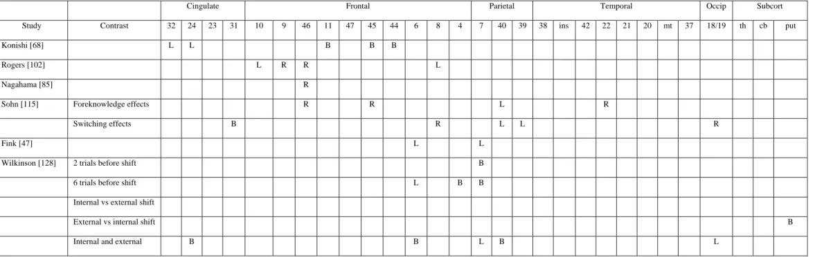
Cognitive set-shifting was frequently explored in brain-damaged patients using visual discrimination learning tasks. These tasks consist in the successive presentation of items that can be classified according to several perceptual dimensions (for example, colour, shape, value,…). On each trial, participants have to select one of the stimuli according to a specified perceptual dimension. After a pre-determined number of correct responses, the discrimination rule changes and participants have to shift from the processing of the relevant perceptual dimension to a new one that was previously considered as irrelevant. Using TEP, Rogers et al. [102] explored cerebral areas involved when participants have to shift from the processing of one stimulus dimension to another. Two conditions were administered: intra-dimensional shift condition, requiring the application of a previously learned discrimination decision rule to a new set of stimuli; and extra-dimensional shift condition, in which participants have to identify a new discrimination rule, again with novel test stimuli. This latter condition required participants to shift from the processing of one specific stimulus dimension (i.e., shape discrimination) to another (i.e., colour discrimination), and produced, in comparison to the intra-dimensional shift condition, cerebral activations in prefrontal regions, including the left anterior prefrontal cortex (BA 10 and BA 8) and right dorsolateral prefrontal cortex (BA 9/46). The results of this study demonstrate that shifting attention away from a previously reinforced stimulus dimension is particularly associated with rCBF increases in the prefrontal cortex.
However, one criticism that can be made about that study is that the shifting process cannot be easily explored with PET since the process is not continuous but rather occurs at specific (and relatively brief) periods of the task. Consequently, Nagahama et al. [80] employed event-related fMRI to re-examine cerebral areas involved in cognitive set-shifting. A significant transient increase in neural activitywas observed in the right antero-dorsal part (BA 46) of the prefrontal cortex, near the frontal pole, when participants had to shift an attentional set between different perceptual dimensions. Konishi et al. [68] used a similar procedure to dissociate the shifting processes from the inhibitory ones in the Wisconsin Card Sorting Test. Transient shift-related signals after each dimensional shift were found in the posterior part of the bilateral inferior frontal sulci (BA 45/44) but foci of activation were also found in the right and left supramarginal gyrus (BA 40) and the anterior cingulate cortex (BA 24/32), although these regions were less reproducibly activated across the participants.
The ability to switch from one cognitive task to another involves both endogenous preparation and exogenous adjustment [76]. Endogenous preparation was demonstrated by a reduction in the shifting cost when participants had foreknowledge about the upcoming task. This reduction indicates that participants could prepare themselves for the upcoming task before the stimulus for the task was presented. However, even in foreknowledge conditions with a very long interval between tasks, the shifting cost is never completely eliminated. The persistence of a switching cost in the case of foreknowledge implies that switching to a new task can only be achieved with the presentation of the first stimulus of the new task. This process is called exogenous adjustment for a new task, in the sense that the process is completed on the basis of the external stimulus. Sohn et al. [115] explored these two processes also using event-related fMRI. Tasks consisted in a simple classification of letters (consonant/vowel) or digits (even/odd). For each task, a stimulus consisted of one letter and one digit. The task which had to be performed was indicated by the colour of the stimulus. Participants were scanned in three conditions: foreknowledge and repetition-task (no switch); foreknowledge and switch-task; no-foreknowledge (task repetition and task switch mixed). Results indicate that endogenous preparation seems to involve the right lateral prefrontal cortex (BA 46/45), left superior posterior parietal cortex (BA 40) and right temporal area (BA 22). Indeed, higher activation increases in these areas were more associated with foreknowledge than with no foreknowledge. Otherwise, relatively higher activations in the right superior prefrontal cortex (BA 8), left posterior parietal cortex
(BA 39/40), posterior cingulate cortex (BA 31) and right occipital cortex (BA 19) were found during the task-switch than during the task-repetition when participants had no foreknowledge about the forthcoming task. Thus, these areas could be selectively involved in exogenous adjustment. Moreover, higher activity in BA 46/45 during the preparation period was associated with faster reaction times in the task in the foreknowledge switch condition. In contrast, higher activation in superior prefrontal cortex (BA 8) during the switch period was associated with slower performance in the task. The authors attributed this different pattern of correlations to the existence of several cognitive processes in the task-shifting paradigm. So, higher activity in the lateral prefrontal cortex (BA 46/45) would be associated with an efficient preparation for the forthcoming task (i.e., active maintenance of information that is newly loaded into working memory to perform the task). On the contrary, higher activation during the switch period in the superior prefrontal cortex (BA 8) was associated to the selection of task-relevant information against task-irrelevant information.
Shifting processes can also intervene between different hierarchical levels of a stimulus. Indeed, visual targets can be coded, in relative terms, at either the global or local level (i.e., a large “s” composed of small “a”). Several experiments demonstrated a time cost associated with identifying targets which appear at a changed hierarchical level, compared to when they reappear at the same hierarchical level as on the previous trial [e.g., 66]. In a first study, Fink et al. [47] explored global/local switching by using a task requiring to report at which level (local or global) a predesignated target had appeared, with target level alternating 1 to 34 times a minute. This contrasted with a directed attention task in which participants were required to name the letter appearing at the same level throughout. Changes in regional cerebral blood flow correlated with an increasing number of switches in left supplementary (BA 6) and medial parietal (BA 7) cortices, while sustained attention to local and global levels activated left and right temporoparietal regions, respectively.
Several other variables were found to influence the cerebral areas involved in shifting processes. For example, Wilkinson et al. [128] demonstrated an effect of the number of trials preceding a shift. Indeed, the presentation of two trials at a same hierarchical level (global or local) before a shift preferentially activated the precuneus while those performed after both four and six trials activated the bilateral inferior parietal cortex (BA 7) and motor and premotor areas (BA 4 and 6). Moreover, these authors compared cerebral activity when shift trials were externally or internally mediated. This was done by controlling the level (global or local) that was first acceded following the presentation of the items: the positioning of local elements very closely together will induce a first appearance of the global hierarchical level while the first appearance of local hierarchical level will be induced by stimuli spaced wide apart. In externally mediated shifts, the appearance of the stimulus corresponds to the novel hierarchical level to process (i.e., a shift local-global with the item appearing at first glance at the global level) while not in the internally mediated shift (i.e., a shift local-global with the item appearing at the first glance at the local level). Relative to external switches, internal switches activated the putamen bilaterally, while no specific foci of activity were found for external switches. More generally, both kinds of switch conjointly activated (in comparison to non-switch trials) a large-scale network including the anterior cingulate (BA 24), pre-SMA bilaterally (BA 6), left precuneus (BA 7) and fusiform gyrus (BA 19), left and right inferior parietal cortices (BA 40).
[Insert table 4 near here]
These neuroimaging data suggest that the shifting process involved not only the prefrontal cortex (as already evidenced in brain-damaged patients, [42]) but also posterior and subcortical areas (see Table 4). However, this process, identified by Miyake [78] as reflecting a specific executive process is in fact influenced by several factors, such as endogenous and exogenous preparation, frequency of shifts, top-down and bottom-up processes. Functional imagery studies demonstrated that these factors influence cerebral areas underlying the shifting process. However, all the executive and non-executive processes involved in a shifting task are not well identified at the present time. Consequently, results of these neuroimaging studies remain ambiguous concerning the cerebral areas that can be specifically associated to the executive requirements of shifting tasks. More generally, there still exist some uncertainties concerning the relationships between task-shifting and stimulus-shifting, as well as between task-shifting and dual-task coordination (e.g. [57]). Concerning this last
point, it is interesting to note that increases of activity associated with task-shifting processes were generally found in cerebral areas which were also activated during the simultaneous execution of two tasks (see below).
2.4. Dual-task coordination
Because the central executive system is thought to be crucial for the coordination of concurrent processing, it has commonly been investigated by using dual-task paradigms in which two behavioural tasks, often with disparate sensory and cognitive processing, are performed concurrently. Although dual-task coordination is considered as a main function of the central executive of working memory, this coordination process is not limited to tasks requiring storage of information but also intervenes for perceptual tasks requiring a low memory load
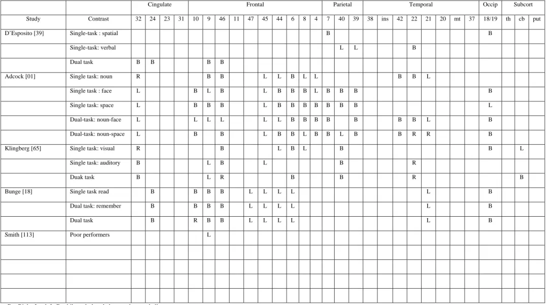
The first neuroimaging study which explored dual-task coordination was that of D’Esposito et al. [39]. Participants were administered two tasks predominantly involving posterior brain regions and requiring no storage in working memory: a semantic judgement task (to identify exemplars of a target category in series of orally presented words) and a spatial-rotation task (to indicate which of two squares had a dot in the same location, relative to a double line, as a spatially rotated target square). In order to determine the cerebral area involved in dual-task coordination, changes in the metabolic activity when participants have to perform the tasks in isolation were compared to cerebral activity when the two tasks are performed simultaneously. Comparison of the single task versus resting baseline conditions showed activation in the bilateral parieto-occipital regions for the dot-location task and in the left temporal region for the semantic judgement task. The comparison of the dual-task condition to the single-task conditions showed significantly increased activity bilaterally in the dorsolateral prefrontal cortex (BA 9 and 46) and the anterior cingulate region. Such a pattern of activity cannot be attributed to task difficulty since increasing difficulty in the single task at the level of the dual task (as attested by the accuracy of performance) resulted in increased activation in posterior brain regions although no additional prefrontal activation was observed. These data support the hypothesis that the dorsolateral prefrontal cortex is involved in the allocation and coordination of attentional resources. Activity of the anterior cingulate was attributed to response selection among competing, complex contingencies.
However, different results were found by Adcock, Constable, Gore and Goldman-Rakic [1] who suggested that dual-task coordination may be mediated by interactions between anatomically and functionally distinct systems engaged in executing component tasks, as opposed to the hypothesis of a dorsolateral prefrontal cortex activity dedicated to a generic executive system. To test that hypothesis, the authors compared changes of cerebral activity in two dual-task paradigms. The first combined an auditory verbal categorization task and a visual mental rotation task (similar to those used in the D’Esposito et al. study). The second combined the same auditory task with a different visual task involving object identification rather than mental rotation. Results indicated that the activated areas in single conditions in comparison to resting baseline varied with the component tasks, except for the dorsomedial frontal cortex and anterior cingulate that were activated in all component tasks. The verbal task activated the left inferior frontal gyrus, bilateral middle frontal gyrus, left inferior precentral sulcus, and left inferior insula. Object identification and mental rotation activated the bilateral frontal gyrus (BA 10/46), bilateral inferior frontal/inferior precentral sulcus, and regions near the junction of the superior frontal sulcus and the precentral sulcus. Finally, the mental rotation task additionally activated the right anterior insula. More interestingly, all of these areas were also activated during the execution of the dual-task condition and no supplementary activation foci were found during this latter condition. These data provide no evidence of a neural locus for a specific function of dual-task coordination of the central executive in the prefrontal cortex but are consistent with the hypothesis that the various specialized information-processing systems in the human brain may, by their interplay, accomplish the regulation of complex operations such as multitasking.
Globally, similar results were found by Klingberg [65] who used working memory paradigms to explore the concurrent performance of two tasks. That experiment involved concurrent delayed matching of pitch and luminance. In the visual working memory task, a circular field changed in luminance at various intervals and participants were asked to compare each target luminance level to
the previous one. In the auditory working-memory task, a series of high-pitched tones of different frequencies were presented and participants were asked to determine if each target tone was lower in frequency than the previous one. In the dual-task condition, the auditory and visual working memory tasks were presented simultaneously. In comparison to a baseline task in which participants received passively sensory information, the auditory and visual working memory tasks activated sensory-specific cortical areas in the temporal and occipital lobes respectively. In addition, both tasks activated overlapping areas in the right prefrontal, left middle frontal, left insula/frontal operculum, cingulate and bilateral inferior parietal cortex. When the two tasks were performed simultaneously, no supplementary activated cerebral area was found. Therefore, the results of Klingberg [65] seem to indicate that no specific cortical area could be associated with any specific cognitive process for dual-task performance and that dual-dual-task coordination depends mainly upon interactions between cerebral areas already activated in the single task. Moreover, since the frontal and parietal inferior areas were engaged by the two tasks at the same time, these results are in agreement with the hypothesis that overlapping cerebral activity is the physiological basis for interference in the dual-task condition. This hypothesis is furthermore supported by the observation that practice is associated with a decrease in interference between two tasks, as well as decreases in prefrontal and cingulate activity and thus presumably a decrease in overlapping activation [59,92].
The question of interference between apparently dissociable neural systems was directly explored by Just et al. [62]. In their fMRI study, participants performed an auditory sentence comprehension task alone (judging general-knowledge sentences), a visual mental rotation task alone (judging similarity of rotated abstract 3-D figures), or both tasks simultaneously. These tasks were chosen because they are supported by non-overlapping neural system (left superiolateral cortex and left inferior frontal gyrus for the comprehension task; bilateral parietal and inferior temporal regions for the mental rotation task). The main results of that study were that in the dual task, the activation in association areas (primarily temporal and parietal cortex) was substantially less than the sum of the activation when the two tasks were performed alone. A similar pattern of results was also obtained in the primary and secondary sensory areas. These results indicate that the simultaneous execution of two tasks requiring non-overlapping cerebral areas is not simply the addictive effect of each task performed alone. Instead, the dual-task condition induces some mutual constraints among cortical areas. The authors interpreted these data by suggesting the existence of biological mechanisms that place an upper limit on the amount of cortical tissue which can be activated at any given time, leading to a limit on how much attention is available to distribute over more than one task. This limit on the availability of attentional resources is responsible for poorer performance in dual-task than in single-task conditions.
Finally, another recent study of Bunge et al. [18] also demonstrated no specific prefrontal activity during dual-task coordination. This study was based on the principle of the reading span test [37] which requires concurrent processing (reading sentences for comprehension) and short-term maintenance (remembering the last word of each sentence) of a same material. Cerebral activity was measured by fMRI in four conditions: (1) to read and evaluate five statements as true or false, (2) to remember the last word of five sequentially presented sentences, (3) to evaluate five sentences and remember the final word of each sentence (dual condition) and (4) a control task in which participants viewed meaningless consonant strings. The third condition required both concurrent processing and maintenance of information. Activations were similar across the three conditions when compared to the control task, with bilateral activations of the prefrontal cortex (inferior: BA 44/45/47, middle: BA 9/46, superior: BA 10), left middle temporal gyrus (BA 21) and anterior cingulate (BA 24). The remember-only and dual-task condition additionally resulted in activations of bilateral parietal, occipital and cerebellar regions. These results also indicate that dual-task performance did not recruit additional brain regions relative to the component tasks but was associated with increased activation in areas already activated by one or both component tasks. These multi-focal activations suggest that the central executive of working memory involves interactions between these different brain regions.
However, a criticism that can be made about the study by Bunge et al. [18] is that their dual task may not have been sufficiently demanding, because performance on memory and sentence evaluation was as accurate in the dual task as when tested alone. In order to explore this possibility, Smith et al. [113] administered a globally similar dual-task paradigm, the Operation Span. In this dual
task, participants perform a memory task while simultaneously verifying simple equations. Cerebral activity in that condition was compared to that obtained when participants performed each component task alone. No supplementary foci of cerebral activity were found in the prefrontal cortex in the group of participants performing relatively well in the dual task (as demonstrated by accuracy and latency of the responses) while participants with a lower performance in the dual-task condition demonstrated increases of cerebral activity in the left dorsolateral prefrontal cortex (BA 9). The authors interpreted these results by suggesting that dual-task coordination per se does not require a specific prefrontal region (since good performers activate any of these regions during the operation span) but rather may require supplementary processes (such as selective attention) when the task is particularly demanding (for example, in the case of poor performers). However, such an explanation is not in agreement with the results of D'Esposito et al. (1995) showing that an increase in task difficulty did not involve increases of activity in prefrontal cerebral areas found during the execution of dual tasks.
In summary (see Table 5), all these studies except that by D'Esposito et al. [39] demonstrated that dual-task coordination (that participants have or have not to store the items in short-term memory) is not dependent upon a specific prefrontal area but rather involves interplay of various specialized information-processing systems. For example, the study by Adcock et al. [1] used the same tasks as D’Esposito et al. [39] and found no specific prefrontal areas during the execution of the dual-task paradigm. Although the reason for these discrepancies is not really clear, one explanation proposed by Klingberg [65] is that activation of the dorsolateral prefrontal and cingulate cortex found by D'Esposito et al. [39] are not specific to dual-task performance and could be due to an increase in working memory demand caused by the simultaneous performance of two non working-memory tasks. For example, it cannot be excluded that the stimulus processing or response in one task is delayed while the other task is given priority, thus inducing a working memory requirement that does not exist in the single task condition. However, such an explanation is not consistent with the study by Adcock et al. [1] who used tasks similar to those used by D’Esposito et al. and found no specific prefrontal area linked to dual-task coordination (although prefrontal activity was already found during single-task performance). According to Bunge et al. [18], contradictory results between these two studies could be reconciled by considering that, in the D’Esposito et al. study, a prefrontal activation was present during the single task but was not detected because of a higher statistical threshold. However, that activity became significant in the dual-task condition, because activation in that condition was much greater in magnitude than for either component task. In conclusion, it appears that the neural substrates of coordination processes are not really clear at the present time. Further studies, in which the single tasks do not require prefrontal involvement at all, are necessary in order to decide between the conception of a specific prefrontal area responsible for dual-task coordination or a greater interplay and requirement of various specialized information-processing systems. Moreover, we could hypothesise that discrepancies between these studies come from the shifting requirements of the different tasks. Indeed, the overlapping in the items to process in the dual-task condition was not controlled between the studies and activation of the dorsolateral prefrontal cortex in some of them may reflect a coordinating role in rapidly switching between the items of the two tasks.
[Insert Table 5 near here]
Conclusions
For some fifteen years, a great number of functional imagery studies have explored the neural substrates of the different functions attributed to the central executive. However, given the highly integrated aspect of central executive functioning and the frequent use of complex and multi-compound tasks to explore that system, most of these studies have produced results which are difficult to interpret. We have presented here studies that specifically explored the different executive processes separated at a cognitive level by Miyake et al. [78] and/or that used a procedure allowing to isolate clearly the executive requirements from the storage aspects of the task. Most of these studies demonstrated the involvement of prefrontal areas in the various kinds of executive processes explored.
The existence of a link between executive functioning and the frontal lobes is now well established. Indeed, many patients who demonstrated severe problems in the control and regulation of