ISSN 0284-186X print/ISSN 1651-226X online © 20010 Informa UK Ltd. (Informa Healthcare, Taylor & Francis AS) DOI: 10.3109/02841860903352959
†Deceased
Correspondence: Oscar Matzinger, Department of Radiation Oncology, Centre Hospitalier Universitaire Vaudois (CHUV), Rue du Bugnon 46, 1010 Lausanne, Switzerland. E-mail: [email protected]
(Received 21 July 2009; accepted 18 September 2009) ORIGINAL ARTICLE
Toxicity at three years with and without irradiation of the internal
mammary and medial supraclavicular lymph node chain in stage I to
III breast cancer (EORTC trial 22922/10925)
OSCAR MATZINGER1,2, IRMA HEIMSOTH3, PHILIP POORTMANS4,
LAURENCE COLLETTE1, HENK STRUIKMANS5, WALTER VAN DEN BOGAERT6,
ALAIN FOURQUET7, HARRY BARTELINK8, FATMA ATAMAN9 †, AKOS GULYBAN1,
MARIANNE PIERART1 AND GEERTJAN VAN TIENHOVEN10 FOR THE EORTC
RADIATION ONCOLOGY & BREAST CANCER GROUPS
1European Organisation for Research and Treatment of Cancer (EORTC), Headquarters, Brussels, Belgium, 2Department of Radiation Oncology, Centre Hospitalier Universitaire Vaudois (CHUV), Lausanne, Switzerland, 3Department of Radiation Oncology, University Medical Centre Utrecht, Utrecht, The Netherlands, 4Department of Radiation Oncology, Dr. Bernard Verbeeten Institut, Tilburg, The Netherlands, 5Department of Radiation Oncology, Leiden University Medical Centre, Leiden, The Netherlands, 6Department of Radiation Oncology, University Hospitals Leuven, Leuven, Belgium, 7Department of Radiation Oncology, Institut Curie, Paris, France, 8Department of Radiation Oncology, The Netherlands Cancer Institute, Amsterdam, The Netherlands, 9Department of Radiation Oncology, Süleyman Demirel University, Isparta, Turkey and 10Department of Radiation Oncology, Academic Medical Centre, Amsterdam, The Netherlands.
Abstract
Introduction. The EORTC 22922/10925 trial investigated the potential survival benefi t and toxicity of elective irradiation
of the internal mammary and medial supraclavicular (IM-MS) nodes Accrual completed in January 2004 and fi rst results are expected in 2012. We present the toxicity reported until year 3 after treatment. Patients and methods. At each visit, toxicity was reported but severity was not graded routinely. Toxicity rates and performance status (PS) changes at three years were compared by χ2 tests and logistic regression models in all the 3 866 of 4 004 patients eligible to the trial
who received the allocated treatment. Results. Only lung (fi brosis; dyspnoea; pneumonitis; any lung toxicities) (4.3% vs. 1.3%; p ⬍ 0.0001) but not cardiac toxicity (0.3% vs. 0.4%; p ⫽ 0.55) signifi cantly increased with IM-MS treatment. No signifi cant worsening of the PS was observed (p ⫽ 0.79), suggesting that treatment-related toxicity does not impair patient’s daily activities. Conclusions. IM-MS irradiation seems well tolerated and does not signifi cantly impair WHO PS at three years. A follow-up period of at least 10 years is needed to determine whether cardiac toxicity is increased after radiotherapy.
Many studies on lymphatic drainage of the breast confi rmed the importance of the Internal Mammary (IM) basin as a second draining route in breast cancer [1–3]. The incidence of lymph node (LN) metastasis of the internal mammary and medial supraclavicular (IM-MS) lymph node chain ranges between 4–9% in axillary node negative patients and 16–52% for axil-lary node positive patients [4–6]. A recent analysis of 2 269 Chinese breast cancer patients examined the
subpopulation with high risk of internal mammary lymph nodes metastasis [7]. They described inci-dences of IM metastasis in more than 20% of patients with the following conditions: (1) patients with four or more positive axillary LN, (2) Patients with medial tumour and positive axillary LN, (3) Patients with T3 tumour and younger than 35 years, (4) Patients with T2 tumour and positive axillary LN and (5) Patients with T2 tumour and medial tumour.
Acta Oncol Downloaded from informahealthcare.com by Cantonale et Universitaire
and 24 Gy was delivered with electrons. To enable many radiotherapy institutes to participate in the trial and to accrue a large and representative sam-ple of patients, a standard treatment technique with one anterior fi eld for the IM-MS irradiation was recommended. The IM-MS lymph node area had to be treated with mixed photon and electron beams matched to the tangential field borders of the breast or thoracic wall (which could alterna-tively be treated with a direct electron field). Sev-eral institutes had developed specific irradiation techniques in this indication [12]. These more complex treatment set-ups were accepted in the trial, provided that they took into account the indi-vidual localisation of the internal mammary nodes [12,13].
The defi ned organs at risk were the lungs and the heart. No specifi c constraints were however defi ned in the protocol as the irradiated lung and heart vol-umes were considered as limited by the use of the mixed beam technique.
This study was subjected to an intensive quality assurance programme consisting of a dummy run and individual case review. The results of this proce-dure were already previously reported [11–15].
The protocol contained no guidelines which patients were to receive adjuvant treatment (hor-monotherapy, chemotherapy).
Data collection
At the time of randomisation, data on WHO perfor-mance status (PS), tumour characteristics, number of positive axillary nodes and on adjuvant systemic treatment were collected for each patient. The fol-lowing details concerning the radiotherapy were col-lected after completion of treatment: duration and interruption of radiotherapy, total dose, number of fractions and the technique used for IM-MS chain treatment. Yearly follow-up visit documented PS, presence of lung fi brosis, presence of cardiac fi brosis, presence of other toxicity and evidence of cardiac disease. Other toxicities and cardiac disease were to be detailed in free text.
Thoracic x-ray was obtained as a part of the yearly loco-regional evaluation in the protocol. Ejec-tion fracEjec-tion study was opEjec-tional.
Statistical methods
The analysis was conducted in the per protocol pop-ulation of patients who were eligible to the protocol and followed the randomly allocated IM-MS treat-ment policy. Patients with partial IM-MS irradiation were also excluded.
IM lymph node dissection was therefore per-formed in a number of institutes in the 1950s and 1960s. This radical surgical procedure was aban-doned in the 1970s because several studies showed that this approach did not improve survival [8]. How to interpret today the clinical relevance of the historic rates of IM lymph node involvement in view of the greater proportion of screen-detected cancers, the improved imaging for detection of IM-nodes involvement, the increasing use of adjuvant systemic therapy and the newer radiotherapy techniques is unclear.
The interest for an elective treatment of the IM-MS nodes was renewed after the publication of several prospective randomised trials that demon-strated a favourable outcome after elective loco-regional irradiation [9,10]. In operable breast cancer however, the role of regional radiotherapy of the IM-MS chain remains controversial [8] since defi nite evidence supporting that elective irradiation of espe-cially the IM nodes improves overall survival is lack-ing. Whether the expected benefi t of elective irradiation of the IM-MS nodes counterbalances a possible increase of the risk of late toxicity is still unresolved [5].
The Radiation Oncology Group and the Breast Cancer Group of the European Organisation for Research and Treatment of Cancer (EORTC) there-fore initiated a large randomised phase III multi-centre trial (EORTC 22922/10925) assessing the impact of elective IM-MS lymph node irradiation on overall survival in patients with localised, stage I–III, breast cancer with medially or centrally located tumours and/or axillary lymph node invasion. This trial that recruited patients between July 1996 and January 2004, enrolled 4 004 women with unilateral breast cancer after breast and axillary surgery. The fi rst analysis of the primary endpoint, overall survival at 10 years, will be performed about eight years after recruitment of the last patient, which is expected to be in 2012. Extensive reviews on the study popula-tion [11], on the radiotherapy techniques [12] as well as on the quality assurance program [13–15] have already been published.
We present here the toxicity reported up to three years after treatment as well as the change in WHO performance status (PS) between entry and year 3 post-treatment.
Patients and methods
Radiotherapy protocol
The prescribed dose was 50 Gy in 25 fractions of 2 Gy; 26 Gy was delivered with photons (minimum energy of Co-60 and maximum energy of 10 MV),
Acta Oncol Downloaded from informahealthcare.com by Cantonale et Universitaire
The percentage of patients with any toxicity reported in the fi rst three years after treatment was compared between the two treatment arms by means of χ2 tests. The events reported as free text were also
grouped by category for descriptive purposes. The rates of individual events related to “lung toxicity” as well as the rates of any lung-related toxicity, the rates of cardiac disease, cardiac fi brosis and those of any skin-related toxicities were also formally compared by means of χ2 tests between the treatment groups.
To adjust the risk of false positive fi ndings for the multiplicity of the tests, a nominal signifi cance level of 0.01 was used.
Furthermore, changes in WHO performance sta-tus between randomisation and year 3 of the fol-low-up were assessed as no change, improvement or worsening. To avoid confounding the worsening of the PS related by the deterioration due to progressive disease, patients with disease progression reported within three years of entry on study were excluded from the analysis. Because patients with WHO PS of 0 at entry could not improve their performance sta-tus, we studied the probability of PS deterioration. Univariate and multivariate logistic regression models were used to assess the impact of treatment arm (IM-MS vs. no IM-MS), laterality of the breast cancer (left vs. right), adjuvant hormonotherapy (yes vs. none reported), neo-adjuvant chemotherapy (yes vs. no), age (⬍45 years vs. 45-⬍55 years vs. 55-⬍65 vs. ⱖ65 y), type of surgery (lumpectomy vs. mastectomy), tumour size (in centimetres), patho-logical axillary nodal status (pN⫹ vs. pN0), oestro-gen and progesterone receptor status (positive vs. negative) and menopausal status (post-menopausal/ artifi cial menopause vs. pre-menopausal) as possible predictors of the worsening of the PS. Statistical signifi cance in these models was set at 5%.
Furthermore, we assessed the correlation between the presence of toxicity and the deterioration of the performance status using univariate logistic regres-sion models as above mentioned.
Because of the very large sample size, the statisti-cal power is very high and statististatisti-cal signifi cance may not necessarily indicate clinically meaningful differences.
Results
We report on the prospectively collected data from 3 866 of the 4 004 patients randomised (97%) in the IM-MS EORTC study 22922/10925 who were eligible and followed the allocated treatment policy (no IM-MS irradiation: N ⫽ 1944 vs. IM-MS irra-diation: N ⫽ 1922). Complete follow-up documen-tation up to year 3 was available for 95.3% of the
Table I. Patient characteristics.
Per protocol population
Treatment No IM-MS (N⫽1944) IM-MS (N⫽1922) Age (years) Median 54.0 54.0 Range 22.0 - 75.0 19.0 - 75.0 ⬍ 45 346 (17.8) 354 (18.4) 45-⬍55 680 (35.0) 661 (34.4) 55-⬍65 582 (29.9) 579 (30.1) ⬎⫽65 336 (17.3) 328 (17.1) Performance Status (PS) PS 0 1733 (89.1) 1725 (89.8) PS 1 198 (10.2) 181 (9.4) PS 2 7 (0.4) 5 (0.3) Missing 6 (0.3) 11 (0.6) Menopausal status Pre-menopausal 650 (33.4) 657 (34.2) Peri-menopausal 151 (7.8) 133 (6.9) Post menopausal 1080 (55.6) 1071 (55.7)
Artifi cial menopause 63 (3.2) 61 (3.2)
Type of breast surgery
Mastectomy 447 (23.0) 454 (23.6) Breast conserving 1497 (77.0) 1468 (76.4) Pathological T * pT1 1180 (60.7) 1160 (60.4) pT2 689 (35.4) 685 (35.6) pT3 65 (3.3) 70 (3.6) Missing 10 (0.5) 7 (0.4) Pathological N (axilla)∗ pN0 877 (45.1) 855 (44.5) pN1 848 (43.6) 822 (42.8) pN2 182 (9.4) 188 (9.8) pN3 37 (1.9) 57 (3.0) Tumour stage∗ Stage I 658 (33.8) 654 (34.0) Stage IIa 647 (33.3) 612 (31.8) Stage IIb 376 (19.3) 374 (19.5) Stage III 253 (13.0) 276 (14.4) Missing 10 (0.5) 6 (0.3)
Combination of ER/PR status
ER⫹, PR⫹ 1066 (54.8) 1055 (54.9)
ER⫹, PR-/unknown 359 (18.5) 380 (19.8)
PG⫹, ER-/unknown 82 (4.2) 66 (3.4)
ER-, PR- 320 (16.5) 295 (15.3)
Missing 117 (6.0) 126 (6.6)
Adjuvant hormonal therapy
None reported 775 (39.9) 786 (40.9) yes 1169 (60.1) 1136 (59.1) Adjvuant chemotherapy No chemotherapy 886 (45.6) 871 (45.3) Adjuvant 243 (12.5) 242 (12.6) Neo-adjuvant 815 (41.9) 809 (42.1) Adjuvant treatment None 294 (15.1) 316 (16.4) Chemotherapy 481 (24.7) 470 (24.5) Hormonal therapy 592 (30.5) 555 (28.9) Both 577 (29.7) 581 (30.2)
∗Staging is according to UICC 1992
ER⫽Estrogen receptor, PR⫽progesterone receptor
Acta Oncol Downloaded from informahealthcare.com by Cantonale et Universitaire
treatment arm vs. 84.1% after IM-MS irradiation. It deteriorated in 141 of 1 944 (8.4%) vs. 144 of 1 657 (8.7%) of the patients in the standard and IM-MS treatment arm, respectively and improved in 8.3% vs. 7.2%, respectively. There was no signifi cant difference between the two treatment arms (p ⫽ 0.79). Respectively 133 of 141 and 134 of 144 of the deteriorations of the PS were in the form of an increase from PS 0 to PS 1. Conversely, all improvements (139 and 119 patients, respectively) were in the form of decrease of an initial PS 1 to a PS of 0.
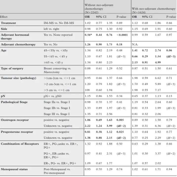
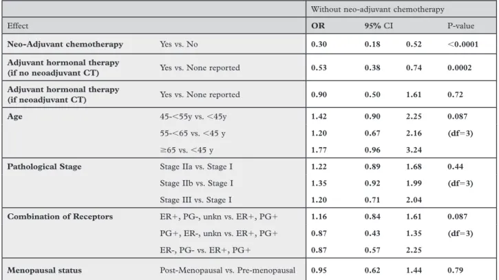
In the whole group, the univariate analysis revealed a statistically signifi cant impact of the application of any adjuvant systemic treatment (OR ⫽ 0.39, CI: 0.30–0.52; p ⬍ 0.0001) on the risk of deterioration of the WHO PS and no signifi cant difference between the two randomised treatment arms (OR⫽1.03, CI: 0.81–1.32, p ⫽ 0.79). In order to elucidate the appar-ently protective impact of adjuvant systemic treat-ment, we then separated the patient group who had received neo-adjuvant chemotherapy) from the others (i.e. no adjuvant chemotherapy and adjuvant chemo-therapy). This was in order to avoid a possible differ-ential effect of other factors in the group neo-adjuvant chemotherapy, which might have had an acute and temporary PS deterioration at the time of randomisa-tion, due to the neo-adjuvant chemotherapy (see Table IV). These analyses revealed a statistically signifi cant impact of adjuvant hormonotherapy in the patient group that did not receive neo-adjuvant chemotherapy (OR ⫽ 0.56, CI: 0.44–0.76; p ⬍ 0.0001) indicating a lower risk of deterioration of the WHO PS for the patients who received adjuvant hormonotherapy. This parameter was the only one that remained signifi cant in a multivariate model. Oestrogen and progesterone receptor status were also signifi cant in the univariate model (OR ⫽ 1.06, CI: 0.69–1.63; p ⬍ 0.005 vs. OR ⫽ 0.81, CI: 0.58–1.12; p ⬍ 0.025 respectively) but their effect vanished in the multivariate model. The other tested variables did not infl uence the evolution of the PS. In the group who received neo-adjuvant chemotherapy, only age was (borderline) statistically signifi cant in the univariate analysis, and none of the factors was signifi cant at the p ⬍ 0.05 signifi cance level in the multivariate model. Table V displays a mul-tivariate model combining all patients in one model, that also includes the factors that are nowadays con-sidered in the decision to deliver neo-adjuvant chemo-therapy or adjuvant hormonochemo-therapy, namely disease stage, age, hormone receptor statuses, and meno-pausal status. The model confi rms that in patients who have otherwise similar age, menopausal status, oestro-gen and progesterone receptor status and disease stage, those who did receive either adjuvant hormono-therapy or neo-adjuvant chemohormono-therapy, as appropriate patients. The characteristics of the patients included
in this analysis are presented in Table I. At entry on study, 89.4% of the patients presented a WHO per-formance status of 0, 9.8% with WHO PS 1 and 12 patients had a WHO PS of 2 (0.3%), WHO PS was missing in 17 (0.4%). Further details on the total study population were previously published [11].
Toxicity within three years of treatment
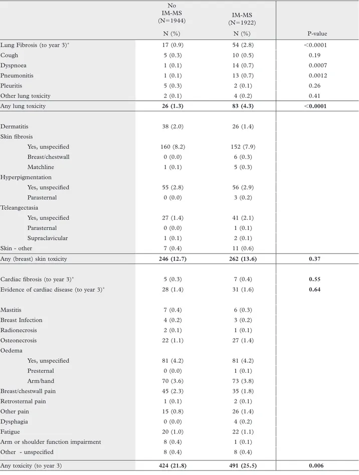
The reported toxicity per treatment arm is sum-marised in Table II. Both study treatment arms were well tolerated with little toxicity: the most frequent reported toxicities were oedema (7.8% vs. 8.1%), skin fi brosis (8.3% vs. 8.5%), teleangectasia (1.5% vs. 2.3%) and lung fi brosis (0.9% vs. 2.8%) in the standard and the IM-MS arm, respectively.
There were no statistically signifi cant differences between the two randomised groups in terms of car-diac fi brosis (0.3% vs. 0.4%; p ⫽ 0.55) nor in terms of presence of “cardiac disease” (1.4% vs. 1.6%; p ⫽ 0.64). Lung fi brosis (0.9% vs. 2.8%; p ⬍ 0.0001), dyspnoea (0.1% vs. 0.7%; p ⫽ 0.0007) and pneu-monitis (0.1% vs. 0.7%; p ⬍ 0.0012) were statisti-cally signifi cantly increased in the IM-MS treatment arm. This translated into a signifi cantly higher rate of “any lung” toxicities in the IM-MS treatment arm as compared to the control arm (4.3% vs. 1.3%; p ⬍ 0.0001). The observed difference represents an addi-tional 57 cases of lung toxicity in the IM-MS arm.
No statistically signifi cant difference could be observed in skin toxicity (including fi brosis, hyper-pigmentation, teleangectasia as well as other skin toxicities; p ⫽ 0.37). The total number of events of toxicity reported up to year 3 amounts 21.8% in the standard treatment arm vs. 25.5% in the IM-MS arm (⫹67 cases, ⫹ 3.7%). This difference is statisti-cally signifi cant (p ⫽ 0.006).
All other reported toxicities (mastitis, breast infection, radionecrosis, osteonecrosis, oedema, pain, dysphagia, fatigue, arm/shoulder function impair-ment, other) were equally distributed between the two treatment arms.
Change in performance status at three years after randomisation
The PS at baseline and at year 3 is summarised in Table III for all 3 866 patients. Since those whose disease progressed or were lost to follow-up were censored for the assessment of WHO PS at 3 years, 3 341 patients are included in this analysis (1 684 and 1 657, respectively). At year 3, the WHO PS was unchanged compared to baseline in the majority of patients in both arms: 83.4% in the standard
Acta Oncol Downloaded from informahealthcare.com by Cantonale et Universitaire
Table II. Toxicity up to year three according to treatment arm No IM-MS
(N⫽1944) (N⫽1922)IM-MS
N (%) N (%) P-value
Lung Fibrosis (to year 3)∗ 17 (0.9) 54 (2.8) ⬍0.0001
Cough 5 (0.3) 10 (0.5) 0.19
Dyspnoea 1 (0.1) 14 (0.7) 0.0007
Pneumonitis 1 (0.1) 13 (0.7) 0.0012
Pleuritis 5 (0.3) 2 (0.1) 0.26
Other lung toxicity 2 (0.1) 4 (0.2) 0.41
Any lung toxicity 26 (1.3) 83 (4.3) ⬍0.0001
Dermatitis 38 (2.0) 26 (1.4) Skin fi brosis Yes, unspecifi ed 160 (8.2) 152 (7.9) Breast/chestwall 0 (0.0) 6 (0.3) Matchline 1 (0.1) 5 (0.3) Hyperpigmentation Yes, unspecifi ed 55 (2.8) 56 (2.9) Parasternal 0 (0.0) 3 (0.2) Teleangectasia Yes, unspecifi ed 27 (1.4) 41 (2.1) Parasternal 0 (0.0) 1 (0.1) Supraclavicular 1 (0.1) 2 (0.1) Skin - other 7 (0.4) 11 (0.6)
Any (breast) skin toxicity 246 (12.7) 262 (13.6) 0.37
Cardiac fi brosis (to year 3)∗ 5 (0.3) 7 (0.4) 0.55
Evidence of cardiac disease (to year 3)∗ 28 (1.4) 31 (1.6) 0.64
Mastitis 7 (0.4) 6 (0.3) Breast Infection 4 (0.2) 3 (0.2) Radionecrosis 2 (0.1) 1 (0.1) Osteonecrosis 22 (1.1) 27 (1.4) Oedema Yes, unspecifi ed 81 (4.2) 81 (4.2) Presternal 0 (0.0) 1 (0.1) Arm/hand 70 (3.6) 73 (3.8) Breast/chestwall pain 45 (2.3) 35 (1.8) Retrosternal pain 1 (0.1) 2 (0.1) Other pain 15 (0.8) 26 (1.4) Dysphagia 0 (0.0) 4 (0.2) Fatigue 20 (1.0) 22 (1.1)
Arm or shoulder function impairment 8 (0.4) 1 (0.1)
Other - unspecifi ed 8 (0.4) 8 (0.4)
Any toxicity (to year 3) 424 (21.8) 491 (25.5) 0.006
∗pre printed item on case report forms
Acta Oncol Downloaded from informahealthcare.com by Cantonale et Universitaire
are at lower risk of PS deterioration at year 3 than those who did not. This PS deterioration however, was seen in only about 8.5% of the assessable patients who for the vast majority had a worsening from PS 0 to PS 1 (see Table III).
Correlation between toxicity and change in performance status at three years after randomisation
Table VI shows the univariate analysis relating the presence of any lung toxicity, lung fi brosis or evidence of cardiac disease within the fi rst three years to the risk of deterioration of the WHO PS. The results indi-cate no statistically signifi cant relationship between lung toxicity or lung fi brosis and the risk of deteriora-tion of the performance status. Cardiac diseases on the contrary seem signifi cantly correlated with a high risk of WHO PS deterioration (OR ⫽ 3.71, CI: 1.90–7.24, p ⬍ 0.0001). The impact of cardiac fi brosis could not be assessed because only 11 patients were reported as having this event. None of these 11 patients had a deterioration of the PS at three years.
Discussion
Radiation therapy is an integral part of the multimo-dality treatment of breast cancer. The Danish and the British Columbia trials have fi rmly established the survival advantage following radiotherapy in post mastectomy patients [10,16]. Furthermore, the EBCTCG meta-analysis has demonstrated that radiotherapy, besides improving local control rates, confers a survival benefi t in breast conservation treatment as well as in post mastectomy patients [15]. Although they showed the importance of loco-regional control on survival outcomes, the Danish and British Columbia trials and the EBCTCG meta-analysis were unable to discern the direct con-tribution from IM-MS treatment. It was noted as well that the survival gains associated with improve-ments in loco-regional control may be diminished by RT-associated cardiac mortality [17–20]. The EORTC 22922/10925 trial investigates therefore if elective irradiation of the IM-MS chain improves overall survival at 10 years.
In our report of the fi rst three years of follow-up in this study, we did not observe any difference
Table III. WHO performance status at baseline and at year for the non-progressive patients Performance status at
year 3 Baseline Performance status
No IM-MS Group Missing Performance
status 0 Performance status 1 Performance status 2 Total Missing 1 223 29 2 255 Performance status 0 5 1370 135 3 1513 Performance status 1 0 133 33 1 167 Performance status 2 0 4 0 1 5 Performance status 3 0 1 1 0 2 Performance status 4 0 2 0 0 2 Total 6 1733 198 7 1944
IM-MS Group Missing Performance
status 0 Performance status 1 Performance status 2 Missing 4 224 30 0 258 Performance status 0 4 1361 114 4 1483 Performance status 1 3 134 33 1 171 Performance status 2 0 4 4 0 8 Performance status 3 0 1 0 0 1 Performance status 4 0 1 0 0 1 Total 11 1725 181 5 1922
(Missing: patients with missing performance status or with progression before the assessment time point Blue⫽deterioration, orange⫽improvement
Acta Oncol Downloaded from informahealthcare.com by Cantonale et Universitaire
Table IV. Infl uence of various parameters on WHO performance status deterioration
Without neo-adjuvant chemotherapy
(N⫽2242)
With neo-adjuvant chemotherapy
(N⫽1624)
Effect OR 95% CI P-value OR 95% CI P-value
Treatment IM-MS vs. No IM-MS 1.02 0.77 1.35 0.89 1.12 0.68 1.86 0.66
Side left vs. right 0.98 0.75 1.30 0.92 1.15 0.69 1.91 0.60
Adjuvant hormonal therapy
Yes vs. None reported 0.56* 0.44 0.76 ⬍0.0001 0.99 0.59 1.67 0.97
Adjuvant chemotherapy Yes vs. No 1.26 0.90 1.75 0.18 N.A.
Age 45-⬍55y vs. ⬍45y 1.34 0.82 2.19 0.48 1.41 0.72 2.74 0.06
55-⬍65 vs. ⬍45 y 1.10 0.67 1.81 (df⫽3) 0.66 0.29 1.54 (df⫽3)
ⱖ65 vs. ⬍45 y 1.34 0.80 2.23 2.13 0.91 4.99
Type of surgery Breast conserving vs. Mastectomy
0.88 0.61 1.29 0.52 0.87 0.51 1.50 0.61
Tumour size (pathology) ⬎1cm-2cm vs. ⬍⫽1 cm 0.95 0.66 1.37 0.66 1.98 0.59 6.62 0.71
⬎2 cm-3cm vs. ⬍⫽1 cm 1.20 0.79 1.82 (df⫽3) 1.70 0.49 5.89 (df⫽3)
⬎3 cm vs. ⬍⫽1 cm 108 0.60 1.94 1.98 0.55 7.17
pN pN⫹ vs. pN0 1.15 0.86 1.53 0.34 0.65 0.37 1.13 0.13
Pathological Stage Stage IIa vs. Stage I 0.98 0.70 1.37 0.41 1.19 0.54 2.64 0.60
Stage IIb vs. Stage I 1.33 0.89 1.97 (df⫽3) 0.81 0.33 1.99 (df⫽3)
Stage III vs. Stage I 1.35 0.71 2.56 0.81 0.32 2.06
Oestrogen receptor positive vs. negative 1.06 0.69 1.63 0.005 0.89 0.50 1.58 0.79
Unknown vs. negative 2.22 1.24 3.99 (df⫽2) 1.39 0.31 6.36 (df⫽2)
Progesterone receptor positive vs. negative 0.81 0.58 1.12 0.025 1.10 0.64 1.92 0.77
Unknown vs. negative 1.38 0.88 2.15 (df⫽2) 0.77 0.25 2.29 (df⫽2)
Combination of Receptors ER⫹, PG-,unkn vs. ER⫹,
PG⫹ 1.32 0.92 1.88 0.50 0.63 0.29 1.38 0.66 PG⫹, ER-,unkn vs. ER⫹, PG⫹ 0.97 0.41 2.31 (df⫽3) 1.01 0.30 3.37 (df⫽2) ER-, PG- vs. ER⫹, PG⫹ 1.09 0.67 1.77 1.07 0.57 2.02
Menopausal status Post-Menopausal vs. Pre-menopausal
0.95 0.70 1.29 0.74 1.02 0.61 1.71 0.94
∗Signifi cant in multivariate model
between the two randomised groups in terms of “cardiac fi brosis” or “evidence of cardiac disease”. However, further follow-up is required to confi rm the absence of any deleterious impact of IM-MS treatment on cardiac function because late cardiac toxicity often appears 10 or even 15 years after treat-ment [17–29] and because we observed already a signifi cant detrimental impact of the presence of cardiac disease on the PS of the patients. A limita-tion of this study will however remain that there was no precise defi nition of cardiac fi brosis in the
protocol and that there was no specifi c investigation planned.
Pulmonary fi brosis was observed in 0.9% and 2.8% of patients in respectively the standard and IM-MS treatment arm. Lung toxicity of any kind was observed in only 1.3% vs. 4.3% of patients, representing an increase of only 57 cases of lung toxicity with IMMS irradiation. These results are in line with recent reports on pulmonary toxicity of breast radiotherapy [29–33]. This increased lung toxicity with IM-MS radiation was the only
Acta Oncol Downloaded from informahealthcare.com by Cantonale et Universitaire
treatments. Although we observed that neo-adju-vant chemotherapy was given more frequently to young premenopausal patients with negative hor-mone receptors and high disease stage, whereas hormonotherapy was given to postmenopausal older women, these patients did not all receive the adjuvant therapy. The observed effects of adjuvant therapies thus likely refl ect, for specifi ed patient subgroups, the benefi t of having actually received the adjuvant therapy that would be recommended in today’s practice (respectively neo-adjuvant chemotherapy in young patients and adjuvant hor-monotherapy in post menopausal women). However, these protective effects can not be explained by an impact on tumour progression as the patients with early relapse were censored in this analysis. It is also important to note that the PS deteriorated in only 8.5% of the patients, mostly as a deterioration from PS 0 to PS 1.which may not be very clinical relevant.
Conclusion
From this study, we conclude that IM-MS irradia-tion seems well tolerated and does not signifi cantly impair WHO PS nor induces excess toxicity within the fi rst three years after treatment. Longer fol-low-up is needed to further document cardiac toxic-ity and the impact of IM-MS irradiation on clinical outcome.
statistically signifi cant difference between the toxic-ity of the two treatment groups. Caution is however needed in the interpretation of these results the very large sample size of this study: due to the very high statistical power, statistically signifi cant differ-ences may not always be clinically relevant. It is important to note that the overall rate of lung tox-icity remained below 5% in both treatment arms. In order to assess the impact of this toxicity on the patient’s every day living, we correlated the changes of PS to the treatment arm. Despite the observed increase of lung toxicity with IM-MS irradiation, we could not demonstrate any signifi cant difference in the risk of decreased PS between the randomised treatments nor could we demonstrate any signifi -cant correlation between the deterioration of the PS and the risk of lung toxicity. This suggests that these lung toxicities remain mainly subclinical or disap-pear with follow-up as is often seen with limited to moderate radio-pneumonitis.
The other reported toxicities are all well known toxicities of breast cancer radiotherapy and were not signifi cantly increased with IM-MS irradiation.
An unexpected observation was that patients treated with neo-adjuvant chemotherapy had a lower risk of worsening their PS. A similar risk reduction was for patients who received adjuvant hormonotherapy. It is important to recognise that the trial protocol contained no strict guidelines as to which patients were to receive these adjuvant
Table V. Infl uence of various parameters in the multivariate analysis on WHO performance status deterioration
Without neo-adjuvant chemotherapy
Effect OR 95% CI P-value
Neo-Adjuvant chemotherapy Yes vs. No 0.30 0.18 0.52 ⬍0.0001
Adjuvant hormonal therapy
(if no neoadjuvant CT) Yes vs. None reported 0.53 0.38 0.74 0.0002
Adjuvant hormonal therapy
(if neoadjuvant CT) Yes vs. None reported 0.90 0.50 1.61 0.72
Age 45-⬍55y vs. ⬍45y 1.42 0.90 2.25 0.087
55-⬍65 vs. ⬍45 y 1.20 0.67 2.16 (df⫽3)
ⱖ65 vs. ⬍45 y 1.77 0.96 3.24
Pathological Stage Stage IIa vs. Stage I 1.22 0.89 1.68 0.44
Stage IIb vs. Stage I 1.35 0.92 1.99 (df⫽3)
Stage III vs. Stage I 1.20 0.71 2.04
Combination of Receptors ER⫹, PG-, unkn vs. ER⫹, PG⫹ 1.16 0.84 1.61 0.087
PG⫹, ER-, unkn vs. ER⫹, PG⫹ 0.87 0.43 1.35 (df⫽3)
ER-, PG- vs. ER⫹, PG⫹ 0.87 0.57 2.25
Menopausal status Post-Menopausal vs. Pre-menopausal 0.95 0.62 1.44 0.79
Acta Oncol Downloaded from informahealthcare.com by Cantonale et Universitaire
Acknowledgements
This study was supported by grants number 2U10 CA11488-22 through 5U10 CA011488-39 from the National Cancer Institute (Bethesda, Maryland, USA). Its contents are solely the responsibility of the authors and do not necessarily represent the offi -cial views of the National Cancer Institute. The work of Dr. O. Matzinger as Emmanuel Vander-schueren Fellow at the EORTC Headquarters was supported by the Vlaamse Liga Tegen Kanker.
The following institutions and physicians contri-buted patients to EORTC trial 22922-10925: Dr. P. H. P. Poortmans (Bernard Verbeeten Instituut, Tilburg, The Netherlands); Pr. W. Van Den Bogaert (Universitair Ziekenhuis Gasthuisberg, Leuven, Belgium); Dr. C. Kirkove (Cliniques Universitaires Saint Luc, Brussels, Belgium); Pr. V. G. Budach (Charite Universitaetsmedizin Berlin – Campus Mitte, Berlin, Germany); Pr. Philippe Maingon (Centre George François.Leclerc, Dijon; France); Dr. Maria Carla Valli (Ospedale Sant Anna, Como, Italy); Dr. J. J. F. M. Immerzeel (Radiotherapeutisch Insti-tuut Stedendriehoek En Omstreken, Deventer, The Netherlands); Dr C. Warlam-Rodenhuis (Universitair Medisch Centrum, Academisch Ziekenhuis, Utrecht, The Netherlands); Dr. C. Le Pechoux (Institut
Gustave Roussy, Villejuif, France); Pr. M. Bamberg (Universitaetsklinikum Tuebingen, Tuebingen, Ger-many); Dr. G. Van Tienhoven (Academisch Medisch Centrum, Amsterdam, The Netherlands); Pr. C. Glanzmann, Pr. U. M Lutolf (Universitaetsspital, Zurich, Switzerland); Dr. A. Fourquet (Institut Curie, Paris, France); Pr. A. Kuten (Rambam Med-ical Center, Haifa, Israël); Dr. P. Quetin (Centre Paul Strauss, Strasbourg, France); Dr. R. Baeza (Insti-tuto de Radiomedicina, Santiago, Chile); Dr. M. David (Nottingham City Hospital, Nottingham, United Kingdom); Dr. J. Jobsen (Medisch Spectrum Twente, Enschede, The Netherlands); Dr. M.-P. Sunyach (Centre Leon Berard, Lyon, France); Pr. H. Bartelink, Dr. N. Russell (The Netherlands Cancer Institute, Antoni Van Leeuwenhoekziekenhuis, Amsterdam, The Netherlands); Pr. R. O. Mirimanoff (Centre Hospitalier Universitaire Vaudois, Lau-sanne, Switzerland); Pr. M. Bolla (C. H. R. De Grenoble-La Tronche, Grenoble, France); Pr. M. Beauduin (Hospital De Jolimont, Haine St. Paul, Belgium); Dr. R. Pfeffer (Chaim Sheba Medical Center, Tel-Hashomer, Israël); Dr. J.-L. Lagrange (C. H. U. Henri Mondor, Creteil, France); Dr. J.-M. Dilhuydy (Institut Bergonie, Bordeaux, France); Dr Chenal, Dr. M. Benchalal (Centre Eugene Marquis, Table VI.Correlation between toxicity and WHO performance status deterioration at three years
Deterioration by year 3 No Change/ improvement (N⫽3056) Deterioration (N⫽285) N (%) N (%) OR 95% CI P-value
Any lung toxicity
No 2972 (91.5) 276 (8.5)
Yes 84 (90.3) 9 (9.7) 1.19 0.59-2.41 0.62
Lung Fibrosis (to year 3)
No 2996 (91.5) 280 (8.5)
Yes 56 (91.8) 5 (8.2) 1.02 0.40-2.59 0.96
Missing 4 (100.0) 0 (0.0)
Cardiac Fibrosis (to year 3)
No 3038 (91.4) 285 (8.6) Too small
Yes 11 (100.0) 0 (0.0) sample for
Missing 7 (100.0) 0 (0.0) testing
Evidence of cardiac disease (to year 3)
No 2989 (91.9) 265 (8.1)
Yes 38 (76.0) 12 (24.0) 3.71 1.90-7.24 ⬍0.0001
Missing 29 (78.4) 8 (21.6)
Acta Oncol Downloaded from informahealthcare.com by Cantonale et Universitaire
Rennes, France); Dr. P. Huget (Algemeen Ziekenhuis Sint, Augustinus, Wilrijk, Belgium); Dr. A. Courdi (Centre Antoine Lacassagne, Nice, France); Pr. J.-F. Bosset (C. H. R. De Besancon-Hospital Jean Mini-joz, Besancon, France); Dr. A. Kobierska (Medical University of Gdansk, Gdansk, Poland); Dr. E. Sen-kus-Konefka (Medical University of Gdansk, Gdansk, Poland); Pr J. Kurtz, Dr. S. Balmer-Majno (Hospital Cantonal Universitaier de Geneve, Geneve, Switzerland); Dr. S. Koswig, Dr. H. Krebs (Charité-Universitaetsmedizin Berlin-Campus Berlin-Buch, Berlin, Germany); Dr. R. Bongartz (Universi-taetskliniken Koeln, Koeln, Germany); Dr. H. Struik-mans (Medisch Centrum Haaglanden, Den Haag, The Netherlands); Dr. F. Amalric (Polyclinique Clairval, Marseille, France); Dr. W. V. Dolsma (University Medical Center Groningen, Groningen, The Nether-lands); Dr. J. J. Jager (Maastro-Maastricht Radiation Oncology, Maastricht, The Netherlands); Dr. K. Stel-lamans (Cazk Groeninghe-Campus Maria’s Voorzie-nigheid, Kortrijk, Belgium); Dr. M. Guenzi (Istituto Nazionale Per Ricerca Sul Cancro, Genova, Italy); Pr. M. Stuschke (Universitaetsklinikum-Essen, Essen, Germany); Dr S. Villa, Dr. A. Boladeras (Institut Cat-ala d’Oncologia, Barcelona, Spain); Dr. H. Basic (Clinical Center University Sarajevo, Sarajevo, Bos-nia); Dr. L. Badinez (Clinica Alemana de Santiago, Santiago, Chile); Dr. I. Silva (Instituto Portugues de Oncologia, Porto, Portugal); Dr. H. M. Muller (Streekziekenhuizen Gooi-Noord Blaricum, The Netherlands); Dr. V. Strnad (Universitaet Erlangen-Nurnberg, Erlangen, Germany).
Declaration of interest: The authors report no
confl icts of interest. The authors alone are responsible for the content and writing of the paper.
References
Heuts EM, van der Ent FWC, von Meyenfeldt MF, Voogd [1]
AC. Internal mammary lymph drainage and sentinel node biopsy in breast cancer – A study on 1008 patients. Eur J Surg Oncol 2008.
Farrús B, Vidal-Sicart S, Velasco M, Zanón G, Fernández PL, [2]
Muñoz M, et al. Incidence of internal mammary node metas-tases after a sentinel lymph node technique in breast cancer and its implication in the radiotherapy plan. Int J Radiat Oncol Biol Phys 2004;60:715–21.
Turner-Warwick RT. The lymphatics of the breast. Br J Surg [3]
1959;46:574–82.
Veronesi U, Arnone P, Veronesi P, Galimberti V, Luini A, [4]
Rotmensz N, et al. The value of radiotherapy on metastatic internal mammary nodes in breast cancer. Results on a large series. Ann Oncol 2008;19:1553–60.
Freedman GM, Fowble BL, Nicolaou N, Sigurdson ER, [5]
Torosian MH, Boraas MC, et al. Should internal mam-mary lymph nodes in breast cancer be a target for the radi-ation oncologist? Int J Radiat Oncol Biol Phys 2000;46: 805–14.
Veronesi U, Marubini E, Mariani L, Valagussa P, Zucali R. [6]
The dissection of internal mammary nodes does not improve the survival of breast cancer patients. 30-year results of a randomised trial. Eur J Cancer 1999;35:1320–5.
Huang O, Wang L, Shen K, Lin H, Hu Z, Liu G, et al. Breast [7]
cancer subpopulation with high risk of internal mammary lymph nodes metastasis: Analysis of 2,269 Chinese breast cancer patients treated with extended radical mastectomy. Breast Cancer Res Treat 2008;107:379–87.
Chen RC, Lin NU, Golshan M, Harris JR, Bellon JR. [8]
Internal mammary nodes in breast cancer: Diagnosis and implications for patient management – a systematic review. J Clin Oncol 2008;26:4981–9.
Overgaard M, Jensen MB, Overgaard J, Hansen PS, Rose C, [9]
Andersson M, et al. Postoperative radiotherapy in high-risk postmenopausal breast-cancer patients given adjuvant tamoxifen: Danish Breast Cancer Cooperative Group DBCG 82c randomised trial. Lancet 1999;353(9165):1641–8. Overgaard M, Hansen PS, Overgaard J, Rose C, Andersson [10]
M, Bach F, et al. Postoperative radiotherapy in high-risk pre-menopausal women with breast cancer who receive adjuvant chemotherapy. Danish Breast Cancer Cooperative Group 82b Trial. N Engl J Med 1997;337:949–55.
Musat E, Poortmans P, Van den Bogaert W, Struikmans H, [11]
Fourquet A, Bartelink H, et al. Quality assurance in breast cancer: EORTC experiences in the phase III trial on irra-diation of the internal mammary nodes. Eur J Cancer 2007;43:718–24.
Lievens Y, Poortmans P, Van den Bogaert W. A glance on [12]
quality assurance in EORTC study 22922 evaluating tech-niques for internal mammary and medial supraclavicular lymph node chain irradiation in breast cancer. Radiother Oncol 2001;60:257–65.
Poortmans P, Kouloulias V, van Tienhoven G, Collette L, [13]
Struikmans H, Venselaar JLM, et al. Quality assurance in the EORTC randomized trial 22922/10925 investigating the role of irradiation of the internal mammary and medial supr-aclavicular lymph node chain works. Strahlenther Onkol 2006;182:576–82.
Poortmans P, Kouloulias VE, Venselaar JL, Struikmans H, [14]
Davis JB, Huyskens D, et al. Quality assurance of EORTC trial 22922/10925 investigating the role of internal mammary– medial supraclavicular irradiation in stage I–III breast cancer: The individual case review. Eur J Cancer 2003;39:2035–42. Poortmans PM, Venselaar JL, Struikmans H, Hurkmans CW, [15]
Davis JB, Huyskens D, et al. The potential impact of treat-ment variations on the results of radiotherapy of the internal mammary lymph node chain: A quality-assurance report on the dummy run of EORTC Phase III randomized trial 22922/10925 in Stage I–III breast cancer (1). Int J Radiat Oncol Biol Phys 2001;49:1399–408.
Ragaz J, Jackson SM, Le N, Plenderleith IH, Spinelli JJ, [16]
Basco VE, et al. Adjuvant radiotherapy and chemotherapy in node-positive premenopausal women with breast cancer. N Engl J Med 1997;337:956–62.
Prosnitz RG, Hubbs JL, Evans ES, Zhou S-M, Yu X, Blazing [17]
MA, et al. Prospective assessment of radiotherapy-associated cardiac toxicity in breast cancer patients: Analysis of data 3 to 6 years after treatment. Cancer 2007;110:1840–50. Roychoudhuri R, Robinson D, Putcha V, Cuzick J, Darby S, [18]
Møller H. Increased cardiovascular mortality more than fi f-teen years after radiotherapy for breast cancer: A population-based study. BMC Cancer 2007;7:9.
Taylor CW, McGale P, Povall JM, Thomas E, Kumar S, Dodwell [19]
D, et al. Estimating cardiac exposure from breast cancer radio-therapy in clinical practice. Int J Radiat Oncol Biol Phys 2008. Harris EE. Cardiac mortality and morbidity after breast can-[20]
cer treatment. Cancer Control 2008;15:120–9.
Acta Oncol Downloaded from informahealthcare.com by Cantonale et Universitaire
breast cancer patients after breast-conservation treatment. J Clin Oncol 2006;24:4100–6.
Patt DA, Goodwin JS, Kuo Y-F, Freeman JL, Zhang DD, [28]
Buchholz TA, et al. Cardiac morbidity of adjuvant radio-therapy for breast cancer. J Clin Oncol 2005;23:7475–82. Prosnitz RG, Chen YH, Marks LB. Cardiac toxicity follow-[29]
ing thoracic radiation. Semin Oncol 2005;32(2 Suppl 3): S71–S80.
Katayama N, Sato S, Katsui K, Takemoto M, Tsuda T, [30]
Yoshida A, et al. Analysis of factors associated with radiation-induced bronchiolitis obliterans organizing pneumonia syn-drome after breast-conserving therapy. Int J Radiat Oncol Biol Phys 2008.
Krengli M, Sacco M, Loi G, Masini L, Ferrante D, [31]
Gambaro G, et al. Pulmonary changes after radiotherapy for conservative treatment of breast cancer: A prospective study. Int J Radiat Oncol Biol Phys 2008;70:1460–7.
Lind P. Clinical relevance of pulmonary toxicity in adjuvant [32]
breast cancer irradiation. Acta Oncol 2006;45:13–5. Muren LP, Maurstad G, Hafslund R, Anker G, Dahl O. [33]
Cardiac and pulmonary doses and complication probabilities in standard and conformal tangential irradiation in conserv-ative management of breast cancer. Radiother Oncol 2002; 62:173–83.
Bird BRJH, Swain SM. Cardiac toxicity in breast cancer sur-[21]
vivors: Review of potential cardiac problems. Clin Cancer Res 2008;14:14–24.
Taylor CW, Povall JM, McGale P, Nisbet A, Dodwell D, [22]
Smith JT, et al. Cardiac dose from tangential breast cancer radiotherapy in the year 2006. Int J Radiat Oncol Biol Phys 2008;72:501–7.
Senkus-Konefka E, Jassem J. Cardiovascular effects of [23]
breast cancer radiotherapy. Cancer Treat Rev 2007;33: 578–93.
Doyle JJ, Neugut AI, Jacobson JS, Wang J, McBride R, Grann [24]
A, et al. Radiation therapy, cardiac risk factors, and cardiac toxicity in early-stage breast cancer patients. Int J Radiat Oncol Biol Phys 2007;68:82–93.
Correa CR, Litt HI, Hwang W-T, Ferrari VA, Solin LJ, [25]
Harris EE. Coronary artery fi ndings after left-sided compared with right-sided radiation treatment for early-stage breast cancer. J Clin Oncol 2007;25:3031–7.
Hooning MJ, Botma A, Aleman BMP, Baaijens MHA, [26]
Bartelink H, Klijn JGM, et al. Long-term risk of cardiovas-cular disease in 10-year survivors of breast cancer. J Natl Cancer Inst 2007;99:365–75.
Harris EER, Correa C, Hwang W-T, Liao J, Litt HI, Ferrari [27]
VA, et al. Late cardiac mortality and morbidity in early-stage
Acta Oncol Downloaded from informahealthcare.com by Cantonale et Universitaire