Université de Montréal
Haptic discrimination of two-dimensional angles: influence of exploratory strategy
Par Myriam Levy
Groupe de recherche sur le système nerveux central Département de Physiologie
faculté de médecine
Mémoire présenté à la faculté des études supérieures en vue de l’obtention du grade de maîtrise (Msc.)
en Sciences Neurologiques
avril, 2006
Ij
Direction des bibliothèques
AVIS
L’auteur a autorisé l’Université de Montréal à reproduire et diffuser, en totalité ou en partie, par quelque moyen que ce soit et sur quelque support que ce soit, et exclusivement à des fins non lucratives d’enseignement et de recherche, des copies de ce mémoire ou de cette thèse.
L’auteur et les coauteurs le cas échéant conservent la propriété du droit d’auteur et des droits moraux qui protègent ce document. Ni la thèse ou le mémoire, ni des extraits substantiels de ce document, ne doivent être imprimés ou autrement reproduits sans l’autorisation de l’auteur.
Afin de se conformer à la Loi canadienne sur la protection des renseignements personnels, quelques formulaires secondaires, coordonnées ou signatures intégrées au texte ont pu être enlevés de ce document. Bien que cela ait pu affecter la pagination, il n’y a aucun contenu manquant. NOTICE
The author of this thesis or dissertation has granted a nonexclusive license allowing Université de Montréal ta reproduce and publish the document, in part or in whole, and in any format, solely for noncommercial educational and research purposes.
The author and co-authors if applicable retain copyright ownership and moral rights in this document. Neither the whole thesis or dissertation, nor substantial extracts from it, may be printed or otherwise reproduced without the author’s permission.
In compliance with the Canadian Privacy Act some supporting forms, contact information or signatures may have been removed from the document. While this may affect the document page count. t does flot represent any loss of content from the document.
Université de Montréal faculté des études supérieures
Ce mémoire intitulé:
Haptic discrimination of two-dimensional angles: influence of exploratory strategy
présenté par: Myriam Levy
à été évaluée par un jury composé des personnes suivantes:
Allan M Smith président-rapporteur Elaine Chapman directrice de recherche Pierre Rainville membre du jury Grade conféré . à Compter du ‘
The aim of this study was to define the relative contribution of self-generated cutaneous and proprioceptive feedback to haptic shape discrimination by systernatically constraining the exploratory strategy. Subjects (n=1 7) explored pairs oftwo-dimensional, 2-D, angles (standard angle, 900; comparison angles, 91° -103°) placed at arrn’s length from the subject, and identified the larger angle of each pair. The exploratory strategies included: static touch of the intersection of the two bars that fonued the angle using the index finger, D2 (cutaneous feedback), dynamic scan of D2 over the entire object [combined cutaneous and proprioccptive (shoulder) feedback], and dynamic scans of the object using a hand-held tool (propnoceptive feedback, shoulder). When using D2 for exploration, discrimination thresholds (75% correct) were very sirnilar for dynarnic touch (5.1 ± 0.09°) and static touch, with thresholds in the latter case being independent of the duration of the static contact (< 1 s, 5.4 + 0.9°; 3 s, 5.7 ± 0.8°). These observations suggested that cutaneous feedback alone may be sufficient to explain 2-D angle discrimination, because the added proprioceptive feedback did not improve performance. Threshold also did flot vary with the number of dynarnic scans (one or two), suggesting that the critical information is gathered on the first pass over the angle. In contrast, when the angles were explored with the tool, threshold was increased, 10.8 + 1.9°, in relation to the corresponding reference condition from the same session (dynamic scan with D2, 4.5 + 0.8°). Thus, performance was poorer when only proprioceptive feedback was present, consistent with cutaneous feedback being relatively more important than proprioceptive feedback for 2-D haptic angle discrimination, at least in sorne conditions. The resuits are discussed in relation to clinical sensory testing and the development of haptic interfaces.
Le but de cette étude était de définir la contribution relative du feedback cutané et proprioceptive auto-généré dans la discrimination haptique de la forme en contraignant systématiquement les stratégies d’exploration. Les sujets (n17) ont exploré des paires d’angles bidimensionnel, 2-D, (angle standard, 900; angles de comparaison, 91° - 103°) placées à la longueur de leur bras en identifiant l’angle le
plus grand de chaque paire. Les stratégies d’explorations incluaient: un contact statique court de l’intersection des deux barres qui formaient l’angle à l’aide du doigt d’index, D2 (rétroaction cutanée), ainsi qu’un toucher dynamique D2 au-dessus de l’objet en entier (rétroaction cutanée et proprioceptive combinée) et un toucher dynamique de l’objet a l’aide d’un outil tenu dans la main (rétroaction proprioceptive, épaule). En utilisant D2 pour l’exploration, le seuil de discrimination (75% correctes) était très similaires pour le toucher dynamique (5.1 ± 0.09°) et le toucher statique, avec des seuils dans le dernier cas indépendant du temps de contact statique (< I s, 5.4 ± 0.9e; —‘3 s, 5.7 ± 0.8°). Ces observations suggèrent que le feedback cutané seul est suffisant afin d’expliquer une discrimination d’angle 2-D car le feedback proprioceptif ajoutée n’a pas amélioré la performance. Aussi, le seuil ne varie pas avec le nombre de scan dynamique (un ou deux), suggérant que l’information critique est récolté dans le premier scan de l’angle. En contraste, lorsque les angles furent explorés avec l’outil, le seuil a été augmenté (10.8 ± 1.9°), en relation avec la condition référence de la même session (scan dynamique avec D2, 4.5 ± 0.8°). L’observation suggère que la performance est inferieure lorsque seul la rétroaction proprioceptive est présente, mais non modulée avec setilement la rétroaction cutanée, que le feedback cutané est relativement plus important que la rétroaction proprioceptive pour la discrimination haptique d’un angle 2-D, au moins dans certaines conditions. Les résultats sont discutés en relation à l’examen clinique sensoriel et au développement d’interfaces haptique.
TABLE 0F CONTENTS
Identification ofjury ii
Abstract iii
Résumé iv
Table of contents y
List of figures vii
List of abbreviations viii
Acknowledgements ix
CHAPTER I: Introduction and Literature Review 1
1.1. Introduction 2
1.2. Literature Review 4
1.2.1. Mechanoreceptors involved in haptic touch 4
1.2.1.1. Cutaneous mechanoreceptors 4
1.2.1.2. Proprioceptors in muscles and joint $
1.2.2. Exploration Strategies 11
1.2.3. Modes oftouch 14
1.2.4. Haptic shape discrimination 15
1.3. Aim ofthe study 18
CHAPTER II: Haptic discrimination of two-dimensional angles:
influence of cxploratory strategy 19
Abstract 21
Introduction 22
Materials and Methods 25
Resuits 29
Discussion 33
Acknowledgements 40
References 41
CHAPTER III: General Discussion 47
111.1. Methodological considerations 49
111.2. Potential clinical applications ofthe 2D angle discrimination task 50 111.2.1. Functional localization in the parietal lobe 50 111.2.2 Brief description of tests used for sornatosensory function 53 111.2.3. The benefits ofusing a 2D angle discrimination task 54 111.3. Potential application ofthe results to the design ofhaptic interfaces 54
CHAPTER IV: Conclusions 5$
CHAPTER V: Biblïography 61
CHAPTER VI: Annexe 69
Annexe A: Certificat d’éthique 70
Annexe B: Contribution ofthe different authors to the thesis 71
Annexe C: Accord des coauteurs 72
LIST 0F FIGURES
FIGURE
1. Experimental set-up 43
2. Results ofexperiment 1, session A 44
3. Resuits ofexperiment 1, session B 45
LIST 0F ABBREVIATIONS
2-D 2 dirnensional
D2 index finger
dB decibel
EP exploratory procedure
fMRJ functional magnetic resonance imaging
GTO Golgi tendon organ
HZ Hertz
MIS Minimally invasive surgery
PET Position emission tornography
PPC Posterior parietal cortex
RA Rapidly adapting
s seconds
S Stimulus (Weber fraction AS/S)
SI Primary somatosensory cortex
Su Secondary somatosensory cortex
SA Slowly adapting
ACKNOWLEDGMENTS
I would like to express my sincere thanks to my director of studies, Dr. C. Elaine Chapman for lier valuable and precious guidance throughout the course of this project. Her devotion, competence and support have been of great help. Also, I would like to mention that my lab experiments were very enriching. Once again, I thank ber very much.
Many thanks to ail the persons that contributed to the successful completion of this project: René Albert, Stéphanie Bourgeon, Claude Gauthier, El Mehdi Mefiah, Marie-thérêse Parent, Joanne Payette as well as Dr. Allan Smith and Dr. Pierre Rainville for evaluating my thesis. I would also like to thank them for their helpful comments.
I would like to take the opportunity to thank ail of my seventeen subjects for their contribution to the advancement ofknowledge.
My most heartfelt thanks to my parents Eliane and Charles, to my sisters and brother who have made my life a rewarding experience through their encouragement and devotion.
The skin, whichfrom head tofoot relates us sensitively to the world in which we live, our matrix, is indeed our most consistentÏy active and informing organ ofsense. In a dark vacuum where only minimal sight, hearing, taste, smell and muscle activily would be possible, the skin cotdd stilÏ report something of the nature of the surroundings: dry, cold, wet, hot, soft, hard, pressure. This was at one time our total awareness ofthe nature ofthe sheltering womb.
Joan M. Erikson, Wisdom and the $enses; the Way of Creativity, 1988
1.1. INTRODUCTION
The capacity of humans to recognize an object on the basis of its shape using somesthetic inputs from the hand is a compiex abiiity. Sensory signais aiiow one to appreciate many parameters of an object such as its size, forrn, texture, consistency and temperature. The relevant sensory information is itseif derived from multiple sources inciuding receptors localized in the skin and in deep structures (muscles and joints). According to Gibson (1966), tactile abilities can be separated into three categories: 1) Cutaneons touch. This implies stimulation of skin and subcutaneous tissues, without joint movement. An example would be touching a surface, or being touched by an object; 2) Haptic totich implies stimulation of cutaneous structures (skin and adjacent tissues) together with movement of the joints. “Haptic”, according to Gibson (1966), is derived from Greek word ‘haptikos” which means “to take hold of“. An example would be grasping and exploring any 3D object and; 3) Dynamic
touch which is similar to haptic touch, with the added elernent being the sense of effort, as for example. when an obj cet is lified to estimate its weight.
The aim of this study was to investigate the effects of modifying the exploratory strategy on the ability of subjects to discriminate small differences in shape, specifically two-dimensional (2-D) angles, using haptic touch. Exploration was systematically constrained to determine the relative contribution of cutaneous and proprioceptive feedback to shape discrimination.
1.2. LITERATURE REVIEW
This review concentrates on four subjects that are important for haptic touch. These include a description of 1) the various mechanoreceptors involved; 2) the exploration strategies used to extract specific object properties; 3) the different modes of touch and 4) our current knowledge of the human ability to discriminate differences in shape.
1.2.1. Mechanoreceptors involved in haptic touch
1.2.1.1. Cutaneous mechanoreccptors
Our current knowledge of the cutaneous mechanoreceptors involved in discriminative touch is based on an extensive body of literature from both animal and human studies (reviewed in Darian-Smith 1984), derived in great part from studies of the glabrous, or hairless, skin ofthe hand. Cutaneous afferents have been categorized according to: 1) their speed of adaptation to mechanical stimulation. Slowly adapting, or SA, afferents discharge continuously during maintained pressure (over many seconds); in contrast, rapidly adapting, or RA, afferents respond only to the application and removal ofmechanical stimulation, falling silent during the period of maintained stimulation; 2) their location on the skin (superficial or deep). The
terminais of SAI (type I) and RAI afferents are superficially located while SAlI and RAIl endings are located in deeper layers of the dermis and; 3) the size of their receptive field. SAI and RAI afferents have small receptive fields while SAIT and RAIl afferents have large receptive fieids. Ail four types of cutaneous afferents are thought to end in relation to specialized end-organs, and ail are innervated by large diameter, myelinated afferents (A beta).
It is presumed that SAI afferents end in relation to Merkel celis superficiaily located in the glabrous skin, in the basal lamina of the epidermal ridges. Individual SAI afferents brandi over an area of approximately 5mm2 in the deepest layers of the epidermis (Iggo et al. 1982). They are densely distributed over the fingertip. Their receptive fleld is small and well-defined, approximately 2-3 mm in diameter. In terms of the transmission of information, recordings from monkey SAI afferents indicate that they faithfully signal detailed information about the spatial structure of surfaces (Johnson 2001). They have a high spatial resolution and are extremely sensitive to local curvature (Lamotte et al.1987 a,b, Srinivasan et al. 1987, Goodwin 1997). In fact, they are the only type of utaneous afferent that responds with sufficient acuity to explain human performance in spatial form (e.g. Braille characters) and texture recognition tasks. Their innervation density on the fingertip is approximately 12-3$/mm2 in humans (Bolton et al. 1966) and 47-60/mm2 in monkeys (Paré et al. 2002). The responses of SAI units to repeated skin indentation are invariant (the variability is around 1 impulse/trial). and they are the only cutaneous afferents that respond linearly to skin indentation, up to 1.5 mm (Blake et aÏ. 1997). Overall, these afferents have properties that are consistent with an important role in signailing surface texture,
spatial form and also local shape. Along with SAlI afferents, they are the only cutaneous mechanoreceptors that can signal maintained touchlpressure. finally, recent resuits indicate that their discharge also contributes to the sense of effort (Jones 2006).
RAI afferents are presumed to branch to innervate Meissner’s corpuscles. These end-organs are superficially located in the dermal papillae protruding up into the epidermis (reviewed in Johnson et al. 2000). Each RA afferent innervates a number of corpuscles. The receptive field is relatively smali (around 3-5 mm in diameter) on the fingerpads but larger receptive fields are found on the proximal phalanges and the paim (Darian-Smith 1984). RAIs are thought to be critical for the detection of light touch (Johansson and Vailbo 1979) and respond weil to transient deformation, particularly to low-frequency vibration (flutter) on the skin surface, <60 Hz (Taibot et al. 1968). The most important function of RAIs may be the provision of feedback signais for grip control (Johansson 1996, Johnson et ai. 2000).
RAIl afferents innervate Pacinian corpuscies (onè afferentlcorpuscle). The most sensitive mechanoreceptors, RAils have a large, poorly defined receptive field. Less densely distributed in the hand than either SAI or RAI units (around 350/finger and 800 in the paim), they are found in the deep dermis (Johnson 2001). They have three important characteristics:1)they can respond to lOnm of stain motion at 200 Hz (Brisben et ai. 1999); 2) they have a powerful filtration system (60/dB/decade) so that low frequencies are filtered out; 3) they can follow (one-to-one) vibration frequencies
of 100-150Hz. Because ofthese response properties, RAils produce a faithful neural image of transient stimuli transmitted through objects held in the hand and so may play an important role in perceiving objects through the use of hand-held tools (Johnson et al. 2000).
• SAlis are thought to end in relation to Ruffini endings but this has recently been challenged (Paré et al., 2002). SAlI afferents are reported in human glabrous skin (Knibestol et al.1970) but flot in monkey glabrous skin (Johnson et al.2000). Slowly adapting, SAlI receptive fields are five times larger than for SAI afferents. SAIT sensitivity to skin indentationldeformation is six times poorer than for SAI afferents. They are more sensitive to lateral skin stretch than the SAI units. SAIT units are thought to play an important role in signalling the position of the fingers and the hand, and so potentially contributing to global shape perception (Edin and Abbs 1991; Edin 1992; Edin and Johansson 1995). Cutaneous afferents including SAH afferents likely play a role in signalling movement about other joints (elbow, knee) as well (Collins et al. 2005). Microstimulation of SAI, RAI and RAIl afferents elicits conscious sensations but activation of single SAIT afferents in general does flot evoke any sensation (Valibo et al.1984).The latter observation, along with their poor spatial resolution (Phillips et al. 1990), makes it unlikely that SAIT afferents contribute to discriminative touch.
1.2.1.2. Proprioceptors in muscle and joint
The term proprioception is derived from the Latin word “proprius”, meaning “one’s own”. Proprioceptive feedback plays an important foie ifi motor controi, and this at multiple levels, including the spinal cord (reflexes), and higher centers (balance, coordination, movement). This term also refers to the perception of three variables: movement (both amplitude and anguiar veiocity), position and force (Jones 1994). Information about these parameters is largeiy, but flot exciusively (see above), signaiied by receptors located within the deeper tissues (muscle, tendon, joint capsules and ligaments).
Skeletal muscles contain two types of slowiy adapting mechanoreceptors: muscle spindles and Goigi tendon organs.
Muscles spindies, known since before the time of Shenington, are the most compiex and studied mammalian proprioceptor (Gandevia et al. 1996). They are in fact considcred the most important source of proprioception (Matthewsl98$). Anatomicaily, muscle spindles are smaii, encapsulated sensory receptors. Muscle spindies are found in the body of a muscle, aligned in parallel with, and embedded in, extrafusal muscle fibers. The muscle spindie is composed of small, specialized muscle fibers known as intrafusal muscle fibers, of which there are three types: dynamic nuclear bag fibers (bagi fibres), static nuclear bag fibers (bag2 fibres), and nuclear chain fibers. Muscle spindles have both a sensory and motor innervation. There are two types of sensory endings: the primary and secondary endings which
are found in the central region of the intrafusal fibers. A single group la afferent (the largest myelinated afferent) innervates ail three types of intrafusal fibers, fonning the annulospiral ending. A group Il sensoty
fi
ber (intermediate size of afferents) innervates both the nuclear chain and static bag fibers forming the secondary endings (flower-spray endings on either side of the primary ending) (reviewed in Clark et al. 1986, Gandevia 1996). The motor innervation of the muscle spindle is provided mainly by the gamma motoneurons, or fusimotor axons that terminate exclusively on intrafusal fibers. Some spindies are innervated by beta motoneurons (skeletofusirnotor) that branch to innervate both extra and intrafusal muscle fibers. Muscle spindie stretch sensitivity is controlled by gamma motoneurons: shortening the polar regions of the intrafusal fibers leads to stretching of the noncontractile central region (region where the sensory endings are located) and increased firing of the group Ta and fi afferents.As reviewed by Matthews (1988), during muscle stretch, primary endings (group Ta afferents) fire more than the secondary endings. la afferents are sensitive to both muscle length changes (movernent) and the velocity of stretch (dynamic sensitivity), while the secondary endings (group fi afferents) are sensitive to muscle length only (length detector/degree of stretch). Thus, the primary ending is more a movement detector whule the secondary ending is more a length detector. The perception of limb position and movement are both thought to depend primarily on muscle spindie signais (Clark et al.1986. Jones 1994; Scott and Loeb 1994; Gandevia 1996). A contribution from the motor command, or efference copy, lias heen suggested (Gandevia et al. 2006).
• Golgi tendon organs (GTOs) ai-e another type of encapsulated sensory receptor located in series with muscle fibers (unlike the muscle spindies which are located in parallel with muscle fibers) at the junction of the muscle fibers with the tendon. for the OTOs located at the muculo-tendinous junction, cadi tendon organ typically bas
10-20 muscle fibers inserting into it. It has been shown that more than 90% of Golgi tendon organs are located at the musculo-tendinous junction, while the remainder are found in the tendon (Gandevia 1996). The GTO is innervated by a single large diameter group lb afferent. Tic terminal branches of the afferent are intertwined with the collagen bundles that form the tendon. When the muscle (and tendon) are stretched, the terminals are compressed, and the 010 discharges. Golgi tendon organs respond to stretch of tic tendon fascicles, but are most sensitive to chaiges in contractile force when the muscle contracts (Jami 1992, Clark and Horch 1986, Gandevia 1996). Their primary role is to signal active muscle tension and flot passive tension, je. muscle stretch (Jami 1992). GTOs are considered to play an important role in the sense of effort, along with contributions from cutaneous afferents and the motor command or efference copy (Gandevia 1996, Jones 1994).
• Joint receptors are found both in the joint capsule and associated ligaments
(Matthews 198$). A varietv of specialized end-organs have been identified including Ruffini (especially in the capsule), Golgi (ligaments) and paciniform endings. These are innervated by larger, myelinated afferents. Both slowly and rapidly adapting response properties have been reported. In earlier studies. it was the joint receptors
that were considered the most important proprioceptors for the sense of position and movement (reviewed in Matthews 1988). But, subsequent studies showed that joint afferents are most sensitive during extreme positions (flexion and extension) with only a few responding in the midrange (Clark and Horch 1986, Gandevia 1996). Muscular contraction can, however, change their operating range (Gandevia 1996). In general, joint afferents are now considered to play a protective role by signaling and preventing hyperextension and hyperfiexion of aj oint.
1.2.2. Exploration Strategies
We know that a variety of object properties can be recognized using haptic exploration. The haptic system uses both cutaneous and kinesthetic inputs generated during manual exploration and, possibly. knowledge of the motor commands generating the exploration (corolÏary discharge). Although movement is not essential, relative motion between objects and the skin improves the perception of qualities such as texture (Katz 1925). Lederman and Klatzky (1987, 1990) documented a number of pattems of exploratory procedures (EPs) used by subjects that depend on the attributes the subjects were instructed to explore: texture, hardness, thermal properties, weight, volume, and/or object function etc. Different stereotypical movements are used, depending on the distinct dimensions of knowledge sought. for example, when extracting information about surface texture the subject moves their hand repetitively, in a back-and-forth manner across a surface (“lateral motion” EP). When hardness is explored, the subject applies a force to one part ofthe object while another part of the object is stabilized with an opposing force. In the same pattem of
thought, thermal properties are evaluated by “static contact” (an object is supported extemally while one of the hands rests on the object passively). Weight is explored by an unsupported holding procedure and the subject lifts the object. The global shape and the volume of an object are extracted through an enclosure procedure, whereby the fingers and the paim of he hand are molded around the contours of the object. To extract global and precise shape, subjects use a contour following movement. Lederman and Klatzky noted that manual exploration of object shape consists of a two-stage sequence. The first stage is a highly generalized routine (“grasp and lift”) followed by a series of more specialized hand movement pattems. Thus a wide range of EPs are employed when exploring objects using touch, each being specialized to extract specific object characteristics.
Turvey (1996) adopted a different conceptual approach towards analysing haptic exploration. 11e focused on dynamic or effortful touch as a haptic subsystem. Dynamic touch was defined as the process that occurs when an object is grasped and wielded in different ways, such as pushed, raised, turned, lowered or transported, in
order to judge the dimensions of the (unseen) wielded object (such as weight, length or width).
Another way to explore an object is by using some form of interface such as a hand-held tool. Recently there has been increased interest in determining perceptual abilities associated with tool use with a view to developing haptic interfaces for applications such as laparoscopie surgery and surgical training (cg. Kim et al.2004; Tholey et al.2005; Weiss et al. 2003). for example, Klatzky and Lederman (1999)
studied the perception of roughness with the bare finger and also with a hand-held probe. They found that subjects can discriminate differences in surface texture (roughness) when this is explored using a hand-held probe, but performance was flot as good as with the bare fingertip. In a similar way, Lederman and Klatzky (2004) reported that haptic recognition of familiar objects is poor when the objects are explored indirectly (probe or a rigid sheath) as compared to a bare finger.
In contrast to the resuits obtained with texture and object recognition, Lamotte (2000) found that subjects can discriminate the softness ofrubber objects equally well with either the fingertip or a hand-held stylus. $ubsequently, however, Tholey et al. (2005) reported diminished compliance discrimination when using a hand-held laparoscopic tool, possibly because of differences in the quality of the feedback with the tool. Further experiments are thtis needed in order to determine, using other tasks, the extent to which performance is modified using a hand-held tool.
It is only recently that investigators have began to systematically explore the limits of human sensory capacities when interacting remotely with the environment. Soechting and collaborators (Henriques and Soechting 2003, Henriques and Soechting 2005, Henriques et al. 2004) have been using a 2-jointed arm with a programmable force field to produce shapes and assess how well subjects can synthesize information about shape. Their resuits have shown that subjects make consistent errors when reproducing haptic shapes, including errors in length estimations and angles (Henriques et al.2004), and distortions in overail shape depending on the complexity ofthe explored shape (Henriques and Soechting .2005).
1.2.3. Modes of touch
Sensory information can be gathered using active or passive touch, and there is an ongoing controversy as to whether the two modes of touch are equivalent. Gibson (1962) in particular proposed that passive touch should be regarded as an atypical or unnatural experience because the sensory signais are generated by external sources. He feit that active touch, being seif-generated and involving simultaneous activation of deep and cutaneous receptors, is a different and richer sensory experience. In support of this, he showed that active touch is superior to passive touch in the tactile recognition of 2-D shapes.
Since Gibson there have been numerous studies comparing active and passive toucli. Many tactile abilities are similar with active and passive touch (reviewed in
Chaprnan 1994), altbough a few studies have reported a superiority for active touch (Relier 1984, 1986). Studies that found no difference did not controi exploration time (Grunwald 1966, Vega-Bermudez et al. 1991), and this may be an important factor (Sinclair et al. 1991).
The absence of large differences between active and passive touch bas led rnany authors to assume that sensory processing is the saine in both situations (Vega Bermudez et al. 1991). This conclusion is difficult to accept because there is
active movement (reviewed in Chapman 1994, see also Seki et al. 2003; Bays et al. 2005).
How can we explain why studies do not find evidence that passive touch is actually better than active? In fact, there are three explanations for this paradoxical observation. firstly, most psychophysical studies used discrimination tasks. Relative differences are, flot too surprisingly, preserved with gating. Secondly, psychophysical studies comparing active and passive touch have generally used very slow movements. It may be that the movements were so slow that there was no, or minimal gating during active movement (Chapman et al. 198$). Thirdly, few studies controlled the length of time that subjects explored the stimuli.
It seems clear, on the other hand, that active touch enjoys an advantage over passive touch in that subjects collect their own sensory impressions, orienting the exploring digits so that the most sensitive skin areas are in contact with the object. Movement may also be slowed as critical features are explored (Chapman 1994). Together, these factors help to explain the perceptual equivalence of active and passive touch.
1.2.4. Haptic shape discrimination
Studies of haptic discrimination of object shape are difficult because shape is multidimensional. Extraction of global shape is difflcult because multiple somatosensory mechanisms are involved in the extraction of 3-D information. The
information can corne frorn position cues involving multiple joints, and from a wide range of skin contact patterns with an object. One approach to the problem has been to study local curvatures, which are sensed with the fingertips. Goodwin et al (1991, 1992) reported that subjects can discriminate differences in local curvature about
10-18% (Weber fraction, AS/S) and this ability is independent of the area of skin contact. Gordon and Morison (1982) reported much less precision (differences of 83-86%) in discrimination performance with larger macro curvatures.
Another approach to define haptic abilities was taken by Roland and Mortensen (1987). They fabricated 3 series of solid objects (spheres, ellipsoids and rectangular parallelepipeda), systematically varying their dimensions. They found that the mean discrimination threshold for size decreased with decreased size of the spheres, fotiowing the law of Weber (AS/S) constant. The Weber fraction was of the order of 1.6%-3.9%. Subjects were ten times better at detecting differences in curvature (ellipsoids pairs) than differences in size (spheres). The sensitivities for curvature and size were in turn better than sensitivity for linear variables e.g. side length ofthe parallelepipeda. It is striking that for atask that recruit’s only cutaneous feedback (local curvature) the Weber fraction is much larger (10-18%) than for this task, exploring 3-D objects, that recruits both cutaneous and proprioceptive feedback is Weber fraction 0f (1.6%-3.9%). These results suggest that sensory performance is better in tasks that recruit multiple sources of sensory receptors (cutaneous, muscle, tendon ami joint receptors) and where both cutaneous and proprioceptive feedbacks are present.
More recently, Voisin et al (2002a) measured the hurnan ability to discriminate 2D angles, using a contour following EP that involved scanning the angles by moving the digit (D2) of the outstretched arrn over the angle in a single to and fro motion. It was shown that subjects can discrirninate angular differences as small as 50 (900
vs. 95°). Subsequently, they showed that that both cutaneous feedback from the skin of the finger and kinaesthetic feedback from the shoulder joints contribute equally to the performance of the task (Voisin et al.2002b). Cutaneous feedback was elirninated with the use of ]ocal anaesthesia of D2; proprioceptive feedback was elirninated by displacing the angles over the immobile finger. Threshold was increased when either source of feedback was suppressed.
Subjects could no longer perform the task when both sources of feedback were eliminated. These findings indicated that 2D angle discrimination is critically dependent on both cutaneous and proprioceptive feedback, i.e. haptic inputs. Cutaneous feedback cornes from the pattern of the skin in contact with the angle, especially at the intersection of the angle. Proprioceptive feedback. in contrast, reflects the orientation of the two bars that forrn the angle. There was, however, sorne indication that proprioceptive feedback rnight be more important than cutaneous feedback because there was a larger increase in threshold when only cutaneous feedback was available (4.5°) as cornpared to when only proprioceptive feedback vas present (3.2°). The authors concluded, conservatively, that both sources of information were equally important for haptic angle discrimination.
1.3. AIM 0F THE STUDY
This study is a logical extension of previous work, 110W concentrating on
defining the relative contribution of self-generated cutaneous and proprioceptive feedback to haptic shape discrimination by systematically constraining the exploratory strategy. In order to determine the contribution of cutaneous feedback to 2-D angle discrimination, we tested the ability of subjects to discriminate 2-D angles when the exploration was restricted to the angle of intersection. In this study, in contrast to Voisin et al. (2002b) which used passive touch, exploration was controlled by the subject (active touch), and consisted of either a short static touch (<1 s, reproducing the pattem of contact during scans of the whole angle), or long static touch
(—
3s) to eliminate the possible contribution of any movement-related gating of sensory input to the results (Chapman 1994; Williams and Chapman 2002). In order to determine the contribution ofproprioceptive feedback to 2-D angle discrimination, exploration was performed using a hand-held tool instead of the finger. Using a tool, subjects evaluated either the orientation ofthe modified bar (so reducing the task to a single dimension) or the global 2-D form. for ail experiments, the reference condition corresponded to an active scan of the index finger over the whole angle, providing both cutaneous feedback from the finger, and proprioceptive feedback from shoulder rotation.ilaptic discrimination of bvo-dimentional angles : influence of
Haptic discrimination of two-dimensional angles: influence of exploratory strategy
by
Myriam Levy’, Stéphanie Bourgeon1 and C. Elaine Chapman”2 Groupe de recherche sur le système nerveux central’, Département de physiologie’, and Ecole de réadaptation2,
Faculté de Médecine, Université de Montréal, Montréal, Québec Canada 9 pages, 4 figures Correspondence to: C. Elaine Chapman, Dépt de physiologie, faculté de Médecine, Université de Montréal,
P0 Box 6128, Station Centre Ville Montréal, Québec H3C 3J7 CANADA TEL: (514) 343-2304
FAX: (514) 343-6113 e-mail:
Abstract
The aim of this study was to define the relative contribution of self-generated cutaneous and proprioceptive feedback to haptic shape discrimination by systematically constraining the exploratory strategy. Subjects (n17) explored pairs oftwo-dimensional, 2-D, angles (standard angle, 90°; comparison angles, 91° -103°) placed at arm’s length from the subject, and identified the larger angle of each pair. The exploratory strategies included: static touch of the intersection of the two bars that formed the angle using the index finger, D2 (cutaneous feedback), dynamic scan of D2 over the entire object [combined cutaneous and proprioceptive (shoulder) feedback], and dynamic scans of the object using a hand-held tool (proprioceptive feedback, shoulder). Discrimination thresholds (75% correct) were very similar for dynamic touch (5.1 ± 0.9°) and static touch (D2). In the latter case, the thresholds were independent of the static contact duration (< 1 s, 5.4 ± 0.9°; 3 s, 5.7 ± 0.8°). These observations suggested that cutaneous feedback alone may be sufficient to explain 2-D angle discrimination, because the added proprioceptive feedback did flot improve performance. Also, threshold did not vary with the number of dynamic scans (one or two), suggesting that the critical information was gathered on the first pass over the angle. In contrast, when the angles were explored with the tool, the threshold increased, 10.7 ± 1 .8°, in relation to the corresponding reference condition from the same session (dynamic scan with D2, 5.2 ± 1.4°). Performance was poorer with proprioceptive feedback alone, which suggests that cutaneous feedback was relatively more important for 2-D haptic angle discrimination in the present experiment.
Introduction
The human capacity to recognize objects on the basis of their shape, as defined by active exploration using the hand, is a complex ability. The actual exploration generally requires active movements, and so involves the motor system. The sensory information itself is derived from multiple sources, including receptors located in the skin and in deep structures (muscles, joints). Together this is referred to as haptic feedback (Gibson 1962). This laboratory recently developed a novel sensory task, two-dimensional (2-D) angle discrimination (Voisin et al. 2002a,b), with the aim of describing the sensitivity of the haptic system to features that contribute to defining haptic shape. A series of 2-D angles (900 to 103°), consisting oftwo bars and an intersection, were constructed, and subjects were asked to scan pairs of angles by sliding the index finger (D2) ofthe outstretched arm over the angles using a single to-and-fro movement. To date, we have shown that humans can discriminate angular differences of the order of 5°, 90° versus 95° (Voisin et al. 2002a), and that both cutaneous and proprioceptive (deep) feedback contribute to this ability (Voisin et al. 2002b). In ail cases, the motor strategy was defined for the subjects (above).
This study is a logical extension of our previous work, now concentrating on defining the relative contribution of self-generated cutaneous and proprioceptive feedback to haptic shape discrimination by systematically constraining the exploratory strategy. Based on the results of our previous study (Voisin et al. 2002b), we concluded that cutaneous and proprioceptive feedback contribute in equal measure to 2-D angle discrimination, since threshold was systematically higher when either source of feedback was disrupted (local anaesthesia to block cutaneous feedback; passive movement of the angles over the immobile finger to eliminate
proprioceptive feedback). These were, however, fairly dramatic disruptions in sensory feedback, and we could flot rule out the possibility that the quality of the remalmng sensory feedback was compromised. That is, the scanning movements might have been modified by the cutaneous anaesthesia of the index finger (D2), thus modifying the quality of the proprioceptive feedback. Similarly, the pattem of skin contact during passive scanning of the angles over the immobile D2 may flot have been identical to that generated during active to-and-fro scans over the 2-D angle. furthermore, the exploratory strategy was changed from active, self-generated feedback during the reference condition to passive touch (subject immobile) in the modified condition. Although we matched the parameters of movement used by the subjects themselves, it remains that the subject was immobile so that the sensory input may not have been optimized as in active touch (e.g. small adjustments in digit orientation that may occur during active scans).
In this study, we took another approach, systematically modifying the exploratoîy strategy in order to limit the source of sensory feedback available for 2-D angle discrimination. The aim was to determine whether one source of feedback is more precise than the other, or altemately whether there is some redundancy in the encoding of 2-D angles across the two modalities, cutaneous and proprioceptive. In ail cases, the explorations themselves were active so that the inputs were self generated.
In our task, salient cutaneous feedback is generated by the pattem of skin contact when D2 is scanned over the two bars that form the angle, but most particularly when the finger contacts the intersection of the two bars. Indeed, most subjects report using the pattem of cutaneous feedback from the intersection to
perform the task, along with a mental image of the whole angle (Voisin et al. 2002a). In this study, we tested the ability of subjects to discriminate 2-D angles when the exploration was restricted to the angle of intersection (range, 90 to 103 o) a simple static touch (< 1 s), approximating the time spent in contact with the intersection during active to-and-fro scans. For comparison, a longer static touch (—.3 s) was also tested to eliminate the possible contribution of any movement-related gating of sensory input to the results (Chapman 1994; Williams and Chapman 2002).
In our original design, movement was limited to the shoulder by placing the apparatus at arm’s length from the subject. This was repeated here, but cutaneous feedback from the finger was now eliminated by substituting a hand-held tool for the finger. Using the tool, subjects evaluated either the orientation ofthe modified bar (so reducing the task to a single dimension) or the global 2-D form. For ail experiments, the reference condition corresponded to an active scan of the whole angle, with cutaneous feedback from D2 and proprioceptive feedback from shoulder rotation.
Surprisingly, the results suggest that, at least for this range of 2-D angles, cutaneous feedback alone can be as good as combined cutaneous + proprioceptive feedback. In contrast, performance in conditions with only proprioceptive feedback was poorer than the combined condition. Aitogether the results are consistent with cutaneous feedback making a relatively larger contribution to 2-D angle discrimination than proprioceptive feedback.
Materials and Methods
Subjects
Subjects were 17 healthy aduits (11 women and 6 men; 19-32 years of age). Ail subjects were right-handed with the exception of one left-handed subject in experiment 2. Participation was voluntary and compensated. The institutional ethics committee approved the experimental protocoi, and ail subjects gave their informed consent before participating in the experiment. The first experiment (n1 1) consisted of two sessions (60 and 90 min long). The second experiment (n=6) comprised a single $0-min session.
Angles
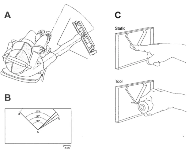
The angles were constructed of 1cm thick Plexigiass, as described by Voisin et al (2002a). The angles were formed by the intersection of two 8-cm long arms. The range of angles varied from 90° to 103°. During each trial, a pair of angles was presented consisting of the standard 90° angle and one of four comparison angles of 91°, 95°, 99°, or 103° (Fig lB) inserted into an apparatus (Fig lA). The first angle presented of each pair was either the standard or the comparison angle (order quasi random and counterbaianced).
Haptic discrimination task
Subjects were seated in a chair with the experimental apparatus positioned at arm’s length, at the level ofthe shoulder. The experimenter adjusted the apparatus SO
outstretched arm. Ail testing was performed with the angles positioned at 300 to the
right ofthe subject (Fig lA). View ofthe apparatus was blocked with a mask attached to a hat (shaded area, F ig lA). Auditory eues were blocked by having the subjects wear ear muffs (20dB noise reduction). At the beginning of the session, each subject received written and verbal instructions indicating that they were going to explore pairs of two-dimensional (2-D) angles and that their task would be to identify which angle of each pair was larger. Subjects were asked to keep their arm and finger straight throughout the scan, in order to limit angular changes to the shoulder joint, and were directed to position their finger on the angle such that the glabrous skin of the middle phalanx of the right index finger (D2) contacted the angle during the scan (nail up).
The general sequence ofevents in each condition was as follows: (1) the first
angle was installed in the apparatus; (2) the experimenter guided the subject’s finger
to the starting position; (3) the subject explored the first angle then withdrew from the angle; (4) the second angle was installed in the apparatus and the exploration
sequence was repeated
(—
5-s delay between scans); and (5) the subject then verballyreported which angle was greater, and the experimenter recorded this response. No feedback on performance was given. For the experimentai conditions that involved dynamic scanning of the entire angle (see beiow), one angle of cadi pair was slightly
shifted 40
on its vertical axis to encourage the subjects to evaluate the whole angle rather than only the second arm of the angle (Voisin et ai. 2002a). Subjects were not informed of this shifi, and the order of the shifted angle was counterbalanced across trials. Before starting a condition, the exploratory strategy vas explained to the subject. This was then practised. The perceptual task was tien described and
practised by having the subject scan a pair of angles with a large difference (900 and 103°). Data collection began afier subjects made two correct discriminations (2-6 trials). To minimize the subject’s fatigue, there was a short pause between each condition. There was a total of 40 trials (10 trials for each pair of angles) per condition, order randomized according to a preset list (same for ail conditions and subj ects).
Afier each condition, subjects were asked to rate the difficulty of the condition using a scale of O (flot at ail difficuit) to 10 (very difficuit). At the end of each session, subjects were posed a series of questions regarding the strategy used to represent the angles. They were aiso asked to estimate the range of angles presented.
In order to describe the physical contact between D2 and the angles, subjects scanned several angles (90°, 95°, 990
and 103°) coated with ink and an imprint was then taken. Two conditions were tested: long static touch of the intersection and a one-pass dynarnic scan over the angles (see below).
Experiment 1
The type of feedback, cutaneous (from the index finger) and/or proprioceptive (from the shoulder joint), was modified across six conditions tested over two sessions (A and B, order counterbalanced) separated by an interval of one week. For ail sessions, testing in one condition was completed before proceding to the next. The order ofthe conditions was counterbalanced across ah sessions and experiments.
The reference condition in Session A consisted of a single dynamic scan over each angle tising the right D2 following the sequence abc (Fig lB). Cutaneous feedback from the index finger and proprioceptive feedback from the shoulder
contributed to the performance of the task in this condition. Three modified conditions were tested: two static and one dynamic. For the static exploration (fig 1C), the subject was guided to contact the intersection, b, using either a brief touch (haptic glance: Klatzky and Lederman 1995) or a longer touch
(
3 s). In both cases, only cutaneous feedback was available and subjects were specffically instructed to remain motionless during the contact. The dynamic modified condition substituted a tool (fig 1C) for the finger, so that only proprioceptive feedback from the shoulder was available. The scan was Iimited to one sweep over the modified arm (bc, f ig lB). The subject held a molded hand grip (fig lC), with a rigid circular rod (2-mm diameter, length adjusted to the length ofthe subject’s D2) extending out between D2 and D3.In session B, the reference condition was the same as that of session A, with the exception that subjects scanned each angle with two passes over the intersection, following the sequence abcba (figlB). This was identical to the exploratory strategy used in previous experiments from our laboratory (Voisin et al. 2002a,b; 2005). In the modified condition, the two-pass exploration was repeated using the tool instead of the index finger, in order to test performance in the presence of proprioceptive feedback only.
Experiment 2
Six additional subjects were recruited to repeat a combination of the conditions tested in sessions A and B of experiment 1. This approach was necessary as subjects show considerable intersession variability in threshold (Voisin et al
2002a). The testing in session B was repeated, adding up the reference condition from session A (single scan with D2, abc).
Data analysis
for each subject and each condition, (40 trials; 4 comparison angles paired with the standard angle), the discrimination performance was calculated by computing the proportion of correct responses (PC) for each angle pair. The resuits were fittedto the following logistic function (Voisin et al 2002a)
PC1/ (1+ e U (comparison angle-standard angle)
In this equation d is the unique degree of freedom of the logistic curve that was adjusted to fit the raw data. Discrimination threshold, T (75% correct), was then computed as follows:
T=d -l ln 1/3
The data from each session were analysed separately using either a repeated measures analysis of variance (ANOVA, Expt lA and 2) andpost hoc contrasts, or a paired t-test (Expt lB). Ah analysis was done with Systat 9.0 (SPSS, Chicago, IL). The level of significance xvas set at P< 0.05.
Resuits
Experiment 1
Data were collected in two sessions from 11 subjects. Discrimination thresholds for short and long duration static touch of the angle of intersection, b, are plotted in f ig. 2A as a function of threshold measures obtained using dynamic toucli
(one-pass with D2, abc). Inspection shows that the data points are distributed equally on either side ofthe diagonal une (equality), i.e. there was little difference across the three conditions [short, 5.4± 0.9° (mean± SEM) and long static touch, 5.7 ± 0.8°; D2 one pass, 5.1 ± O.9°J. An ANOVA confirmed the lack of difference across the four conditions tested in session A (P=0.908; see also below). The similarity of the resuits, static versus dynamic, suggests that cutaneous feedback alone is sufficient to explain 2-D angle discrimination. Moreover, this information appears to be gleaned from the pattern of initial contact with the intersection, since there was no difference as a function of the duration of the static contact.
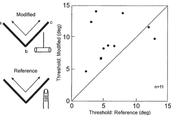
figure 2B plots the resuits obtained using the tool (one pass, bc) as a function of the threshold measures obtained using D2 (one pass, abc; same as in Fig 2A). Inspection shows that the majority of data points are located above the diagonal,. consistent with a modest but non significant increase in threshold from 5.1 + 0.9° to 6.6 ± 1.0° when only proprioceptive feedback related to the orientation of the second arm of the angle was available. It should be noted, however, that the proprioceptive feedback when using the tool differed from that in the reference condition because the first arm ofthe angle was not explored. This was addressed in session B: both arms of the angle were scanned using either the tool or D2. The results (f ig 3) showed a significant increase in threshold when the tool was used (9.6 + 1.0°) as compared to D2 (6.2± 1.00, P=0.018).
The pattems of contact between the glabrous skin of D2 and the angles (90
-103 0)
were characterized in six subjects. for both static (long) and dynamic (one pass) scans, each imprint consisted of two distinct skin areas on the middle phalanx (the radial and ulnar sides). The prints were digitized and a number of parameters
measured: width and area of each skin area in contact; and the distance between the radial and ulnar contacts. No differences were observed as a function of the mode of exploration (static versus dynamic, independent t-tests). Two of the parameters were significantly correlated with angle (P<0.01): as the angle value increased: the distance between the radial and ulnar skin contacts declined as did the width of the ulnar skin area. In contrast, none of the parameters showed a significant difference when
comparing measures at900 with those obtained at950
(conesponding, approximately,
to threshold in these experiments).
Experiment 2
One possible explanation for the failure to obtain a significant difference when using either static touch or the tool in the first experiment (session A), as compared to the reference condition (one dynamic scan with D2, abc), was that performance in the reference condition may flot have been optimal. In other words, threshold may have been higher in this condition as compared to when two scans
(abcba) were used. This was addressed by recruiting a further six naive subjects and
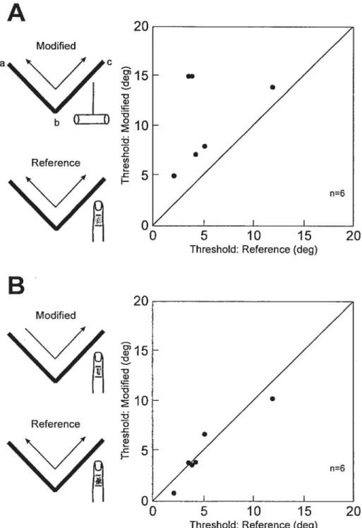
having them perform the conditions tested in session B of experiment 1 along with the reference condition from session A. The resuits confirmed those obtained in session B. Figure 4A shows that the thresholds were increased when the tool was
substituted for D2 (10.7 ± 1.80 versus 5.2 ± 1.4° respectively). In contrast, no
difference was seen for one-pass (4.9 ± 1.3°) versus two-pass scans using D2 (Fig.
45). These impressions were confirmed with an ANOVA: there was a significant
difference across the three conditions (P=0.005). Post hoc analyses indicated that thresholds were higher using the tool (P=0.03) as compared to the reference
condition. No difference was seen between a single or double pass using D2 (P=0.49), indicating that a single pass provided sufficient information on which to base the perceptual response.
Subject reports
The perceived difficulty of the tasks varied considerably between subjects, from a Iow of 3/10 (relatively easy) to 10/10 (very difficuit). Overail, the difficulty ratings showed no systematic changes across the various test conditions. For example, in session A (expt 1), subjects rated long static touch as the easiest of the four conditions (mean, 5.6 ± 0.5) and the tool as the most difficult (6.8 ± 0.5). The differences were flot, however, significant (ANOVA, P0.437). Although there was a weak trend for thresholds to covary with difficulty (linear regression, P0.056), this was not conflrmed in the other sessions.
For the conditions in which subjects explored both arms of the angle, most subjects used some form of mental imagery, visual or otherwise, to representthe angles (15/17). A majority of subjects recognized that one ofthe angles was 90°. The estimated range of angles was much larger (mean, 50°) than the actual range presented (13°, from 90°-103°) and the majority of subjects (9/17) thought that angles smafler than 90° had been presented.
Discussion
11e present study showed that haptic discrimination of 2-D angles is surprisingly insensitive to large changes in the exploratory strategy, including restricting the exploration to a single pass over the angle or a single static contact with the intersection. Only one strategy, substituting a tool for the exploring finger, produced an increase in threshold. Taken together, the results suggest that cutaneous feedback may be relatively more important than proprioceptive feedback for haptic angle discrimination, at least in the experimental conditions studied here.
One- versus two-pass haptic exploration
The approach taken in this study was to strictly control the exploration strategy, to ensure that between-subject differences in search strategy did flot contribute to the resuits. Many studies put no, or minimal, lirhits on exploration time, and there is evidence that some tactile sensory abilities are improved with increased exploration. for example, Sinclair and Burton (1991) fottnd that texture discrimination threshold decreases when the number of passes over the surfaces is increased. Soechting et al (Epub 2005) more recently reported a similar observation for haptic recognition of shape, whereby accuracy in reproducing the contours of virtual objects is highest for contours that are explored the most. The present resuits, in contrast, showed no difference as a function of the number of passes over the angle, one or two. One explanation for this resuit is that it may be that further exploration (three or more passes) is required in order to demonstrate an advantage for haptic discrimination of 2-D angles with increased exploration. We would argue
against this interpretation because similar resuits were achieved with an even more limited exploration strategy, static touch. An altemate explanation is that the critical information for haptic discrimination of angles is obtained during the first pass over the angle, with the second pass contributing littie or nothing to task performance. Indeed, several subjects in one of our previous studies (Voisin et al. 2002a) reported using sensory cues generated mainly on the first pass over these same angles. Further to this, we have also found that the motor strategy used during haptic categorization of these same 2-D angles is characterized by slower movements on the flrst as compared to the second pass over the angles (unpublished observations, G Michaud, J Voisin, CE Chapman). The latter observation may, on the other hand, be task specific: Voisin et al (2002a) reported that in the task used here there is no significant difference in speed across the first and second spans. Nevertheless, they pointed out that there was a trend for slower movements on the flrst (versus the second) pass.
Static versus dynamic haptic touch
No difference was found in 2-D angle discrimination threshold when static touch (cutaneous feedback from the intersection) was compared with dynamic touch (proprioceptive + cutaneous feedback as the subject scanned the two bars and the intersection). Thus, the presence of proprioceptive feedback did flot appear to provide any additional information, so that cutaneous feedback alone may be sufficient for the task of 2-D angle discrimination. At first gtance, this finding contradicts our previous conclusion that both proprioceptive and cutaneous feedback contribute to 2-D angle discrimination (Voisin et al 2002b). The latter conclusion was based on the demonstration that threshold increases when either source of feedback is selectively
eliminated (see Introduction). Thus, logically, we expected to see an increase in threshold during static touch. Several factors may have contributed to our resuits. First, the resuits obtained with static touch may have been biased to lower values by the fact there was no shifi applied to one of the pair of angles explored. In contrast, the reference condition testing (dynamic scan with D2) included a small 40
shifi applied to the orientation of one of each pair of angles presented. We previously showed that threshold tends to be lower when no shift is applied (Voisin et al 2002a). Second, the results may reflect that fact that there is redundancy in the coding of 2-D angles — that both sources contribute equally, but the relative weight of each contribution may vary with the testing condition. At a minimum, the resuits confirm that cutaneous afferent feedback is important for 2-D haptic discrimination. Third, and perhaps rnost importantly, the methods for acquiring the cutaneous feedback were very different in the two studies. In this study, the cutaneous feedback was self generated (active touch): subjects actively lowered their finger onto the unseen intersection. In our previous study, the cutaneous input vas externally generated (passive touch): the subject was immobile, and the angles were displaced over the passive finger. Sensory feedback may flot have been optimized in the latter study, as the subjects did flot make the normal small adjustments in digit orientation and/or contact force that may occur during active scans. following this reasoning, our failure to find the expected increase in threshold with static touch may, in fact, reflect the superiority of active touch over passive touch. This topic has been the subject of debate since the time of Gibson (1962), with some authors arguing that the two modes of touch are equivalent (Vega-Bermudez et al 1991). There is, on the other hand, considerable evidence that tactile inputs are gated during active movement, yet
paradoxicaÏly there is almost no evidence to show that passive touch is superior to active touch (Chapman 1994).
Further to this, the long static touch condition (-3 s) was included to determine whether movement-related gating of cutaneous feedback contributed to haptic angle discrimination. We reasoned that cutaneous feedback during short touch (< 1 s), or what Lederman and Klatzky (1995) terrned a “haptic glance”, might be compromised by the presence of movernent-related gating (Chapman 1994; Williams and Chapman 2002). As threshold was similar in both conditions, we conclude that gating did flot contribute significantly to the results. A role for gating cannot, on the other hand, be cornpletely excluded because subjects tended to rate the long touch condition as less djfflcult than the short touch condition.
Tool use and haptics
When only proprioceptive feedback was available (tool substituted for the finger), threshold increased. This observation was in marked contrast with the lack of any change in 2-D angle discrimination threshold when only cutaneous feedback was available (static vs dynamic touch). Although we believe that the static results may have been modestly biased by the lack of a shift (see above), the results suggest overali that cutaneous feedback may be relatively more important than proprioceptive feedback for this task of 2-D angle discrimination.
Two factors may have contributed to the reduced performance when the subject wielded the tool. f irst, the cognitive demands of the task were modified by requiring that the subject manipulate the tool. The increased attentional demands when using the tool may have contributed to the increased threshold (Post and
Chapman 1991; Zompa and Chapman 1995). Second, the increased threshold may have reflected an interaction between the proprioceptive feedback and the diffuse cutaneous feedback from the hand holding the foam-covered handie of the tool. The latter may have served as a source of noise, degrading the quality of the proprioceptive signais generated during the exploration. Such a possibility could be tested by systematically changing the quality of this diffuse feedback, for example by replacing the polished surfaces of the angles with textured surfaces varying in, for example, spatial period. An increase in non-specific feedback would be expected to worsen performance.
The influence of tool use on other sensory abilities has been addressed by others, and mixed results have been obtained. Katz (1925), for example, showed that some textures can be discriminated as weli with a hand-held probe as with the bare finger. More recently, Klatzky and Lederman (1999) reported that roughness discrimination is impaired when using a probe. They pointed out, however, that their resuits may have reflected some confusion as to what the subjects meant by their “rougher” judgments, given that the spacing of the tactile elements on the surfaces employed varied in more than one dimension (Coimor et al 1990; Mefiah et al 2000). As regards softness or compliance of objects, LaMotte (2000) found that subjects could discriminate the sofiness of rubber objects (with variable compiiance) equally well with direct contact through the fingertip and through a hand-heid stylus. Diminished performance was, on the other hand, reported by Tholey et al (2005). Finally, Lederman and Klatzky (2004) found that haptic recognition of familiar objects is reduced when a probe is substituted for the finger, or if the feedback is degraded (e.g. encasing the exploring finger in a rigid sheath). Clearly further
.4-experiments are needed in order to more fully characterize the human ability to explore the environment indirectly, using hand-heId tools.
The importance of this issue cornes from the fact that there is now considerable interest in developing haptic interfaces for different applications, including the remote operation of instruments and training surgical skills within virtual-reality environments. The various approaches taken ail share a common theme in using force-feedback through a hand-held tool or implement to guide the operator in interacting with the remote or virtual environruent. Studies have shown that force feedback can significantly improve human performance in real and virtual environments (Kim et al. 2004; Tholey et al 2005; Yao and Hayward 2005). Nevertheiess, it does flot appear that performance with the types of haptic feedback generated now is as good as with the bare hand (Tholey et al 2005), so that future applications may need to consider methods to amplify the feedback in order to enhance performance (e.g. Yao and Hayward 2005). Certainly one implication of the present restiits is that haptic interfaces should concentrate in providing cues transduced by cutaneous mechanoreceptors.
Nature of the sensory signais
Another implication of the present resuits is that the pattern of skin contact from the intersection of the two bars forming the angle provides sufficient information on which to base the discrimination. Although our imprint measures were not sufficiently precise to show differences across the pattems for the standard and modified angles at threshold level, the critical parameters appeared to be the distance between the radial and ulnar contact sites on the middle phalanx and the width of the
ulnar contact, corresponding to the side of D2 that contacted the modified bar.
further studies are needed to clarify the important parameters. These observations do, however, provide some insight into the necessary response properties of cutaneous afferents that contribute to haptic 2-D angle discrimination. Thus, the afferents must have small, discrete receptive fields that can reliably discriminate small changes in the pattem of contact. The rnost likely candidates are slowly adapting type I (SAI) afferents andlor rapidly adapting (RA) afferents (see review by Johnson 2001), as both have been characterized as having small receptive fields. 0f particular interest for this study, the spatiotemporal response profile of SAI and RA afferents elicited by scanning wavy surfaces (alternating convex and concave bars) and other shapes such as ellipsoids over their receptive field is relatively independent of the way in which the object cornes into contact with the skin (contact force, orientation of the shape) (LaMotte et al 1994; LaMotte and Srinivasan 1996; cf Goodwin et al. 1995). Such characteristics would contribute to generating an invariant central representation of 2-D shape.
The latter suggestion is complementary to our recent proposal (Voisin et al 2005) that regional variations in proprioceptive acuity (proximal joints more sensitive than distal joints: reviewed in Clark and Horch 1986) may reflect an adaptation to generate an invariant central representation of haptic shape. This suggestion was based on our demonstration that 2-D angle discrimination is identical for explorations made with proximal and distal joints. Given that both explorations involved the index finger, however, we need to consider whether the previous findings should be reinterpreted. Could cutaneous feedback from the exploring digit have been responsible for the similar thresholds? While cutaneous inputs undoubtedly
contributed, this was flot the sole source of information because threshold increased when the proprioceptive feedback from the shoulder was modified by dispiacing the angles to a more eccentric position, further from the midiine. Under the same testing conditions, threshold declined when the exploration was restricted to distal movements, consistent with both cutaneous and proprioceptive feedback contributing to 2-D haptic discrimination.
Acknowledgements. The authors would like to thank the following for the excellent technical assistance provided: René Albert, Jacques Bérichon, Marie-Thérèse Parent, Gaétan Richard, and Christian Valiquette. We also thank Trevor Drew and Allan Smith for helpful comments on the manuscript, and El-Mehdi Meftah for helpful suggestions throughout the course of the experiments. Funding from the Natural Sciences and Engineering Research Council of Canada (NSERC) and the Canadian Institutes of Health Research (CIIIR: individual and group grants) are gratefully acknowledged. Stéphanie Bourgeon was supported, in part, by a bursary from the GRSNC (Groupe de la recherche sur le système nerveux central, a group supported by the Fonds de la recherche en santé du Québec, FRSQ, and the Université de Montréal).
References
Chapman CE (1994) Active versus passive touch: factors influencing the transmission of somatosensory signais to primary somatosensory cortex. Can J Physiol Pharmacol 72: 55$-570
Clark FJ, Horch KW (1986) Kinesthesia. In: Handbook of Perception and Human Performance, eds K Boff, L Kaufman, J Thomas, Vol 1, chapter 13, Wiley, New York, pp 13-1 — 13-62
Connor CE, Hsiao SS, Philips JR, Johnson KO (1990) Tactile rouglmess: neural codes that account for psychophysical magnitude estimates. J Neurosci 10: 3823-3836
Gibson JJ (1962) Observations on active touch. Psychol Rev 69: 477-49 1
Goodwin AW, Browning AS, Wheat HE (1995) Representation ofcurved surfaces in responses of mechanoreceptive afferent fibers innervating the monkey’s fingerpad. J Neurosci 15: 79$-810
Johnson KO (2001) The roles and function of cutaneous mechanoreceptors. Curr Opinion Neurobiol 11:455-461
Katz D The World of Touch. Hillsdale, NJ: Erlbaum (translated by LE Krueger, published originally in 1925), 1989
Kim I-1K, Rattner DW, Srinivasan MA (2004) Virtual-reality-based laparoscopic surgical training: the role of simulation fidelity in haptic feedback. Comput Aid Surg 9: 227-234
Klatzky RL, Lederman SJ (1995) Identifying objects from a haptic glance. Percep Psychophys 57: 1111-1123
Klatzky RL, Lederman SJ (1999) Tactile roughness perception with a rigid link interposed between skin and surface. Percep Psychophys 61: 591-607
LaMotte RH (2000) Softness discrimination with a tool. J Neurophysiol $3: 1777-1786
LaMotte RH, Srinivasan MA (1996) Neural encoding of shape: responses of cutaneous mechanoreceptors to a wavy surface stroked across the monkey fingerpad. J Neurophysiol 76: 3787-3797
LaMotte RI-1, Srinivasan MA, Lu C, Kusch-Petersen A (1994) Cutaneous neural codes for shape. Can J Physiol Pharmacol 72: 498-505