U
NIVERSITÉ DEM
ONTRÉALA study of K
Vchannel dynamics using a fluorescent
unnatural amino acid
par Tanja Kalstrup
Département de Pharmacologie et Physiologie Faculté de Médecine
Thèse présentée en vue de l’obtention du grade de philosophiæ doctor (Ph.D.) en physiologie
option biophysique et physiologie moléculaires
Octobre, 2017
U
NIVERSITÉ DEM
ONTRÉALFaculté de Médecine
Cette thèse intitulée :
A study of KV channel dynamics characterized by a fluorescent unnatural amino acid
présenté par Tanja Kalstrup
a été évaluée par un jury composé des personnes suivantes :
Lucie Parent, Président rapporteur Rikard Blunck, Directeur de recherche Roberto Araya, Membre du jury Francisco Bezanilla, Examinateur externe Daniel Zenklusen, Représentant du doyen
I
Les protéines sont les nanomachines moléculaires de la nature et leurs diverses fonctions sont essentielles au fonctionnement de tous les mécanismes cellulaires. Une grande partie de la recherche fondamentale en physiologie se concentre présentement sur l’étude de la structure et de la fonction des protéines. L'objectif général de cette thèse est d'élargir la compréhension des canaux potassiques voltage-dépendants (KV), un groupe de protéines gouvernant la signalisation "électrique de divers aspects physiologiques, en particulier la propagation de potentiels d'action dans le système nerveux central et dans les cellules musculaires.
Dans le but d'obtenir des indices quant à la dynamique des canaux KV, la thèse met l´enphase sur l'utilisation d'un acide aminé non naturel et fluorescent (Anap) comme outil moléculaire pour étudier les changements de conformation protéique avec la fluorimétrie en voltage imposé (FVI). L'avantage de la technique Anap-FVI par rapport aux techniques traditionnelles de marquage fluorescent, tient au fait qu'il n'y a pas de restriction sur le site d'intérêt. Ainsi, Anap est incorporé génétiquement dans des régions clés du canal Shaker KV en utilisant la technique de substitution du "codon-stop amber", en utilisant une paire orthogonale d'ARNt/synthétase avec les ovocytes de Xenopus laevis comme modèle d’expression protéique.
Tout d'abord, la comparaison directe des deux extrémités du senseur de voltage (SV) est rendue possible par FVI bicolore dans lequel une cystéine externe est marquée avec TMR (tetramethylrhodamine), tandis que l'acide aminé (Anap) est incorporé du côté intracellulaire. On constate que la partie intracellulaire du SV s'active de façon similaire à la partie externe (chapitre 3). En outre, on constate que les portes S6 intracellulaires s'ouvrent essentiellement en deux étapes.
Ensuite, Anap a été inséré aux deux extrémités de la boucle cytoplasmique S4-S5 afin d'en étudier le mouvement pendant le couplage électromécanique (chapitre 4). Les expériences FVI bicolores démontrent que la partie N-terminale de la boucle se déplace avec le senseur de voltage alors que le mouvement de la partie C-terminale est retardé. Les résultats supportent un modèle de couplage électromécanique dans lequel l'énergie est stockée au centre du de la
II le transfert d'énergie du senseur de voltage vers le pore.
Dans un troisième projet, Anap a été inséré dans diverses positions de l'extrémité N-terminale cytosolique afin d'en sonder le mouvement pendant l'inactivation de type N (chapitre 5). Les données FVI montrent que la région hydrophile de la balle d'inactivation (BI) subit un mouvement qui est étroitement lié à l'inactivation mais qui ne joue pas un rôle dans l'obstruction finale du pore. Par contre, la pointe hydrophobe de la BI, subit un mouvement supplémentaire qui est sensible à l'état du pore, suggérant qu'il subit un mouvement qui impacte l'obstruction du pore. Les résultats supportent un modèle d'inactivation sous la forme de mécanisme d'étapes séquentielles d'au moins deux transitions.
Enfin, un quatrième projet se distingue de l'objectif général de la thèse. Il est démontré que les canaux avec un codon-stop N-terminal s’expriment malgré l'absence d'Anap (chapitre 6). Cette expression de fuite est causée par la réinitiation de la traduction de l’ARN messager, à des codons d’initiations non-canoniques en aval, et peut être réduite en supprimant ces codons. Les résultats mettent en évidence l'importance des expériences de contrôles lors d'utilisation d'acides aminés non naturels.
III
Proteins are the nature’s molecular nanomachines and their diverse set of functions are crucial to the workings of all cellular mechanisms. A great part of fundamental research today is therefore focusing on the elucidation of protein structure and function. The general objective of this thesis is to expand the understanding of voltage-gated potassium (KV) channels, a group of proteins which governs electrical signalling in various physiological aspects, in particular the propagation of action potentials in the central nervous system and in muscle cells.
In the pursuit of obtaining dynamic information of KV channels, the focus in this thesis has been on exploring the applicability of a fluorescent unnatural amino acid (Anap) as a molecular tool to study protein conformational changes using voltage clamp fluorometry (VCF). The advantage of the Anap-VCF technique over traditional post-translational fluorescence labeling techniques is that there are no restrictions regarding the choice on the site of interest. Anap is genetically incorporated into key regions in the Shaker KV channel by using the amber stop codon suppression technique using an orthogonal tRNA/synthetase pair, and Xenopus laevis oocytes are used as expression system.
The first project involves direct comparison of both ends of the voltage sensor (VS) and is made possible by two-color VCF, in which an external cysteine is labeled with TMR (tetramethylrhodamine) while Anap is incorporated on the intracellular side (chapter 3). We found that the intracellular part of VS activates together with the external part. Moreover, it is found that the intracellular S6 gates open in a sequential two-step transition.
Next, to investigate electromechanical coupling, Anap was inserted into both ends of the S4-S5 linker (S4-S5L, chapter 4). Two-color VCF experiments demonstrates that the N-terminal part of S4-S5L moves with the VS. On the other hand, the movement of the C-terminal part of the linker is delayed with respect to the VS. The findings support a model for electromechanical coupling in which energy is stored in the middle of the S4-S5L, and not in the C- or N-termini. Moreover, it is found that the S4-S5L undergoes both independent and cooperative movements – a finding which agrees with a pivotal role of the S4-S5 linker in energy transfer from the VS to the pore.
IV
hydrophilic chain region of the inactivation particle (IP) undergoes a motion which is closely related to inactivation but is not involved in the final step of pore block. The hydrophobic tip of the IP, on the other hand, undergoes an additional motion which is sensitive to the state of the pore, suggesting that it causes pore block. The findings support a model for inactivation as a sequential step mechanism of at least two transitions.
Finally, a fourth project stands out from the general objective of the thesis. It is demonstrated that channels with N-terminal stop codons still express despite the absence of Anap (chapter 6). This leak expression is caused by translation reinitiation at downstream non-canonical start codons and can be reduced by removing the start codons. The findings highlight the importance of control experiments when using unnatural amino acids.
V
Résumé ... I Abstract ... III Table of figures ... VIII List of abbreviations ... X Thesis acknowledgements ... XI
Chapter 1 ... 1
Introduction ... 1
1.1 Potassium channels ... 3
1.2 Voltage-gated potassium channels ... 5
1.3 KV1 channel family ... 8
1.4 KV1 channel structure ... 9
1.5 Mechanism of KV channel activation ... 14
1.6 Mechanism of KV channel inactivation ... 18
1.7 Genetic incorporation of unnatural amino acids ... 22
1.8 Protein translation ... 24
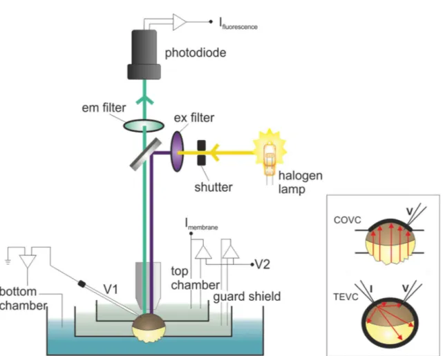
1.9 Cut-open oocyte voltage clamp ... 27
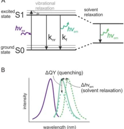
1.10 Voltage clamp fluorometry ... 29
1.11 Thesis objectives ... 34
1.12 References ... 38
Chapter 2 ... 47
Methodology ... 47
2.1 Oocyte handling and injection ... 47
2.2 Incorporation of Anap into Shaker expressed in Xenopus oocytes ... 48
2.3 Experimental procedure ... 49
2.4 Data analysis ... 50
2.5 References ... 54
Chapter 3 ... 55
Dynamics of internal pore opening in K(V) channels probed by a fluorescent unnatural amino acid ... 55
VI
3.3 Results... 59
3.4 Discussion ... 64
3.5 Materials and Methods ... 66
3.6 Acknowledgements ... 67
3.7 Supporting information ... 68
3.8 References ... 68
Chapter 4 ... 72
The S4-S5 linker movement during activation and inactivation in voltage-gated K+ channels . 72 4.1 Abstract... 73
4.2 Introduction ... 73
4.3 Results... 75
4.4 Model for the cytosolic gating machinery ... 85
4.5 Discussion ... 87
4.6 Methods and Materials ... 88
4.7 Acknowledgements ... 89
4.8 Supplementary Material ... 89
4.9 References ... 94
Chapter 5 ... 97
Probing dynamics of the ball and chain in KV channels during N-type inactivation ... 97
5.1 Abstract... 98
5.2 Introduction ... 98
5.3 Results... 99
5.4 Discussion ... 105
5.5 Methods and materials... 108
5.6 Supporting information ... 109
5.7 Acknowledgements ... 110
5.8 References ... 110
Chapter 6 ... 112
Reinitiation at non-canonical start codons leads to leak expression when incorporating unnatural amino acids ... 112
VII
6.4 Discussion ... 122
6.5 Methods and materials... 124
6.6 Acknowledgements ... 126
6.7 References ... 126
Chapter 7 ... 130
Discussion ... 130
6.8 Dynamics of internal pore opening probed by a fluorescent unnatural amino acid ... 130
6.9 The S4-S5 linker movement during activation and inactivation in voltage-gated K+ channels ... 132
6.10 Probing dynamics of the ball and chain in KV channels during N-type inactivation ... 135
6.11 Reinitiation at non-canonical start codons leads to leak expression when incorporating unnatural amino acids... 136
7.1 VCF data interpretation ... 137
7.2 Anap incorporation ... 140
7.3 Perspectives ... 142
VIII
Figure 1.1 Cartoon of neurotransmitter release in the neuromuscular junction ... 7
Figure 1.2 Amino acid sequence alignment of Kv channels ... 11
Figure 1.3 Overview of KV channel structure and function ... 13
Figure 1.4 Main conformational states of Shaker ... 14
Figure 1.5 Shaker channel topology and gating residues ... 18
Figure 1.6 N-type inactivation in KV channels ... 20
Figure 1.7 Cartoon of genetic incorporation of UAAs via in vivo aminoacylation. ... 23
Figure 1.8 Protein translation ... 25
Figure 1.9 mRNA open reading frames and premature stop codons ... 26
Figure 1.10 Illustration of the COVC and VCF setup stration of the COVC and VCF setup. ... 28
Figure 1.11 Fluorescence parameters ... 31
Figure 1.12 fUAA timeline for expression in Xenopus oocytes ... 49
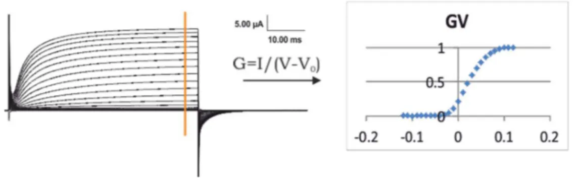
Figure 2.1 Ionic currents and GV... 51
Figure 2.2 Gating currents and QV ... 52
Figure 2.3 Fluorescence intensity and FV ... 53
Figure 2.4 Exponential fit of a fluorescence time course ... 54
Figure 3.1 Incorporation of Anap into Shaker. ... 58
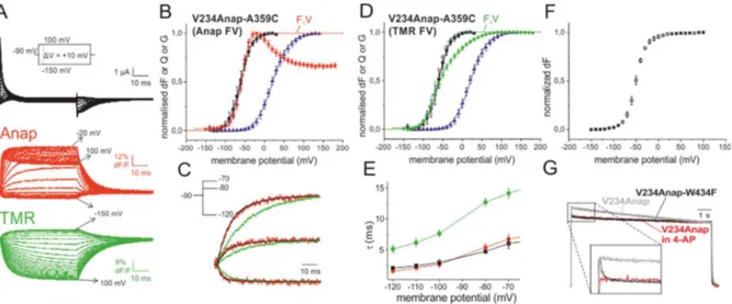
Figure 3.2 Two-colour VCF results of V234Anap-A359C... 61
Figure 3.3 Incorporation of Anap into the C-terminal S6 ... 63
Figure S3.1 Supplementary figure of conducting mutants ... 68
IX
Figure 4.4 Kinetical analysis of fluorescence and gating current time course. ... 81
Figure 4.5 Comparison of onset of TMR and Anap fluorescence signal ... 82
Figure 4.6 Separation of the final gating transition using F290A ... 84
Figure 4.7 C-type inactivation of H486Anap ... 87
Figure S4.1 iVSD expression does not interfere with the function of full length Shaker channels 91 Figure S4.2 Supplementary figure 2. Anap fluorescence in K390Anap oocytes is not affected by iVSD ... 93
Figure S4.3 Characterization of the final gating transition separated by F290A. ... 93
Figure 5.1 Functional expression with Anap in N-terminus ... 100
Figure 5.2 VCF results for the tip region mutants A3Anap and Y8Anap ... 102
Figure 5.3 VCF results for the chain region mutants K19Anap and E35Anap ... 103
Figure 5.4 VCF results for the receptor site mutant E201Anap in the T1-S1 linker ... 105
Figure 5.5 Proposed model for N-type inactivation ... 106
Figure 5.6 Overview of residues selected for Trp insertion. ... 108
Figure S5.1 Two fluorescence components present in A3Anap and Y8Anap ... 109
Figure 6.1 Translation reinitiation, Shaker channel topology, and current phenotypes ... 115
Figure 6.2 Expression of Shaker channels with N-terminal stop codons ... 117
Figure 6.3 Identification of non-canonical start codons ... 120
Figure 7.1 Anap and TMR-maleimide ... 131
Figure 7.2 Sequence alignment of the S4-S5 linker. ... 134
Figure 7.3 Overview of residues used for Anap incorporation ... 139
X 4-AP 4-aminopyridine
aaRS aminoacyl tRNA synthethase
Anap 3-(6-acetylnaphthalen-2-ylamino)-2-aminopropanoic acid BCN bicyclononynes
COVC cut-open oocyte voltage clamp E. coli Escherichia coli
GV conductance-voltage relationship FRET förster resonance energy transfer fUAA fluorescent unnatural amino acid IP inactivation particle
KV voltage-gated potassium channel LRET lanthanide resonance energy transfer NaV voltage-gated sodium channel
pAnap plasmid encoding for Anap-synthetase and tRNA QV charge-voltage relationship
QY quantum yield S4-S5L S4-S5 linker TCO trans-cyclooctene TEA tetraethylammonium TEVC two electrode voltage clamp TMR
TMRM tetramethylrhodamine tetramethylrhodamine maleimide TTX tetradotoxin
UAA unnatural amino acid VCF voltage clamp fluorometry VS voltage sensor
WT wild type X. laevis Xenopus laevis
XI
I wish to thank my supervisor Dr. Rikard Blunck for giving me the opportunity to investigate innovative approaches at an early stage, and for believing in my qualifications as a researcher. His confidence and guidance has truly contributed to my development as an independent scientist.
1
Chapter 1
Introduction
The electrical signalling which governs cell-cell communication in the nervous system, heart and muscle, form the molecular foundation of our heartbeat and how we store memory, perceive, react and move our limbs, among many other physiological aspects. Excitable cells convert chemical or mechanical signals intro electrical signals by means of ion channels which are proteins that selectively conduct potassium, sodium or calcium ions across the cell membrane. The neuronal electrical signaling is generated by action potentials which are millisecond-long signals that propagate along the nerve fiber.
Excitable cells have a negative resting membrane potential of about -70 mV which arises from the activity of molecular pumps which transport sodium ions out of the cell and potassium into the cell. This creates an electrochemical gradient where, at rest, the concentration of potassium is high inside, and the concentration of sodium is high outside. When the membrane potential reaches a threshold above -50 mV, rapid activation of voltage-dependent sodium (NaV) channels leads to a Na+ influx which depolarizes the cell membrane. Voltage-dependent potassium (KV) channels also activate leading to the influx of K+ which repolarizes the membrane back to rest. These components of an action potential, or the nerve impulse, constitutes the fundamental brain signalling throughout the animal kingdom.
The story of ion channels begins with the famous experiments performed by Alan Hodgkin and Andrew Huxley in 1952. Their discovery of Na+ and K+ currents as the fundamental events underlying the nerve impulse, were made possible by the development of the voltage-clamp concept as introduced to them by Kenneth Cole [1]. By insertion of axial wires into a giant squid axon, a feed-back system could maintain the membrane potential and circumvent the unstable character of the action potential [2]. This way, Hodgkin and Huxley identified independent Na+
2
and K+ currents [3], characterized their kinetic properties [4, 5], and developed a model which predicted the action potential from the regulation of conductance by four sets of charged particles [6] - a gating process which today is known to be controlled by the four voltage sensors in NaV and KV channels. The pioneering work of Hodgkin and Huxley awarded them the Nobel prize in 1963.
The field of ion channels as we know it today is based on several key discoveries in the following decades. First, the notion that ions would cross the membrane through transmembrane proteins in a channel-like manner was suggested in the 60’s by the findings that tetradotoxin (TTX) and tetraethylammonium (TEA) selectively blocked Na+ and K+ currents, respectively [7, 8]. Furthermore, the presence of ion-conducting channels was confirmed by the invention of the patch clamp technique which enabled measurements of electrical activity in small areas of membrane giving rise to single channel recordings [9]. The development of this technique by Drs. Neher and Sakmann lead to the Nobel prize in 1991. Then, in the 80’s, gene cloning and recombinant manipulation began to be applied in the field of ion channel research. The cloning of voltage-gated ion channels [10, 11] paved the way for emerging molecular biology methods to be used to identify the role of individual amino acids. Site-directed mutagenesis not only allowed for better understanding of how point mutations could alter channel function and result in diseases, but also became a useful method in the elucidation of fundamental workings of ion channels. The 90’s highlight a decade during which biophysical properties of the voltage sensor and the pore were established using electrophysiology and mutagenesis (gating currents, cooperativity, ion selectivity) [12-16]. Finally, atomic structures of voltage-dependent bacterial and eukaryotic potassium channels solved by X-ray crystallography [17, 18] made it possible to relate structural data with gating function, and questions on subunit assembly, pore conformation and permeation could now be addressed structurally. Dr Mackinnon was awarded a share of the Nobel Prize in 2003 for the work. The crystal structures also confirmed a number of structural features postulated by Bertil Hille more than 20 years earlier [19]: These ion channels contain a selectivity filter located towards the extracellular end of the pore, with a voltage sensor controlling a cytoplasmic gate.
Although a wealth of functional and structural data has been provided by electrophysiology and X-ray crystallography in the field of ion channels, they are limited when it comes to unfolding the
3
molecular dynamics which drive the channel from one state to another. Research in dynamics today is characterized by the parallel emerging of two technological areas. First, experimental findings based on advances in fluorescence-based techniques have yielded kinetic information on conformational changes and relative distance changes, such as voltage clamp fluorometry (VCF), förster resonance energy transfer and lanthanide resonance energy transfer. Second, important advances in highly sophisticated computational approaches is continuously expanding, allowing for the elucidation of microscopic factors governing structural dynamics and ion permeation. There is no doubt that as computational power grows, the theoretic prediction of channel behaviour will play a key role in the future. Also, the recent advances in cryo-electron microscopy which resulted in snapshots of various conformational states of a sodium-activated K+ channel, surely has shed light on cryo-EM as a valuable tool in capturing transitions [20].
1.1 Potassium channels
The importance of understanding the detailed molecular basis of ion channel function is highlighted by their widespread roles in numerous physiological aspects. The group of K+ channels is, with 78 members, the largest within the ion channel family [21]. The group is divided into 4 subfamilies based on function and homology: Calcium- and sodium-activated K+ channels (KCa and KNa), inwardly rectifying K+ channels (Kir), two P domain K+ channels (K2P), and voltage-gated K+ channels (KV). The majority of the potassium channel subunits consists of 6 transmembrane domains, but Kir and K2P consists of 2 and 4 transmembrane domains, respectively, and some KCa channels have 7 transmembrane domains. There is one structural feature which all K+ channels have in common, and that is a GXG signature sequence which is responsible for the selective permeation of K+ ions over other ions (X is a tyrosine except in some K2P channels where it can also be a phenylalanine).
KCa channels are present in a large range of both excitable and non-excitable cells, where they are involved in neuronal excitability, transmitter release and Ca2+ homeostasis [22]. They are tightly coupled to, and regulate, the amount of intracellular Ca2+. When activated by Ca2+, the channels open and repolarize the membrane, which in turn causes the closing of voltage-gated calcium channels thus limiting the influx of Ca2+.
4
KNa channels are predominantly expressed in neuronal tissue. They are activated by high concentrations of intracellular Na2+, suggesting that they are coupled to local increase of sodium in connection with opening of voltage-gated sodium channels and/or in restricted compartments like dendritic spines. Single amino acid mutations in some KNa channels result in devastating effects causing epilepsy and intellectual and physical disabilities [23].
Kir channels allow ions to move into the cell rather than out, due to intracellular block by Mg2+ or polyamines at positive potentials. Their physiological function is diverse and depend on type and location. When potassium flows at negative potentials it allows the channels to control the resting membrane potential. Some Kir channels are regulated by G protein-coupled receptors and others are ATP-sensitive and are directly involved in regulation of insulin secretion in pancreatic beta cells.
The structure of K2P channels is markedly different from the other potassium channels in that it is a homodimer of two 4 transmembrane subunits. They play several roles in excitable cells where they give rise to leak K+ currents to stabilize the negative resting membrane potential. They are regulated by voltage-independent factors like pH, stretch and temperature and a range of intracellular signaling pathways.
Finally, KV channels are found in all excitable cells with channel open probabilities which depend on changes in the membrane potential, and they carry a voltage sensor which is central to their function. The channels generate action potentials where they are responsible for returning the depolarized cell to resting state. With their different voltage-dependencies and inactivation properties, they regulate action potential duration and firing patterns and sets the resting membrane potential.
The molecular diversity of potassium channels is enhanced by the possibility of heteromeric assembly of different subunits within a subfamily. Furthermore, post-translational modifications (splicing, phosphorylation, glycosylation) and regulation by auxiliary subunits also diversifies the channel properties, creating a wide and complex distribution of K+ channel function. To better understand the function of KV channels, which are the subject of the thesis, they are described in more detail in the following section.
5
1.2 Voltage-gated potassium channels
As Hodgkin and Huxley showed in the squid axon, the action potential consists of rapid feedback processes involving voltage-gated ion channels: NaV+ channels which first activate by positive voltage causing sodium ions flowing into the cell which depolarizes the membrane. This step is the rising phase of an action potential. Then, activation of KV+ channels causes potassium ions to flow out of cell which allows the cell to return to the resting potential. The different current profiles of KV channels makes them capable of shaping and regulating the action potential (amplitude, duration, frequency) leading to different patterns of action potentials. This way, in neurons, the types of ion channels in the membrane can vary across the cell, which gives the dendrites, axon and cell body their different electrical properties. KV channels play an active role in action potentials of heart, brain, spinal cord, sensory neurons and muscle, but are also involved in regulation of cell volume, proliferation, apoptosis and migration of a wide range of cell types.
The KV channel subfamily are encoded by >40 genes in the human genome and make up half of the potassium channel family. KV channels are divided into 12 molecular subgroups (KV1-12) which display different voltage-dependent activation and inactivation properties. They open upon membrane depolarisation and selectively conducts potassium ions across the cell membrane according to the electrochemical gradient of potassium. KV5, KV6, KV8 and KV9 families give rise to homomeric channels that are electrically silent likely due to their retention in the endoplasmic reticulum. Instead, these silent subunits assemble with members of other KV groups to form heteromeric channels.
1.2.1 Why is it important to understand the function of KV channels?
KV channels shape and regulate neuronal and cardiac action potentials [24], and participate in apoptosis [25] and cell differentiation [26] among other functions. Furthermore, KV channels are necessary for the release of neurotransmitters and hormones [27, 28] and regulate cell volume, proliferation and migration [24]. The critical roles and physiologically diverse implication of KV channels makes them a major therapeutic target for treatment of many neurological, metabolic and cardiovascular disorders, in which the cause of the disease affects the function of these channels.
6
Another reason for studying KV channels is that malfunction due to genetic mutations result in channelopathies such as cardiac arrhythmias (short and long QT syndromes), episodic ataxia, epilepsy, and deafness, and has also been associated with impaired glucose tolerance, insulin insensitivity and atrial fibrillation [29].
Small molecules and peptide toxins have been developed and identified as drugs for targeting KV channels as a pharmaceutical strategy, and are also being used as research tools to characterize channel structure and function [29]. Peptide toxins can bind to the outer vestibule of the ion conduction path and block K+ flow (e.g. charybdotoxin) [30], or they can interact with the voltage sensor to favor the closed state of the channel (e.g. hanatoxin) [31]. Most binding sites for small molecules reside in the inner pore region or are located on the intracellular side, where they can act as channel openers (e.g. retigabine) [32] or blockers (e.g. 4-aminopyridine) [33]. The current U.S. Food and Drug Administration (FDA) requires that all drug candidates for human use are evaluated for potential KV11.1 activity (encoded by the human Ether-à-go-go related gene, hERG) to prevent arrhythmia side effects [34]. The broad pharmacological aspect of KV channels makes them a group of proteins with great therapeutic potential.
Human health has benefited directly from detailed knowledge about ion channel function because channelopathy diseases can now be readily diagnosed. For example, genetic testing of the cystic fibrosis transmembrane conductance regulator, the CFTR ion channel, helps diagnosis of the cystic fibrosis disease which affects about 1 in 3000 new born in Northern Europe, giving the possibility of early treatment. The continued advances in the field of ion channels from researchers in academia and pharmaceutical companies, will extend this benefit to diagnose and prevent diseases, and develop highly selective drugs targeting ion channels.
Overall, the therapeutic importance and social benefit of understanding KV channel function is evident. From a biophysical point of view, the elucidation of voltage sensing mechanisms and dynamics of conformational changes is first and foremost driven by our scientific curiosity and fascination by nature. As humans we wish to know how nature works to ultimately answer what we are made of and where we come from. The need to understand nature’s origin constitutes the main motivation factor in fundamental research. KV channels are highly evolutionarily
7
conserved ion channel families playing fundamental roles in all living organisms, making the study of these channels a prerequisite to understand life and its origin in full.
1.2.2 The Drosophila Shaker channel
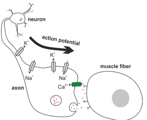
The first cloned potassium channel was the Drosophila Shaker KV channel [11], which was discovered based on a leg-shaking phenotype under ether-induced anesthesia [35]. The shaking is caused by neurons which fail to repolarize as quickly as normal neurons (figure 1.1), making them exceptionally excitable, resulting in abnormal muscle contractions. What happens on the molecular level is that in the absence of KV channels at the axon of the neuromuscular junction, the action potential is prolonged. In turn, voltage-gated calcium channels at the axon terminal which close upon repolarization, remain open for a longer time than normal, thus increasing the time for Ca2+ influx. The increase of intracellular Ca2+ stimulates the neurotransmitter release from the synaptic vesicles into the synaptic cleft. This way, postsynaptic receptors are excessively triggered, resulting in abnormal action potentials in the muscle fibers.
Figure 1.1 Cartoon of neurotransmitter release in the neuromuscular junction
When the neuronal action potential comes down the axon and reaches the axon terminal, it activates CaV channels which open upon depolarization. The increase of intracellular Ca2+ leads to fusing of the synaptic vesicles with the membrane, so that neurotransmitter molecules diffuse into the synaptic cleft and activate neurotransmitter activated receptors on the target cell, which is muscle fiber cells in the case of the
8
neuromuscular junction. The voltage-gated potassium channel at the axon are representative of KV1 channels in human and Shaker channels in Drosophila.
Recently, the Shaker channel has been shown to play a role in controlling Drosophila sleep, in which a point mutation in the first transmembrane helix S1 (a threonine to isoleucine substitution) was identified to cause short-sleeping phenotypes with preserved performance [36]. The molecular link between neuronal firing and sleep in relation to Shaker channels is not known, but it highlights the fundamental and diverse implication of Shaker-related channels in Drosophila, as it also is in mammalians.
1.3 K
V1 channel family
Mammalian KV1 channels are homologues of the Drosophila Shaker channel and are also called the Shaker-related KV channels. The group consists of 8 members (KV1.1, KV1.2, KV1.3, KV1.4, KV1.5, KV 1.6, KV1.7, KV1.8) displaying sustained potassium currents, except KV1.4 which display a transient “A-type” (fast inactivation) current, when heterologously expressed as homomeric tetramers.
KV1.1, KV1.2 and KV1.4 are those which are most abundant in the mammalian brain, localized to axons and nerve terminals where they exist in a complex heterogenous subunit association controlling neuronal action potentials and presynaptic transmitter release [37]. Episodic ataxia 1 is a neurological disorder which leads to myokymia and episodes of spastic contractions of skeletal muscle. 30 genetic mutations in the KCNA1 gene encoding for KV1.1 have been identified to be the underlying cause for episodic ataxia 1 [38]. Most of the mutations lead to altered biophysical properties such as positive shift of activation voltage dependency or slower activation kinetics among others, while some abolish channel activity. Several drugs improve symptoms, but no single medication has proven efficient. How KV1.1 mutations result in episodic ataxia 1 phenotypes are not clearly understood. Patients having the same mutation do not necessarily respond to the same drugs, nor do they necessarily demonstrate the same disease characteristics, which means that there are likely other factors than KV1.1 mutations which are involved. Mutations in KV1.1 and KV1.2 have also been identified in a subset of patients to be involved in certain epileptic disorders with seizures associated to episodic ataxia 1 (KV1.1) and epileptic encephalopathy (KV1.2).
9
KV1.6 exist in interneurons in the spinal cord, and also in dendrites together with KV1.1 and KV1.2 [37], and KV1.3 is predominantly expressed in the cerebellum. In the brain, KV1.5 is restricted to glial and endothelial cells, but is also expressed in vascular smooth muscle cells together with KV1.2 and KV1.4 [39]. KV1.7 and KV1.8 have not been detected in the brain, but KV1.7 exist in skeletal muscle and heart, and has also been suggested to play an active role in insulin secretion in pancreatic beta cells [40]. KV1.3 and KV1.8 are highly expressed in the kidney where they stabilize the membrane potential in the renal tubule [41].
Finally, in the heart, KV1.4 and KV1.5 play critical roles in the cardiac action potential [42]. One loss-of-function mutation in KV1.5 has been shown to cause atrial fibrillation [43], which is a condition characterized by abnormal electrical activity in the heart, and predisposes the patient to stroke and heart failure .
The expression pattern listed here is not comprehensive but highlights the predominant localization of members in the KV1 subfamily. It remains a challenging task to make a complete list of KV channel tissue distribution and to identify the molecular function and cellular role of each localised potassium channel gene.
1.4 K
V1 channel structure
The full amino acid sequence of Shaker is ∼70% identical to the KV1 channels, with no homology in the N- and C-termini, whereas the sequence of the transmembrane regions and the intracellular tetramerization domain (T1) is ∼90% identical (figure 1.2). Due to the early cloning of Shaker in 1987 [11] and its high expression efficiency in Xenopus oocytes, the majority of interpreted gating data is based on measurements obtained from Shaker. The first mammalian KV channel crystal structure, which was determined in 2005, was KV1.2 from rat in complex with a beta subunit (β2) [18], and was followed by a complete structure determination in 2007 at 2.4 Å of the KV1.2/2.1 chimera [44]. Finally, a complete structure of the native KV1.2 channel was obtained by using a refinement method in 2010 [45]. These structures are the only available mammalian KV structures and have thus often served as models to explain functional data obtained from Shaker. Overall, the KV channel field has two structures, the bacterial KvAP and the mammalian KV1.2 channels and both are in the open state. The validity of interpretation of functional measurements from Shaker using the KV1.2 crystal structures is that both channels are
10
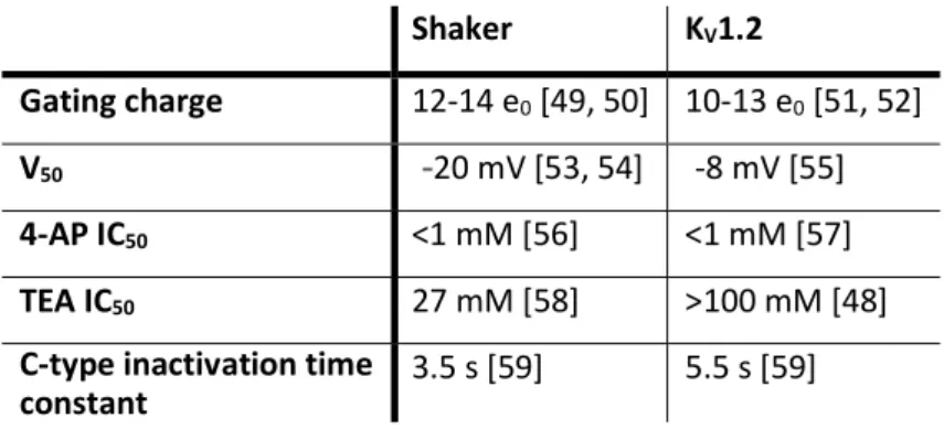
related closely enough such that correlation of their data is appropriate. The high sequence similarity between Shaker and KV1 channels (figure 1.2), as well as the correlation between predicted structural features of Shaker and the KV channel crystal structures, indicate that functional Shaker data can be interpreted with KV structural data. Shaker and KV1.2 both exhibit high sensitivity to channel block by 4-aminopyridine (4-AP) (Table 1) and voltage clamp fluorometry studies show that the top of the S4 helix correlates with charge movement in both channels [46, 47]. The two channels differ only slightly in gating charge, activation midpoint voltage (V50) and in the time course of C-type inactivation (Table 1). Considering the dissimilarity in the loop regions (figure 1.2) such differences would be anticipated. However, Shaker and KV1.2 differ in sensitivity to tetraethylammonium (TEA) block, which is due to a single amino acid difference in the outer pore region [48].
Table 1 Comparison of selected functional parameters for Shaker and KV1.2 Shaker KV1.2
Gating charge 12-14 e0 [49, 50] 10-13 e0 [51, 52]
V50 -20 mV [53, 54] -8 mV [55]
4-AP IC50 <1 mM [56] <1 mM [57]
TEA IC50 27 mM [58] >100 mM [48]
C-type inactivation time
11 Figure 1.2 Amino acid sequence alignment of Kv channels
Amino acid sequence alignment of Shaker and the human KV1 subfamily with locations of secondary structures. Green residues are identical and blue residues indicate similarity. The C- and N-termini have been omitted for space-saving purposes. Alignment is generated using T-coffee (http://tcoffee.crg.cat/apps/tcoffee/index.html) with input sequence IDs: Shaker-P08510, Kv1.1-Q09470, Kv1.2-P16389, Kv1.3-P22001, Kv1.4-P22459, Kv1.5-P22460, Kv1.6-P17658, Kv1.7-Q96RP8, Kv1.8-Q16322.
12
Common to all Shaker-related channels is the structural architecture of tetrameric symmetry. Each monomer consists of six transmembrane helices (S1-S6). A top-view of the channel shows that the first four helices (S1-S4) assemble in the periphery, linked to the pore domain by the intracellular S4-S5 linker that behaves like a protruding arm, and S5 and S6 assemble in the center to form the pore domain (A-B). This organization allows the S1-S4 region to act as independent voltage sensors controlling the ionic pathway. A KV signature sequence, PXP, is located at the intracellular end of S6 and constitutes the main activation gate which bends open upon pore opening (red residues in figure 1.3C) [18, 60]. A re-entrant loop of the S5-S6 linker forms a narrow selectivity filter which contains the GYG signature sequence [18]. High K+ selectivity is obtained by the unique ion binding sites which consist of carbonyl oxygens oriented towards the pore (dark blue residues in figure 1.3C). When entering the selectivity filter, the K+ ion’s interaction with water molecules is replaced by interaction with the carbonyl groups [14, 61]. The ions pass through the selectivity filter in a single file diffusion arrangement where each ion binds strongly to the selectivity filter but at the same time is destabilized by repulsion of the neighboring ion [61]. This is how the channel exhibit both high K+ selectivity and a high K+ permeation rate. The flexible glycines in the GYG motif allows the selectivity filter to adopt different conformations which prevent ion conduction (C-type inactivation).
13 Figure 1.2 Overview of KV channel structure and function
A) Crystal structure of the tetrameric KV channel showing the top view (PDB: 2R9R). The four voltage sensors are symmetrically arranged as modules around the central pore which contains the selectivity filter. B) Cartoon of the relative rearrangement of the four subunits shows how this organisation allows independent VS movement and cooperative pore movement C) KV1.2 crystal structure showing the S5-S6 helices of two subunits. Potassium ions are shown as grey spheres and the selectivity filter residues are highlighted in dark blue. The PXP motif at the bundlecross is highlighted in red, and in yellow is shown the interaction between Trp434 and Asp477. There exist four main conformations of the Shaker channel which dictates the flow of K+ ions (figure 1.4). In resting state or when deactivated, the channel is closed by the bundlecrossing of the four S6 intracellular C-termini. In the open activated state, the bundlecrossing widens and allows K+ ions to pass. A third state is the type inactivated state in which the channel’s N-terminus functions as a plug that inserts into the intracellular opening and blocks the pore. Finally, there is the C-type inactivated state in which a gate at the extracellular portion of the pore obstructs the ionic pathway.
14 Figure 1.3 Main conformational states of Shaker
Cartoon illustrating the different states which regulate the flow of K+ ions through the pore. When closed, the voltage sensors are deactivated and the S6 bundlecross blocks the intracellular pathway. In the open state, the voltage sensors are activated and the S6 bundlecross has widened to allow K+ to flow through. The intracellular N-terminus of either the α subunit (Shaker) or the β subunit (KV channels) enters the pore and blocks the ionic pathway leading to N-type inactivation. In absence of N-type inactivation, the selectivity filter rearranges which also blocks the ionic pathway and results in C-type inactivation. For clarity, only two subunits are shown.
1.5 Mechanism of K
Vchannel activation
The process by which KV channels transition from the deactivated state to the open activated state can be divided into three distinct but tightly coupled mechanisms: Voltage sensing, electromechanical coupling and pore opening. Each mechanism is accomplished by rearrangements in different parts of the channel which are connected structurally and/or energetically. The probability of an open channel is regulated by the voltage sensor which in turn depends on the membrane potential.
The voltage-dependency originates primarily from four arginines in the fourth transmembrane helix, S4. The number of translocated charges per channel has been determined experimentally [49] and theoretically [50] to be 13 elementary charges. Numerous biophysical and computational studies have investigated the resulting motion of the S4 helix, and although they differ in relative distances, a current understanding of the voltage sensor movement has
15
converged towards a general consensus model [62, 63]. Upon membrane depolarisation, the S4 helix undergoes a 7-10 Å vertical displacement to accommodate the change in the electric field. The electric field has been shown to be highly focused in water-exposed crevices in the VS domain facing the intracellular and extracellular compartments during hyperpolarization and depolarisation, respectively [64]. This explains why the arginines do not need to traverse the bilayer completely to account for the displacement of 13 elementary charges. A second S4 helix movement is accompanied by a tilt and a rotation around its helical axis. The first movement (Q1) occur independently in each VS, applying a force onto the S4-S5 linker. Then, during the second S4 charge movement (Q2), energy is released to the pore domain in a cooperative conformational change which finally results in widening of the bundlecross at the intracellular S6 gates.
The VS movement does not exclusively depend on the rearrangement of charges but requires also a network of stabilizing residues. Substitution studies, performed in Shaker, in the region surrounding the gating charges have identified several hydrophobic residues to play a key role in shaping the steric and energetic landscape as the voltage sensor moves from resting to activated state [65]. These residues are collectively called gating pore residues and are situated close to the membrane center within S1-S3. While one residue (Ser240) allows the passage of arginines, others make up a narrow constriction forming a barrier as a hydrophobic plug (Ile237 and Ile287) [65]. Finally, two phenylalanines form molecular clamps and stabilize the arginines in the activated state via cation-pi interactions (Phe244 and Phe290) [66, 67].
Although complex, the described VS process is relatively well understood. However, when it comes to how the VS governs the state of the pore, the underlying mechanism remains less clear. A fraction of the displaced charges has been shown to be associated with pore opening [67-69] during which the S4 helix also moves [70]. Since pore opening is a transition that requires a cooperative movement of all four subunits, it means that the VS movement is not exclusively independent but also includes a cooperative component. In agreement with this, VS movement has been found to consist of the two major components, Q1 and Q2, which accounts for approximately 80% and 20% of the total displaced charge, respectively [13]. Electromechanical coupling of voltage sensor movement to pore opening is an ongoing subject as the molecular mechanism behind the cooperative motion is not fully understood. The tetrameric organisation
16
of the channel shown in figure 1.2B, gives an idea of how cooperativity regulates pore opening. The pore region (S5-S6) of each subunit overlaps with that of the adjacent subunit suggesting that intersubunit interactions are required for the pore to open.
Box 1.1 Two-state closed-open channel
When considering the state of the channel pore as a two-state closed-open process in which the voltage sensor either resides in the closed deactivated state or in the open activated state, the transition barrier depends on the relative free energy,
∆𝐺 = ∆𝐺 + ∆𝐺 , (1.1) where
∆𝐺 = RT ∙ ln (K) (1.2) is the voltage-independent chemical energy difference with the equilibrium constant K, and
∆𝐺 = zFV (1.3) is the voltage-dependent electrical work required to activate the voltage sensor, where z is the displaced charge in response to the voltage V, and F is the Faraday constant. The probability of being in the open state PO, is given by:
𝑃 = 𝑃 𝑃 + 𝑃 = 1 1 + 𝑃 𝑃⁄ = 1 1 + exp ∆G𝑅𝑇 = 1 1 + exp 𝑅𝑇 ∙ ln(𝐾) + zFV𝑅𝑇 (1.4)
At equilibrium, K can be written as zFV1/2, where V1/2 is the voltage at which 50% of the voltage sensors have activated (or 50% of the channels are open). Equation 1.2 then rearranges to:
𝑃 = 1
1 + exp 𝑉 − 𝑉𝑑𝑉 /
, (1.5)
where the midpoint value of activation is V1/2, and the steepness of the curve dV=RT/zF which is related to the number of displaced charges, are useful comparative parameters in characterizing effects of mutants on the open probability (Po). Voltage sensor activation is experimentally measured as gating currents which are caused by movement of charges within the membrane [12]. The voltage dependency of charge displacement is then obtained by plotting the integrated gating currents as a function of voltage (QV curve) which then can be fitted to the Boltzmann function, equation 1.3, to obtain dV and V1/2.
17 1.5.1 Electromechanical coupling
The S4-S5 linker is the covalent link between the VS and the pore region (figure 1.5A). Early indications on its pivotal role in coupling VS movements to pore movements showed that a voltage-dependent channel was functional as long as the S4-S5 linker and the S6 tail were compatible, as in from the same channel [71, 72]. The S4-S5 linker lines the intracellular membrane leaflet in a radial position well situated to translate S4 motions to pore movements (figure 1.5A). A general understanding of the coupling process is that the combined movement of the S4 helix during activation exerts a mechanical force on the S4-S5 linker which in turn acts on the S6 bundlecross. In the KV crystal structure monomer, the S4-S5 linker is closely situated in parallel to the bundlecross, which could explain how the linker mechanically affects the state of the pore (red subunit, figure 1.5B). Indeed, it has been shown that hydrophobic interactions between residues of each region are critical for coupling VS movement to pore opening (light-pink residues in Figure 1.5B) [73, 74]. However, such intrasubunit interactions are not exclusively responsible for electromechanical coupling and cannot account for cooperativity. In fact, the coupling of movements is more complex than a simple push-and-pull mechanism. Substitution studies show that the ILT triple mutant (V369I, I372L, S376T) situated in the S4 helix, disrupt an energetic coupling between the VS and the pore, such that the final cooperative step is isolated from the early independent VS movements, and is shifted to very positive voltages [68, 70]. Another group of mutations situated on the inner S5 helix also perturb the transition towards the concerted opening [75]. Interestingly, these two groups cluster together in close physical proximity in the KV channel crystal structure of adjacent subunits (yellow residues in figure 1.5B). Their individual profound effects on coupling of independent VS movements to concerted pore movement, and their possible intersubunit interactions strongly suggest that they play a key role in electromechanical coupling as well as cooperativity. Finally, an intersubunit interaction between the S6 and the corner of the S4-S5 linker and S5 has been shown to be crucial for stabilization of the open state (blue residues in Figure 1.5B) [76]. Taken together, the transfer of movements between VS and pore involves a network of stabilizing molecular interactions, but the exact structural implications and sequence of events of these interactions remain unresolved.
18 Figure 1.4 Shaker channel topology and gating residues
A) KV channel topology B) KV2.1 crystal structure [18] showing parts of two adjacent subunits (red and green) with gating residues and interactions important for electromechanical coupling and pore opening. The group of hydrophobic residues in the S4 helix (ILT: V369, I372, S376) [68] and the inner S5 residues (L398, F401, F402, I405) [75] are shown in yellow. Residues involved in intersubunit interactions between the S6 tail and lower S5 helix are shown in blue [76]. Annealing of the S4-S5L (L382-T388) to the S6 helix (Y481-H486) is shown in light pink [72, 73, 77]. The voltage sensor of the red subunit has been removed for clarity.
1.6 Mechanism of K
Vchannel inactivation
Voltage-gated ion channels can undergo inactivation, which is a mechanism where the pore becomes non-conducting before the intracellular gates open (closed-state inactivation) or has already opened (open-state inactivation). The various inactivation mechanisms display a wide range of rates (from milliseconds to minutes) and provide functional diversity for cells to shape action potentials and to regulate their physiological roles. KV and NaV channels can be inactivated by binding of a cytoplasmic region to the pore which prevents ion flow. For KV channels, it is the N-terminal region [78, 79] which binds only after pore opening [80] (N-type inactivation), whereas for NaV channels, it is a cytoplasmic loop [81, 82] which can bind to both a closed and an open pore [83]. A second usually slower inactivation mechanism in NaV and KV channels involves global conformational changes surrounding the pore region and the selectivity filter (slow inactivation or C-type inactivation) which in KV channels result in pore constriction (discussed below) whereas the mechanism is less understood in NaV channels [84]. U-type inactivation is a different kind of slow inactivation which has been identified in certain KV channels, including
19
KV1.5, KV2.1, KV3.1 and Shaker [85-87]. U-type inactivation exhibit different properties than C-type inactivation (e.g. in voltage-dependency, sensitivity to external potassium and blockers) and the molecular determinants are much less understood.
1.6.1 N-type inactivation
N-type inactivation in KV channels modulates neuronal excitability and signaling by means of shortening the time that current passes through the channel. In N-type inactivation, the long and flexible N-terminus of one of the four subunits enters the side window of the cytosolic tetramerization T1 domain (T1) and physically occludes the open pore to block the ionic pathway in a ball-and-chain mechanism (Figure 1.6A) [78, 79, 88]. Inactivation can be eliminated by removing the N-terminal sequence and be restored by cytoplasmic application of the N-terminal peptide to the channel [78, 79]. Shaker, KV1.4, KV3.4 and KV4.2 channels are intrinsically inactivated by the N-terminus [78, 89-91], while other KV1 channels are inactivated by the N-terminus of auxiliary β1 or β3 subunits [90, 92] (except KV1.6 which has an inactivation prevention domain [93]). The other KV families do not undergo N-type inactivation. Derived peptides of the inactivating N-terminus, or inactivation particle (IP), of Shaker, KV1.4 and KV3.4 are capable of blocking KV1 channels although there is no sequence similarity in the IPs [94-96] (figure 1.6B). Therefore, in contrast to the molecular mechanisms of VS activation and pore opening, the binding of the N-terminus does not seem to require specific amino acid interactions. Instead, the rate of inactivation depends on long-range electrostatic interactions between the chain and putative receptor sites [18, 79, 97, 98], and final pore block depends on general hydrophobic interactions between the tip and pore region [88]. In agreement with this understanding, the first 5-9 amino acids of the IP (inactivation particle) are mainly hydrophobic, while the downstream 20-40 amino acids, which act as the chain, are mainly hydrophilic carrying a net-positive charge (figure 1.6B).
20 Shaker β MAAVAGLYGLGEDRQHRKKQQQQQQHQKEQLEQKEEQKKIAERKLQLREQQLQRNSL +2 Kv1.4 β MEVAMVSAESSGCNSHMPYGYAAQARARERERLAHSRAAAAAAVAAATAAVEGSGGS 0 Kv3.4 β MISSVCVSSYRGRKSGNKPPSKTCLKEEMAKGEASEKIIINVGGTRHETYRSTLRTL +6 Kvβ3 MQVSIACTEQNLRSRSSEDRLCGPRPGPGGGNGGPAGGGHGNPPGGGGSGPKARAAL +3 Kvβ1.1 MQVSIACTEHNLKSRNGEDRLLSKQSSTAPNVVNAARAKFRTVAIIARSLGTFTPQH +5 Kvβ1.2 MHLYKPACADIPSPKLGLPKSSESALKCRWHLAVTKTQPQAACKPVRPSGAAEQKYV +5 Kvβ1.3 MLAARTGAAGSQISEENTKLRRQSGFSVAGKDKSPKKASENAKDSSLSPSGESQLRA +4 Figure 1.5 N-type inactivation in KV channels
A) Structural overview of the ball-and-chain mechanism where the N-terminus of either the alpha (left) or the beta (right) subunit enters the side window of the T1 subunit (in yellow) and binds to the open pore B) Comparison of N-termini of Shaker and of human KV channels and of β subunits (residues 1-60) which are responsible for N-type inactivation. Hydrophobic residues are colored in red and charged residues in blue. To the right is noted the net charge. Sequence Uniprot IDs: Shaker-P08510, Kv1.4-P22459, Kv3.4-Q03721, Kvβ3-O43448, Kvβ1.1-Q14722.2, Kvβ1.2-Q14722.3, Kvβ1.3-Q14722.
It is interesting that different IPs can block the same channels. It suggests that the inactivation pathway is not identical although the receptor site is the same. As an example, the N-terminal glutamates in KV1.4 (E2 and E9) are of unique importance for efficient inactivation [99], whereas insertion of glutamates in the same region of the Shaker N-terminus has the opposite effect (L7E) [78].
21
In the KV1.2 crystal structures [18, 45], the channel is in complex with a β subunit which do not cause N-type inactivation (β2), so the position and structure of the IP is not known. NMR studies of IPs indicate different conformations ranging from unstructured (Shaker) to structured (KV1.4 and KV3.4) [100, 101]. These observations suggest that the IP could either always be disordered and available to bind inside the channel after the pore opens, or that binding to early receptor sites trigger a structural unfolding. Interestingly, a recent computational study with Shaker shows that an IP adopting a helical structure spontaneously enters the inner cavity of the pore, and then inserts the tip deeply into the pore when driven by voltage [102].
1.6.2 C-type inactivation
The other inactivation mechanism in Shaker is C-type inactivation and occurs at the selectivity filter. The structural basis of the C-type inactivated state is well understood. First, molecular dynamic simulations of the KcsA channel shows that flexibility provided by the glycines in the GYG motif allows the selectivity filter to adopt several conformations which disturb coordination of the ions, which in turn prevents ion conduction [103]. In agreement with this, X-ray crystallography studies of inactivated KcsA channels show a confirmation in which the selectivity filter is pinched inward at the GYG motif [88, 104]. Also, C-type inactivation is highly sensitive to external K+ [105, 106], pore blockers [107, 108], cations [109, 110] and mutations in the region surrounding the pore [105, 111, 112], but the mechanism which initially drive the filter to rearrange and cause inactivation is less clear. One hypothesis is that K+ dissociation from the pore caused by the occasional entry of cations favors the inactivated state [110]. Alternatively, opening of the S6 helical bundlecross has shown to be allosterically coupled to the inactivation gate via a network of steric contacts which in the end cause the filter to collapse [112]. C-type inactivation is a complex process which may involve both mechanisms. Possibly, coupling of the S6 gates to the inactivation gates constitute an intrinsic way for the channel to ensure inactivation, whereas ion vacancy is an extrinsically regulated pathway which accelerates the transition into inactivation further.
An interesting feature of the C-type inactivated state is the presence of water molecules behind the selectivity filter, as demonstrated by molecular dynamic simulations on the KcsA structures [113]. It is shown how recovery of the pinched GYG motif requires release of the water molecules
22
to the extracellular side. However, the molecular determinants behind the initial entry of the water molecules is not corroborated, yet, and it is not certain whether the water molecules are in fact causing the pinched state. To get a complete understanding of C-type inactivation, it is necessary to find out how and if, activation of the S6 helical bundle and/or K+ dissociation leads to entry of the inactivating water molecules behind the filter.
Molecular dynamic simulations on KV1.2 [114] support a link between C-type inactivation and water molecules as “gate keepers”. A hydrogen bond between Trp434 and Asp477 (yellow residues in figure 1.3C) keeps water away from entering behind the selectivity filter [114]. The W434F mutation is a well known mutant used to abolish ionic currents without affecting the intracellular gate [115] and it has been shown that this mutation results in a constitutively C-type inactivated channel [111]. W434F has therefore been widely used to investigate gating currents. The absence of a hydrogen bond between Trp434 and Asp477 in W434F channels is probably what is accelerating the transition into the C-type inactivated state, by allowing water molecules to enter from the external solution.
1.7 Genetic incorporation of unnatural amino acids
Given the important roles of proteins in biology, it is desirable to be able to manipulate proteins for understanding structure-function relationships and for generating proteins with new properties. Many efforts have been made to incorporate UAAs into proteins to introduce new functional groups different from those found in canonical amino acids. To add a new amino acid to the genetic repertoire, a codon is needed that uniquely specifies that amino acid. The 20 canonical amino acids are encoded by 61 triplet codons, leaving the remaining three stop-codons (TAG, amber; TAA, ochre; and TGA, opal) as candidates for suppression for the incorporation of an UAA. The amber stop codon has been the most used for UAA incorporation as it is the least frequent stop codon in E.coli (9%), yeast (24%) and mammalian cells (~18%) [116] thus limiting UAA insertion at endogenous sites.
In 1989, Dr. Schultz reported for the first time the incorporation of UAAs into a protein in a cell-free system via chemical aminoacylation of a suppressor tRNA [117]. In the following decade it was established that the nonsense suppression method could become a general tool in modifying proteins enabling unprecedented structure-function studies. Indeed, in 1995 the approach was
23
expanded to function in living cells in Xenopus oocytes where the amino-acylated tRNA was injected with the UAA and protein-encoding mRNA [118]. The high expression efficiency of Xenopus oocytes combined with the high sensitivity of electrophysiology made it possible to probe UAA insertion, and once again did Xenopus oocytes play a valuable role in developing a tool for the understanding of ion channels. Today, more than 20 channels and receptors have been expressed with more than 100 different UAAs using the chemical aminoacylation strategy [119]. Although successful, the technique is limiting as the stoichiometry of injected amino-acylated tRNAs still produces relatively small amounts of protein.
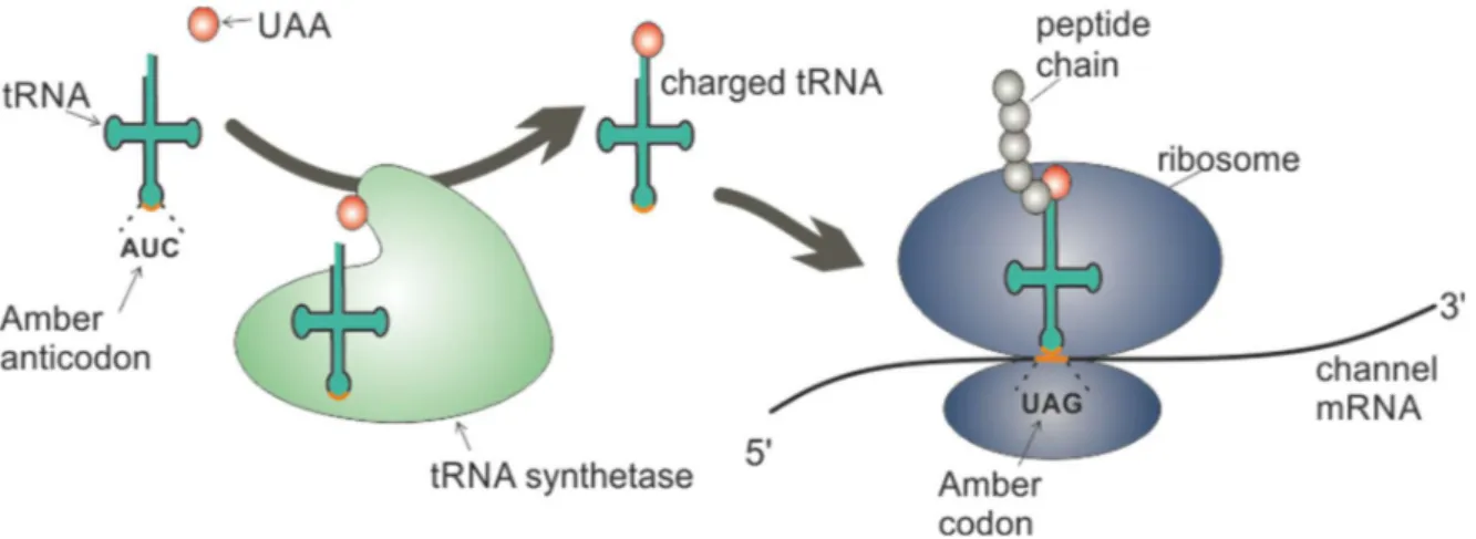
Figure 1.6 Cartoon of genetic incorporation of UAAs via in vivo aminoacylation.
The UAA is specifically aminoacylated to the tRNAAUC by the tRNA synthetase. The charged UAA-tRNACUA is then recognized by the ribosome when translation of the protein mRNA reaches the amber codon, and the UAA is then inserted into the growing peptide chain.
An alternative technique relies on in vivo aminoacylation using an engineered aminoacyl tRNA synthetase (aaRS) which specifically charges the tRNA with the UAA without cross-reaction with endogenous tRNAs or amino acids (orthogonality) (figure 1.7). Orthogonality is achieved by identifying aaRS/tRNA pairs from other organisms, which then are altered to specifically recognize an UAA via directive evolution using large libraries of mutant aaRS’s. In 2001, the Schultz laboratory generated an orthogonal tRNA/synthetase pair to function in E. coli by importing it from the archaea Methoanoccus jannaschii whose tRNATyr identity elements differ from those in E.coli [120]. In the following years, the genetic code expansion advanced rapidly to also include yeast and mammalian cells, in which bacterial aaRS/tRNA pairs are generally orthogonal [121]. So far, three bacterial (tyrosyl, leucyl, tryptophanyl) and one archaea (pyrrolysyl) aaRS/tRNA pair have been used to incorporate distinct UAAs in mammalian cells
24
[121]. Today, almost 200 different UAAs have been incorporated into proteins in prokaryotic and eukaryotic organisms using the in vivo aminoacylation strategy [122].
1.8 Protein translation
While orthogonality between the UAA and the engineered synthetase/tRNA pair is necessary to obtain specificity and high incorporation fidelity, it is not a guarantee. There are other mechanisms of the translational machinery in the cell, which can cause unwanted expression of proteins lacking the UAA.
The process of translation is a step in gene expression during which the single stranded mRNA is translated into protein, according to the genetic code. Each group of three bases in the mRNA molecule codes for an amino acid. Within all eukaryotic cells, translation occurs in a specialized complex called the ribosome, which is located in the cytoplasm, and consists of two large RNA molecules (40S and 60S) and a subset of ribosomal proteins.
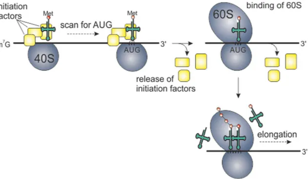
When the mature mRNA has left the nucleus, translation begins at the 5’end of the mRNA and ends at the 3’end. An initial complex structure of three initiation factor proteins and the small 40S ribosomal subunit assembles and binds a methionine-charged tRNA. Subsequently, this complex binds to the mRNA 5’end (figure 1.8) and starts to scan the mRNA for the 5’-proximal AUG codon. The large 60S ribosomal subunit binds to the complex and initiation factors are released (figure 1.8). The next step is elongation during which incoming charged tRNAs are recognized by the ribosome according to the mRNA codon, and peptide bonds are formed between the amino acids to create a growing polypeptide chain (figure 1.8). Elongation continues until the ribosome encounters a stop codon, and release factors then binds to facilitate release of the ribosome from the mRNA.
Most mammalian mRNAs follow the straightforward model of linear ribosome scanning, in which initiation occurs exclusively at the 5′-proximal AUG codon, and termination occurs at the downstream stop codon at the end of the open reading frame (ORF). Factors such as sequence context and RNA structure can influence the scanning efficiency and cause alternative translation resulting in different proteins [123]. Both start and stop codons can be “ignored” by the ribosome (readthrough and leaky scanning) resulting in alternative ORFs [123, 124]. Resumption of scanning after termination on the same mRNA is also a mechanism in eukaryotic ribosomes
25
(reinitiation), adding possibilities to the mRNA coding potential [124]. In this case, the 40S subunit is not released from the mRNA, but stays in a complex with the mRNA. This only happens when the first ORF is short (e.g. 10-20 codons), such that initiation factors are still connected to the ribosome, thus allowing a resumption of scanning for another start codon further downstream.
Figure 1.7 Protein translation
Cartoon of the eukaryotic translation machinery. Yellow boxes represent initiation factors which form an initiation complex with a methionine-tRNA, the mRNA and the 40S ribosomal subunit. When scanning for the 5’-proximal AUG start codon, the 60S subunit binds to the complex and initiation factors are released which marks the beginning of the next step, elongation. The ribosome reads the mRNA codons in a base-by-base fashion, binds to the corresponding tRNAs and creates peptide bonds of the incoming amino acids to form a polypeptide chain.
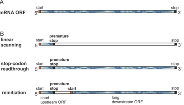
If a stop codon resides near the initiation site (by spontaneous mutation or naturally occurring), there is a possibility that gene expression is compromised. In figure 1.9A is shown an example of an mRNA ORF (blue frame is translated). Then, in figure 1.9B is shown three translation scenarios, in the case where a premature stop codon is mutated in close vicinity to the start site. Linear scanning would result in ribosomal release and give one short ORF. On the other hand, stop-codon readthrough would ignore the stop stop-codon and yield a full length ORF. Since the stop stop-codon is close to the first initiation site, it is also likely that initiation factors are still bound and allows for resumption of initiation at an alternative downstream start codon, thus yielding two ORFs.
26
When using UAA incorporation with non-available UAA-charged tRNAs, a number of things can happen depending on the position of the stop codon. Ideally, the ribosome stops translation when it encounters the amber stop codon and releases from the mRNA such that no functional protein is translated. This scenario constitutes an intrinsic control for UAA expression, but only if the stop codon is not situated in the C-terminal portion such that a C-terminal truncated and functional protein would be expressed. On the other hand, if the stop codon is situated in the near N-terminal portion, the ribosome may reinitiate translation and result in N-terminal truncated proteins. The latter scenario is demonstrated in chapter 6 where we shed light in important translational factors. Therefore, for each amber stop mutation for any UAA incorporation in any protein, it is important to consider all translational scenarios and measure the amount of leak expression in absence of the UAA to be able to evaluate potential effects of a mixed population during UAA experiments.
Figure 1.8 mRNA open reading frames and premature stop codons
A) Example of an mRNA ORF where the blue frame indicates translated area. In B) are shown three translation scenarios in case of a premature stop codon. Normal linear scanning results in a short ORF, whereas readthrough of the premature stop codon would result in a full length ORF as in A). Reinitiation of translation at a downstream start codon would result in two ORFs.