Burkholderia Diversity and Versatility: An Inventory of the Extracellular
Products
VIAL, LUDOVIC, MARIE-CHRISTINE GROLEAU, VALÉRIE DEKIMPE, AND ERIC DÉZIEL*
INRS-Institut Armand-Frappier, Laval, Québec, Canada H7V 1B7
Received: July 1, 2007 Accepted: July 31, 2007 Abstract The Burkholderia genus consists of over 40 Gram-negative, β-proteobacteria species that occupy remarkably diverse ecological niches. This genus contains species pathogenic to human, animals, and plants, as well as species involved in promoting plant growth and biodegradation of pollutants. This is largely explained by the extraordinary versatility of Burkholderia, as reflected by the remarkable diversity of extracellular products released by these bacteria. We exhaustively surveyed the extracellular enzymes, siderophores, toxins, antimicrobials, and other secondary metabolites produced by the members of this very diverse genus. Available information on regulation, especially quorum sensing mechanisms, and secretion is highlighted.
Keywords: Burkholderia, enzymes, siderophores, toxins, antimicrobials, quorum sensing
The Burkholderia genus, β subdivision ofthe proteobacteria,
comprises more than 40 species that inhabit remarkably diverse ecological niches, as they have been isolated from soil, plant rhizosphere, water, insects, fungus, and hospital environments and from infected humans (Table 1). The genus Burkholderia was proposed by Yabuuchi et al. [218] to accommodate the former rRNA group II pseudomonads [218]. In fact, confusion with Pseudomonas has impeded knowledge progression.
Traditionally, Burkholderia species have been known as plant pathogens. B. cepacia was first described by Burkholder in 1950 as a plant pathogen causing sour skin of onion [22]. For example, B. glumae causes rot of rice grains and seedlings (panicle blight) [88]. Several
Burkholderia species have developed beneficial interactions with their plant hosts. Somes species are able to fix atmospheric nitrogen, including B. vietnamiensis, B. unamae, and B. tropica [23, 157, 188]. Legumes are also
nodulated by several Burkholderia species such as B. mimosarum, B. nodosa, and B. phymatum [28, 29, 154]. Several Burkholderia species have considerable commercial and ecological importance. Certain species of Burkholderia have proved to be very efficient in biocontrol, bioremediation, and plant growth promotion. For example, B. xenovorans strain LB400 is one of the most effective polychlorinated biphenyl(PCB) degraders known [27].
In contrast, several Burkholderia species are also opportunistic human pathogens. These species include all
Burkholderia cepacia complex (Bcc) bacteria, B. gladioli, and B. fungorum. The Bcc contains (at least) nine closely related species or genomovars, which can cause severe respiratory infections in people suffering from cystic fibrosis (CF) or chronic granulomatous disease [39]. All nine species have been recovered from CF patients, but B. cenocepacia and B. multivorans are the dominant Bcc species [119]. B. cenocepacia comprises the most virulent clones and has been associated with higher mortality rates among CF patients [119].
B. pseudomallei and B. mallei are the only known members of the genus Burkholderia that are primary pathogens in humans and animals. The saprophyte B. pseudomallei is the causative agent of melioidosis, a potentially fatalsepticemic infection of animals and humans [30]. Melioidosis is endemic in tropical and subtropical areas of Southeast Asia and Northern Australia [30]. B. pseudomallei has been classified as a potential biological weapon and has been used by Germany during World War I [160]. B. mallei is the etiologic agent of glanders, which is mainly a horse disease but in rare cases affects humans [138]. B. thailandensis is closely related to B. pseudomallei
and is generally considered avirulent [68].
The ecological versatility of Burkholderia is likely due to their large genomes, which are often comprised of several large replicons (two to four circular chromosomes or large plasmids). An important variation in genome size from 4.7 to 9 Mb is observed in the Burkholderia genus
*Corresponding author
Phone: 450-687-5010; Fax: 450-686-5501; E-mail: eric.deziel@iaf.inrs.ca
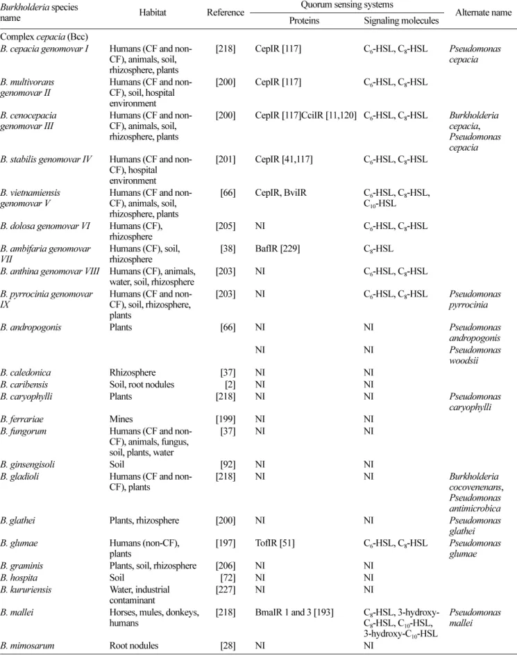
Table 1. List of known Burkholderia species, with their reported QS systems.
Burkholderia species
name Habitat Reference ProteinsQuorum sensing systemsSignaling molecules Alternate name Complex cepacia (Bcc)
B. cepacia genomovar I Humans (CF and non-CF), animals, soil, rhizosphere, plants
[218] CepIR [117] C6-HSL, C8-HSL Pseudomonas
cepacia B. multivorans
genomovar II Humans (CF and non-CF), soil, hospital environment
[200] CepIR [117] C6-HSL, C8-HSL
B. cenocepacia
genomovar III Humans (CF and non-CF), animals, soil, rhizosphere, plants
[200] CepIR [117]CciIR [11,120] C6-HSL, C8-HSL Burkholderia
cepacia, Pseudomonas cepacia B. stabilis genomovar IV Humans (CF and
non-CF), hospital environment
[201] CepIR [41,117] C6-HSL, C8-HSL
B. vietnamiensis
genomovar V Humans (CF and non-CF), animals, soil, rhizosphere, plants
0[66] CepIR, BviIR C6-HSL, C8-HSL,
C10-HSL
B. dolosa genomovar VI Humans (CF),
rhizosphere [205] NI C6-HSL, C8-HSL
B. ambifaria genomovar
VII Humans (CF), soil,rhizosphere 0[38] BafIR [229] C8-HSL B. anthina genomovar VIII Humans (CF), animals,
water, soil, rhizosphere [203] NI C6-HSL, C8-HSL B. pyrrocinia genomovar
IX Humans (CF and non-CF), soil, rhizosphere, plants
[203] NI C6-HSL, C8-HSL Pseudomonas
pyrrocinia
B. andropogonis Plants 0[66] NI NI Pseudomonas
andropogonis
NI NI Pseudomonas
woodsii
B. caledonica Rhizosphere 0[37] NI NI
B. caribensis Soil, root nodules 00[2] NI NI
B. caryophylli Plants [218] NI NI Pseudomonas
caryophylli
B. ferrariae Mines [199] NI NI
B. fungorum Humans (CF and non-CF), animals, fungus, soil, plants, water
0[37] NI NI
B. ginsengisoli Soil 0[92] NI NI
B. gladioli Humans (CF and
non-CF), plants [218] NI NI Burkholderiacocovenenans, Pseudomonas antimicrobica
B.glathei Plants, rhizosphere [200] NI NI Pseudomonas
glathei B. glumae Humans (non-CF),
plants [197] TofIR [51] C6-HSL, C8-HSL Pseudomonasglumae
B. graminis Plants, soil, rhizosphere [206] NI NI
B. hospita Soil 0[72] NI NI
B. kururiensis Water, industrial
contaminant [227] NI NI
B. mallei Horses, mules, donkeys,
humans [218] BmaIR 1 and 3 [193] CC88-HSL, 3-hydroxy--HSL, C10-HSL,
3-hydroxy-C10-HSL
Pseudomonas mallei
[215]. The presence of multiple insertion sequences that confer genome plasticity could also explain the versatility of the genus Burkholderia [130].
Burkholderia species secrete a variety of extracellular enzymes with proteolytic, lipolytic, and hemolytic activities. Several strains secrete also toxins, antibiotics, and siderophores. The extracellular products of Burkholderia
represent admirably the diversity and versatility of that genus. In this review, we present a survey of the extracellular products of Burkholderia species and their role in interaction with their hosts. Regulation and secretion of these products are also reported.
T
RANSCRIPTIONALR
EGULATION OFE
XOPRODUCTS:
Q
UORUMS
ENSINGBacteria closely regulate the synthesis and release of extracellular products. Besides environmental and physiological conditions that influence expression of exoproducts, another type of global regulation is widespread among bacterial populations and is called quorum sensing (QS) [156]. Quorum sensing is fundamental to the ability of many bacterial species to create coordinated cell-to-cell interactions. Quorum sensing is a cell-to-cell communication system used by bacteria to perceive and respond to their population
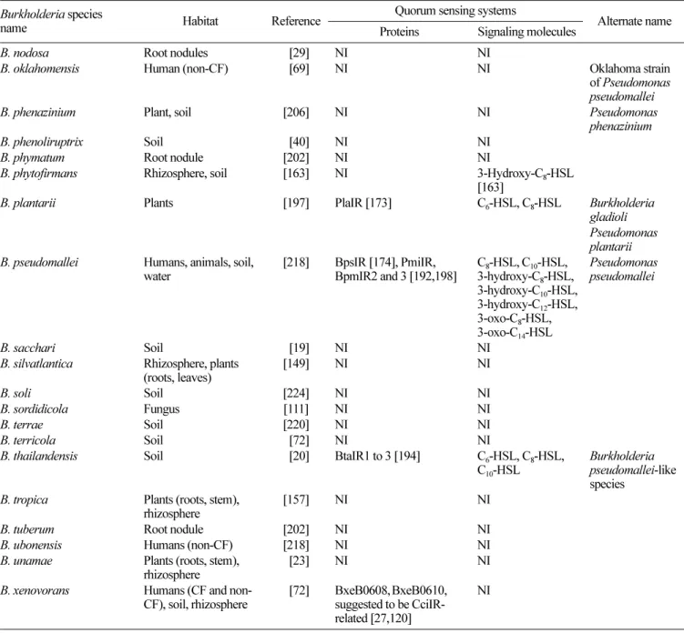
Table 1. Continued.
Burkholderia species
name Habitat Reference ProteinsQuorum sensing systemsSignaling molecules Alternate name
B. nodosa Root nodules 0[29] NI NI
B. oklahomensis Human (non-CF) 0[69] NI NI Oklahoma strain
of Pseudomonas pseudomallei
B. phenazinium Plant, soil [206] NI NI Pseudomonas
phenazinium
B. phenoliruptrix Soil 0[40] NI NI
B. phymatum Root nodule [202] NI NI
B. phytofirmans Rhizosphere, soil [163] NI 3-Hydroxy-C8-HSL
[163]
B. plantarii Plants [197] PlaIR [173] C6-HSL, C8-HSL Burkholderia
gladioli Pseudomonas plantarii B. pseudomallei Humans, animals, soil,
water [218] BpsIR [174], PmiIR,BpmIR2 and 3 [192,198] C3-hydroxy-C8-HSL, C10-HSL,8-HSL,
3-hydroxy-C10-HSL, 3-hydroxy-C12-HSL, 3-oxo-C8-HSL, 3-oxo-C14-HSL Pseudomonas pseudomallei B. sacchari Soil 0[19] NI NI
B. silvatlantica Rhizosphere, plants
(roots, leaves) [149] NI NI
B. soli Soil [224] NI NI
B. sordidicola Fungus [111] NI NI
B. terrae Soil [220] NI NI
B. terricola Soil 0[72] NI NI
B. thailandensis Soil 0[20] BtaIR1 to 3 [194] C6-HSL, C8-HSL,
C10-HSL
Burkholderia pseudomallei-like species
B. tropica Plants (roots, stem),
rhizosphere [157] NI NI
B. tuberum Root nodule [202] NI NI
B. ubonensis Humans (non-CF) [218] NI NI
B. unamae Plants (roots, stem),
rhizosphere 0[23] NI NI
B. xenovorans Humans (CF and
non-CF), soil, rhizosphere 0[72] BxeB0608,suggested to be CciIR-BxeB0610, related [27,120]
NI
density in order to coordinate gene expression. In Gram-negative bacteria, the most widespread QS mechanism is based on LuxR-type transcriptional regulators and their cognate N-acyl-homoserine lactones (AHLs) ligands [63]. These molecules, when they reach a threshold reflecting cell density, activate LuxR-type transcriptional regulators that specifically regulate bacterial gene expression.
Quorum sensing has been well studied among Bcc bacteria, where it was first identified from the clinical isolate
B. cenocepacia strain K56-2 [108], and comprises the LuxIR-type homologs CepI and CepR (Table 1). The CepI synthase is responsible for the production of two AHLs: N -hexanoyl-HSL (C6-HSL) and the most abundant N-octanoyl-HSL
(C8-HSL). The transcriptional regulator CepR responds most
efficiently to C8-HSL [82]. The cepI and cepR genes have
been found in the 9 species comprised in the Bcc [117]. In B. ambifaria, the CepIR system has been renamed BafIR [229].
Some Bcc species possess more than one QS circuitry. For example, some B. vietnamiensis strains, as well as
producing C6- and C8-HSLs, display additional AHLs, C10
-HSL being the most abundant [41]. In these strains, another QS system is responsible for the production of these additional AHLs, named BviIR. BviI and BviR are only 36% identical to CepIand CepR, respectively. Whether the BviI/BviR system interacts with the CepI/CepR system remains to be investigated. In a particulary virulent strain of B. cenocepacia lineage ET12, another QS system is also present and is encoded in a pathogenicity island [11]. It is yet unclear whether this QS system, consisting of the AHL synthase CciI and its cognate receptor protein CciR, operates independently of CepI/CepR.
Although QS has been less studied outside the Bcc, it appears present in every Burkholderia strain where it was investigated. In B. glumae and B. plantarii, QS systems have been identified and named TofIR [93] and PlaIR [173], respectively. These two systems share more than 99% identity, and 75% identity with the CepIR system [173].
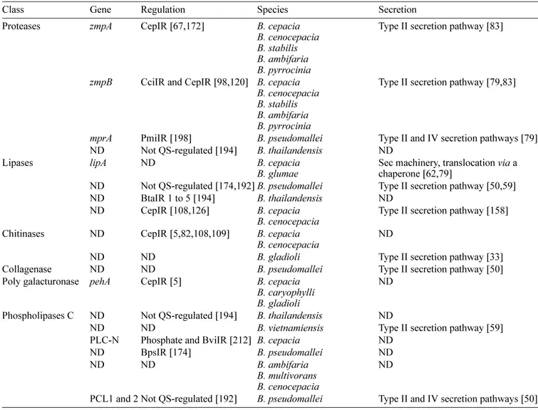
Table 2. Extracellular enzymes.
Class Gene Regulation Species Secretion
Proteases zmpA CepIR [67,172] B. cepacia B. cenocepacia B. stabilis B. ambifaria B. pyrrocinia
Type II secretion pathway [83]
zmpB CciIR and CepIR [98,120] B. cepacia B. cenocepacia B. stabilis B. ambifaria B. pyrrocinia
Type II secretion pathway [79,83]
mprA PmiIR [198] B. pseudomallei Type II and IV secretion pathways [79] ND Not QS-regulated [194] B. thailandensis ND
Lipases lipA ND B. cepacia
B. glumae Sec machinery, translocation chaperone [62,79] via a ND Not QS-regulated [174,192]B. pseudomallei Type II secretion pathway [50,59] ND BtaIR 1 to 5 [194] B. thailandensis ND
ND CepIR [108,126] B. cepacia
B. cenocepacia Type II secretion pathway [158] Chitinases ND CepIR [5,82,108,109] B. cepacia
B. cenocepacia ND
ND ND B. gladioli Type II secretion pathway [33]
Collagenase ND ND B. pseudomallei Type II secretion pathway [50] Poly galacturonase pehA CepIR [5] B. cepacia
B. caryophylli B. gladioli
ND Phospholipases C ND Not QS-regulated [194] B. thailandensis ND
ND ND B. vietnamiensis Type II secretion pathway [59]
PLC-N Phosphate and BviIR [212] B. cepacia ND
ND BpsIR [174] B. pseudomallei ND
ND ND B. ambifaria
B. multivorans B. cenocepacia
ND
PCL1 and 2 Not QS-regulated [192] B. pseudomallei Type II and IV secretion pathways [50]
Quorum sensing in the pathogens B. pseudomallei and
B. mallei is more complex than in other Burkholderia
species. Cell-to-cell communication networks in these organisms comprise multiple LuxIR homologs utilizing numerous AHL signal molecules. In various B. pseudomallei
strains, four LuxI and six LuxR homologs were identified [174, 192, 198]. In B. mallei,the QS circuitry consists of two LuxIR-like homologs, but in contrast with B. pseudomallei, lacks the BpmIR2, which shares the highest degree of homology with the BviIR system of B. vietnamiensis [193].
Although little is known about the regulation of extracellular molecules such as antibiotics and toxins produced by
Burkholderia spp., multiple extracellular enzymes have been shown to be under the control of QS. In addition, a secretory machinery is needed to export many high molecular weight exoproducts in the extracellular environment. However, only a few studies have focused on their secretion pathways in the Burkholderia genus. Regulation and secretion of each category presented here are reported in the different tables within this review.
E
XTRACELLULARE
NZYMESThe severity of infections by Bcc strains may be related to their ability to secrete a huge diversity of extracellular proteins. Production of enzymes by environmental strains can also be interesting for industrial purposes. The large variety of enzymes secreted by the Burkholderia correlates with the diversity of their ecological niches (Table 2).
Proteases
A majority of Bcc isolates produce extracellular proteases. In a study on exoenzymes production in CF isolates of
B. cenocepacia, B. multivorans, B. ambifaria, and B. vietnamiensis, only B. ambifaria and B. cenocepacia had proteolytic activity on BHI-milk agar [26]. A 34 kDa protease formerly named PSCP (Pseudomonas cepacia
protease) was purified from B. cenocepacia strain Pc715j supernatant [127]. This protease, now known as ZmpA, is a zinc metalloprotease. Expression of zmpA was detected in B. cepacia, B. cenocepacia, B. stabilis, B. ambifaria, and B. pyrrocinia, which correlated with a strong protease activity when tested on BHI-milk agar. Strains in which the
zmpA gene was absent (B. multivorans, B. vietnamiensis, B. dolosa, and B. anthina) showed no proteolytic activity. B. cepacia genomovar I type strain ATCC 25416, an onion pathogen, shows proteolytic activity and expresses ZmpA [4,67]. ZmpA is important for extracellular protease activity in Bcc strains [67]. The mature ZmpA is proteolytically active against hide powder azure, type IV collagen, fibronectin, neutrophil alpha-1 proteinase inhibitor, alpha(2)-macroglobulin, and gamma interferon [97]. The impact of ZmpA on persistence was investigated. A zmpA- mutant of B. cenocepacia
K56-2 was less able to persist in the lungs of infected rats [4K56-2]. However, in B. cenocepacia Pc715j, the same mutation caused no difference in persistence when compared with the parent strain [42]. Quorum sensing cepI- and
cepR-mutants of this strain showed less proteolytic activity than the parent strain [4]. ZmpA expression also seems to be controlled by the CepIR QS system in B. cenocepacia
K56-2 and, accordingly, a possible Cep box is found in the promoter region [181].
A second metalloprotease called ZmpB was identified in the same species carrying ZmpA. ZmpB has proteolytic activity against α-1 proteinase inhibitor, α2-macrogobulin,
type IV collagen, fibronectin, lactoferrin, transferrin, and immunoglobulins [98]. zmpB- mutants and zmpA-zmpB
-double mutants show no proteolytic activity against casein and are less virulent in the agar bead rat lung infection model, indicating that proteolytic activity is involved in virulence of the bacteria. zmpB expression is controlled by CepIR and CciIR quorum sensing systems in B. cenocepacia [98].
Most B. pseudomallei isolates show protease activity on BHI-milk agar [9, 50]. Strains deficient in protease production are less virulent than the parental strain in diabetes-induced Sprague-Dawley rats, a lung infection model [164]. However, Gauthier et al. [64] reported no correlation between virulence and proteolytic activity when human isolated B. pseudomallei ATCC 23353 was injected intraperitoneally in SWISS mice, although this strain is a low protease producer [64, 107]. In addition, a deficiency in the type II secretion pathway inhibits protease activity but does not affect virulence of a B. pseudomallei mutant in Syrian hamsters, which are usually very susceptible to B. pseudomallei infections [50].
The mprA gene, encoding a metalloprotease expressed by B. pseudomallei, was cloned and sequenced [107]. An
mprA- mutant has very low protease activity compared with
wild type, indicating production of MprA is essential to
B. pseudomallei’s proteolytic activity [198]. The SWISS mice were used to test the mprA- mutant’s virulence compared with
parental strain B. pseudomallei 008. No significant differences were noted, indicating that MprA did not act as a virulence determinant in this model. [198]. The PmlIR QS system downregulates expression of MprA during the stationary phase in B. pseudomallei [198]. B. thailandensis, although closely related to B. pseudomallei, does not seem to regulate its proteolytic activity by QS, as shown by Ulrich et al. [194].
Lipases
Many bacterial species produce lipases, enzymes that catalyze the hydrolysis and synthesis of esters of glycerol with long-chain fatty acids. Lipases of microbial origin have found great application in many industrial sectors since they are able to catalyze a large variety of reactions.
Ten B. cepacia (originally P. cepacia) isolates from the sputum of CF patients were tested for their lipolytic
activity against various substrates. All of them produced variable activities when substrates of different chain lengths were used [113]. A similar screening was performed on various Bcc CF isolates of B. cenocepacia, B. ambifaria, B. vietnamiensis, and B. multivorans. All the strains were lipolytic against various substrates, although the activity from B. multivorans was the strongest [26].
A 25-kDa lipase has been purified by gel filtration from the clinical isolate B. cepacia 90ee supernatant and tested on the lungs of rats [114], resulting in a large amount of proteinaceous exudates, accumulation of polymorphonuclear leucocytes and red blood cells, and disorganization of the alveolar structure [114]. B. cepacia DSM3959 produces an extracellular lipase, LipA [89], whose activity depends on the presence of the chaperone, LimA [1, 79]. An extracellular 37-kDa cholesterol esterase was purified from B. cepacia
strain ST-200, which shows 87% similarity to LipA from
B. cepacia DSM3959 and preferentially hydrolyzes long-chain fatty acid esters of cholesterol, except cholesteryl palmitate. This enzyme also displays lipolytic activity toward various
p-nitrophenyl esters [184]. B. glumae strain PG1, a rice pathogen, also produces a LipA lipase [60,158]. The chaperone LipB is essential for folding of the LipA precursor into an active enzyme and acts on LipA stability by protecting it against proteolysis [55,61,62]. the presence of hexadecane and Tween 80 in the medium was shown to enhance LipA production and activity, depending on the carbon source [18].
In B. pseudomallei, lipase activity was detected in 96% of strains investigated for their extracellular production [9]. Quorum sensing seems not to control lipase production in this bacterium [192]. However, in B. thailandensis, which is genetically closely related to B. pseudomallei, lipase production is positively regulated by LuxIR homolog BtaIR2 and negatively regulated by BtaIR1 and BtaIR3. Mutations in luxR homologs btaR4 and btaR5 increase lipase production when compared with the wild-type strain [194]. A study on Tn5 mutants of B. pseudomallei showed that gspC, a centrally located gene of a genetic locus implicated in secretion, is essential for exoproduction of protease, lipase, and phospholipase C, suggesting this bacterium secretes these products by the same pathway [50].
Chitinases
Chitin is a homopolymer of N-acetyl-D-glucosamine (GlcNAc) residues linked by β-1,4 bonds, found in the
exoskeletons of arthropods, coelenterates, nematodes, protozoa, and molluscs and in the cell walls of many fungi [222]. Chitosan is a deacetylated derivative of chitin. Low-molecular-weight chitosan oligomers are studied for their beneficial biological activities such as their antifungal and antibacterial potentials. Chitinases are able to hydrolyze the 1,4-beta-linkages of chitin [151, 222].
B. cepacia KH2, a strain isolated from the bed log of Shiitake mushrooms, secretes a 34-kDa chitinase. This
enzyme exhibits higher activity against 62% deacetylated chitosan (Chitosan 7B) and colloidal chitin than toward highly deacetylated chitosan substrates such as Chitosan EL, 10, 9B, and 8B [139]. B. gladioli can be found in many CF patients’ lungs [34, 216] or in some chronic granulomatous disease (CGD) cases [159], but is primarily known as a plant pathogen responsible for infection of Gladioli [125]. Two chitosanases (I and II), which have respectively 37 and 30 kDa, were purified from B. gladioli
CHB101 cultures [165]. Most bacterial chitosanases are induced by the presence of chitosan in the medium, although these two enzymes are produced in a constitutive manner. They are responsible for 90% of the chitolytic activity in the medium when chitosan (D.A. 30%) is used as a substrate. More recently, a third chitosanase (chitosanase A) with a molecular mass of 28 kDa and strong activity against chitosans with a low degree of acetylation (D.A. between 0 and 30%) was purified from the same strain. However, it has no activity against colloidal chitin and carboxymethyl cellulose [166]. Chitosanase A can fully hydrolyze chitosan (D.A. 0%), chitopentaose, and chitohexaose, whereas chitosanases I and II cannot. Chitosanase A seems to be responsible for the degradation and utilization of GlnN oligomers produced by the action of chitosanases I and II on partially acetylated chitosan [166]. Chitinase activity is also detected in B. cenocepacia H111 supernatant, and it seems to be regulated by the QS CepIR system [83, 181].
Collagenase
When comparing the secretomes from genetically closely related B. thailandensis and B. pseudomallei, a 65-kDa protein was identified as being a collagenase. Its absence in the B. thailandensis profile was suggested to contribute to the virulence of B. pseudomallei [153].
Polygalacturonase
Pectic substances are found in the cell walls of plants. Plant pathogens secrete a variety of cell wall-degrading enzymes responsible for the breakdown of polysaccharides that compose cell walls, which helps them invade plant tissues [123, 210]. Polygalacturonase are pectin-degrading enzymes. B. cepacia gen. I-induced maceration of the onion is possibly related to polygalacturonase secretion, which is known to be implicatedin onion disease development [4, 71]. Polygalacturonase activity was detected in B. cepacia
ATCC 25416 supernatant in the presence of polygalacturonic acid [71, 123]. A cepIR- mutant shows diminished production
of polygalacturonase, and this correlates with attenuated maceration of onion [4]. Polygalacturonase production was also found in B. gladioli and B. caryophylli [71].
Phospholipase C
Phospholipases of the C type (PLC) are enzymes that cleave the phosphodiester bond of phospholipids to yield
diacylglycerol and a water-soluble phosphate ester. PLC are present in both Gram-positive and Gram-negative bacteria [175]. Two types of PLC can be distinguished: hemolytic and nonhemolytic. For example, in P. aeruginosa, the hemolytic PLC lyzes red blood cells, hydrolyzing phosphatidylcholine and sphingomyelin. The nonhemolytic PLC hydrolyzes phosphatidylcholine and phosphatidylserine, but not sphingomyelin [142]. PLC might be important for interaction with the phospholipids of eukaryotic cell membranes during infections [175].
B. cenocepacia, B. multivorans, B. ambifaria, and B. vietnamiensis isolates from CF patients produce PLC [26]. A 54-kDa nonhemolytic PLC was purified from B. cepacia
strain Pc224c [213]. Expression of this PLC was repressed by high phosphate concentrations [212]. Two nonhemolytic PLC (Plc1 and Plc2) have been characterized in B. pseudomallei K96243. These PLC are able to hydrolyze phospholipid phosphatidylcholine and sphingomyelin [100]. Plc1, together with Plc2, contributes to the plaque formation assay used to detect cell-to-cell spread. B. pseudomallei
K96243 requires Plc2 for cell cytotoxicity, but not Plc1 [100]. They have no effect on induction of apoptosis [100]. These two PLC present on chromosome 1 share strong homology with the two PLC from B. thailandensis [99, 100]. Only a gene homologous to plc1 is present in the B. mallei genome [100]. The published B. pseudomallei K96243 genome sequence reveals a third PLC on chromosome 2 (Plc3) [80,100]. In B. pseudomallei 1026B, this PLC is involved in virulence in the hamster model of acute melioidosis, as pcl3 is strongly upregulated in several infected hamster organs [191]. Quorum sensing mutants of
B. pseudomallei 1026B are not altered in PLC production [192]. However, PLC production by B. pseudomallei
KHW is dependent on the growth phase and is positively regulated by the BpsIR QS system [174]. On the other hand, disruption of the B. thailandensisQS system had no effect on PLC activity [194].
There are few reports on the hemolytic PLC activity in
Burkholderia. Two genes (with one gene that shares homology with PLC from P. aeruginosa) from B. cepacia
PC-69 are required for expression of hemolytic and PLC activities (when only one gene is expressed, hemolytic and PLC activities are not detected) [204]. The phospholipase activity of B. cepacia does not correlate with hemolytic activity. Nagazawa et al. [137] reported that 70% of B. cepacia strains produce lecithinase but only 4% produce hemolysin.
H
EMOLYTICA
CTIVITYHemolytic activity is very often found in Burkholderia
[15, 26]. Bevivino et al. [15] reported that several strains belonging to the Bcc exhibit hemolytic activity, B. ambifaria and B. pyrrocinia being the species with the highest percentage of strains positive for this activity [15]. Surprisingly, in that study, a much higher percentage of environmental than clinical isolates showed hemolytic activity [15]. Hemolytic activity is also reported in B. pseudomallei, but B. mallei and B. thailandensis are normally negative (see Rhamnolipids section) [9]. B. cenocepacia J2315 secretes a lipopeptide with a hemolytic activity against horse and human erythrocytes [84]. At low concentration, this toxin is able to induce apoptosis in human neutrophils. At high concentration, this toxin induces degranulation of mammalian phagocytes [84].
S
IDEROPHORESIron is one of the most abundant element on Earth and one of the most important nutrients of bacteria. However, in the presence of oxygen and at neutral pH, Fe2+ is rapidly
oxidized to Fe3+, which is not readily available to bacteria.
Bacteria have developed ways to scavenge iron with high affinity by producing siderophores, low-molecular-weight chelating molecules that sequester iron from other iron-containing molecules present in the surroundings. For example, in animal hosts, little free iron is available to bacteria as it is bound by lactoferrin, transferrin, and heme
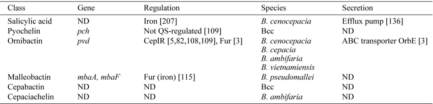
Table 3. Siderophores.
Class Gene Regulation Species Secretion
Salicylic acid ND Iron [207] B. cenocepacia Efflux pump [136]
Pyochelin pch Not QS-regulated [109] Bcc ND
Ornibactin pvd CepIR [5,82,108,109], Fur [3] B. cenocepacia B. cepacia B. ambifaria B. vietnamiensis
ABC transporter OrbE [3] Malleobactin mbaA, mbaF Fur (iron) [115] B. pseudomallei ND
Cepabactin ND ND Bcc ND
Cepaciachelin ND ND B. ambifaria ND
[211]. Members of the Burkholderia genus produce many kinds of siderophores which, depending on the chemical structure of their chelating group, are mostly classified into hydroxamates (based on hydroxamic acid) and catecholates (containing a catechol ring) [155, 186] (Table 3).
Salicylic Acid
Salicylic acid, or 2-hydroxybenzoic acid, which serves as a precursor for many siderophores such as pyochelin, was initially identified in many B. cepacia isolates and was then called azurechelin [170]. It has iron-binding properties and appears to promote iron uptake as well as growth of many bacteria in iron-limiting conditions, including B. cenocepacia [171]. Its production is regulated by the availability of iron [170]. In B. cenocepacia, salicylate was found to induce an antibiotic efflux pump, conferring resistance to chloramphenicol, trimethoprim, and ciprofloxacin [136]. Since very low iron is present in CF lungs, it was proposed that salicylate might induce efflux-mediated resistance, even in the absence of antibiotic selective pressure, and then contribute to the virulence of this bacterium [136]. The role of salicylate as a siderophore is often questioned. Bacteria that produce salicylate but no other siderophore are negative in the standard CAS plate assay, even when it is overproduced or when a shuttle between the dye complex and the siderophore is added [171]. Moreover, mutants producing salicylate as the sole potential siderophore are growth limited under iron-restrictive conditions [3, 49].
Pyochelin
The siderophore pyochelin was first described in studies on P. aeruginosa grown under iron-limiting conditions [46]. It is derived from the condensation of two molecules of cysteine with salicylic acid, which requires the presence of non-ribosomal peptide synthetases (NRPS), PchE and PchF. PchE is necessary for the synthesis of the first intermediate, and PchF is responsible for synthesis of the core structure. These two enzymes are coded by the
pchEFGHI operon, which is regulated by iron-regulated repressor Fur and by PchR, an AraC family transcriptional regulator [77, 116]. Details on the biosynthesis and regulation of pyochelin have been recently reviewed [186].
Pyochelin is produced by some members of the Bcc, including B. cepacia strains (e.g., ATCC 25416 and ATCC 17759) [129]. However, in a study screening Bcc clinical isolates, about 50% produced little or no pyochelin [47, 169]. Strain K56-2, now confirmed to be B. cenocepacia, produces negligible amounts of pyochelin, whereas the clinical isolate Pc715j produces significant amounts [209]. The pch genes coding for the biosynthesis machinery and transport of pyochelin are found in both B. pseudomallei
and B. thailandensis, but not in B. mallei [138]. B. cenocepacia Tn5 mutants were screened for the inability to
produce pyochelin. Two mutants were identified. In both mutants, the transposon had integrated into the cysW gene coding for a component of the sulfate/thiosulfate transporter. Both mutants were also defective in sulfate transport, and the ability to produce pyochelin was restored in the presence of cysteine, a bioprecursor of pyochelin [58].
Ornibactin
Ornibactin is a linear hydroxamate-hydroxycarboxylate siderophore, related in its peptide structure to pyoverdine, which is produced by P. aeruginosa and P. fluorescens
[129, 176].It is composed of a conserved tetrapeptide, L -Orn1(Nδ-OH,Nδ-acyl)-D-threo-Asp(β-OH)-L-Ser-L-Orn4(Nδ
-OH,Nδ-formyl)-1,4-diaminobutane and it provides three
bidentate metal chelation groups. These three groups (two hydroxamates and one α-hydroxycarboxylate) are obtained
by modification of the side chains of the N- and C-terminal ornithines, and the D-aspartate found in the peptide. Depending on the chain length of the acid binding to Orn1,
three species of ornibactin are generated: ornibactin-C4,
ornibactin-C6, and ornibactin-C8 [176]. During a study on
clinical Bcc strains, ornibactin was shown to be produced by 87% of tested isolates [47]. It has been isolated from culture supernatants of B. vietnamiensis and was also identified in B. ambifaria PHP7, in two clinical strains of
B. cepacia (ATCC 17759 and ATCC 25416), and in many other Bcc strains including B. cenocepacia [12, 129]. Ornibactin appears to be the most prevalent siderophore among the Burkholderia. The genes required for ornibactin biosynthesis and transport have been identified in B. cenocepacia K56-2 and shown to be negatively regulated by the QS system CepIR, in contrast to pyochelin and salicylic acid [109, 171]. In a proposed synthesis pathway model, precursors are modified by the products of pvdA,
pvdF, orbG, orbK, and orbL genes and assembled into ornibactin by NRPSs OrbI and OrbJ. PvdA encodes for L -ornithine N5-oxygenase, an enzyme responsible for catalyzing
the hydroxylation of the δ-amino group nitrogen of
ornithine, the first step in the formation of a hydroxamate siderophore [208]. B. cenocepacia K56-2 pvdA- mutants
display no siderophore activity, but can be restored by addition of the precursor L-N5-OH-Orn. The pvdA- mutants are less virulent than the parent strain in chronic and acute models of respiratory infection in rats [171]. Details about the biosynthesis and transport of ornibactin across the cytoplasmic membrane for Bcc members were recently reviewed [186].
B. cenocepacia Pc715j produces both ornibactin and pyochelin [47, 109, 209]. When orbA, a gene encoding for the outer membrane receptor of ferric-ornibactin, is inactivated, the mutant strain shows less virulence than the parent strain in a rat agar bead lung infection model [209]. The orbA- mutant is cleared from the lungs, showing that
pyochelin uptake cannot compensate for the ornibactin system. However, a ptaA- mutant from the same B. cenocepacia
strain, which is deficient in ferric-pyochelin uptake, persists in the lung in the same animal model, showing that ornibactin uptake is able to compensate a deficiency in the pyochelin system. Both mutants can grow in iron-starvation conditions in vitro and produce smaller amounts of the corresponding siderophore [209].
Malleobactin
Malleobactin, a hydroxamate siderophore purified from B. pseudomallei K96243presents similarities with ornibactins. MbaA, a putative ornithine-N5-oxygenase, and MbaF, a
putative NRPS, are involved in its biosynthesis, whereas
fmtA is coding for the malleobactin receptor and is involved in the uptake [7]. These genes are part of an operon with mbaJ and mbaI, which are under the control of the sigma factor MbaS [7]. Malleobactin is able to acquire iron from both transferrin and lactoferrin. Because
B. pseudomallei initiates infection at the mucosal surface, its ability to take iron from lactoferrin could play an important role in infections, as it is found in most secretions bathing mucosal surfaces [219]. The FmtA receptor can also recognize ornibactin (not produced by B. pseudomallei), and B. cenocepacia’s purified ornibactins can cross-feed mbaA- mutants [7].
Cepabactin
Cepabactin, or 1-hydroxy-5-methoxy-6-methyl-2(1H)-pyridinone, is a cyclic hydroxamate siderophore that was first identified as a metal-binding antibiotic secreted in the culture medium of Pseudomonas alcaligenes NCIB11492 [128]. Some Bcc clinical strains (ATCC 17759 and ATCC 25416 gen. I) were shown to produce cepabactin, although it never was the only siderophore produced [47, 128]. In fact, cepabactin is not widely produced among Burkholderia
strains, clinical or environmental, which does not preclude its utilization.
Cepaciachelin
Another siderophore, cepaciachelin, was isolated from a culture supernatant of B. cepacia PHP7 (now B. ambifaria) grown under iron-limiting conditions [12]. Cepaciachelin seems to represent the third subunit of protochelin, a siderophore identified in Azotobacter vinelandii [12, 44]. This type of siderophore is among the most effective because it contains three complexing sites for Fe3+. The
other two subunits of protochelin are azotochelin and aminochelin and have not yet been reported in Burkholderia
species.
R
HAMNOLIPIDSRhamnolipids are extracellular glycolipidic surface-active molecules produced by many bacteria. Rhamnolipids have
several potential biotechnological applications owing to their tensioactive properties [168].
A rhamnolipid was detected in the supernatant of
B. pseudomallei cultures. This rhamnolipid is composed of two molecules of rhamnoses and two molecules of β
-hydroxytetradecanoic acid to give 2-O-α-L
-rhamnopyranosyl-α-L-rhamnopyranosyl-β-hydroxytetradecanoyl-β -hydroxytetradecanoate (Rha-Rha-C14-C14). At high
concentrations, this rhamnolipid is toxic to nonphagocytic and phagocytic cell lines and displays hemolytic activity on erythrocytes from different species [75]. The hemolytic and cytotoxic activities of the B. pseudomallei rhamnolipid are probably due to its detergent-like properties. At low concentrations, this rhamnolipid changes the cell morphology with modifications of the cytoskeleton organization [76].
B. thailandensis synthesizes a rhamnolipid that is structurally analogous to the B. pseudomallei rhamnolipid [194]. Disruption of the B. thailandensis QS system results in hyper-beta-hemolysis of erythrocytes, which was suggested to result from increased rhamnolipid production [194]. Genes involved in rhamnolipid synthesis have not yet been identified.
T
OXINSPhytopathogenic Burkholderia
Phytopathogenic bacteria produce phytotoxins that are toxic to plant cells and influence symptoms development.
Toxoflavin
B. glumae causes rot of rice grains and seedlings (panicle blight) and is a limiting factor of rice yield throughout rice-growing countries (United States, Japan, the Philippines, and Korea) [45, 182]. Moreover, B. glumae can also cause wilting symptoms in solanaceous crops [88]. B. glumae
produces bright yellow pigments, identified as toxoflavin {1.6-dimethylpyrimido[5,4-e]-1,2,4-triazine-5,7(1H,6H )-dione} and fervenulin (a tautomeric isomer of toxoflavin), which are essential for the pathogenicity of rice seedling rot and grain rot [93, 182]. A toxoflavin-deficient mutant fails to cause disease symptoms in plants. Toxoflavin also has antibacterial, antifungal, and herbicidal activities [135]. Recently, toxoflavin was identified as a potential novel anticancer agent by its action against Polo-like kinase (involved in signal transduction) [70]. Toxoflavin acts in various microorganisms and cell extracts as an electron carrier between NADH and oxygen, and it can produce hydrogen peroxide or superoxide anion [102]. Toxoflavin is also produced by several strains of B. gladioli [196].
In B. glumae, the biosynthesis and transport of toxoflavin involve, respectively, the toxABCDE and the toxFGHI
operons, which are regulated by the LysR family regulator ToxR, with toxoflavin acting as a co-inducer [93]. The TofI-TofR(C8-HSL) QS system regulates toxoflavin production
B. glumae, a tofI mutant fails to produce phytotoxins and causes much less severe panicle blight than that produced by the wild type. Kim et al. [93] suggested that since toxoflavin production consumes large amounts of energy,
B. glumae cells must ensure that they reach a critical cell density before they start to produce it [93]. This is the only study demonstrating that QS is involved in the pathogenicity of phytopathogenic bacteria by regulating a phytotoxin production. Toxoflavin production is also dependent on growth temperature, which is maximal at 37oC [124].
Tropolone
B. plantarii (also known as Pseudomonas plantarii), a rice phytopathogen, is often co-isolated with B. glumae, suggesting that they may share similar transmission paths and life cycle [36, 197]. B. plantarii and B. glumae induce similar symptoms on rice. The virulence of B. plantarii was associated with production of the phytotoxin tropolone, which is also produced by some other Pseudomonas and
Burkholderia spp. including B. glumae. Tropolone is a non-benzenoid aromatic compound that has properties characteristic of phenol and acids. Tropolone causes root growth inhibition and wilting of seedlings, symptoms that are typically caused by the pathogen itself [10]. Its activity is inhibited by the presence of iron [10]. This compound is toxic to rice seedlings but also exhibits antimicrobial and antifungal activities, especially against
Pythium aphanidermatum [133, 189]. Tropolone displays strong insecticidal activity on Tyrophagus putrescentiae
(mould mite) and Dermatophagoides farinae. This activity is even higher than that of N, N-diethyl-m-toluamide [133]. The PlaI-PlaR QS system plays an important role in the ability of B. plantarii to cause rice seedling blight disease: a plaI mutant is less virulent than the wild type. However, it is not known whether QS regulates tropolone synthesis [173].
Rhizobitoxine
Rhizobitoxine, an enol-ether amino acid [2-amino-4-(2-amino-3-hydroxypropoxy)-trans-but-3-enoic acid] is synthesized by the legume symbiont Bradyrhizobium elkanii and the plant pathogen B. andropogonis [131]. These strains also produce dihydrorhizobitoxine [131]. B. andropogonis causes chlorotic symptoms in corn and sorghum, presumably as a result of rhizobitoxine production
in planta [131]. Rhizobitoxine also plays a positive role in establishing symbiosis between B. elkanii and host legumes [53]. Rhizobitoxine inhibits cystathionine-β-lyase
in methionine biosynthesis and 1-aminocyclopropane-1-carboxylate (ACC) synthase in the ethylene biosynthesis pathway [225]. ACC synthase is the rate-limiting enzyme in the ethylene biosynthesis pathway in plants [118]. Inhibition of ethylene biosynthesis by rhizobitoxine enhances the nodulation process and nodulation competitivenes [53]. B. andropogonis probably produces rhizobitoxine to
inhibit ethylene biosynthesis and reduce defence reactions by the host plants [141]. However, in B. elkanii, it was shown recently that rhizobitoxine-induced foliar chlorosis is the result of methionine deficiency due to inhibition of cystathione-β-lyase [141].
The genes involved in rhizobitoxine biosynthesis have been identified in B. elkanii as rtxA and rtxC [221]. However, these genes have not been isolated from B. andropogonis.
Rhizoxin
Rhizoxin, a macrocyclic polyketide, is the causative agent of the rice seedling blight. This phytotoxin exerts its effect by binding to rice β-tubulin, which results in inhibition
of mitosis and cell cycle arrest [96]. The blockage of microtubule formation has been observed in many other eukaryotic cells, including human and murine tumor cells. Additionally, rhizoxin can depolymerize assembled microtubules [183]. Rhizoxin demonstrates broad antitumor activity in vitro [187, 190]. Rhizoxin has undergone extensive clinical trials as a potential antitumor drug candidate [101]. For many years, the fungus Rhizopus microsporus was assumed to synthesize rhizoxin. However, recent evidence indicate that it is instead produced by a symbiotic bacteria of the genus Burkholderia residing within the fungal mycelium [146]. Curing the fungus of the bacteria with an antibiotic treatment, which resulted in a nonproducing phenotype, unequivocally established the role of the rhizoxin. In pure culture, the endosymbiont produces rhizoxin, and the re-inoculation of the cured fungal strain with the symbiotic Burkholderia re-establishes a rhizoxin-produced fungal bacterial symbiosis. The name
Burkholderia rhizoxina was proposed for this symbiotic bacterium [148]. Novel rhizoxin derivatives isolated from a scaled-up fermentation of the cultured B. rhizoxina strain are 1,000 times more active than rhizoxin in antimitotic bioassays [162]. A gene cluster encoding rhizoxin biosynthesis has been isolated in the genome of B. rhizoxina [148]. Recently, Partida-Martinez et al. [147] found that rhizonin is also produced by an endosymbiont Burkholderia (see below).
O
THERT
OXINSBongkrek Acid
Tempe bongkrek is an Indonesian food made by fermentation of coconut. Consumption of tempe bongkrek is associated with a foodborne human intoxication and significant numbers of mortalities. The main symptom is a strong hyperglycemia followed by hypoglycemia [78]. The causative organism B. gladioli pathovar cocovenenans (also referred to as Pseudomonas cocovenenans and B. cocovenenans) produces two toxins, toxoflavin (discussed above) and bongkrekic acid (also commonly referred to as bongkrek acid) [36]. Bongkrekic acid is an inhibitor of adenine
nucleotide translocase, which is a component of the mitochondrial permeability transition pore complex [78]. It is also an inhibitor of apoptosis by preventing a number of phenomena: generation of reactive oxygen species, chromatin condensation, and cytoplasmic vacuolization [78, 122]. Pseudomonas farinofermentans strains isolated from acase of food poisoning caused by the consumption of fermented corn flour in China are now know to be B. gladioli pathovar cocovenenans [228].
Rhizonin
Rhizonins A and B were known as mycotoxins from
Rhizopus microsporus, a fungus that is traditionally used in many food fermentations; for example, for soybean
tempeh production in Indonesia [87]. Rhizonins A and B have a strong hepatotoxic activity [217]. In animal tests, rhizonins cause hepatic lesions and induce acute and chronic failure of the liver [217]. However, this toxin, like rhizoxin, is not produced by the fungus but by bacteria that reside within the fungal mycelium. Phylogenetic analyses revealed that this symbiont belongs to the genus Burkholderia
[147]. Pure rhizonin A can be isolated from a scaled-up culture of this Burkholderia strain [147].
A
NTIFUNGALS ANDO
THERA
NTIMICROBIALSOrganisms such as Burkholderia produce a wide range of antifungals and other compounds that are able to suppress many soilborne plant pathogens (e.g., R. solani, Pythium
spp., Fusarium spp.) and in doing so improve plant health. For example, B. ambifaria LMG 19182 (B. ambifaria
AMMD) is very effective in controlling phytopathogenic
Pythium species and Aphanomyceseuteiches [38]. Various strains of Burkholderia have been reported to produce a large variety of antifungals such as altericidins, pyrrolnitrin, and xylocandins (also called cepacidines) [16, 54, 94].
Pyrrolnitrin
Pyrrolnitrin [3-chloro-4-(2'-nitro-3'-chlorophenyl)-pyrrole] is a secondary metabolite derived from tryptophan, first
isolated from B. pyrrocinia (originally Pseudomonas pyrrocinia) [8]. It is produced by several strains of
Pseudomonas and Burkholderia [21, 56] and also
Enterobacter agglomerans, Serratia plymuthica, and
Myxococcus isolates [31, 65, 143].
This active metabolite has been used as a clinical antifungal agent to treat humans infected by opportunistic fungi. Phenylpyrrole derivatives (fenpiclonil and fludioxonil) have been developed by Norvatis as agricultural fungicides [185]. Pyrrolnitrin prevents fungal growth by inhibiting the respiratory electron transport system [54].
Pyrrolnitrin produced by B. cepacia NB-1 exhibits a broad spectrum of activity against phytopathogenic fungi and Gram-positive bacteria, with a particular efficiency against Streptomyces, whereas Gram-negative bacteria, except Proteus vulgaris, are resistant [54]. Pyrrolnitrin production by B. cepacia NB-1 is influenced by nutritional and environmental factors, with glycerol strongly enhancing the antifungal production [54]. Pyrrolnitrin produced by B. cepacia B37w exhibits antifungal activity against the potato dry rot fungus Fusarium sambucinum [21].
The prnABCD gene cluster encodes the four biochemical steps to produce pyrrolnitrin from tryptophan by P. fluorescens [95]. This cluster was identified in B. cepacia
LT4-12W and in B. pyrrocinia [73]. In P. fluorescens, B. pyrrocinia, and B. cepacia LT4-12W, genes are arranged identically and in a linear relationship to the order of the biochemical reactions for pyrrolnitrin synthesis [73]. De Souza and Raaijmakers [48] suggested that the Burkholderia
pyrrolnitrin synthase gene was acquired from Pseudomonas
by horizontal transfer.
Regulation of pyrrolnitrin biosynthesis is not well documented in Burkholderia strains. It was reported that QS is required for its production in a rhizospheric biocontrol strain of Serratia plymuthica [112]. It is therefore possible that QS is required also for production in Burkholderia strains.
Xylocandin Complex
Xylocandin (also called cepacidines A and B) is a complex of peptides with potent anticandidal and antidermatophytic
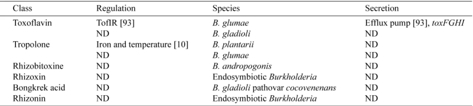
Table 4. Toxins.
Class Regulation Species Secretion
Toxoflavin TofIR [93] B. glumae Efflux pump [93], toxFGHI
ND B. gladioli ND
Tropolone Iron and temperature [10] B. plantarii ND
ND B. glumae ND
Rhizobitoxine ND B. andropogonis ND
Rhizoxin ND Endosymbiotic Burkholderia ND
Bongkrek acid ND B. gladioli pathovar cocovenenans ND
Rhizonin ND Endosymbiotic Burkholderia ND
activities that is produced by B. pyrrocinia ATCC 39277 (originally P. cepacia), a strain isolated from cornfield soil in New Jersey (U.S.A.) [16]. Cepacidine A has been found in culture broth of B. cepacia AF2001 (originally P. cepacia) and exhibits high in vitro antifungal activity against pathogenic fungi, that showing no activity against bacteria [106]. In semi-greenhouse biocontrol assays, this strain displayed excellent biological activity against
Pythium ultimum on cotton and cucumbers [105]. Moreover, cepacidine A was found to have potent immunosuppressive activity, significantly suppressing the activation of B lymphocytes [104].
B. cepacia BC11 inhibits the growth of R. solani
and Sclerotium rolfsii in soils and enhances the yield of peanuts [91]. This strain produces a lipopeptide, called AFC-BC11, with characteristics of members of the xylocandin family. However, unlike xylocandins, AFC-BC11 is not active against Candida spp. Nonetheless, AFC-BC11 is very active against various plant pathogenic fungi [91]. A region of the genome of strain BC11 that is required for production of this antifungal metabolite was characterized. This region encodes proteins involved in the production of a nonribosomally synthesized lipopeptide [91].
Quinoline Derivatives
The phytophthora blight of red pepper is a plant disease caused by Phytophtora capsici. B. cepacia PCII inhibits the mycelial growth and zoosporangial germination of P. capsici. Inoculation of this strain promotes red pepper plant growth [132]. B. cepacia PCII produces several 4-quinolinone metabolites (or pseudanes): [2-(2-heptenyl)-3-methyl-4-quinolinone] (HMQ), and 3-methyl-2-(2-nonenyl)-4-quinolinone (NMQ). HMQ exhibits in vitro antifungal activity against P. capsici, F. oxysporum, and R. solani
[132]. Treatment of red pepper seeds with HMQ resulted in an increase in weight and height of plants after 30 days [132]. HMQ and NMQ are also synthetized by B. cepacia
RB245, a strain isolated from a lettuce root and showing activity against several fungal pathogens including Pyricularia oryzae, and R. solani, but relatively limited antibacterial activity [81].
Glidobactins
Burkholderia spp. K481-B101 (originally assigned as
Polyangium brachysporum) produces glidobactins [161], acylated tripeptides that contain a 12-membered ring consisting in most variants of the two nonproteinogenic amino acids erythro-4-hydroxy-L-lysine and 4(S)-amino-2(E)-pentenoic acid. The ring structure is linked to an L -threonine residue, which in turn is acylated by different variant-specific unsaturated fatty acids [161]. These compounds were also isolated from B. cepacia and designated cepafungin [167]. Glidobactins exhibit broad inhibitory
action against fungi and yeasts, as well as antitumor activity [140, 167].
A gene cluster (glbA-glbH) involved in glidobactin biosynthesis has been identified in Burkholderia strain K481-B101. This gene cluster was also found in the ten completely sequenced B. pseudomallei strains. Interestingly, in the nine sequenced B. mallei strains, this cluster is inactivated by a transposon [161].
CF661
B. cepacia CF66 produces the antifungal compound CF661,which inhibits the growth of some soilborne fungi such as R. solani, Aspergillus flavus, and F. oxysporum
[150]. Strain CF66 exhibits strong antimicrobial activity against yeasts such as C. albicans, but no activity against bacteria. CF661 completely inhibits the hyphal growth of R. solani. The exact structure of this compound is not known, but based on nuclear magnetic resonance analysis, and GC-MS spectral and infrared spectral data, CF661 is confirmed to have amide bonds, α-metyl fatty
acid, bromine, and some structural units such as CH2CH2O
[150].
Antifungal Compounds from Burkholderia Strain MP-1
Burkholderia strain MP-1, isolated from the rhizosphere, exhibits antifungal activities against various filamentous plant pathogenic fungi(F. oyxsporum, R. solani). This strain produces at least four antifungal substances: phenylacetic acid, hydrocinnamic acid, 4-hydroxyphenylacetic acid, and 4-hydroxyphenylacetate methyl ester [121]. These four substances exhibit antifungal activity against several pathogenic fungi. Phenylacetic acid, a deamination product of phenylalanine, is known as a plant growth regulator (auxin activity) and it displays growth-inhibitory activity towards bacteria and fungi (R. solani, Pythium ultimum) [85, 134]. It may be involved in defence mechanisms, protecting the producing strain from competing cells.
O
THERA
NTIFUNGALS2-Hydroxymethyl-chroman-4-one
Burkholderia sp. MSSP was isolated from roots of
Mimosa pudica and secretes an antifungal compound against R. solani, P. ultimum, and Botrytis cinerea [91]. An antifungal compound was identified as 2-hydroxymethyl-chroman-4-one and exhibits activities against several plant pathogenic fungi [91].
Altericidins
Altericidins, a complex of closely related oligopeptides, were isolated from the culture broth of B. cepacia KB-1. The altericidin complex inhibits the germination of Alternaria kikuchiana conidia (a black spot fungus of pear). It was
proposed that altericidins might act on the cytoplasmic membrane [94].
Cepacins A and B
Cepacins A and B have been isolated from B. cepacia SC 11 783 (originally P. cepacia). These antibiotics exhibit only antibacterial activity against staphylococci and Gram-negative microorganisms [145].
Cepaciamides A and B
B. cepacia D-202 is a biological control agent against
Botrytis cinerea and Penicillium expansum, which causes beet roots rot in Japan. This strain produces the 3-amino-2-piperidinone-containing lipids cepaciamides A and B, which exhibit an activity against B. cinerea [223].
Hydrogen Cyanide
Several bacterial species are known to produce and excrete hydrogen cyanide (HCN), a potent inhibitor of cytochrome C oxidase [17]. For example, HCN production by strain
P. fluorescens CHAO suppresses black root rot of tobacco caused by Thielaviopsis basicola [17]. The production of HCN has been rarely described in the genus
Burkholderia. The endophytic plant growth-promoting
Burkholderia sp. strain MSSP, isolated from root nodules of Mimosa pudica, produces HCN, but its role is unknown [144].
Phenazines
Phenazines are naturally occurring pigments produced by bacteria and are known for their antibiotic properties, and antitumor and antiparasitic activities [103].For example, phenazine-1-carboxylic acid produced by P. fluorescens is important for the control of take-all disease of wheat, which is caused by Gaeumannomyces graminis var. tritici
[32]. Phenazine producers have been identified as organisms belonging to a range of species like Pseudomonads, members of the Streptomyces genus, B. cepacia, and B. phenazinium (for a recent review see [103]).
B. phenazinium (previously known as Pseudomonas phenazinium) produces ten phenazine pigments, predominantly iodinin [13]. B. cepacia strain 5.5B, isolated from soil, produces a purple pigment identified as 4,9-dihydroxyphenazine-1,6-dicarboxylic acid dimethyl ester, a phenazine [25]. This compound inhibits in vitro the phytopathogenic fungi R. solani [25].
Volatile Compounds
Volatile compounds (molecular weight less than 300, low polarity, and a high vapor pressure) can act as antibiotics and affect fungal mycelial growth [86, 214]. B. cepacia
RJ3 and ATCC 52796 (originally P. cepacia) inhibit several fungal plant pathogens. These strains inhibit the fungi by producing unidentified inhibitory volatile compound(s).
The volatile compound(s) of B. cepacia moderately inhibits the growth of R. solani [90].
Unknown Antifungals
The biocontrol strain B. ambifaria BC-F exhibits broad-spectrum antifungal activity against important soilborne pathogens and suppresses diseases caused by fungal pathogens on a number of important crop plants [110]. The nature of this antifungal compound is not known. Interestingly, QS deficient mutants of B. ambifaria BC-F have decreased antifungal activity [229].
Burkholderia strain 2.2 N isolated from soil is capable of inhibiting the growth of plant-pathogenic fungi, yeasts, and protozoa. An extracellular compound seems to be responsible for this activity [24].
P
HYTOHORMONESBeneficial bacteria that stimulate growth of cereals and grasses are usually referred to as plant growth promoting Rhizobacteria (PGPR), a group that includes different bacterial species belonging to genera such as Acetobacter,
Azospirillum, Bacillus, Pseudomonas, Herbaspirillum, and
Burkholderia [52]. PGPR exert their beneficial effects on plant growth directly or indirectly through various mechanisms. Indirect effects rely on preventing deleterious functions of pathogenic microorganisms, generally by the production of antibiotics or antifungal compounds or by competing for nutrients like iron [52]. Several strains of
Burkholderia can antagonize and repress the growth of many soilborne plant pathogens (see above). Direct mechanisms include the synthesis of phytohormones and vitamins, inhibition of plant ethylene synthesis, improved nutrient uptake, solubilization of inorganic phosphate, or mineralization of organic phosphate.
Several Burkholderia species exert beneficial effects on their plant hosts. For example, B. vietnamiensis is recognized for its abilities to promote rice plant growth: the inoculation of strain TVV75 resulted in a final 13% to 22% increase in grain yield [188]. Similarly, B. phytofirmans
strain PsJN inoculation stimulates grapevine growth and enhances resistance to cold stress [6]. B. ambifaria MCI-7 enhances the growth of Zea mays [35].
Bacterial production of phytohormones can explain the changes in root morphology following Burkholderia
inoculation. The plant hormones auxins and cytokinins are involved in several stages of plant growth and development, such as cell elongation, cell division, tissue differentiation, and apical dominance [226]. However, there are only a few reports on the phytohormone biosynthetic capacities of Burkholderia. The most important auxin, indole-3 acetic acid, is produced by several strains of B. cepacia isolated from the rhizosphere, by an endophytic
Burkholderia isolated from root nodules of Mimosa piduca, and by B. vietnamiensis MGK3 isolated from rice root [14, 144].
E
FFECTORP
ROTEINSBurkholderia Secretion Apparatus: Effector Proteins
B. pseudomallei is a facultative intracellular pathogen; it can invade nonphagocytic host cells and survive and replicate within phagocytes. B. pseudomallei contains at least three loci encoding putative type III secretion systems (TTSS) [152, 178]. TTSS, central to the virulence of many Gram-negative pathogens, resembles molecular syringes for the injection of multiple bacterial effector proteins directly into the host cell cytoplasm [43]. Effector proteins subvert host cell processes to the benefit of the invader. For example, the type III protein secretion apparatus BSA (Burkholderia secretion apparatus) is required for full virulence of B. pseudomallei in mice [177]. The bsa locus is also present in B. mallei and B. cenocepacia genomes [177]. Several effector proteins encoded within the bsa
locus have been identified in the B. pseudomallei genome (BopA, BopB, and BopE) [177] (Table 5).
BopE facilitates the invasion of nonphagocytic epithelial cells. BopE shares sequence homology with the translocated effector proteins SopE and SopE2 of Salmonella, proteins that play an important role in Salmonella invasion of nonphagocytic intestinal epithelial cells. In eukaroytic cells (HeLa cells), BopE induces rearrangements in the subcortical actin cytoskeleton, likely by acting as a guanine nucleotide exchange factor for RhoGTPases, that regulate the actin network [195]. The bopE gene is also present in the B. mallei and B. thailendensis genomes [177].
BopB contains an amino acid motif (CX5R) that is conserved in the catalytic domains of numerous phosphatases, like the type III secreted protein of Salmonella SobB. The latter influences inositol phosphate signalling pathways in eukaryotic cells as well as bacterial invasion [179]. BopA is a homolog of the Shigella type III secreted protein IscB, which mediates cell-to-cell spread of Shigella [179]. The exact role of BopA is not known, but B. pseudomallei is also capable of cell-to-cell spread. In mice, B. pseudomallei bopA- and bopB- mutants are less virulent than wild type,
causing a delay in median time to death [179].
Other Proteins
BimA (Burkholderia intracellular motility A), an autosecreted protein, is localized at the pole of the bacteria and is required for actin-based motility in a macrophage-like cell line [180]. Mutation of bimA abolishes the actin-based motility of B. pseudomallei in a cell line [180].
B. cenocepacia strain K56-2 is capable of causing a plant tissue watersoaking phenotype (ptw) that results from loss of cell membrane integrity and the accumulation of fluids in the intracellular spaces of plant tissue [57]. In strain K56-2, a type IV secretion system is responsible for the secretion of a plant cytotoxic protein, which causes plasmolysis of plant protoplasts [57].
C
ONCLUSIONThe large variety of extracellular products synthesized by Burkholderia species correlates with its important ecological diversity and might explain its versatility. Although a lot of information is published on Burkholderia secondary metabolites, effectors, and other exoproducts, sequencing of the genomes will improve knowledge on that matter. Studies on regulation of exoproduction in Burkholderia
will introduce interesting perspectives about the adaptation, survival, and pathogenesis of these bacteria. This will also favor the optimization of production of various enzymes for industrial purposes.
Acknowledgrment
Ludovic Vial and Marie-Christine Groleau contributed equally to this work.
R
EFERENCES1. Aamand, J. L., A. H. Hobson, C. M. Buckley, S. T. Jorgensen, B. Diderichsen, and D. J. McConnell. 1994. Chaperone-mediated activation in vivo of a Pseudomonas cepacia lipase. Mol. Gen. Genet.245: 556-564.
2. Achouak, W., R. Christen, M. Barakat, M. H. Martel, and T. Heulin. 1999. Burkholderia caribensis sp. nov., an exopolysaccharide-producing bacterium isolated from vertisol microaggregates in Martinique. Int. J. Syst. Bacteriol. 49:
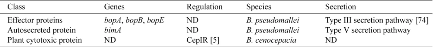
787-794. Table 5. Effector proteins.
Class Genes Regulation Species Secretion
Effector proteins bopA, bopB, bopE ND B. pseudomallei Type III secretion pathway [74] Autosecreted protein bimA ND B. pseudomallei Type V secretion pathway Plant cytotoxic protein ND CepIR [5] B. cenocepacia ND
3. Agnoli, K., C. A. Lowe, K. L. Farmer, S. I. Husnain, and M. S. Thomas. 2006. The ornibactin biosynthesis and transport genes of Burkholderia cenocepacia are regulated by an extracytoplasmic function sigma factor which is a part of the Fur regulon. J. Bacteriol.188: 3631-3644.
4. Aguilar, C., I. Bertani, and V. Venturi. 2003. Quorum-sensing system and stationary-phase sigma factor (rpoS) of the onion pathogen Burkholderia cepacia genomovar I type strain, ATCC 25416. Appl. Environ. Microbiol. 69: 1739
-1747.
5. Aguilar, C., A. Friscina, G. Devescovi, M. Kojic, and V. Venturi. 2003. Identification of quorum-sensing-regulated genes of Burkholderia cepacia. J. Bacteriol. 185: 6456
-6462.
6. Ait Barka, E., J. Nowak, and C. Clement. 2006. Enhancement of chilling resistance of inoculated grapevine plantlets with a plant growth-promoting rhizobacterium, Burkholderia phytofirmans strain PsJN. Appl. Environ. Microbiol.72: 7246-7252.
7. Alice, A. F., C. S. Lopez, C. A. Lowe, M. A. Ledesma, and J. H. Crosa. 2006. Genetic and transcriptional analysis of the siderophore malleobactin biosynthesis and transport genes in the human pathogen Burkholderia pseudomallei K96243. J. Bacteriol.188: 1551-1566.
8. Arima, K., H. Imanaka, M. Kousaka, A. Fukuda, and G. Tamura. 1964. Pyrrolnitrin, a new antibiotic substance, produced by Pseudomonas. Agric. Biol. Chem.28: 575-576.
9. Ashdown, L. R. and J. M. Koehler. 1990. Production of hemolysin and other extracellular enzymes by clinical isolates of Pseudomonas pseudomallei. J. Clin. Microbiol. 28:
2331-2334.
10. Azegami, K., K. Nishiyama, and H. Kato. 1988. Effect of iron limitation on “Pseudomonas plantarii” growth and tropolone and protein production. Appl. Environ. Microbiol.
54: 844-847.
11. Baldwin, A., P. A. Sokol, J. Parkhill, and E. Mahenthiralingam. 2004. The Burkholderia cepacia epidemic strain marker is part of a novel genomic island encoding both virulence and metabolism-associated genes in Burkholderia cenocepacia. Infect. Immun. 72: 1537-1547.
12. Barelmann, I., J. M. Meyer, K. Taraz, and H. Budzikiewicz. 1996. Cepaciachelin, a new catecholate siderophore from Burkholderia (Pseudomonas) cepacia. Z. Naturforsch. C J. Biosci.51: 627-630.
13. Bell, S. C. and J. M. Turner. 1973. Iodinin biosynthesis by a Pseudomonad. Biochem. Soc. Trans.1: 751-753.
14. Bevivino, A., S. Tabacchioni, L. Chiarini, M. V. Carusi, M. Del Gallo, and P. Visca. 1994. Phenotypic comparison between rhizosphere and clinical isolates of Burkholderia cepacia. Microbiology140(Pt 5): 1069-1077.
15. Bevivino, A., C. Dalmastri, S. Tabacchioni, L. Chiarini, M. L. Belli, S. Piana, A. Materazzo, P. Vandamme, and G. Manno. 2002. Burkholderia cepacia complex bacteria from clinical and environmental sources in Italy: Genomovar status and distribution of traits related to virulence and transmissibility. J. Clin. Microbiol.40: 846-851.
16. Bisacchi, G. S., D. R. Hockstein, W. H. Koster, W. L. Parker, M. L. Rathnum, and S. E. Unger. 1987. Xylocandin: A new
complex of antifungal peptides. II. Structural studies and chemical modifications. J. Antibiot. (Tokyo) 40: 1520
-1529.
17. Blumer, C. and D. Haas. 2000. Mechanism, regulation, and ecological role of bacterial cyanide biosynthesis. Arch. Microbiol.173: 170-177.
18. Boekema, B. K., A. Beselin, M. Breuer, B. Hauer, M. Koster, F. Rosenau, K. E. Jaeger, and J. Tommassen. 2007. Hexadecane and Tween 80 stimulate lipase production in Burkholderia glumae by different mechanisms. Appl. Environ. Microbiol. 73: 3838-3844.
19. Bramer, C., P. Vandamme, L. da Silva, J. Gomez, and A. Steinbuchel. 2001. Burkholderia sacchari sp. nov., a polyhydroxyalkanoate-accumulating bacterium isolated from soil of a sugar-cane plantation in Brazil. Int. J. Syst. Evol. Microbiol.51: 1709-1713.
20. Brett, P. J., D. DeShazer, and D. E. Woods. 1998. Burkholderia thailandensis sp. nov., a Burkholderia pseudomallei-like species. Int. J. Syst. Bacteriol.48: 317-320.
21. Burkhead, K. D., D. A. Schisler, and P. J. Slininger. 1994. Pyrrolnitrin production by biological control agent Pseudomonas cepacia B37w in culture and in colonized wounds of potatoes. Appl. Environ. Microbiol.60: 2031-2039.
22. Burkholder, W. H. 1950. Sour skin, a bacterial rot of onions bulbs. Phytopathology40: 115-117.
23. Caballero-Mellado, J., L. Martinez-Aguilar, G. Paredes-Valdez, and P. E. Santos. 2004. Burkholderia unamae sp. nov., an N2-fixing rhizospheric and endophytic species. Int.
J. Syst. Evol. Microbiol.54: 1165-1172.
24. Cain, C. C., A. T. Henry, R. H. Waldo 3rd, L. J. Casida Jr., and J. O. Falkinham 3rd. 2000. Identification and characteristics of a novel Burkholderia strain with broad-spectrum antimicrobial activity. Appl. Environ. Microbiol.
66: 4139-4141.
25. Cartwright, D. K., W. S. Chilton, and D. M. Benson. 1995. Pyrrolnitrin and phenazine production by Pseudomonas cepacia, strain 5.5B, a biocontrol agent of Rhizoctonia solani. Appl. Microbiol. Biotechnol.43: 211-216.
26. Carvalho, A. P., G. M. Ventura, C. B. Pereira, R. S. Leao, T. W. Folescu, L. Higa, L. M. Teixeira, M. C. Plotkowski, V. L. Merquior, R. M. Albano, and E. A. Marques. 2007. Burkholderia cenocepacia, B. multivorans, B. ambifaria, and B. vietnamiensis isolates from cystic fibrosis patients have different profiles of exoenzyme production. APMIS
115: 311-318.
27. Chain, P. S., V. J. Denef, K. T. Konstantinidis, L. M. Vergez, L. Agullo, V. L. Reyes, L. Hauser, M. Cordova, L. Gomez, M. Gonzalez, M. Land, V. Lao, F. Larimer, J. J. LiPuma, E. Mahenthiralingam, S. A. Malfatti, C. J. Marx, J. J. Parnell, A. Ramette, P. Richardson, M. Seeger, D. Smith, T. Spilker, W. J. Sul, T. V. Tsoi, L. E. Ulrich, I. B. Zhulin, and J. M. Tiedje. 2006. Burkholderia xenovorans LB400 harbors a multi-replicon, 9.73-Mbp genome shaped for versatility. Proc. Natl. Acad. Sci. USA103: 15280-15287.
28. Chen, W. M., E. K. James, T. Coenye, J. H. Chou, E. Barrios, S. M. de Faria, G. N. Elliott, S. Y. Sheu, J. I. Sprent, and P. Vandamme. 2006. Burkholderia mimosarum sp. nov., isolated from root nodules of Mimosa spp. from Taiwan