Ultrastructural Characterization of Turnip Mosaic Virus-Induced
Cellular Rearrangements Reveals Membrane-Bound Viral Particles
Accumulating in Vacuoles
Juan Wan,aKaustuv Basu,bJeannie Mui,bHojatollah Vali,b,cHuanquan Zheng,dJean-François Lalibertéa INRS-Institut Armand-Frappier, Laval, Québec, Canadaa
; Facility for Electron Microscopy Research, McGill University, Montréal, Québec, Canadab
; Department of Anatomy & Cell Biology, McGill University, Montréal, Québec, Canadac
; Department of Biology, McGill University, Montréal, Québec, Canadad ABSTRACT
Positive-strand RNA [(
ⴙ) RNA] viruses remodel cellular membranes to facilitate virus replication and assembly. In the case of
turnip mosaic virus (TuMV), the viral membrane protein 6K
2plays an essential role in endomembrane alterations. Although
6K
2-induced membrane dynamics have been widely studied by confocal microscopy, the ultrastructure of this remodeling has
not been extensively examined. In this study, we investigated the formation of TuMV-induced membrane changes by chemical
fixation and high-pressure freezing/freeze substitution (HPF/FS) for transmission electron microscopy at different times of
fection. We observed the formation of convoluted membranes connected to rough endoplasmic reticulum (rER) early in the
in-fection process, followed by the production of single-membrane vesicle-like (SMVL) structures at the midstage of inin-fection. Both
SMVL and double-membrane vesicle-like structures with electron-dense cores, as well as electron-dense bodies, were found late
in the infection process. Immunogold labeling results showed that the vesicle-like structures were 6K
2tagged and suggested that
only the SMVL structures were viral RNA replication sites. Electron tomography (ET) was used to regenerate a
three-dimen-sional model of these vesicle-like structures, which showed that they were, in fact, tubules. Late in infection, we observed
fila-mentous particle bundles associated with electron-dense bodies, which suggests that these are sites for viral particle assembly. In
addition, TuMV particles were observed to accumulate in the central vacuole as membrane-associated linear arrays. Our work
thus unravels the sequential appearance of distinct TuMV-induced membrane structures for viral RNA replication, viral particle
assembly, and accumulation.
IMPORTANCE
Positive-strand RNA viruses remodel cellular membranes for different stages of the infection process, such as protein translation
and processing, viral RNA synthesis, particle assembly, and virus transmission. The ultrastructure of turnip mosaic virus
(TuMV)-induced membrane remodeling was investigated over several days of infection. The first change that was observed
in-volved endoplasmic reticulum-connected convoluted membrane accumulation. This was followed by the formation of
single-membrane tubules, which were shown to be viral RNA replication sites. Later in the infection process, double-single-membrane
tubu-lar structures were observed and were associated with viral particle bundles. In addition, TuMV particles were observed to
accumulate in the central vacuole as membrane-associated linear arrays. This work thus unravels the sequential appearance of
distinct TuMV-induced membrane structures for viral RNA replication, viral particle assembly, and accumulation.
P
ositive-strand RNA [(
⫹) RNA] viruses remodel cellular
membranes for different stages of the infection process, such
as protein translation and processing, viral RNA (vRNA)
synthe-sis, particle assembly, and virus transmission. Ultrastructural
studies showed that distinct membrane structures could be
simul-taneously generated in one cell that was infected with animal (
⫹)
RNA viruses. For instance, severe acute respiratory syndrome (SARS)
coronavirus-induced membrane structures are a unique
reticulo-vesicular network of modified endoplasmic reticulum (ER) that
integrates convoluted membranes (CMs) for polyprotein
synthe-sis and processing, numerous interconnected double-membrane
vesicles (DMVs) for vRNA synthesis, and vesicle packets (VPs) for
virus assembly and budding (
1
). (
⫹) RNA virus-induced distinct
membrane structures can also be observed during the time course
of infection. Coxsackievirus B3 (CVB3) induces single-membrane
tubules (SMTs) at an early stage of infection for vRNA synthesis,
followed by the formation of DMVs and multilamellar structures
late in infection (
2
).
Plant (⫹) RNA viruses also manipulate host membranes for
their replication (reviewed in reference
3
). However, limited
transmission electron microscopy (TEM) data on the remodeling
of the cellular membrane during vRNA replication and
encapsi-dation have been generated. Several plant (⫹) RNA viruses have
been observed to produce spherule-like structures on different
Received 24 August 2015 Accepted 26 September 2015 Accepted manuscript posted online 30 September 2015
Citation Wan J, Basu K, Mui J, Vali H, Zheng H, Laliberté J-F. 2015. Ultrastructural characterization of turnip mosaic virus-induced cellular rearrangements reveals membrane-bound viral particles accumulating in vacuoles. J Virol
89:12441–12456.doi:10.1128/JVI.02138-15. Editor: A. Simon
Address correspondence to Jean-François Laliberté, [email protected].
Supplemental material for this article may be found athttp://dx.doi.org/10.1128 /JVI.02138-15.
Copyright © 2015, American Society for Microbiology. All Rights Reserved.
on January 18, 2016 by INRS-Institut Armand-Frappier
http://jvi.asm.org/
membrane-bound organelles, such as the ER (brome mosaic virus
[BMV] [
4
]), vacuole (cucumber mosaic virus [CMV] [
5
]),
perox-isome (tomato bushy stunt virus [TBSV] [
6
,
7
]), mitochondrion
(carnation Italian ringspot virus [CIRV] [
8
,
9
]), and chloroplast
(turnip yellow mosaic virus [TYMV; Tymovirus, Tymoviridae]
[
10
,
11
]). These spherule-like structures are similar to those
in-duced by some animal (
⫹) RNA viruses for vRNA synthesis, as
they are all formed by membrane invagination of the outer
organ-elle membrane and have a pore-like opening allowing the
ex-change of materials between the lumen of the spherule and the
cytoplasm. These spherule-like structures are static and reside
in-side the different membrane-bound organelles. Conversely, other
types of plant (
⫹) RNA virus-induced membrane structures are
ER-derived vesicles, which are highly motile and morphologically
dynamic, such as in cells infected with cowpea mosaic virus
(CPMV) (
12
), grapevine fanleaf virus (GFLV) (
13
), potato virus X
(PVX) (
14
,
15
), and turnip mosaic virus (TuMV) (
16
). However,
not much is known about the ultrastructure of these ER-derived
vesicles.
TuMV is a (⫹) RNA virus that belongs to the genus Potyvirus
in the family Potyviridae. Potyviruses are the largest genera of
plant viruses and are responsible for more than half of the viral
crop damage in the world (reviewed in reference
17
). Potyviral
particles are flexuous rods of approximately 680 to 900 nm in
length and 11 to 15 nm in diameter. The viral genome is a single
RNA molecule of approximately 10 kb. The 5= end is covalently
linked to a protein, VPg (for viral protein genome-linked), and the
3= end has a poly(A) tail. The vRNA codes for a polyprotein that is
processed by three viral proteinases into at least 12 proteins.
TuMV infection leads to the formation of numerous vesicles
that originate from the ER (
16
,
18
,
19
). The viral membrane
pro-tein 6K
2is responsible for vesicle formation (
18
). 6K
2-induced
vesicles contain vRNA and several replication-related viral and
host proteins (
18–21
) and are thus considered to be the site of
vRNA replication. These vesicles also participate in the cell-to-cell
movement of the vRNA. They are motile (
19
), traffic through the
secretory pathway, and use myosin motors (
22
). Ultimately, the
vesicles associate with plasmodesmata (PD) and are then released
into neighboring cells (
23
) for a new round of infection. They are
also involved in long-distance virus movement, as they are present
in phloem sieve elements and xylem vessels (
24
).
The studies cited above relied mainly on observations made by
light microscopy. TEM images of virus-induced vesicles have also
been reported for TuMV-infected (
16
) and potato virus Y
(PVY)-infected cells (
25
). These images showed the presence of
numer-ous single-membrane vesicles (SMVs) or what appeared to be
ves-icles, and on some occasions, the vesicles were found to be in
direct continuity with the rough endoplasmic reticulum (rER).
However, no study has looked at the biogenesis of these apparent
vesicles over the time course of infection and their relationship
with viral particle assembly.
In this study, we investigated the formation and maturation of
the TuMV-induced membrane remodeling that took place over
several days of infection. The first change that was observed
in-volved rER-connected convoluted membrane accumulation. This
was followed by the formation of SMV-like (SMVL) structures,
which were shown to be vRNA replication sites. Later in the
infec-tion process, double-membrane vesicle-like (DMVL) structures
having an electron-dense core, along with electron-dense bodies
associated with viral particle bundles, were observed. Electron
to-mography (ET) showed that the vesicle-like structures were in fact
tubules. In addition, TuMV particles were observed to accumulate
in the central vacuole as membrane-associated linear arrays.
MATERIALS AND METHODS
Plasmid DNA and plant inoculation. The infectious clone
pCambia-TuMV/6K2:GFP has been described previously (26). The plasmid pCambia-TuMV/6K2:GFP:HA was constructed as follows. The green fluorescent protein (GFP) fragment was amplified by PCR from pGreen/PABP:GFP (20) by using the forward primer 5=-CGGGATCCATGGTGAGCAAGG GCGA-3= and the reverse primer 5=-CGGAATTCTTACTTGTACAGCT CGTCCA-3= (the restriction sites are underlined) and digested with BamHI and EcoRI. The digested GFP fragment was then used to replace the mCherry fragment of pCambia/6K2:mCherry (23), and the resulting construct was named pCambia/6K2:GFP. The 6K2:GFP:hemagglutinin (HA)-coding region was amplified by PCR from pCambia/6K2:GFP by using the forward primer 5=-GCTCTAGAATGAACACCAGCGACATGA GC-3= and the reverse primer 5=-CGGAATTCTTAAGCGTAGTCTGGA
ACGTCGTATGGGTACTTGTACAGCTCGTCCATGCC-3= (the
restric-tion sites are underlined, the HA tag-coding sequence is highlighted in bold italics) and digested with XbaI and EcoRI. The digested 6K2:GFP:HA fragment was then used to replace the 6K2:GFP fragment of pCambia/6K2: GFP, and the resulting construct was named pCambia/6K2:GFP:HA. The 6K2:GFP:HA-coding region was again amplified by PCR from pCambia/ 6K2:GFP:HA by using the forward primer 5=-TCCCCGCGGGAAACAC CAGCGACATGAGC-3= and the reverse primer 5=-TCCCCGCGGCT
GCCTGGTGATAGACACAAGCAGCGTAGTCTGGAACGTC-3= (the
restriction sites are underlined, the proteinase cleavage site-coding se-quence is highlighted in bold italics). The PCR product was digested with SacII and introduced into the SacII site of p35Tunos/SacII (27) to obtain p35Tunos/6K2:GFP:HA. This plasmid was then cut with SmaI and KpnI and ligated in pCambiaTunos (23), which was cut with the same enzymes. Kanamycin-resistant Escherichia coli colonies were screened for plasmids containing the fragment encoding 6K2:GFP:HA, and the resulting con-struct was pCambiaTuMV/6K2:GFP:HA. All plasmid constructs were ver-ified by sequencing.
TuMV infectious clones and the mock-infected empty vector pCambia 0390 were electroporated into Agrobacterium tumefaciens strain AGL1 and selected on Luria broth (LB) plates containing ampicillin and kana-mycin. The pellet of an overnight culture was resuspended in water sup-plemented with 10 mM MgCl2and 150M acetosyringone and kept at room temperature (RT) for 2 to 4 h. The solution was then diluted to an optical density at 600 nm of 0.3. Agroinfiltration was performed with 4-week-old Nicotiana benthamiana plants, which were grown under a 16-h light and 8-h dark photoperiod, at temperatures of 22°C during the day and 20°C at night.
Histological preparation and confocal microscopy. The midrib area
of N. benthamiana leaves systemically infected with TuMV/6K2:GFP was cut and fixed in 4% (wt/vol) paraformaldehyde plus 0.5% (wt/vol) glutar-aldehyde at different days postinfection (dpi) for more than 24 h at 4°C. The air between the intercellular spaces was removed by infiltration with the fixative before the leaves were cut. The fixed samples were treated with 100 mM glycine in phosphate-buffered saline (PBS) for 1 h to reduce the background fluorescence, followed by sucrose gradient infiltration and cryosectioning as described previously (28). The sections were observed using a 20⫻ objective (numerical aperture ⫽ 0.8) on an LSM-780 confo-cal microscope (Carl Zeiss Microscopy). Zen 2011 software (Carl Zeiss Microscopy) was used for image acquisition. Excitation and emission wavelengths were 405 and 410 to 440 nm, respectively, for Fluorescent Brightener 28 and 488 and 490 to 560 nm, respectively, for GFP.
Chemical fixation. Small pieces (1.5 mm by 2 mm) of mock- and
TuMV-infected leaf midrib were cut and fixed in 2.5% (wt/vol) glutaral-dehyde in 0.1 M sodium cacodylate buffer, pH 7.4, for 24 h at 4°C. After rinsing 3 times for 10 min each time in washing buffer at RT, the samples were postfixed in 1% (wt/vol) osmium tetroxide with 1.5% (wt/vol)
po-Wan et al.
on January 18, 2016 by INRS-Institut Armand-Frappier
http://jvi.asm.org/
tassium ferrocyanide in sodium cacodylate buffer for 2 h at 4°C. The samples were then rinsed in washing buffer at RT (3 times for 10 min each time) and stained with 1% (wt/vol) tannic acid for 1 h at 4°C. After rinsing 3 times in water at RT, the samples were dehydrated in a graded acetone series (30, 50, 70, 80, 90, and 100%) for 20 min at each step at RT. A rinse with 100% acetone was repeated two more times. The samples were then gradually infiltrated with increasing concentrations (50, 66, 75, and 100%) of Epon 812 resin mixed with acetone for a minimum of 8 h for each step. A vacuum of 25 lb/in2was applied when the samples were in pure Epon 812 resin. The samples were finally embedded in pure, fresh Epon 812 resin and polymerized at 60°C for 48 h. After polymerization, 90- to 100-nm ultrathin sections were obtained and stained with 4% (wt/ vol) uranyl acetate for 8 min and Reynolds lead citrate for 5 min. The sections were examined in a Tecnai T12 transmission electron microscope (FEI) operating at 120 kV. Images were recorded using an AMT XR80C charge-coupled-device (CCD) camera system (Advanced Microscopy Techniques Corp.).
Immunogold labeling. Large pieces (1.5 mm by 5 mm) of mock- and
TuMV-infected leaf midrib were cut and fixed in 4% (wt/vol) formalde-hyde and 0.25% (wt/vol) glutaraldeformalde-hyde in 0.1 M Sorensen’s phosphate buffer, pH 7.4, for 4 h at 4°C. The samples were then rinsed 3 times for 10 min each time in washing buffer at RT. For anti-double-stranded RNA (anti-dsRNA) antibody-treated samples, the samples were postfixed in 0.1% (wt/vol) reduced osmium tetroxide for 15 min at 4°C (29). The samples were then rinsed in water at RT (3 times for 10 min each time) and dehydrated in a graded alcohol series (30, 50, 70, 80, 90, 95, and 100%) for 20 min at each step at 4°C on a rotator. Rinsing in 100% alcohol was repeated one more time. The samples were then gradually infiltrated with increasing concentrations of LR White resin (50, 75, and 100%) mixed with alcohol for a minimum of 8 h for each step at 4°C on a rotator. The samples were finally embedded in pure LR White resin and polymerized at 50°C for 48 h. Sections of 90 to 100 nm were incubated in 20 mM glycine in Dulbecco’s phosphate-buffered saline (DPBS; 137 mM NaCl, 2.7 mM KCl, 1.5 mM KH2PO4, 6.5 mM Na2HPO4, 1 mM CaCl2, 0.5 mM MgCl2, pH 7.4) for 10 min to inactivate residual aldehyde groups and then in a blocking solution consisting of 2% (wt/vol) bovine serum albumin (BSA), 2% (wt/vol) casein, and 0.5% (wt/vol) ovalbumin in DPBS (DPBS-BCO) for 5 min. Sections were then incubated with primary antibody diluted in DPBS-BCO for 1 h at RT. After 6 washings (5 min each time) in DPBS, the sections were incubated with secondary antibodies diluted in DPBS-BCO. After washing with DPBS and distilled water, grids were stained with 4% (wt/vol) uranyl acetate for 5 min and Reynolds lead citrate for 3 min. Background labeling was determined using mock-infected leaf cross sec-tions. The number of gold particles per square micrometer and the rela-tive labeling distribution over mock-infected and TuMV-infected sec-tions were determined as described previously (30).
For the primary antibodies, the mouse monoclonal dsRNA anti-body J2 (stock solution concentration, 1 mg ml⫺1; English and Scientific Consulting Bt.) was diluted in DPBS-BCO to 1:40, the rat monoclonal anti-HA antibody 3F10 (stock solution concentration, 0.1 mg ml⫺1; Roche) was diluted in DPBS-BCO to 1:10, and rabbit polyclonal anti-RNA-dependent RNA polymerase (anti-RdRp) antibodies (21) were di-luted in DPBS-BCO to 1:20. For the secondary antibodies, goat anti-mouse and anti-rat antibodies conjugated to 10-nm gold particles (Sigma-Aldrich), as well as goat anti-rabbit antibodies conjugated to 12-nm gold particles (Jackson ImmunoResearch), were used at a dilu-tion of 1:20.
HPF/FS. For high-pressure freezing (HPF), discs of mock- and
TuMV-infected leaf tissues (rib tissue was avoided) with a diameter of 1.2 mm were punched out with a punching device for flat specimen carriers (Leica Microsystems) in a drop of 1-hexadecene on a soft piece of rubber. Sub-sequently, the samples were transferred into the cavity (diameter, 1.2 mm) of gold-plated flat specimen carriers that were 200m in depth and that had been prefilled with 1-hexadecene as a cryoprotectant. The samples were immediately frozen in a high-pressure freezer (Leica EM PACT2;
Leica Microsystems). Platelets containing frozen samples were then trans-ferred and stored under liquid nitrogen conditions in transfer boxes.
Freeze substitution (FS) was carried out using an automatic FS system (Leica EM AFS2; Leica Microsystems) at⫺80°C (72 h), ⫺65°C (24 h), ⫺40°C (12 h), ⫺20°C (12 h), 0°C (6 h), and RT (2 h) in anhydrous acetone containing 2% osmium tetroxide and 0.2% uranyl acetate. After the sam-ples were rinsed three times in anhydrous acetone for 15 min each time, they were infiltrated with increasing concentrations of Epon 812 resin (5% [4 h], 10% [8 h], 25% [8 h], 50% [18 h], 75% [24 h], 100% [24 h]) mixed with acetone at RT. The embedded samples were then polymerized in pure, fresh Epon 812 resin for 48 h at 60°C. Ultrathin sections were cut, poststained as described above, and observed in the TEM.
Electron tomography. Chemical-fixed sections 90 nm thick and 200
nm thick were collected on Formvar-coated copper grids. A series of sin-gle-axis-tilt images was collected with a Tecnai G2F20 cryo-S/TEM (FEI) operated at an accelerating voltage of 200 kV, and these images were recorded with a Gatan Ultrascan 4000 4k⫻ 4k digital CCD camera system (model 895). Images were captured over a tilt range of⫾4° (1° incre-ments) on the 90-nm-thick section, and the resulting images had a pixel size of 0.5 nm. Images were captured over a tilt range of⫾60° (2° incre-ments in low tilts [up to a tilt angle of⫾30°] and 1° increments at high tilts [from tilt angles of⫾30° to ⫾60°]) on the 200-nm-thick sections, and the resulting images had pixel sizes of 1.0 nm (seeFig. 6) and 0.78 nm (seeFig. 7). The images from the tilt series were aligned and reconstructed into a series of tomographic slices using the IMOD software package (31). The three-dimensional (3-D) surface models were created with the Amira (version 6.0) visualization package (FEI Visualization Sciences Group) by manually selecting areas of interest.
Vacuole isolation. The vacuole isolation was done as described
previ-ously (32) with some modifications. Four-week-old N. benthamiana plants were agroinfiltrated with the infectious clone TuMV/6K2:GFP or the mock-infected empty vector, pCambia 0390. Leaves systemically in-fected with TuMV or mock-inin-fected young leaves were collected at 9 dpi and sliced into 1-mm strips using a razor blade. The processed leaves were placed in protoplast enzyme solution (0.4 M mannitol, 20 mM MES [morpholineethanesulfonic acid], pH 5.7, 20 mM KCl, 1.5% [wt/vol] Cel-lulase R-10, 0.2% [wt/vol] Macerozyme R-10 macerating enzyme, 0.1% [wt/vol] BSA, 10 mM CaCl2). A vacuum was applied for 20 min to remove the air within the leaf tissues, and then the vacuum was slowly released for about 10 min to allow the protoplast enzyme solution to enter into the leaf tissues. The leaf strips were kept in the dark at RT for another 3.5 h. The released protoplasts were filtered with a 41-m-pore-size filter and cen-trifuged at 100⫻ g for 3 min at 4°C. Protoplasts were washed two times in washing buffer (0.4 M mannitol, 20 mM MES, pH 5.7) and then resus-pended in 10 ml prewarmed (37°C) lysis buffer (0.2 M mannitol, 10% [wt/vol] Ficoll, 10 mM EDTA, pH 8.0, 5 mM sodium phosphate, pH 8.0). After 5 min, 5 ml of the solution was overlaid with 3 ml 4% (wt/vol) Ficoll solution and 1 ml ice-cold vacuole buffer (0.2 M mannitol, 2 mM EDTA, pH 8.0, 5 mM sodium phosphate, pH 7.5). The gradient was centrifuged at 1,500⫻ g for 20 min at 10°C, and the vacuoles were located at the interface between 0 and 4% Ficoll.
RESULTS
TEM protocol for improved membrane contrast. To obtain a
well-defined description of TuMV-induced cellular
reorganiza-tion, we optimized several steps in sample preparation to enhance
the contrast of the membrane structure in TEM. Different
fixa-tives (2.5% glutaraldehyde or 4% paraformaldehyde plus 2%
glu-taraldehyde), postfixation solutions (1% osmium tetroxide or
reduced osmium–1% osmium tetroxide plus 1.5% potassium
ferrocyanide), dehydration solutions (ethanol or acetone), and
embedding media (Epon or Spurr) were tested. Tannic acid was
also added before dehydration, since tannic acid-treated samples
showed increased contrast and the greater delineation of cell
TuMV-Induced Cellular Rearrangements
on January 18, 2016 by INRS-Institut Armand-Frappier
http://jvi.asm.org/
membranes (
33
). Epon tended to give a higher image contrast
than Spurr, but its penetration into the sample was inhibited
be-cause of its higher viscosity (
34
). To solve this problem, the time
period for each Epon embedding step was increased and vacuum
was applied during pure Epon embedding to improve solute
pen-etration into the dense cell wall (see Materials and Methods).
Cells from a leaf infiltrated with an A. tumefaciens suspension
that contained pCambiaTuMV/6K
2:GFP or the resulting upper
leaf systemically infected with TuMV were analyzed by TEM. The
cytoplasm of mesophyll cells from an agroinfiltrated leaf was
pushed to the periphery of the cell owing to the presence of the
large central vacuole (
Fig. 1A
, top). Although it was easy to find
organelles, such as the nucleus, chloroplasts, mitochondria, and
peroxisomes, TuMV cytoplasmic inclusions and virus-induced
membrane structures were more difficult to observe, likely
due to the compacted cytosol (
Fig. 1A
, bottom). On the other
hand, TuMV cytoplasmic inclusions (arrows) and
virus-in-duced membrane structures (arrowheads) (
Fig. 1B
, bottom)
were more apparent in the relatively more abundant cytoplasm
of younger mesophyll cells from the systemically infected
leaves (
Fig. 1B
, top). Finally, choosing systemically infected
leaves over agroinfiltrated leaves for TEM characterization
avoided unknown bacterium-induced negative effects, since
agrobacteria were frequently found in the intercellular space of
the infiltrated leaf (
Fig. 1C
, arrow).
Time course analysis of TuMV-induced cellular
reorganiza-tion. The midrib area of systemically infected leaves was collected
at different days postinfection with the infectious clone TuMV/
6K
2:GFP, which produces GFP-fluorescing 6K
2-tagged vesicles
during infection (
Fig. 2A
, white rectangle). No 6K
2:GFP signal was
observed by confocal microscopy in the cross sections of the
mid-rib area at 4 dpi (n
⫽ 8), suggesting that infection had not yet
reached the upper noninfiltrated leaves (
Fig. 2B
, left). Infection of
vascular tissues, however, was noted at 5 dpi (
Fig. 2B
, middle),
which then spread to all the other tissues (e.g., angular
collenc-hyma cells, epidermal cells, and mesophyll cells) at 6 dpi (
Fig. 2B
,
right). This time frame was thus chosen to observe by TEM the
progression of TuMV cellular reorganization.
Since TuMV reached the vascular tissues first and then moved
to the rest of the systemically infected leaf tissues, we initially
fo-FIG 1 Comparison of agroinfiltrated leaves and systemically infected leaves. Cross sections of agroinfiltrated (A, C) and systemicallyTuMV-infected (B) N. benthamiana leaves were collected and observed by TEM. (A and B) Overview of mesophyll cells in TuMV-agroinfiltrated (A) and systemically TuMV-infected (B) leaves. The areas delineated by rectangles in the top panels are shown at higher magnification in the bottom panels, where organelles and TuMV-induced membranous aggregates and cytoplasmic inclusion bodies are highlighted. Arrowheads, TuMV-induced membranous aggregates. (C) Agro-bacteria in the intercellular space of the TuMV-agroinfiltrated leaf. N, nucleus; V, vacuole; CL, chloroplast; CW, cell wall; Pr, peroxisome; M, mitochondrion; G, Golgi apparatus; CI, cytoplasmic inclusion body; IS, intercellular space; A, Agrobacterium.
Wan et al.
on January 18, 2016 by INRS-Institut Armand-Frappier
http://jvi.asm.org/
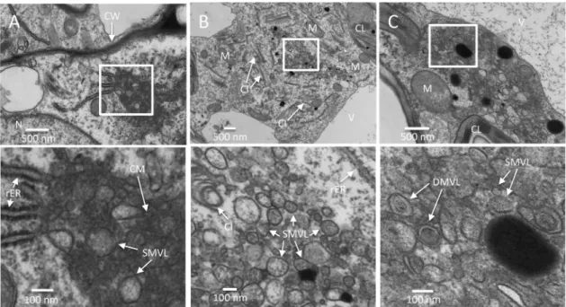
cused on vascular parenchymal cells. Cells were scored positive for
TuMV infection by the presence of cytoplasmic inclusions of
dif-ferent morphologies (e.g., pinwheels, bundles, and short curved
laminated aggregates or scrolls) (
35
). Infected cells were
charac-terized by the accumulation of CM structures that were adjacent
to the rER at 5 dpi (
Fig. 2C
). Few SMVL structures were present at
the periphery of CM amalgams (
Fig. 2C
, bottom). We then
ob-served the presence of numerous heterogeneously sized SMVL
structures in most of the vascular parenchymal cells at 6 dpi (
Fig.
2D
). The shape of the SMVL structures was round or oval, and the
size was estimated to be 113 nm
⫾ 38 nm (n ⫽ 56) in diameter or
in length. Finally, we observed both SMVL structures and DMVL
structures with an electron-dense core in most of the cells at 7 dpi
(
Fig. 2E
). The shape of DMVL structures was also round or oval,
with the outer membrane diameter or length being 244 nm
⫾ 60
nm (n
⫽ 52) and the inner membrane diameter or length being
162 nm
⫾ 48 nm (n ⫽ 52).
As expected, a 1-day delay in membrane modification took
place in mesophyll cells compared to the time of modification in
vascular parenchymal cells. At 6 dpi, the CM structures were
ei-ther located near or connected with the rER, and some SMVL
structures were associated with the CM structures (
Fig. 3A
). At 7
dpi, aggregations of heterogeneously sized SMVL structures of
111 nm
⫾ 42 nm (n ⫽ 72) in diameter or in length were observed
(
Fig. 3B
). At 8 dpi, both SMVL structures and DMVL structures
with an electron-dense core were observed (
Fig. 3C
). DMVL
structures had an outer membrane diameter or length of 172 nm
⫾ 56 nm (n ⫽ 40) and an electron-dense core diameter or length
of 114 nm
⫾ 45 nm (n ⫽ 40).
SMVL structures are RNA replication sites of TuMV. To
con-firm if TuMV-induced membranous aggregates were 6K
2-con-taining structures (
Fig. 2
and
3
), an HA tag was fused to the C
terminus of 6K
2:GFP (6K
2:GFP:HA). The 6K
2:GFP:HA-coding
sequence was thus introduced into the TuMV infectious clone
FIG 2 Time course analysis of TuMV-induced membranous aggregates in N. benthamiana leaf midrib. (A) An upper young leaf of an N. benthamiana plant wasimaged by tile scanning with a Zeiss LSM-780 confocal microscope using a 10⫻ objective. The tile scan was carried out by assembling images of 11 by 13 tiles. (B) Cross sections of a leaf midrib area systemically infected with TuMV/6K2:GFP (marked with a rectangle in panel A) were imaged by confocal microscopy using
a 20⫻ objective on the day postinfection indicated in the upper right. The Fluorescent Brightener 28-stained cell wall is shown in false magenta color. 6K2:GFP
is shown in green. All images are single optical slices. (C to E) Time course analysis of TuMV-induced membranous aggregates in vascular parenchymal cells. N. benthamiana leaf midribs systemically infected with TuMV/6K2:GFP were chemically fixed, processed, and observed by TEM. The lower panels show
higher-magnification images of the areas in the rectangles in the upper panels. (C) TuMV-induced CM structures, which were associated with SMVL structures, were located close to the rER in a vascular parenchymal cell at 5 dpi. (D) TuMV-induced heterogeneous SMVL structures in a vascular parenchymal cell at 6 dpi. (E) TuMV-induced aggregates contain both SMVL structures and DMVL structures with an electron-dense core in a vascular parenchymal cell at 7 dpi. Ue, upper epidermis; Le, lower epidermis; Ac, angular collenchyma cells; Vt, vascular tissue; Me, mesophyll cells; N, nucleus; G, Golgi apparatus; M, mitochondrion; V, vacuole; CL, chloroplast; CW, cell wall; rER, rough endoplasmic reticulum; CI, cytoplasmic inclusion body; CM, convoluted membranes; SMVL, single-membrane vesicle-like structure; DMVL, double-single-membrane vesicle-like structure.
TuMV-Induced Cellular Rearrangements
on January 18, 2016 by INRS-Institut Armand-Frappier
http://jvi.asm.org/
pCambiaTuMV to produce pCambiaTuMV/6K
2:GFP:HA (
Fig.
4A
). Introduction of the 6K
2:GFP:HA fragment into the
polypro-tein produced the expected propolypro-tein with the correct molecular
mass, as shown by Western blot analysis using an anti-HA
mono-clonal antibody (
Fig. 4B
). TuMV infection was not compromised,
as analyzed by Western blot analysis using a rabbit serum raised
against the coat protein (CP) of TuMV (
Fig. 4B
), nor was the
morphology of 6K
2vesicles compromised when they were
ob-served under a confocal microscope (data not shown).
Immuno-gold labeling using an anti-HA monoclonal antibody confirmed
that the TuMV-induced membranous aggregates were truly
tagged with 6K
2(
Fig. 4C
, arrowheads). Immunogold labeling
us-ing anti-RNA-dependent RNA polymerase (anti-RdRp)
poly-clonal antibodies also showed the vesicle aggregates contained
RdRp (
Fig. 4D
, arrowheads).
To better differentiate between the distinctive TuMV-induced
membrane structures (i.e., the CM, SMVL, and DMVL
struc-tures), a 15-min postfixation incubation period with 0.1%
re-duced osmium tetroxide was added before the dehydration step.
This step increased the membrane contrast but also decreased the
antigenicity of the tested proteins. For instance, HA and
anti-RdRp labeling was weak with osmium-treated samples, since only
a few gold particles were found on the sections (data not shown).
However, the anti-dsRNA monoclonal antibody J2 worked well
on the reduced osmium-treated samples (
Fig. 4E
). In mesophyll
cells at 8 dpi, when SMVL structures were distinguishable from
DMVL structures on osmium-treated samples,
anti-dsRNA-specific gold particles mainly decorated the SMVL structures
(
Fig. 4E
).
The number of gold particles per square micrometer and the
relative labeling distribution over mock-infected and
TuMV-in-fected sections were determined. For each of the three different
antibodies, at least 10 sections were observed. Practically no
label-ing with the anti-HA and anti-dsRNA monoclonal antibodies was
observed in mock-infected samples (
Fig. 4F
). On the other hand,
anti-HA and anti-dsRNA labeling was specifically associated with
TuMV-induced membranous aggregates and was essentially
ab-sent from the rest of the cell (
Fig. 4G
). Background labeling was
detected with the polyclonal anti-RdRp antiserum in
mock-infected samples (
Fig. 4F
), but a larger amount of labeling was
observed on TuMV-induced membranous aggregates (
Fig. 4G
).
Quantification of the anti-dsRNA gold particle distribution on
SMVL and DMVL structures in mesophyll cells on
TuMV-in-fected sections was performed at 8 dpi. The results showed that the
gold particles were mainly associated with SMVL structures
(98.5%), with few gold particles being found on the DMVL
struc-tures (
Fig. 4H
). No gold particles decorated CM structures (data
not shown). These data suggest that SMVL structures are TuMV
RNA replication sites.
Comparison of TuMV-induced cellular reorganization in
chemically fixed and HPF/FS-prepared samples. Chemical
fixa-tion may induce ultrastructural artifacts owing to the slow
diffu-sion of chemical fixatives that results in the nonsynchronized
immobilization of cellular macromolecular components and
owing to the selective cross-linking reactions of chemical
fixa-tives, such as aqueous aldehydes and osmium tetroxide.
High-pressure freezing (HPF) is widely regarded as the optimal
fix-ation method for TEM (
36
). HPF is complete within milliseconds
and ensures the simultaneous immobilization of all
macromo-lecular components (
37
). Subsequent processing for
morpho-logical studies is predominantly achieved by freeze substitution
(FS), where ice is removed by organic solvents at about
⫺90°C.
FIG 3 Time course analysis of TuMV-induced membranous aggregates in mesophyll cells. (A to C) N. benthamiana leaf midribs systemically infected with
TuMV/6K2:GFP were chemically fixed, processed, and observed by TEM. Higher-magnification images of the areas in the white rectangles are shown in
the panels below. (A) TuMV-induced CM structures amid SMVL structures were connected with several rERs in a mesophyll cell at 6 dpi. (B) TuMV-induced heterogeneous SMVL structures close to the rER in a mesophyll cell at 7 dpi. (C) TuMV-induced aggregates contain both SMVL structures and DMVL structures with an electron-dense core in a mesophyll cell at 8 dpi. CW, cell wall; M, mitochondrion; CL, chloroplast; V, vacuole; CI, cytoplasmic inclusion body; rER, rough endoplasmic reticulum; CM, convoluted membranes; SMVL, single-membrane vesicle-like structure; DMVL, double-membrane vesicle-like structure.
Wan et al.
on January 18, 2016 by INRS-Institut Armand-Frappier
http://jvi.asm.org/
Freeze-substituted samples appear similar to materials
pre-pared for TEM by conventional chemical fixation methods, but
they have a greater likelihood of displaying structures in their
native state (
38
). Since HPF/FS leads to an improved
ultra-structural preservation, we also conducted HPF/FS for sample
preparation.
Currently, HPF allows a depth of 200
m thick without
detect-able ice crystallization damage (
39
). In systemically infected N.
benthamiana leaves, the thickness of the midrib is 356
m ⫾ 64
m (n ⫽ 35), while it is 97 m ⫾ 23 m (n ⫽ 35) for the other leaf
area. To avoid damage from ice crystallization, the leaf area
with-out the rib tissue was consequently chosen. Consistent with what
FIG 4 Subcellular localization of TuMV RNA replication sites. (A) Schematic representation of the infectious clone TuMV/6K2:GFP:HA that coexpresses 6K2as a GFP:HA protein fusion. Black lines, the plasmid backbone; arrow, the cauliflower mosaic virus 35S promoter; AAAAA, the position of the polyadenylated tail; rectangles, TuMV proteins. 6K2:GFP:HA is inserted between P1 and HCpro. PIPO, pretty interesting Potyviridae ORF. (B) Western blot analysis of
6K2:GFP:HA and CP expression in N. benthamiana plants after TuMV/6K2:GFP:HA infection. (C to E) Immunogold labeling was performed on the cross
sections of mock- and TuMV-infected N. benthamiana leaf tissues by using anti-HA, anti-RdRp, and anti-dsRNA antibodies. Higher-magnification images of the areas in the white rectangles are shown on the right of each panel. Arrowheads, HA-specific (C) and RdRp-specific (D) gold particles, which are located in TuMV-induced membranous aggregates. (E) The dsRNA-specific gold particles are mainly localized to TuMV-induced SMVL structures. (F to H) The number of gold particles per square micrometer in mock-infected versus TuMV-infected cells (F), the relative labeling distribution in infected cells (G), and the relative labeling distribution in TuMV-induced membranous aggregates (H) are shown. The results of two different labeling experiments were considered, and 200 gold particles were counted for each experiment. N, nucleus; CL, chloroplast; CW, cell wall; MA, membranous aggregate; CI, cytoplasmic inclusion body; SMVL, single-membrane vesicle-like structure; DMVL, double-membrane vesicle-like structure; ER, endoplasmic reticulum; M, mitochondrion; PM, plasma mem-brane; C, cytosol; V, vacuole.
TuMV-Induced Cellular Rearrangements
on January 18, 2016 by INRS-Institut Armand-Frappier
http://jvi.asm.org/
we observed in chemically fixed samples, HPF/FS-prepared
mes-ophyll cells showed similar membrane remodeling. CM structures
were observed at 6 dpi (
Fig. 5A
) and SMVL structures of 74 nm
⫾
22 nm (n
⫽ 48) in diameter were observed at 7 dpi (
Fig. 5B
) in
mesophyll cells. However, instead of only SMVL structures and
DMVL structures with an electron-dense core being observed in
chemically fixed mesophyll cells (
Fig. 5D
and
E
), electron-dense
bodies of 172 nm
⫾ 59 nm (n ⫽ 20) in diameter were also
fre-quently observed in HPF/FS-prepared mesophyll cells at 8 dpi
(Fig, 5C, arrows labeled I to III). DMVL structures had an outer
vesicle diameter of 138 nm
⫾ 20 nm (n ⫽ 48) and an
electron-dense core diameter of 93 nm
⫾ 19 nm (n ⫽ 48). Thus, the sizes of
SMVL and DMVL structures in HPF/FS-prepared samples were
smaller than those in chemically fixed samples. Another
notice-able difference was that HPF/FS-prepared vesicles were largely
rounded and had more electron-dense materials within them (
Fig.
5C
to
E
). We have no explanation for the different sizes observed
between the HPF/HS and chemically fixed samples. Likewise,
Royer and Kinnamon (
40
) found that T-cell tubules were more
uniform and somewhat smaller in diameter in HPF/FS-prepared
samples than in chemically fixed samples, but no reason for this
was given.
3-D architecture of TuMV-induced membrane
reorganiza-tion. We performed ET on semithin (200-nm) sections to
gener-ate the three-dimensional (3-D) representation of the numerous
heterogeneously sized SMVL structures that were observed in
vas-cular parenchymal cells at 6 dpi (
Fig. 2D
).
Figures 6I
to
IV
show
the representative tomograms that were generated from a
single-axis-tilt series. SMVL structures associated with the rER, along
with three closely associated cytoplasmic inclusions having a
pin-wheel configuration, were clearly observed.
Figures 6V
and
VI
show the 3-D surface rendering generated from tilted images over
a range of
⫾60° (1° increments in high tilts and 2° increments in
low tilts) and indicate that the SMVL structures were actually
SMTs (represented in yellow), similar to those observed in
CVB3-infected (
2
) and poliovirus-infected (
41
) cells (see also Movie S1
in the supplemental material). The 3-D surface rendering shows
that most SMTs are closely packed together and have a similar
orientation. The SMTs were touching one another, but no holes or
connections were observed. Cytoplasmic inclusions (represented
in magenta in
Fig. 6V
and
VI
) and rER were often found in close
proximity to TuMV-modified membranes, and some dilated rER
(represented in sky blue in
Fig. 6V
and
VI
), including ER
mem-branes and the associated ribosomes, was tightly associated with
the SMTs (
Fig. 6IV
to
VI
, red arrows).
We next investigated the 3-D architecture of the membrane
structures in TuMV-infected vascular parenchymal cells at 7 dpi
(
Fig. 7
; see also Movie S2 in the supplemental material). At this
stage, the membrane structures had various shapes and
complex-ities (
Fig. 7A
), and 3-D renderings were generated from selected
membrane structures (boxed areas in
Fig. 7A
).
Figure 7B
is a
close-up view of an SMVL structure (left) along with its 3-D
surface rendering (right two panels), which shows its tubular
nature (represented in yellow). The 3-D rendering of the
elec-FIG 5 Comparison of TuMV-induced membranous aggregates in HPF/FS-prepared and chemically fixed samples. (A to C) Time course analysis ofTuMV-induced membranous aggregates in mesophyll cells. TuMV/6K2:GFP systemically infected N. benthamiana leaf without rib tissues were fixed by
HPF, processed by FS, and observed by TEM. Higher-magnification images of the areas in the white rectangles are shown on the right of each panel. (A) TuMV-induced CM structures in a mesophyll cell at 6 dpi. (B) TuMV-induced heterogeneous SMVL structures in a mesophyll cell at 7 dpi. (C) TuMV-induced aggregates contain SMVL structures (I), DMVL structures with an electron-dense core (II), and electron-dense bodies (III) in a mesophyll cell at 8 dpi. The images in panels I to III are higher-magnification images of the structures identified by the arrows labeled I to III, respectively, in panel C. (D and E) TuMV-induced heterogeneous SMVL structures (D) and a DMVL structure (E) in chemically fixed mesophyll cells. CL, chloroplast; M, mitochondrion; CM, convoluted membranes; SMVL, single-membrane vesicle-like structure; DMVL, double-membrane vesicle-like structure; EDB, electron-dense body.
Wan et al.
on January 18, 2016 by INRS-Institut Armand-Frappier
http://jvi.asm.org/
tron-dense fibrillar materials inside this SMT (represented in
light red) was also regenerated. These fibrillar materials could
be the replication complexes containing vRNA, as proposed for
beet black scorch virus (BBSV)-induced replication factories
(
42
), which is consistent with our observation that SMVL
structures contain dsRNA (
Fig. 4E
). Adjacent to the SMT is an
irregularly shaped tubule (represented in green) that might
represent an intermediate structure leading to the formation of
DMTs (see below).
The left panel of
Fig. 7C
is a close-up of the area identified by
the solid square depicted in
Fig. 7A
, and the two right panels are
3-D surface renderings for some of the membrane structures.
These membrane structures are SMTs (in yellow) along with
dou-ble-membrane tubules (DMTs) (the outer membrane is
repre-sented in light blue, and the inner membrane is reprerepre-sented in
dark blue) interspersed between them with irregularly shaped
tu-bules (represented in green) with frequent membrane pairing
(
Fig. 7C
, white arrowheads) or curving (
Fig. 7C
, red arrowheads)
(see also Movie S2 in the supplemental material).
Figure 7D
also
shows tubule bending. Finally, the left panel of
Fig. 7E
is a close-up
view of a DMVL structure (left), along with its 3-D surface
ren-dering (right two panels). The DMT contents (represented in dark
red) did not have a fibrillar profile but almost fill the inner core of
the DMT.
In conclusion, the SMVL and DMVL structures that were
ob-served in the two-dimensional (2-D) TEM images of
TuMV-in-fected cells were, in fact, tubules. These SMTs and DMTs were also
interspersed with irregularly shaped tubules, which might be
in-termediate forms leading to the transformation of SMTs into
DMTs.
Viral particle bundles associated with electron-dense bodies.
At 8 dpi, filament bundles in association with electron-dense
bod-ies in the vicinity of TuMV-induced membranous aggregates were
frequently observed in HPF/FS-prepared mesophyll cells (
Fig.
8A
). Individual filaments were 13 nm
⫾ 1 nm (n ⫽ 30) in width,
which corresponds to the thickness of TuMV particles. The
elec-tron-dense bodies had diameters of 64 nm
⫾ 13 nm (n ⫽ 44), and
DMVL structures with an electron-dense content of a similar
in-FIG 6 3-D reconstruction of TuMV-induced membrane rearrangement at midstage of infection. (I to IV) Representative tomogram slices generated on a200-nm-thick section from TuMV-infected vascular parenchymal cell at 6 dpi. The SMVL structures in close proximity to dilated rER and a cytoplasmic inclusion body are shown. (V and VI) A 3-D surface rendering of the closely packed SMTs, dilated rER, and cytoplasmic inclusion body. Red arrows, connection between a SMT and the rER membrane; yellow, SMTs, sky blue, rER; magenta, cytoplasmic inclusion body. CI, cytoplasmic inclusion body; rER, rough endoplasmic reticulum; SMVL, single-membrane vesicle-like structure.
TuMV-Induced Cellular Rearrangements
on January 18, 2016 by INRS-Institut Armand-Frappier
http://jvi.asm.org/
tensity were found close to them (
Fig. 8A
and
B
). These structures
were also seen in chemically fixed samples, although less
fre-quently (
Fig. 8C
). These data suggest that these bodies are
in-volved in viral particle assembly.
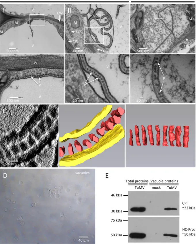
TuMV particles accumulate in vacuoles. In addition to the
above-described vesicle-like structures, linear arrays of dot-like
structures with a diameter of 15 nm
⫾ 1 nm (n ⫽ 30) juxtaposing
the cytoplasmic face of the tonoplast and frequently protruding
into the vacuole were seen (
Fig. 9A
, panel I).
Figure 9A
(panel II)
shows that the aligned dots were enclosed in a membrane sac
within the central vacuole. These structures were also found in
HPF/FS-prepared samples, although the vacuole showed more
electron-dense material (
Fig. 9B
).
The diameter of the dot-like structures is consistent with the
diameter of viral particles. These structures may then represent
the top view of vertically aligned viral particles. To confirm that
FIG 7 3-D architecture of TuMV-induced complex membrane structures at late stage of infection. (A) Overview of a single slice of a tomogram generatedon a 200-nm-thick section in the perinuclear region of a TuMV-infected vascular parenchymal cell at 7 dpi. (B) A higher-magnification image (left) of the area in the dashed white square of panel A is shown, and the 3-D model (middle and right) shows an SMT with fibrillar material inside and with an adjacent intermediate tubular structure. (C) A higher-magnification image (left) of the area in the solid white square in panel A is shown, and the 3-D model (middle and right) shows SMTs, intermediate tubular structures, and DMTs. White arrowheads, intermediate SMTs that paired into flattened cisternae; red arrowheads, intermediate SMTs with a slightly negative curvature that may result in the wrapping of the cytoplasm to form DMTs. (D) A higher-magnification image of the area in the top dashed white rectangle in the rightmost panel of panel C. Red arrow in the upper panel, a slightly bent, intermediate tubular structure (green) located among several SMTs (yellow) with similar orientations; red arrow in the lower panel, a view rotated 43.2° around the y axis, highlighting its tubular nature. (E) A higher-magnification image of the area in the bottom dashed white square in the rightmost panel of panel C. (Left) A DMVL structure and its electron-dense materials; (middle and right) the 3-D model of the DMT with a core of electron-dense materials. Yellow, SMTs; light red, electron-dense materials; green, intermediate tubular structures; light blue, outer membranes of DMTs; dark blue, inner membranes of DMTs; dark red, the electron-dense materials inside DMTs. rER, rough endoplasmic reticulum; M, mitochondrion; NM, nuclear membrane.
Wan et al.
on January 18, 2016 by INRS-Institut Armand-Frappier
http://jvi.asm.org/
this was the case, a 3-D representation was generated by ET. A
series of single-axis-tilt 2-D images was collected on a
90-nm-thick ultrathin section (
Fig. 9C
; see also Movie S3 in the
supple-mental material) over a tilt range of
⫾64° (in 1° increments).
Images were aligned and reconstructed to generate a 3-D
repre-sentation of this structure. The middle and right panels of
Fig. 9C
show the 3-D reconstruction of the complete tomogram of the
image of the left panel. A 3-D surface rendering of the aligned
dot-like structures clearly showed that they were a cross section of
a monolayer of TuMV particles aligned along the same axis and
that they were enclosed with a membrane envelope, likely derived
from the tonoplast.
Vacuoles were isolated from mock-infected leaves and leaves
systemically infected with TuMV to confirm that viral particles
accumulated in these organelles. Vacuoles were released from
protoplasts and isolated by Ficoll gradient centrifugation (
Fig.
9D
). We then performed Western blot analysis with anti-CP and
helper component proteinase (HCpro) polyclonal
anti-bodies. Purified vacuoles from mock- and TuMV-infected leaves
were visualized by phase-contrast microscopy to ascertain that
similar amounts of vacuoles were loaded. The Western blot
anal-ysis results showed that the vacuoles isolated from TuMV-infected
leaves contained CP (
Fig. 9E
), supporting the tomography data
showing that TuMV particles are loaded into the vacuoles. HCpro,
which is involved in aphid transmission of viral particles, was also
detected in purified vacuoles (
Fig. 9E
).
DISCUSSION
In this study, we looked at the cellular remodeling that takes place
during TuMV infection using a time course ultrastructural
anal-ysis. The very first event that was observed in the infected cells was
the accumulation of CM structures close to or connected to the
rER (
Fig. 2C
and
3A
). These CM collections are reminiscent of
those induced during infection by dengue virus (DENV; a
flavivi-rus) (
43
) and the SARS coronavirus (
1
). CM structures have been
proposed to be sites of viral polyprotein processing and storage for
proteins and lipids involved in vRNA replication (
43
). Based on
the morphological similarities between the CM structures
in-duced by TuMV and by flavivirus and their observation early in
the infection process, we propose that TuMV-induced CM
struc-tures are also sites for polyprotein processing and/or for storage of
the proteins and lipids required for vesicle-like structure
biogen-esis. Indeed, the accumulation of large amounts of lipids has been
observed in association with TuMV replication complexes (
24
)
and for other plant viruses as well (
44
).
Few SMVL structures were observed amid CM structures in
vascular parenchymal and mesophyll cells (
Fig. 2C
and
3A
), but
SMVL structures became prevalent 1 day later in these cells (
Fig.
2D
and
3B
). The SMVL structures scored positive for 6K
2and
RdRp, as well as for dsRNA, by immunogold labeling (
Fig. 4C
to
E
), suggesting that these structures are the sites for vRNA
replica-tion. The TuMV-induced SMVL structures are similar to the
FIG 8 TuMV particles are associated with electron-dense bodies. (A and B) In HPF/FS-prepared samples, virus-like particles are found as filament bundlesassociated with electron-dense bodies in the vicinity of TuMV-induced membranous aggregates at a late stage of infection (A), and the DMVL structures are found close to these filament bundle structures (B). (C) In chemically fixed samples, the filament bundles associated with electron-dense bodies are found in the cytoplasm near the tonoplast. CI, cytoplasmic inclusion body; DMVL, double-membrane vesicle-like structure; EDB, electron-dense body; P, particles; Pr, peroxisome; V, vacuole; rER, rough endoplasmic reticulum.
TuMV-Induced Cellular Rearrangements
on January 18, 2016 by INRS-Institut Armand-Frappier
http://jvi.asm.org/
FIG 9 TuMV acquires an envelope by hijacking the tonoplast. (A and B) N. benthamiana leaf midribs systemically infected with TuMV/6K2:GFP were fixed by
chemical fixation (A) or HPF (B), processed, and observed by TEM. A higher-magnification image of the area in the white rectangle is shown at the bottom. Monolayer dot-like structures are aligned along the tonoplast (A, panel I) and loaded into the vacuole from the cytoplasm (A, panel II, and B). (C) 3-D reconstruction of enveloped TuMV particles by ET. (Left) A higher-magnification image of the area in the white square in panel A (panel II) with a 180° rotation showing a single slice of the tomogram generated from the 90-nm-thick section and enveloped dot-like structures in the vacuole; (middle) the 3-D model generated from the whole tomogram of the left panel; (right) the 90° rotation of the dot-like structures in the middle panel. Yellow, tonoplast; red, dot-like structures. (D, E) CP and HCpro are present in purified vacuoles. (D) Vacuoles were isolated by Ficoll gradient centrifugation from N. benthamiana leaves systemically infected with TuMV. (E) Western blot analysis of viral proteins CP and HCpro in vacuoles purified from mock-infected N. benthamiana leaves and N. benthamiana leaves systemically infected with TuMV. The total proteins of N. benthamiana leaves systemically infected with TuMV were used as a positive control. V, vacuole; CW, cell wall; M, mitochondrion; rER, rough endoplasmic reticulum; G, Golgi apparatus; CL, chloroplast; P, particles.
on January 18, 2016 by INRS-Institut Armand-Frappier
http://jvi.asm.org/
membrane structures induced during infection by the
enterovi-ruses CVB3 (
2
) and poliovirus (
45
). Conventional 2-D TEM
im-ages of enterovirus CVB3- and poliovirus-induced membrane
structures have been described to be either SMVs (
46
,
47
) or
DMVs (
48–50
), whereas for both viruses, ET-generated 3-D
mod-els revealed that the SMVs observed by TEM are SMTs (
2
,
41
).
Both potyviruses and enteroviruses belong to the
picornavirus-like virus superfamily, suggesting that they might share similar
membrane modification mechanisms. Similarly, in this study, the
3-D model showed that the TuMV-induced SMVL structures are
SMTs (
Fig. 6
and
7
). Polioviral replication proteins were localized
on the external cytoplasmic surface of the single-membrane
tu-bule/vesicle. This apparently was not the case for TuMV. vRNA
replication probably occurs inside the SMVL structures, since
dsRNA-specific gold particles were located within the SMVL
structures (
Fig. 4E
). In addition, the electron-dense fibrillar
ma-terials inside the SMT that were generated by ET might represent
replication complexes (
Fig. 7B
). The intratubular localization of
the replication complex is further supported by previous
mem-brane fractionation data that showed that the
replication-associ-ated soluble viral and host proteins (VPg-Pro, RdRp, and poly(A)
binding protein 2 [PABP2]) were trapped within the lumen of 6K
2vesicles (
20
).
DMVL structures were observed rather late in the infection
cycle (
Fig. 2E
and
3C
) and were characterized by having an
electron-dense core, which was particularly apparent when
samples were processed by HPF/FS (
Fig. 5C
).
Poliovirus-in-duced DMTs may result from a transformation process of
mem-brane apposition, enwrapping, and fusion of SMTs (
41
). The
3-D model generated at the late stage of TuMV infection also
suggested that the DMVL structures are DMTs and that they
were formed by enwrapping of the SMTs, which resulted in the
engulfment of the cytoplasm (
Fig. 7
). Electron-dense bodies
with a size and intensity similar to those of the electron-dense
core found within DMVL structures were associated with
fila-ment bundles (
Fig. 8
). These bodies may derive from DMVL
structures and may be associated with TuMV particle assembly.
It is important to note that no SMVL structures were found
near these assembly sites. This fits our confocal microscopy
observations that CP did not colocalize with membrane-bound
viral replication complexes (
19
,
24
), suggesting that vRNA
en-capsidation occurs at a site adjacent to membrane-bound
vRNA replication complexes. These tubules are likely the
ultra-structure underlying the perinuclear ER amalgam observed by
confocal microscopy during TuMV infection (
16
). However,
the ultrastructure of the TuMV-induced small motile vesicles
still needs to be defined.
We observed the accumulation of TuMV particles as a linear
array in the central vacuole, with the tonoplast forming an
envelope around them (
Fig. 9A
to
C
). Similar aligned dot-like
structures were observed for cells infected with the potyviruses
pokeweed mosaic virus (
51
) and carnation vein mottle virus
(
52
), but their exact nature was not explained. The tonoplast
has also been shown to be remodeled during several plant virus
infections. TEM images showed that tonoplast-associated
ves-icles protruded into the vacuole in cells infected with the
cucu-moviruses cucumber mosaic virus (CMV) and tomato aspermy
virus (TAV) (
5
) and the alfamovirus alfalfa mosaic virus
(AMV) (
53
). Owing to the presence of replicase proteins, the
authors concluded that these were sites of vRNA synthesis (
54
).
On the other hand, particles of the icosahedral sobemovirus
rice yellow mottle virus (RYMV) have been found in vacuoles
as crystalline arrays (
55
). The authors proposed that the acidic
pH and the presence of Ca
2⫹could facilitate crystal formation
and thus stabilize RYMV particles, which may allow the virus to
accumulate to high levels without having deleterious effects on
cellular viability and integrity. The highly stable and compact
RYMV particles would ultimately be released from the vacuole
during xylem vessel differentiation and trafficking along the
water flow for long-distance movement (
55
). The membrane
envelope around TuMV particles would thus provide a
protec-tive shield against the harsh environmental conditions
preva-lent in the central vacuole for subsequent long-distance
move-ment during xylem vessel differentiation (
24
).
Potyviruses are transmitted from plant to plant by aphids in a
nonpersistent manner (
56
). The interaction between CP and the
receptor in the cuticle of the aphid stylet tip is mediated by the
viral protein HCpro (
57
). HCpro was detected in purified
vacu-oles from TuMV-infected cells (
Fig. 9E
). The accumulation of
TuMV particles in the vacuole may have something to do with
plant-to-plant virus transmission by aphids. Aphids are phloem
feeders, since phloem sap is rich in key nutrients, such as
carbo-hydrates (sucrose), amino acids, and minerals (
58
). Before the
phloem nutrient source is sampled, the aphid performs host plant
selection by probing the peripheral plant tissues. Potyviruses have
been suggested to be acquired and inoculated during brief (⬍1
min) and superficial stylet penetrations in the peripheral plant
tissues (epidermal and mesophyll cells) (
59
). Once the suitable
host plant is detected, the aphid ultimately inserts the stylet along
the cell wall of parenchymatous cells into the phloem sieve
ele-ment for nutrient acquisition. The aphid recognizes the phloem
sap by sensing the pH (7.0 to 7.5) and a high sucrose concentration
(about 400 mmol
⫺1) (
60
). During the search for phloem sap, the
stylet tip is observed to be inserted into the vacuole of
parenchy-matous cells, and owing to the acidic pH (5.0 to 5.5) and low
sucrose concentration, the stylet is withdrawn and changes
direc-tion until the sieve element is reached (
60
). This brief probing of
the aphid stylet in the central vacuole may be sufficient to acquire
the membrane-bound TuMV.
On the basis of the observations described above, we propose
the following model that links the TuMV-induced membrane
remodeling for vRNA replication with virus storage in the
cen-tral vacuole (
Fig. 10
). Membrane remodeling starts with the
accumulation of ER-connected CM structures. This leads to
the formation of SMTs that are involved in vRNA replication.
As infection proceeds, the number of SMTs decreases, and
DMTs with an electron-dense core and electron-dense bodies
are generated. Possibly, at this stage, vRNA and CP have been
produced in sufficient quantities for particle assembly to take
place in the vicinity of DMTs and electron-dense bodies.
Fi-nally, TuMV particles accumulate in a linear array along the
same axis in the central vacuole. Additional steps are likely to
take place between steps 4 and 5 in order to release and close the
continuous double-membrane-enveloped ring of particles.
These steps would involve membrane fusion events, requiring
the participation of host factors, notably, secretory pathway
factors, that might have been co-opted with the SMTs and
DMTs. There is also the possibility of additional unseen
mem-brane connections that increase the complexity of the
relation-ship of the membrane structures that have been observed.
TuMV-Induced Cellular Rearrangements