Abstract. Dysregulation of galectin expression is frequently
observed in cancer tissues. Such an abnormal expression
pattern often correlates with aggressiveness and relapse in
many types of cancer. Because galectins have the ability to
modulate functions that are important for cell survival,
migra-tion and metastasis, they also represent attractive targets for
cancer therapy. This has been well-exploited for extracellular
galectins, which bind glycoconjugates expressed on the
surface of cancer cells. Although the existence of
intracel-lular functions of galectins has been known for many years,
an increasing number of studies indicate that these proteins
can also alter tumor progression through their interaction
with intracellular ligands. In fact, in some instances, the
interactions of galectins with their intracellular ligands seem
to occur independently of their carbohydrate recognition
domain. Such findings call for a change in the basic
assump-tions, or paradigms, concerning the activity of galectins in
cancer and may force us to revisit our strategies to develop
galectin antagonists for the treatment of cancer.
Contents
1. Introduction
2. Where do we find galectins inside the cells?
3. Intracellular functions of galectins in cancer
4. CRD-independent functions for intracellular galectins?
1. Introduction
Galectins represent a family of evolutionarily conserved
animal lectins that are widely distributed from lower
inver-tebrates to higher verinver-tebrates. They were initially described
in the electric eel, Electrophorus electricus, as low molecular
weight,
β-galactoside binding proteins (1). Since then, galectins
have been numbered according to the order of their discovery.
The 15 family members are now classified according to their
structure and number of carbohydrate recognition domain
(CRD). The prototype subfamily of galectins (galectin-1,
-2, -5, -7, -10, -11, -13, -14 and -15) consists of a single CRD
with a short N-terminal sequence. The tandem-repeat type
subfamily (galectin-4, -6, -8, -9 and -12) has two non-identical
CRDs joined by a short linker peptide sequence. There is also
a chimerical form of galectin (galectin-3) that contains one
CRD connected to a non-lectin domain.
One of the first clues that galectins were involved in
cancer was published more than 25 years ago when it was
observed that they were differently regulated in normal and
cancer tissues. Since then, a large number of studies have
focused on the role of galectins in cancer and excellent
reviews on the role of galectins have been published (2-5).
Historically, studies on the role of galectins in cancer have
mostly focused on their ability to bind membrane-anchored
cell surface receptors via their CRD. Their dimeric form (or
multimeric in the case of galectin-3) induces crosslinking
of the receptors and formation of a lattice that triggers a
cascade of transmembrane signaling events. For example,
binding of galectin-3 protects EGF and TGF-
βreceptors
from negative regulation via constitutive endocytosis and
increases sensitivity of tumor cells to growth factors (6).
Binding to cell surface receptors can also induce apoptosis.
This is particularly relevant in the case of galectin-1, which
is capable of inducing apoptosis of T-cells and potentially
create an immunosuppressive tumor microenvironment (7).
Alternatively, binding to cell surface receptors can facilitate
intercellular adhesion (to promote homo- and heterotypic
aggregation) or adhesion of tumor cells to extracellular
matrix proteins. Exhaustive efforts have thus been deployed
for the identification of highly selective and potent galectin
inhibitors. Despite decades of research, the progression
in this field has been relatively slow. In most cases, these
inhibitors are peptides or high molecular weight, naturally
occurring polysaccharides that are used to specifically block
the binding of extracellular galectins to carbohydrate
struc-tures on cell surface receptors. While targeting extracellular
galectins is warranted, such inhibitors are largely if not
completely ineffective at targeting intracellular galectins.
Indeed, most galectins preferentially exist in intracellular
Intracellular galectins in cancer cells:
Potential new targets for therapy (Review)
MARIA C. VLADOIU, MARILYNE LABRIE and YVES ST-PIERRE
INRS-Institut Armand-Frappier, Laval, QC H7V 1B7, Canada
Received November 1, 2013; Accepted December 2, 2013
DOI: 10.3892/ijo.2014.2267
Correspondence to:
Professor Yves St-Pierre, INRS-Institut Armand-Frappier, University of Quebec, 531 Boul. Des Prairies, Laval, QC H7V 1B7, CanadaE-mail: [email protected]
compartments, consistent with the fact that they do not
harbor a signal sequence and are transported outside the
cells via a non-classical secretory pathway, possibly via
galectin-rich vesicles or exosomes. A better understanding
of their intracellular functions in cancer cells is thus critical
to help develop new anticancer therapies directed at these
proteins.
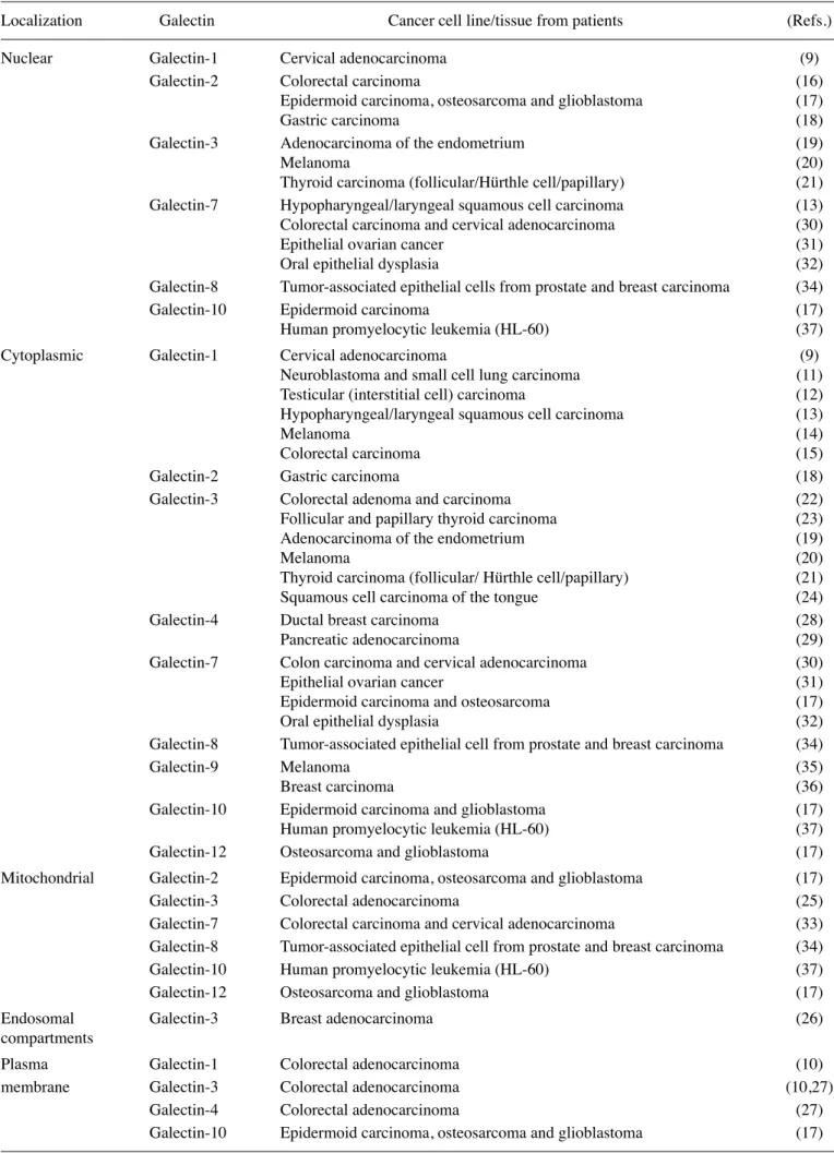
2. Where do we find galectins inside the cells?
The answer to this question is rather simple: almost anywhere
(Fig. 1). They can be detected in various intracellular
compartments of both normal and cancerous cells. Frequently,
modifications in the subcellular localization occur when cells
undergo cell-transformation into malignant phenotypes (4).
It is noteworthy to mention that galectins expression is also
modulated during some of these cell transformation processes,
hence their presence/absence in those subcellular
localiza-tions is not exclusive to protein translocation (8). Up to now,
however, our knowledge of intracellular galectins has mostly
been obtained while studying galectin-3. As we gain more and
more knowledge on other members of the galectin family, we
find overwhelming evidence that most if not all galectins are
often expressed inside the cells. Here we describe the
intra-cellular localization of various galectins with their respective
cancer tissues and/or cell lines (Table I).
Galectin-1 is observed in the nuclear compartment of
transfected HeLa cells (9) and the inner plasma membrane of
colorectal adenocarcinoma cells (HCT116) (10). Moreover,
its presence is also seen in the cytosol of neuroblastoma
and small cell lung carcinoma tissues, testicular interstitial
and cervical carcinoma cell lines (MA-10 and HeLa),
hypo-pharyngeal (HSCCs) and laryngeal (LSCCs) squamous cell
carcinoma tissues, human melanoma cell lines (A375 and
A2058) and colorectal cancer tissues including adenomas,
carcinomas and metastases from patients (9,11-15). Although
fewer studies have been conducted on galectin-2, the
avail-able data indicate its presence in the nucleus of genetically
engineered human colon cancer cells that have ectopic stable
expression (16) in addition to gastric carcinoma tissues,
epidermoid carcinoma, osteosarcoma and glioblastoma cell
lines (A-431, U-2 OS and U-251MG) (17,18). Its presence
has also been reported in the cytosol of gastric carcinoma
tissues and in mitochondria of epidermoid carcinoma,
osteo-sarcoma and glioblastoma cell lines (A-431, U-2 OS and
U-251MG) (17,18). In the case of galectin-3, one of the most
investigated members of the galectin family, its presence is
detected in the nucleus of aggressive endometrial
adenocar-cinoma, melanoma cell lines, malignant thyroid carcinomas
(follicular adenoma, Hürthle cell adenoma and papillary
carcinoma) (19-21). Galectin-3 is also found in the cytosol
of colonic adenomas/carcinomas tissues, follicular/papillary
thyroid carcinomas, endometrial adenocarcinoma, human
melanoma cell lines (MlDo and M4Be), malignant thyroid
carcinoma (follicular adenoma, Hürthle cell adenoma and
papillary carcinoma) and in tongue squamous cell carcinoma
tissues (19-24). Additionally, galectin-3 is found in the
mito-chondria of colorectal adenocarcinoma cell line (SNU-769B),
in endosomal compartments of breast adenocarcinoma cell
line (SKBR3), and in apical membrane regions of human
colon adenocarcinoma cell lines (T84 and HCT116)
(10,25-27). Galectin-4 is detected in the cytosol of human breast
ductal carcinoma tissues (28,29) and pancreatic
adenocar-cinoma cell line (Pa-Tu-8988S) (29) as well as inside the
basal plasma membrane of human colon adenocarcinoma
cells (T84) (27). Galectin-7, which has recently attracted
more interest in cancer because its preferential expression
in epithelial tissues and carcinomas, is seen in the nucleus
of many cancer cells, including hypopharyngeal (HSCCs)
and laryngeal (LSCCs) squamous cell carcinomas tissues,
colon carcinoma cells (DLD-1), cervical adenocarcinoma
(HeLa), epithelial ovarian cancer tissues and oral epithelial
dysplasia tissues (13,30-32). Galectin-7 is also observed in
the cytosol of the colon carcinoma cell line DLD-1, cervical
adenocarcinoma cells (HeLa), epithelial ovarian cancer and
oral epithelial dysplasia tissues (17,30-32). Like galectin-3,
it is also detected in mitochondrial fractions, most notably
in the case of human colorectal carcinoma and cervical
adenocarcinoma cell lines (HCT116, HeLa) and the HaCaT
keratinocyte cell line (33). Galectin-8 expression is detected
in the cytosol, nucleus and mitochondria of tumor-associated
epithelial cells from human prostate and breast tissues (34).
Intracellular galectin-9 is observed in the cytosol of human
melanoma cell lines (MM-BP and MM-RU) and the MCF-7
breast carcinoma cell line (35,36). Galectin-10 is observed
in the nuclei and cytosol of epidermoid carcinoma cells and
in the cytoplasmic compartments of glioblastoma and
osteo-sarcoma. In the human promyelocytic leukemia HL-60 cell
line, it is found in the nucleus, cytosol and mitochondria (37)
while its localization is associated with the inner plasma
membrane of many glioblastoma cell lines (A-431, U-2 OS
and U-251MG) (17). Galectin-12 is observed in the cytosol
and mitochondria of osteosarcoma and glioblastoma cell
lines (U-2 OS and U-251MG) (17).
Although there are no reports yet that other galectins are
present inside cancer cells, there are indications that this may
well be the case given their presence inside normal cells. For
example, galectin-12, a close structural homolog of galectin-7,
has been found in the nucleus and mitochondrial fractions
of adipocytes (38-40). The fact that galectin-12-deficient
mice have abnormal mitochondrial activity is particularly
interesting considering the key role of mitochondria in energy
metabolism of cancer cells (41,42). Galectin-10 is also found
inside human regulatory T-cells and other inflammatory
cells (43) while galectin-13 is found in the perinuclear area of
syncytiotrophoblasts (44). Computational predictions of where
galectins resides in a cell show that it is logical to assume that
many galectins will be present within several intracellular
compartments. For example, using pSORT, a commonly used
tool to predict intracellular localization of proteins, we found
that all galectins have a strong preference for cytoplasmic,
nuclear and mitochondrial compartments (Table II) (45,46).
We have obtained similar results using other computational
tools (unpublished data).
3. Intracellular functions of galectins in cancer
The main challenge in studying the galectin functions in
neoplasms remains their opposing functions in tumor
progression. Depending on the type of cancer, one galectin
Table I. Intracellular localization of galectins in different cancers.
Localization
Galectin
Cancer cell line/tissue from patients
(Refs.)
Nuclear
Galectin-1
Cervical adenocarcinoma
(9)
Galectin-2
Colorectal carcinoma
(16)
Epidermoid carcinoma, osteosarcoma and glioblastoma
(17)
Gastric carcinoma
(18)
Galectin-3
Adenocarcinoma of the endometrium
(19)
Melanoma
(20)
Thyroid carcinoma (follicular/Hürthle cell/papillary)
(21)
Galectin-7
Hypopharyngeal/laryngeal squamous cell carcinoma
(13)
Colorectal carcinoma and cervical adenocarcinoma
(30)
Epithelial ovarian cancer
(31)
Oral epithelial dysplasia
(32)
Galectin-8
Tumor-associated epithelial cells from prostate and breast carcinoma
(34)
Galectin-10
Epidermoid carcinoma
(17)
Human promyelocytic leukemia (HL-60)
(37)
Cytoplasmic
Galectin-1
Cervical adenocarcinoma
(9)
Neuroblastoma and small cell lung carcinoma
(11)
Testicular (interstitial cell) carcinoma
(12)
Hypopharyngeal/laryngeal squamous cell carcinoma
(13)
Melanoma
(14)
Colorectal carcinoma
(15)
Galectin-2
Gastric carcinoma
(18)
Galectin-3
Colorectal adenoma and carcinoma
(22)
Follicular and papillary thyroid carcinoma
(23)
Adenocarcinoma of the endometrium
(19)
Melanoma
(20)
Thyroid carcinoma (follicular/ Hürthle cell/papillary)
(21)
Squamous cell carcinoma of the tongue
(24)
Galectin-4
Ductal breast carcinoma
(28)
Pancreatic adenocarcinoma
(29)
Galectin-7
Colon carcinoma and cervical adenocarcinoma
(30)
Epithelial ovarian cancer
(31)
Epidermoid carcinoma and osteosarcoma
(17)
Oral epithelial dysplasia
(32)
Galectin-8
Tumor-associated epithelial cell from prostate and breast carcinoma
(34)
Galectin-9 Melanoma
(35)
Breast carcinoma
(36)
Galectin-10
Epidermoid carcinoma and glioblastoma
(17)
Human promyelocytic leukemia (HL-60)
(37)
Galectin-12
Osteosarcoma and glioblastoma
(17)
Mitochondrial
Galectin-2
Epidermoid carcinoma, osteosarcoma and glioblastoma
(17)
Galectin-3
Colorectal adenocarcinoma
(25)
Galectin-7
Colorectal carcinoma and cervical adenocarcinoma
(33)
Galectin-8
Tumor-associated epithelial cell from prostate and breast carcinoma
(34)
Galectin-10
Human promyelocytic leukemia (HL-60)
(37)
Galectin-12
Osteosarcoma and glioblastoma
(17)
Endosomal
Galectin-3
Breast adenocarcinoma
(26)
compartments
Plasma
Galectin-1
Colorectal adenocarcinoma
(10)
membrane
Galectin-3
Colorectal adenocarcinoma
(10,27)
Galectin-4
Colorectal adenocarcinoma
(27)
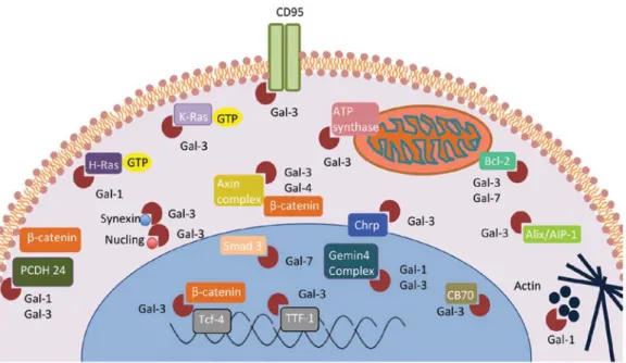
Figure 1. Pro- and anti-tumoral functions of galectins in cancer. Galectins are found in the cytoplasm (C), mitochondria (M), nucleus (N), endosomal compart-ments (EC) and inner plasma membrane (PM). They are capable of modulating many aspects of tumor progression such as cell adhesion and migration, immune escape, cell transformation, apoptosis, angiogenesis, tumor growth, invasion and metastasis.
can either have pro/antitumoral properties (5,47,48) (Fig. 1).
This characteristic of galectins can be explained by the large
diversity of binding partners (Fig. 2) and by the expression
pattern of these partners, which varies contingent to the
cell type. Another proposed hypothesis supporting the dual
functionalities of galectins in cancer is based on the distinct
compartmentalization of the proteins within the cells. In
fact, it was shown that intracellular localization of galectins
differs according to the cell type and tumor progression
stage. Supporting this hypothesis, Califice et al (47)
demon-strated that overexpression of galectin-3 in LnCap prostate
cancer cells in the cytoplasm induces invasion behavior,
anchorage-independant growth, tumor growth and
angio-genesis and reduces apoptosis, while nuclear overexpression
results in the opposite biological activities. Hence, it is of great
interest to take a closer look at the intracellular localization of
these galectins and the impact it has on their biological
func-tions with regards to cancer progression. Here, we discuss the
main findings on the possible roles of intracellular galectins in
cancer. A detailed report of their functions and their putative
ligands is found in Tables III and IV.
Cell transformation. A positive correlation between the
expression of galectin-1 and -3 and malignant transformation
has been established using different cellular models (49-51).
Although the mechanisms involved are not completely clear, it
potentially involves interactions with membrane-bound H-Ras
and K-Ras (52-54). Interestingly, Ras-transformed NIH-3T3
cells have increased expression of galectin-1 and galectin-3
compared to control cells (55). This induction is not necessarily
a consequence of Ras pathway activation but rather a secondary
effect of cell transformation. Hebert et al demonstrated that
Ras transfected cells that have a transformed phenotype,
express galectin-3 while Ras transfected cells that have not
achieved cell transformation do not (56). Another possibility
for galectin-induced malignant transformation might be via
their association with the spliceosome. Indeed, galectin-1 and
-3 are found in Gemin4 (C50)/SMN/Gemin2 complex and play
an important role in spliceosome assembly (57). This
associa-tion suggests that those galectins might regulate the processing
of pre-mRNA during malignant transformation.
Apoptosis. Apoptosis regulation by galectins is probably one
of their most studied intracellular functions. Several studies
have shown that galectins either positively or negatively
regu-late apoptosis in various cancer cell models. Galectin-1 for
example, increases apoptosis of LnCap prostate cancer cells,
CoLo201 colon cancer cells, Leydig tumor cells and B-cell
lymphomas (12,58-61). Conversely, it reduces apoptosis in
gliomas, cervical and lung cancer (62-64). Galectin-3 has
also been shown to modulate apoptosis. In myeloid leukemia,
neuroblastoma, colorectal, breast, prostate, thyroid, bladder,
pancreatic, gastric and some B-cell lymphoma cancer cells it
has been shown to have anti-apoptotic functions (47,65-80).
In contrast, it seems to induce apoptosis in other B-cell
lymphomas (81). Galectin-7 displays a dual functionality
in apoptosis as well since it reduces chemosensitivity in
melanomas, breast and lymphoid cancer cells, yet it
sensi-tizes colon, urothelial and cervical cancer cells to cell death
(82-87). This role of galectin-7 in melanoma cells is clearly
distinct from that of galecin-9 which rather promotes death of
melanoma cells (35,88).
The underlying mechanisms of galectin's regulation of
apoptosis are not fully understood. Nonetheless, many binding
partners implicated in cell fate have been identified. Galectin-3
and -7 have been shown to interact in vitro and in vivo with the
anti-apoptotic B-cell lymphoma-2 (Bcl-2) protein (33,89,90).
The domain of galectin-7 protein implicated in this binding
has not yet been identified. Still, the NWGR motif present at
the N-terminus of galectin-3 protein shows a strong homology
with the BH1 motif of Bcl-2, which appears to be essential
for its anti-apoptotic functions (90). Due to a strong homology
between the different pro- and anti-apoptotic members of the
Bcl-2 family, galectins might also be able to interact with other
members of the family. The modulation of either their stability
or their localization would explain the dual role of galectins
in apoptosis. The members of the Bcl-2 family are probably
not the only galectin-binding partners implicated in apoptosis
regulation. Synexin, a calcium and phospholipid-binding
protein has been shown to drive the perinuclear
transloca-tion of galectin-3, which is essential to its anti-apoptotic
function (91). Galectin-3 also interacts with the intracellular
domain of the CD95 receptor, also known as FAS receptor
Table II. Predicted intracellular localization of galectins.
Galectin (%)
Cellular
---compartment 1 2 3 4 7 8 9 10 12 13
Cytoplasmic 65 52 26 65 65 70
65
52 39 61
Nuclear
22 26 48 17 17 17 17 13 13 17
Mitochondrial
4
9
9
13
17
9
4
4.3
44
13
ER
4
-
-
-
-
4
4
-
-
4
VSS
4
-
-
4
-
-
4
-
-
-Vacuolar
-
4
-
-
-
-
-
-
-
4
Cytoskeletal
-
4
-
-
-
-
-
22
4.3
-Peroxisomal
-
4
-
-
-
-
-
9
-
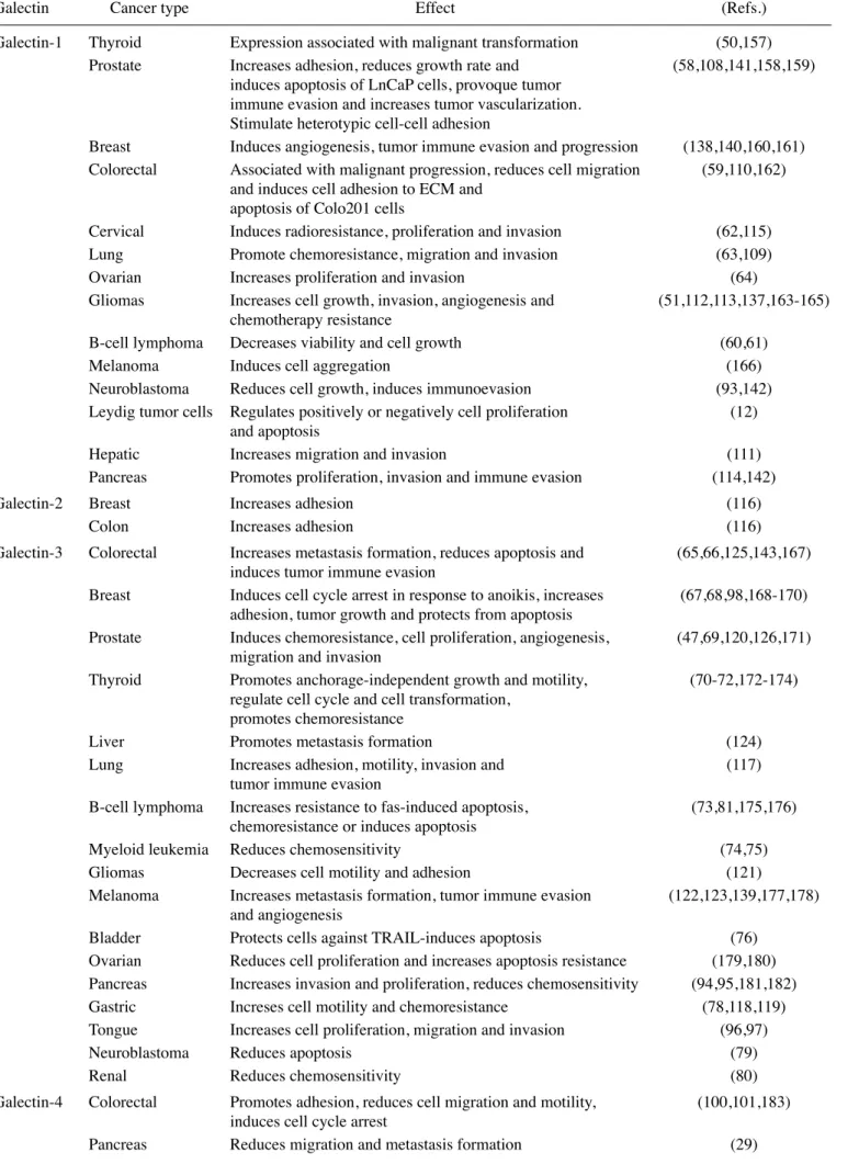
Table III. Intracellular functions of galectins in different cancers.
Galectin
Cancer type
Effect
(Refs.)
Galectin-1 Thyroid
Expression associated with malignant transformation
(50,157)
Prostate
Increases adhesion, reduces growth rate and
(58,108,141,158,159)
induces apoptosis of LnCaP cells, provoque tumor
immune evasion and increases tumor vascularization.
Stimulate heterotypic cell-cell adhesion
Breast
Induces angiogenesis, tumor immune evasion and progression
(138,140,160,161)
Colorectal
Associated with malignant progression, reduces cell migration
(59,110,162)
and induces cell adhesion to ECM and
apoptosis of Colo201 cells
Cervical
Induces radioresistance, proliferation and invasion
(62,115)
Lung
Promote chemoresistance, migration and invasion
(63,109)
Ovarian
Increases proliferation and invasion
(64)
Gliomas
Increases cell growth, invasion, angiogenesis and
(51,112,113,137,163-165)
chemotherapy resistance
B-cell lymphoma Decreases viability and cell growth
(60,61)
Melanoma
Induces cell aggregation
(166)
Neuroblastoma
Reduces cell growth, induces immunoevasion
(93,142)
Leydig tumor cells Regulates positively or negatively cell proliferation
(12)
and apoptosis
Hepatic
Increases migration and invasion
(111)
Pancreas
Promotes proliferation, invasion and immune evasion
(114,142)
Galectin-2 Breast
Increases adhesion
(116)
Colon
Increases adhesion
(116)
Galectin-3 Colorectal
Increases metastasis formation, reduces apoptosis and
(65,66,125,143,167)
induces tumor immune evasion
Breast
Induces cell cycle arrest in response to anoikis, increases
(67,68,98,168-170)
adhesion, tumor growth and protects from apoptosis
Prostate
Induces chemoresistance, cell proliferation, angiogenesis,
(47,69,120,126,171)
migration and invasion
Thyroid
Promotes anchorage-independent growth and motility,
(70-72,172-174)
regulate cell cycle and cell transformation,
promotes chemoresistance
Liver
Promotes metastasis formation
(124)
Lung
Increases adhesion, motility, invasion and
(117)
tumor immune evasion
B-cell lymphoma Increases resistance to fas-induced apoptosis,
(73,81,175,176)
chemoresistance or induces apoptosis
Myeloid leukemia Reduces chemosensitivity
(74,75)
Gliomas
Decreases cell motility and adhesion
(121)
Melanoma
Increases metastasis formation, tumor immune evasion
(122,123,139,177,178)
and angiogenesis
Bladder
Protects cells against TRAIL-induces apoptosis
(76)
Ovarian
Reduces cell proliferation and increases apoptosis resistance
(179,180)
Pancreas
Increases invasion and proliferation, reduces chemosensitivity
(94,95,181,182)
Gastric
Increses cell motility and chemoresistance
(78,118,119)
Tongue
Increases cell proliferation, migration and invasion
(96,97)
Neuroblastoma
Reduces apoptosis
(79)
Renal
Reduces chemosensitivity
(80)
Galectin-4 Colorectal
Promotes adhesion, reduces cell migration and motility,
(100,101,183)
induces cell cycle arrest
(FasR) or apoptosis antigen 1 (APO-1 or APT), leading to
opposing apoptogenic mitochondrial activity (92).
Proliferation. Given their role in apoptosis, it is not surprising
that galectins play a central role in the control of cell
prolif-eration in tumors. This has been well documented in the
case of galectin-1, which reduces proliferation of B-cell
lymphomas, neuroblastoma and LnCap prostate cancer cells
while it increases cell division of glioma, cervical, ovarian
and pancreatic cancer cells (12,51,58,61,62,64,93). A similar
case exists for galectin-3, which displays, once more dual
functionalities in cell proliferation. For instance, galectin-3
increases proliferation of breast, prostate, pancreatic and
tongue tumors (47,94-98). This might be due to the
interac-tion of galectin-3 with the APC/Axin/
β-catenin complex in
the nucleus. This interaction increases the transcriptional
activity of Tcf-4 transcription factor and subsequently
elevates c-myc and cyclin D1 expression (99). In contrast,
cytoplasmic galectin-3, along with galectin-1, bind to
proto-cadherin-24, allowing cytoplasmic localization of
β-catenin,
while decreasing Wnt signaling (10). Ectopic expression of
galectin-4 has also been shown to induce cell cycle arrest
and to reduce cell migration/motility while sensitizing cells
to camptothecin-induced apoptosis in colorectal cancer
(100,101). The data from Satelli et al (101) suggest that
galectin-4 induces downregulation of
β-catenin, Dvl2, TCF1,
TCF4, c-Myc, LRP6 and cyclin D1 expression levels while
upregulating p21, p15 Naked 1 and Ephrin B1 (101,102). An
interaction between galectin-4 and APC/Axin/
β-catenin is
also observed that possibly restricts the translocation of the
complex to the nucleus. This results in a downregulation of
Wnt signaling and a decrease in proliferative potential of
colon cancer cells. In contrast, galectin-7 seems to exhibit
an increased proliferative activity in ovarian cancer cells,
whereas it reduces the proliferation rate of neuroblastomas,
colon and gastric cancer cells (31,103,104). Galectin-8 and -9
have been shown to reduce colon and myelomas tumor growth,
respectively (105,106). Galectin-12, for its part, reduces the
proliferation of T-leukemia and cervical cancer cells (107).
Such contradictory roles for galectins in cell proliferation
suggest that extreme precaution must be taken in order to
target intracellular galectins in cancer.
Adhesion, migration and invasion. The metastatic behavior
of cancer cells is initiated by dysregulation in cell adhesion,
migration and invasion abilities. Alterations of interactions
between extracellular transmembrane receptors and galectins
are often seen in malignancies and late stages of carcinomas.
For instance, galectin-1 increases adhesion of colorectal and
prostate cancer cells (59,108) and stimulates migration of
hepatic and lung cancer cells, while reducing colorectal cell
migration (63,109-111). It also increases the invasive behavior
of gliomas, lung, ovarian, hepatic, pancreatic and cervical
cancer cells (51,63,64,109,111-115). The ability of galectins to
increase adhesion and migration has been well documented
in the case of galectin-2 and most notably in the case of
galectin-3 (47,67,97,116-120). Specifically, galectin-3 reduces
glioma cell migration (121). In general, however, galectin-3
Table III. Continued.
Galectin
Cancer type
Effect
(Refs.)
Galectin-7 Breast
Increases invasion, reduces chemosensitivity
(82)
Lymphoma
Increases metastasis formation
(127-129)
Ovarian
Increases cell proliferation
(31)
Neuroblastoma
Reduces cell growth
(104)
Colon
Increases chemosensitivity and reduces cell growth,
(83)
anchorage-independent cell growth and angiogenesis
Urothelial
Increases chemosensitivity
(85)
Cervical
Increases invasive behavior in vitro, reduces invasion and
(86,87,130)
chemoresistance
Melanoma
Increases chemoresistance
(84)
Gastric
Reduces cell proliferation, migration and invasion
(103)
Galectin-8 Glioblastoma
Stimulates cell migration
(131)
Colon
Reduces tumor growth and cell migration
(106)
Galectin-9 Melanoma
Induces cell aggregation and apoptosis
(35,88,136)
Breast
Increases cell aggregation and reduces adhesion
(36)
Oral
Increases adhesion
(132,133)
Colon
Increases adhesion in vitro but reduces metastasis
(134-136)
formation
in vivo
Myeloma
Reduces cell growth and induces apoptosis
(105,184)
Galectin-12 Cervical
Reduces cell growth
(107)
is mostly associated with increased invasive behavior in
most cancer cell types tested (94,95,117,122-126), supporting
the view that targeting this galectin might be a promising
avenue for the treatment of many types of cancer. Whether
this is also true for other galectins has to be determined. On
the contrary, galectin-4 was found to promote adhesion of
colorectal cells and to reduce migration and metastasis
forma-tion of colorectal and pancreatic cancer cells (29). Further,
conflicting functionalities are once again displayed in the
case of galectin-7 dependent on the cell type. Particularly,
galectin-7 reduces migration of gastric cancer cells and
inva-sion of urothelial and gastric cancer cells (85,103) while it is
associated with increased invasion of other types of cancer,
including breast cancer and T-cell lymphoma (82,127-130).
Galectin-8 also seems to have different abilities to modulate
migration, most notably in glioblastoma and colon cancer
Table IV. Intracellular ligands of galectins.
CRD/non-CRD
Galectin
Binding partners
binding
Effect
(Refs.)
Galectin-1
H-Ras
Increased membrane anchorage of Ras
(52)
and GTP bound state resulting in cell
transformation
Gemin4 (C50)/SMN/
Supply functional snRNPs to the H/E complex
(57)
Gemin2 complex
in the pathway of spliceosome assembly
Protocadherin-24
Localization of
β-catenin to the cell membrane
(10)
resulting in decreased Wnt signaling
Monomeric actin
CRD
Polymerization-depolymerization of actin
(185,186)
in platelet aggregation
Galectin-3
ATP synthase
Inhibition of ATP synthase activity and
(25)
cell cycle progression to G0/G1 phase
Protocadherin-24
Localization
β-catenin to the cell membrane
(10)
resulting in decreased Wnt signaling
CD95 (APO-1/Fas)
Non-CRD
Induction of apoptogenic activity
(92)
at the mitochondria
Nucling
Increase sensitivity to apoptosis
(187)
Synexin
Decrease sensitivity to apoptosis
(91)
CBP70 CRD ND
(188)
β
-catenin/TCF
NH2 and
Induction of transcriptional activity of Tcf-4 with
(99)
complex
COOH termini
an increase in c-Myc + cyclin D1 expression
Axin/
β-catenin/APC
Consensus
Promotion GSK-3
β-dependent phosphorylation
(147)
sequence
of galectin-3/
β-catenin resulting in a decrease
(S92XXXS96)
in Wnt signaling
TTF-1
Upregulation of transcriptional activity of TTF-1
(189)
contributing to cellular proliferation
K-Ras
Increase Raf-1/PI3K signaling and attenuated
(53,54)
ERK signaling
Bcl-2
Non-CRD
Apoptosis-suppressing activity and increase
(89,90)
(NWGR motif)
mitochondrial integrity and decrease
caspase activation
Alix/AIP-1
Facilitation of pro-apoptotic signaling
(190-192)
(Ca
2+dependent)
Gemin4 (C50)/SMN/
Supply functional snRNPs to the H/E complex
(57)
Gemin2 complex
in the pathway of spliceosome assembly
Chrp
CRD ND
(193,194)
Galectin-4
β-catenin/APC/Axin
Increase Naked 1 which destabilizes Dsh/Dvl
(101,102)
proteins resulting in a decreased Wnt signaling
Galectin-7
Bcl-2
Sensitize mitochondria to apoptosis signals
(33)
Smad 3
Decrease expression of TGF-
βresponsive genes
(195)
cells (106,131). A similar scenario exists for galectin-9, which
increases adhesion of melanoma, oral and colon cancer cells,
but reduces adhesion of melanoma and breast cancer cells
and metastasis formation of colon cancer cells
(35,36,132-136). How galectin positively or negatively modulates the
invasive behavior of cancer cells remains largely unknown.
There are some indications that galectins may increase the
secretion of extracellular proteases, remarkably in the case of
galectin-7, which induces the upregulation of matrix
metallo-proteinase-9 (MMP-9) gene expression, possibly through the
p38 mitogenic-activated protein kinase (MAPK) (128,130).
Unlike apoptosis, however, the identification of the
intracel-lular binding partners that are involved in the modulation of
the invasive behavior of cancer cells remains unknown. In
contrast, extracellular galectins and their respective binding
partners have been fairly well characterized.
Other functions of galectins. Angiogenesis is also among
the functions associated with galectin activity. For example,
galectin-1 increases glioma, prostate and breast tumor
vascularisation (108,137,138). Galectin-3 also increases
vascu-larisation of prostate tumors and melanomas, while galectin-7
reduces angiogenesis of colon tumors (47,83,139). Galectins
have been shown to take part in the tumor immune escape.
Indeed, galectin-1 promotes immunoevasion of
neuroblas-toma, prostate, breast and pancreatic cancer cells (140-142).
Galectin-3 also increases tumor immune escape of melanomas,
colorectal and lung cancer cells (117,123,143). Most studies
suggest that extracellular galectins are responsible for these
functions. The involvement of intracellular galectins in these
processes remains unknown.
4. CRD-independent functions for intracellular galectins?
Galectins are primarily known for their ability to bind
to glycans containing lactose or N-aceyllactosamine via
Van der Walls interactions between the carbohydrate and
binding pocket. They have a relatively broad specificity
depending on the type and the length of the carbohydrate and
the mode of presentation of ligand to the CRD. It is thus logical
to assume that inside the cells, they will also preferentially
bind to intracellular glycoconjugates, which are abundantly
found in the cytosol. There is compelling evidence, however,
that galectins might have non-carbohydrate binding partners
and functions. CRD-independent functions have been
particu-larly well documented for intracellular galectins (144-146).
For example, galectins do interact with Bcl-2 family members
via a CRD-independent interaction (33,85,89,90). This
galectin/Bcl-2 interaction is important since the balance of
activity between pro- and anti-apoptotic signals of members of
the Bcl-2 family regulates apoptosis. Other CRD-independent
functions of galectins include RNA processing in the nucleus
(57) and regulation of cell cycle progression (Wnt signaling?)
(25,99,101,102,147). All these galectin functions are
inde-pendent of their saccharidic binding activities and rather
rely on protein-protein interactions. Some galectins, such
as galectin-10, harbor very low affinity for galactosides and
are believed to act mainly through other specificities, while
their CRD binding activity remains debated (148,149). These
CRD-independent functions represent a paradigm shift in our
understanding of galectin function and the development of
galectin-specific antagonists.
A new challenge: studying the redundancy of galectin
func-tions. The existence of redundant or antagonistic functions
between galectins is a major concern because these proteins
can converge under normal or pathological conditions. The
cross-talk between intracellular galectins remains completely
unknown although cells often express more than one
intracel-lular galectin. For example, MCF-7 breast cancer cells express
galectin-3, -8 and -9 (150). MCF-10 and MDA-MB-468, two
other human mammary epithelial cell lines, express both
galectin-3 and -7, but not galectin-8 or -9 (151,152). Moreover,
many galectins could be present within the same intracellular
compartments. A case in point is the mitochondria, where both
galectin-3 and -7 are found. Galectin-12 can also be present in
mitochondria and not surprisingly, it seems to be involved in
the control of cellular metabolism (38-40). Whether galectins
have redundant or opposed functions in the mitochondria
is an interesting question given the critical role of cellular
metabolism in cancer. A better understanding of the functional
redundancy among homologous proteins, which is frequently
observed in eukaryotes, is also critical. Such redundancy often
occurs in order to increase maintenance of important gene
function and to limit losses following mutations/deletions of
specific genes (functional compensation). Lessons learned
from such studies could also bring important insight into
many others fields, from understanding pathologies to general
developmental biology.
Future directions. Because of their critical role in cancer,
considerable efforts have been directed towards the
devel-opment of carbohydrate-based inhibitors that would limit
the binding of galectins to glycosylated residues on cell
surface receptors. For example, GCS-100 is a galectin-3
antagonist with a modified citrus pectin carbohydrate that
has been shown to inhibit tumor growth and metastasis in
several preclinical models (153-155). Others, like OTX008,
a galectin-1 antagonist, act as allosteric CRD-dependent
inhibitors following binding to a site distant from the
carbo-hydrate-binding site (156). Nevertheless, despite almost two
decades of research, the development of effective galectin
antagonists for the treatment of cancer has met with limited
success. The emerging evidence that galectins have critical
intracellular and CRD-independent functions calls for a
refo-cusing of our efforts on development of new galectin-specific
antagonists to modulate apoptosis. Our knowledge of the
subcellular localization of galectins will also significantly
improve target identification during the drug discovery
process. It is thus imperative to better understand the role of
intracellular galectins and to provide novel insight into how
galectins collaboratively modulate cancer progression from
within the cells.
Acknowledgements
This research was supported by a grant from the National
Science and Engineering Research Council of Canada
(NSERC). M.L. is supported by a doctoral studentship from
the Fonds de la Recherche du Québec-Santé (FRQS).
References
1. Levi G and Teichberg VI: Isolation and physicochemical characterization of electrolectin, a beta-D-galactoside binding lectin from the electric organ of Electrophorus electricus. J Biol Chem 256: 5735-5740, 1981.
2. Astorgues-Xerri L, Riveiro ME, Tijeras-Raballand A, et al: Unraveling galectin-1 as a novel therapeutic target for cancer. Cancer Treat Rev: Aug 1, 2013 (Epub ahead of print).
3. Radosavljevic G, Volarevic V, Jovanovic I, et al: The roles of Galectin-3 in autoimmunity and tumor progression. Immunol Res 52: 100-110, 2012.
4. Van den Brule F, Califice S and Castronovo V: Expression of galectins in cancer: a critical review. Glycoconj J 19: 537-542, 2004.
5. Liu FT and Rabinovich GA: Galectins as modulators of tumour progression. Nat Rev Cancer 5: 29-41, 2005.
6. Partridge EA, Le Roy C, Di Guglielmo GM, et al: Regulation of cytokine receptors by Golgi N-glycan processing and endo-cytosis. Science 306: 120-124, 2004.
7. Perillo NL, Uittenbogaart CH, Nguyen JT and Baum LG: Galectin-1, an endogenous lectin produced by thymic epithelial cells, induces apoptosis of human thymocytes. J Exp Med 185: 1851-1858, 1997.
8. Chiariotti L, Salvatore P, Frunzio R and Bruni CB: Galectin genes: regulation of expression. Glycoconj J 19: 441-449, 2004. 9. Vyakarnam A, Lenneman AJ, Lakkides KM, Patterson RJ and
Wang JL: A comparative nuclear localization study of galectin-1 with other splicing components. Exp Cell Res 242: 419-428, 1998. 10. Ose R, Oharaa O and Nagase T: Galectin-1 and galectin-3
mediate protocadherin-24-dependent membrane localization of beta-catenin in colon cancer cell line HCT116. Curr Chem Genomics 6: 18-26, 2012.
11. Gabius HJ, Andre S, Gunsenhauser I, et al: Association of galectin-1- but not galectin-3-dependent parameters with proliferation activity in human neuroblastomas and small cell lung carcinomas. Anticancer Res 22: 405-410, 2002.
12. Biron VA, Iglesias MM, Troncoso MF, et al: Galectin-1: biphasic growth regulation of Leydig tumor cells. Glycobiology 16: 810-821, 2006.
13. Saussez S, Decaestecker C, Lorfevre F, et al: Increased expression and altered intracellular distribution of adhesion/growth-regulatory lectins galectins-1 and -7 during tumour progression in hypopharyngeal and laryngeal squamous cell carcinomas. Histopathology 52: 483-493, 2008.
14. Van den Brule FA, Buicu C, Baldet M, et al: Galectin-1 modulates human melanoma cell adhesion to laminin. Biochem Biophys Res Commun 209: 760-767, 1995.
15. Sanjuan X, Fernandez PL, Castells A, et al: Differential expression of galectin 3 and galectin 1 in colorectal cancer progression. Gastroenterology 113: 1906-1915, 1997.
16. Dvorankova B, Lacina L, Smetana K Jr, et al: Human galectin-2: nuclear presence in vitro and its modulation by quiescence/ stress factors. Histol Histopathol 23: 167-178, 2008.
17. Uhlen M, Oksvold P, Fagerberg L, et al: Towards a knowledge-based Human Protein Atlas. Nat Biotechnol 28: 1248-1250, 2010.
18. Viaene AN, Petrof I and Sherman SM: Properties of the thalamic projection from the posterior medial nucleus to primary and secondary somatosensory cortices in the mouse. Proc Natl Acad Sci USA 108: 18156-18161, 2011.
19. Van den Brule FA, Buicu C, Berchuck A, et al: Expression of the 67-kD laminin receptor, galectin-1, and galectin-3 in advanced human uterine adenocarcinoma. Hum Pathol 27: 1185-1191, 1996.
20. Mey A, Berthier-Vergnes O, Apoil PA, Dore JF and Revillard JP: Expression of the galactose binding protein Mac-2 by human melanoma cell-lines. Cancer Lett 81: 155-163, 1994.
21. Matesa-Anic D, Moslavac S, Matesa N, Cupic H and Kusic Z: Intensity and distribution of immunohistochemical expression of galectin-3 in thyroid neoplasms. Acta Clin Croat 51: 237-241, 2012.
22. Lotz MM, Andrews CW Jr, Korzelius CA, et al: Decreased expression of Mac-2 (carbohydrate binding protein 35) and loss of its nuclear localization are associated with the neoplastic progression of colon carcinoma. Proc Natl Acad Sci USA 90: 3466-3470, 1993.
23. Kawachi K, Matsushita Y, Yonezawa S, et al: Galectin-3 expression in various thyroid neoplasms and its possible role in metastasis formation. Hum Pathol 31: 428-433, 2000.
24. Honjo Y, Inohara H, Akahani S, et al: Expression of cyto-plasmic galectin-3 as a prognostic marker in tongue carcinoma. Clin Cancer Res 6: 4635-4640, 2000.
25. Kim DW, Kim KH, Yoo BC, et al: Identification of mitochon-drial F(1)F(0)-ATP synthase interacting with galectin-3 in colon cancer cells. Cancer Sci 99: 1884-1891, 2008.
26. Lepur A, Carlsson MC, Novak R, Dumic J, Nilsson UJ and Leffler H: Galectin-3 endocytosis by carbohydrate independent and dependent pathways in different macrophage like cell types. Biochim Biophys Acta 1820: 804-818, 2012.
27. Huflejt ME, Jordan ET, Gitt MA, Barondes SH and Leffler H: Strikingly different localization of galectin-3 and galectin-4 in human colon adenocarcinoma T84 cells. Galectin-4 is localized at sites of cell adhesion. J Biol Chem 272: 14294-14303, 1997. 28. Huflejt ME and Leffler H: Galectin-4 in normal tissues and
cancer. Glycoconj J 20: 247-255, 2004.
29. Belo AI, van der Sar AM, Tefsen B and van Die I: Galectin-4 reduces migration and metastasis formation of pancreatic cancer cells. PLoS One 8: e65957, 2013.
30. Kuwabara I, Kuwabara Y, Yang RY, et al: Galectin-7 (PIG1) exhibits pro-apoptotic function through JNK activation and mitochondrial cytochrome c release. J Biol Chem 277: 3487-3497, 2002.
31. Kim HJ, Jeon HK, Lee JK, et al: Clinical significance of galectin-7 in epithelial ovarian cancer. Anticancer Res 33: 1555-1561, 2013.
32. De Vasconcelos Carvalho M, Pereira Jdos S, Alves PM, Silveira EJ, de Souza LB and Queiroz LM: Alterations in the immunoexpression of galectins-1, -3 and -7 between different grades of oral epithelial dysplasia. J Oral Pathol Med 42: 174-179, 2013.
33. Villeneuve C, Baricault L, Canelle L, et al: Mitochondrial proteomic approach reveals galectin-7 as a novel BCL-2 binding protein in human cells. Mol Biol Cell 22: 999-1013, 2011. 34. Delgado VM, Nugnes LG, Colombo LL, et al: Modulation of
endothelial cell migration and angiogenesis: a novel function for the ‘tandem-repeat’ lectin galectin-8. FASEB J 25: 242-254, 2011. 35. Kageshita T, Kashio Y, Yamauchi A, et al: Possible role of
galectin-9 in cell aggregation and apoptosis of human melanoma cell lines and its clinical significance. Int J Cancer 99: 809-816, 2002.
36. Irie A, Yamauchi A, Kontani K, et al: Galectin-9 as a prog-nostic factor with antimetastatic potential in breast cancer. Clin Cancer Res 11: 2962-2968, 2005.
37. Rousseau C, Muriel MP, Musset M, Botti J and Seve AP: Glycosylated nuclear lectin CBP70 also associated with endo-plasmic reticulum and the Golgi apparatus: does the ‘classic pathway’ of glycosylation also apply to nuclear glycoproteins? J Cell Biochem 78: 638-649, 2000.
38. Hotta K, Funahashi T, Matsukawa Y, et al: Galectin-12, an adipose-expressed galectin-like molecule possessing apoptosis-inducing activity. J Biol Chem 276: 34089-34097, 2001.
39. Wang JL, Gray RM, Haudek KC and Patterson RJ: Nucleocytoplasmic lectins. Biochim Biophys Acta 1673: 75-93, 2004.
40. Carlsson S, Carlsson MC and Leffler H: Intracellular sorting of galectin-8 based on carbohydrate fine specificity. Glycobiology 17: 906-912, 2007.
41. Yang RY, Yu L, Graham JL, et al: Ablation of a galectin pref-erentially expressed in adipocytes increases lipolysis, reduces adiposity, and improves insulin sensitivity in mice. Proc Natl Acad Sci USA 108: 18696-18701, 2011.
42. Baum LG: Burn control, an adipocyte-specific function for galectin-12. Proc Natl Acad Sci USA 108: 18575-18576, 2011. 43. Kubach J, Lutter P, Bopp T, et al: Human CD4+CD25+
regula-tory T cells: proteome analysis identifies galectin-10 as a novel marker essential for their anergy and suppressive function. Blood 110: 1550-1558, 2007.
44. Than NG, Pick E, Bellyei S, et al: Functional analyses of placental protein 13/galectin-13. Eur J Biochem 271: 1065-1078, 2004. 45. Nakai K and Horton P: PSORT: a program for detecting sorting
signals in proteins and predicting their subcellular localization. Trends Biochem Sci 24: 34-36, 1999.
46. Nakai K and Kanehisa M: A knowledge base for predicting protein localization sites in eukaryotic cells. Genomics 14: 897-911, 1992.
47. Califice S, Castronovo V, Bracke M and van den Brule F: Dual activities of galectin-3 in human prostate cancer: tumor suppression of nuclear galectin-3 vs tumor promotion of cyto-plasmic galectin-3. Oncogene 23: 7527-7536, 2004.
48. St-Pierre Y, Campion CG and Grosset AA: A distinctive role for galectin-7 in cancer? Front Biosci (Landmark Ed) 17: 438-450, 2012.
49. Chiariotti L, Berlingieri MT, De Rosa P, et al: Increased expression of the negative growth factor, galactoside-binding protein, gene in transformed thyroid cells and in human thyroid carcinomas. Oncogene 7: 2507-2511, 1992.
50. Xu XC, el-Naggar AK and Lotan R: Differential expression of galectin-1 and galectin-3 in thyroid tumors. Potential diagnostic implications. Am J Pathol 147: 815-822, 1995.
51. Yamaoka K, Mishima K, Nagashima Y, Asai A, Sanai Y and Kirino T: Expression of galectin-1 mRNA correlates with the malignant potential of human gliomas and expression of antisense galectin-1 inhibits the growth of 9 glioma cells. J Neurosci Res 59: 722-730, 2000.
52. Paz A, Haklai R, Elad-Sfadia G, Ballan E and Kloog Y: Galectin-1 binds oncogenic H-Ras to mediate Ras membrane anchorage and cell transformation. Oncogene 20: 7486-7493, 2001.
53. Elad-Sfadia G, Haklai R, Balan E and Kloog Y: Galectin-3 augments K-Ras activation and triggers a Ras signal that attenuates ERK but not phosphoinositide 3-kinase activity. J Biol Chem 279: 34922-34930, 2004.
54. Shalom-Feuerstein R, Levy R, Makovski V, Raz A and Kloog Y: Galectin-3 regulates RasGRP4-mediated activation of N-Ras and H-Ras. Biochim Biophys Acta 1783: 985-993, 2008. 55. Hebert E and Monsigny M: Galectin-3 mRNA level depends on
transformation phenotype in ras-transformed NIH 3T3 cells. Biol Cell 81: 73-76, 1994.
56. Hebert E, Roche AC, Nachtigal M and Monsigny M: Transformation but not ras-transfection increases the expres-sion of galectin-3 in human HOS cells. C R Acad Sci III 319: 871-877, 1996.
57. Park JW, Voss PG, Grabski S, Wang JL and Patterson RJ: Association of galectin-1 and galectin-3 with Gemin4 in complexes containing the SMN protein. Nucleic Acids Res 29: 3595-3602, 2001.
58. Ellerhorst J, Nguyen T, Cooper DN, Estrov Y, Lotan D and Lotan R: Induction of differentiation and apoptosis in the prostate cancer cell line LNCaP by sodium butyrate and galectin-1. Int J Oncol 14: 225-232, 1999.
59. Horiguchi N, Arimoto K, Mizutani A, Endo-Ichikawa Y, Nakada H and Taketani S: Galectin-1 induces cell adhesion to the extracellular matrix and apoptosis of non-adherent human colon cancer Colo201 cells. J Biochem 134: 869-874, 2003. 60. Fouillit M, Joubert-Caron R, Poirier F, et al: Regulation of
CD45-induced signaling by galectin-1 in Burkitt lymphoma B cells. Glycobiology 10: 413-419, 2000.
61. Poirier F, Bourin P, Bladier D, Joubert-Caron R and Caron M: Effect of 5-azacytidine and galectin-1 on growth and differen-tiation of the human b lymphoma cell line bl36. Cancer Cell Int 1: 2, 2001.
62. Huang EY, Chen YF, Chen YM, et al: A novel radioresistant mechanism of galectin-1 mediated by H-Ras-dependent pathways in cervical cancer cells. Cell Death Dis 3: e251, 2012. 63. Chung LY, Tang SJ, Sun GH, et al: Galectin-1 promotes lung
cancer progression and chemoresistance by upregulating p38 MAPK, ERK, and cyclooxygenase-2. Clin Cancer Res 18: 4037-4047, 2012.
64. Kim HJ, Jeon HK, Cho YJ, et al: High galectin-1 expression correlates with poor prognosis and is involved in epithelial ovarian cancer proliferation and invasion. Eur J Cancer 48: 1914-1921, 2012.
65. Mazurek N, Byrd JC, Sun Y, et al: Cell-surface galectin-3 confers resistance to TRAIL by impeding trafficking of death receptors in metastatic colon adenocarcinoma cells. Cell Death Differ 19: 523-533, 2012.
66. Shi Y, He B, Kuchenbecker KM, et al: Inhibition of Wnt-2 and galectin-3 synergistically destabilizes beta-catenin and induces apoptosis in human colorectal cancer cells. Int J Cancer 121: 1175-1181, 2007.
67. Matarrese P, Fusco O, Tinari N, et al: Galectin-3 overexpression protects from apoptosis by improving cell adhesion properties. Int J Cancer 85: 545-554, 2000.
68. Moon BK, Lee YJ, Battle P, Jessup JM, Raz A and Kim HR: Galectin-3 protects human breast carcinoma cells against nitric oxide-induced apoptosis: implication of galectin-3 function during metastasis. Am J Pathol 159: 1055-1060, 2001.
69. Fukumori T, Oka N, Takenaka Y, et al: Galectin-3 regulates mitochondrial stability and antiapoptotic function in response to anticancer drug in prostate cancer. Cancer Res 66: 3114-3119, 2006.
70. Lavra L, Ulivieri A, Rinaldo C, et al: Gal-3 is stimulated by gain-of-function p53 mutations and modulates chemoresistance in anaplastic thyroid carcinomas. J Pathol 218: 66-75, 2009. 71. Lin CI, Whang EE, Abramson MA, et al: Galectin-3 regulates
apoptosis and doxorubicin chemoresistance in papillary thyroid cancer cells. Biochem Biophys Res Commun 379: 626-631, 2009.
72. Lin CI, Whang EE, Donner DB, et al: Galectin-3 targeted therapy with a small molecule inhibitor activates apoptosis and enhances both chemosensitivity and radiosensitivity in papillary thyroid cancer. Mol Cancer Res 7: 1655-1662, 2009. 73. Hoyer KK, Pang M, Gui D, et al: An anti-apoptotic role for
galectin-3 in diffuse large B-cell lymphomas. Am J Pathol 164: 893-902, 2004.
74. Cheng YL, Huang WC, Chen CL, et al: Increased galectin-3 facilitates leukemia cell survival from apoptotic stimuli. Biochem Biophys Res Commun 412: 334-340, 2011.
75. Yamamoto-Sugitani M, Kuroda J, Ashihara E, et al: Galectin-3 (Gal-3) induced by leukemia microenvironment promotes drug resistance and bone marrow lodgment in chronic myelogenous leukemia. Proc Natl Acad Sci USA 108: 17468-17473, 2011. 76. Oka N, Nakahara S, Takenaka Y, et al: Galectin-3 inhibits tumor
necrosis factor-related apoptosis-inducing ligand-induced apoptosis by activating Akt in human bladder carcinoma cells. Cancer Res 65: 7546-7553, 2005.
77. Kobayashi T, Shimura T, Yajima T, et al: Transient gene silencing of galectin-3 suppresses pancreatic cancer cell migration and invasion through degradation of beta-catenin. Int J Cancer 129: 2775-2786, 2011.
78. Cheong TC, Shin JY and Chun KH: Silencing of galectin-3 changes the gene expression and augments the sensitivity of gastric cancer cells to chemotherapeutic agents. Cancer Sci 101: 94-102, 2010.
79. Veschi V, Petroni M, Cardinali B, et al: Galectin-3 impairment of MYCN-dependent apoptosis-sensitive phenotype is antagonized by nutlin-3 in neuroblastoma cells. PLoS One 7: e49139, 2012. 80. Xu Y, Gu X, Gong M, Guo G, Han K and An R: Galectin-3
inhi-bition sensitizes human renal cell carcinoma cells to arsenic trioxide treatment. Cancer Biol Ther 14: 897-906, 2013. 81. Suzuki O and Abe M: Cell surface N-glycosylation and
sialylation regulate galectin-3-induced apoptosis in human diffuse large B cell lymphoma. Oncol Rep 19: 743-748, 2008. 82. Demers M, Rose AA, Grosset AA, et al: Overexpression of
galectin-7, a myoepithelial cell marker, enhances spontaneous metastasis of breast cancer cells. Am J Pathol 176: 3023-3031, 2010.
83. Ueda S, Kuwabara I and Liu FT: Suppression of tumor growth by galectin-7 gene transfer. Cancer Res 64: 5672-5676, 2004. 84. Biron-Pain K, Grosset AA, Poirier F, Gaboury L and
St-Pierre Y: Expression and functions of galectin-7 in human and murine melanomas. PLoS One 8: e63307, 2013.
85. Matsui Y, Ueda S, Watanabe J, Kuwabara I, Ogawa O and Nishiyama H: Sensitizing effect of galectin-7 in urothelial cancer to cisplatin through the accumulation of intracellular reactive oxygen species. Cancer Res 67: 1212-1220, 2007. 86. Tsai CJ, Sulman EP, Eifel PJ, et al: Galectin-7 levels predict
radiation response in squamous cell carcinoma of the cervix. Gynecol Oncol: Apr 30, 2013 (Epub ahead of print).
87. Zhu H, Wu TC, Chen WQ, et al: Roles of galectin-7 and S100A9 in cervical squamous carcinoma: Clinicopathological and in vitro evidence. Int J Cancer 132: 1051-1059, 2013.
88. Wiersma VR, de Bruyn M, van Ginkel RJ, et al: The glycan-binding protein galectin-9 has direct apoptotic activity toward melanoma cells. J Invest Dermatol 132: 2302-2305, 2012.
89. Yang RY, Hsu DK and Liu FT: Expression of galectin-3 modulates T-cell growth and apoptosis. Proc Natl Acad Sci USA 93: 6737-6742, 1996.
90. Akahani S, Nangia-Makker P, Inohara H, Kim HR and Raz A: Galectin-3: a novel antiapoptotic molecule with a functional BH1 (NWGR) domain of Bcl-2 family. Cancer Res 57: 5272-5276, 1997.
91. Dumic J, Dabelic S and Flogel M: Galectin-3: an open-ended story. Biochim Biophys Acta 1760: 616-635, 2006.
92. Fukumori T, Takenaka Y, Oka N, et al: Endogenous galectin-3 determines the routing of CD95 apoptotic signaling pathways. Cancer Res 64: 3376-3379, 2004.
93. Kopitz J, von Reitzenstein C, Andre S, et al: Negative regulation of neuroblastoma cell growth by carbohydrate-dependent surface binding of galectin-1 and functional divergence from galectin-3. J Biol Chem 276: 35917-35923, 2001.
94. Jiang HB, Xu M and Wang XP: Pancreatic stellate cells promote proliferation and invasiveness of human pancreatic cancer cells via galectin-3. World J Gastroenterol 14: 2023-2028, 2008. 95. Song S, Ji B, Ramachandran V, et al: Overexpressed galectin-3
in pancreatic cancer induces cell proliferation and invasion by binding Ras and activating Ras signaling. PLoS One 7: e42699, 2012.
96. Wang LP, Chen SW, Zhuang SM, Li H and Song M: Galectin-3 accelerates the progression of oral tongue squamous cell carcinoma via a Wnt/beta-catenin-dependent pathway. Pathol Oncol Res 19: 461-474, 2013.
97. Zhang D, Chen ZG, Liu SH, et al: Galectin-3 gene silencing inhibits migration and invasion of human tongue cancer cells in vitro via downregulating beta-catenin. Acta Pharmacol Sin 34: 176-184, 2013.
98. Kim HR, Lin HM, Biliran H and Raz A: Cell cycle arrest and inhibition of anoikis by galectin-3 in human breast epithelial cells. Cancer Res 59: 4148-4154, 1999.
99. Shimura T, Takenaka Y, Tsutsumi S, Hogan V, Kikuchi A and Raz A: Galectin-3, a novel binding partner of beta-catenin. Cancer Res 64: 6363-6367, 2004.
100. Kim SW, Park KC, Jeon SM, et al: Abrogation of galectin-4 expression promotes tumorigenesis in colorectal cancer. Cell Oncol (Dordr) 36: 169-178, 2013.
101. Satelli A, Rao PS, Thirumala S and Rao US: Galectin-4 functions as a tumor suppressor of human colorectal cancer. Int J Cancer 129: 799-809, 2011.
102. Guo J, Cagatay T, Zhou G, et al: Mutations in the human naked cuticle homolog NKD1 found in colorectal cancer alter Wnt/Dvl/beta-catenin signaling. PLoS One 4: e7982, 2009. 103. Kim SJ, Hwang JA, Ro JY, Lee YS and Chun KH: Galectin-7
is epigenetically-regulated tumor suppressor in gastric cancer. Oncotarget 4: 1461-1471, 2013.
104. Kopitz J, Andre S, von Reitzenstein C, et al: Homodimeric galectin-7 (p53-induced gene 1) is a negative growth regulator for human neuroblastoma cells. Oncogene 22: 6277-6288, 2003.
105. Kobayashi T, Kuroda J, Ashihara E, et al: Galectin-9 exhibits anti-myeloma activity through JNK and p38 MAP kinase pathways. Leukemia 24: 843-850, 2010.
106. Nagy N, Bronckart Y, Camby I, et al: Galectin-8 expression decreases in cancer compared with normal and dysplastic human colon tissue and acts significantly on human colon cancer cell migration as a suppressor. Gut 50: 392-401, 2002. 107. Yang RY, Hsu DK, Yu L, Ni J and Liu FT: Cell cycle
regula-tion by galectin-12, a new member of the galectin superfamily. J Biol Chem 276: 20252-20260, 2001.
108. Clausse N, van den Brule F, Waltregny D, Garnier F and Castronovo V: Galectin-1 expression in prostate tumor-associated capillary endothelial cells is increased by prostate carcinoma cells and modulates heterotypic cell-cell adhesion. Angiogenesis 3: 317-325, 1999.
109. Hsu YL, Wu CY, Hung JY, Lin YS, Huang MS and Kuo PL: Galectin-1 promotes lung cancer tumor metastasis by poten-tiating integrin alpha6beta4 and Notch1/Jagged2 signaling pathway. Carcinogenesis 34: 1370-1381, 2013.
110. Hittelet A, Legendre H, Nagy N, et al: Upregulation of galectins-1 and -3 in human colon cancer and their role in regulating cell migration. Int J Cancer 103: 370-379, 2003. 111. Spano D, Russo R, Di Maso V, et al: Galectin-1 and its
involve-ment in hepatocellular carcinoma aggressiveness. Mol Med 16: 102-115, 2010.
112. Jung TY, Jung S, Ryu HH, et al: Role of galectin-1 in migration and invasion of human glioblastoma multiforme cell lines. J Neurosurg 109: 273-284, 2008.
113. Rorive S, Belot N, Decaestecker C, et al: Galectin-1 is highly expressed in human gliomas with relevance for modulation of invasion of tumor astrocytes into the brain parenchyma. Glia 33: 241-255, 2001.
114. Xue X, Lu Z, Tang D, et al: Galectin-1 secreted by activated stellate cells in pancreatic ductal adenocarcinoma stroma promotes proliferation and invasion of pancreatic cancer cells: an in vitro study on the microenvironment of pancreatic ductal adenocarcinoma. Pancreas 40: 832-839, 2011.
115. Kim HJ, Do IG, Jeon HK, et al: Galectin 1 expression is asso-ciated with tumor invasion and metastasis in stage IB to IIA cervical cancer. Hum Pathol 44: 62-68, 2013.
116. Barrow H, Guo X, Wandall HH, et al: Serum galectin-2, -4, and -8 are greatly increased in colon and breast cancer patients and promote cancer cell adhesion to blood vascular endothelium. Clin Cancer Res 17: 7035-7046, 2011.
117. O'Driscoll L, Linehan R, Liang YH, Joyce H, Oglesby I and Clynes M: Galectin-3 expression alters adhesion, motility and invasion in a lung cell line (DLKP), in vitro. Anticancer Res 22: 3117-3125, 2002.
118. Kim SJ, Choi IJ, Cheong TC, et al: Galectin-3 increases gastric cancer cell motility by up-regulating fascin-1 expression. Gastroenterology 138: 1035-1045.e2, 2010.
119. Kim SJ, Shin JY, Lee KD, et al: Galectin-3 facilitates cell motility in gastric cancer by up-regulating protease-activated receptor-1 (PAR-1) and matrix metalloproteinase-1 (MMP-1). PLoS One 6: e25103, 2011.
120. Wang Y, Nangia-Makker P, Tait L, et al: Regulation of prostate cancer progression by galectin-3. Am J Pathol 174: 1515-1523, 2009.
121. Debray C, Vereecken P, Belot N, et al: Multifaceted role of galectin-3 on human glioblastoma cell motility. Biochem Biophys Res Commun 325: 1393-1398, 2004.
122. Braeuer RR, Zigler M, Kamiya T, et al: Galectin-3 contributes to melanoma growth and metastasis via regulation of NFAT1 and autotaxin. Cancer Res 72: 5757-5766, 2012.
123. Radosavljevic G, Jovanovic I, Majstorovic I, et al: Deletion of galectin-3 in the host attenuates metastasis of murine melanoma by modulating tumor adhesion and NK cell activity. Clin Exp Metastasis 28: 451-462, 2011.
124. Inufusa H, Nakamura M, Adachi T, et al: Role of galectin-3 in adenocarcinoma liver metastasis. Int J Oncol 19: 913-919, 2001. 125. Bresalier RS, Mazurek N, Sternberg LR, et al: Metastasis of
human colon cancer is altered by modifying expression of the beta-galactoside-binding protein galectin 3. Gastroenterology 115: 287-296, 1998.
126. Ellerhorst JA, Stephens LC, Nguyen T and Xu XC: Effects of galectin-3 expression on growth and tumorigenicity of the prostate cancer cell line LNCaP. Prostate 50: 64-70, 2002. 127. Demers M, Biron-Pain K, Hebert J, Lamarre A, Magnaldo T
and St-Pierre Y: Galectin-7 in lymphoma: elevated expression in human lymphoid malignancies and decreased lymphoma dissemination by antisense strategies in experimental model. Cancer Res 67: 2824-2829, 2007.
128. Demers M, Magnaldo T and St-Pierre Y: A novel function for galectin-7: promoting tumorigenesis by up-regulating MMP-9 gene expression. Cancer Res 65: 5205-5210, 2005.
129. Moisan S, Demers M, Mercier J, Magnaldo T, Potworowski EF and St-Pierre Y: Upregulation of galectin-7 in murine lymphoma cells is associated with progression toward an aggressive phenotype. Leukemia 17: 751-759, 2003.
130. Park JE, Chang WY and Cho M: Induction of matrix metal-loproteinase-9 by galectin-7 through p38 MAPK signaling in HeLa human cervical epithelial adenocarcinoma cells. Oncol Rep 22: 1373-1379, 2009.
131. Camby I, Belot N, Rorive S, et al: Galectins are differentially expressed in supratentorial pilocytic astrocytomas, astro-cytomas, anaplastic astrocytomas and glioblastomas, and significantly modulate tumor astrocyte migration. Brain Pathol 11: 12-26, 2001.
132. Kasamatsu A, Uzawa K, Nakashima D, et al: Galectin-9 as a regulator of cellular adhesion in human oral squamous cell carcinoma cell lines. Int J Mol Med 16: 269-273, 2005. 133. Yamauchi A, Kontani K, Kihara M, Nishi N, Yokomise H and
Hirashima M: Galectin-9, a novel prognostic factor with anti-metastatic potential in breast cancer. Breast J 12: S196-S200, 2006.
134. Zhang F, Zheng M, Qu Y, et al: Different roles of galectin-9 isoforms in modulating E-selectin expression and adhesion function in LoVo colon carcinoma cells. Mol Biol Rep 36: 823-830, 2009.
135. Zhang F, Zheng MH, Qu Y, et al: Galectin-9 isoforms influence the adhesion between colon carcinoma LoVo cells and human umbilical vein endothelial cells in vitro by regulating the expression of E-selectin in LoVo cells. Zhonghua Zhong Liu Za Zhi 31: 95-98, 2009 (In Chinese).
136. Nobumoto A, Nagahara K, Oomizu S, et al: Galectin-9 suppresses tumor metastasis by blocking adhesion to endothelium and extra-cellular matrices. Glycobiology 18: 735-744, 2008.
137. Le Mercier M, Mathieu V, Haibe-Kains B, et al: Knocking down galectin 1 in human hs683 glioblastoma cells impairs both angiogenesis and endoplasmic reticulum stress responses. J Neuropathol Exp Neurol 67: 456-469, 2008.
138. Ito K, Scott SA, Cutler S, et al: Thiodigalactoside inhibits murine cancers by concurrently blocking effects of galectin-1 on immune dysregulation, angiogenesis and protection against oxidative stress. Angiogenesis 14: 293-307, 2011.