Science Arts & Métiers (SAM)
is an open access repository that collects the work of Arts et Métiers Institute of
Technology researchers and makes it freely available over the web where possible.
This is an author-deposited version published in: https://sam.ensam.eu
Handle ID: .http://hdl.handle.net/10985/18636
To cite this version :
Jean-Yves LAZENNEC, Henri D'ASTORG, Marc-Antoine ROUSSEAU - Cervical spine surgery in
ankylosing spondylitis : review and current concept - Orthopaedics & Traumatology : Surgery &
Research - Vol. 101, n°4, p.507-513 - 2015
Any correspondence concerning this service should be sent to the repository
Administrator : [email protected]
Cervical
spine surgery in ankylosing spondylitis: Review and current
concept
J.-Y.
Lazennec
a,b,
H. d’Astorg
c,
M.-A. Rousseau
b,d,∗a Service de chirurgie orthopédique et traumatologique, hôpital Pitié-Salpetrière, AP–HP, 47, boulevard de l’Hôpital, 75013 Paris, France b Laboratoire de biomécanique, arts et métiers Paristech, 151, boulevard de l’Hôpital, 75013 Paris, France
c Service de chirurgie orthopédique et traumatologique, hôpital Beaujon, AP–HP, 100, boulevard du Général-Leclerc, 92110 Clichy, France d Service de chirurgie orthopédique et traumatologique, hôpital Avicenne, AP–HP, 125, rue de Stalingrad, 93000 Bobigny, France
Keywords: Cervical spine Ankylosing spondylitis Fracture DISH
a b s t r a c t
Ankylosing spondylitis of the cervical spine is associated with stiff kyphosis and increased risk of transver-sal unstable fracture. A spine surgeon may be involved mainly in the management of trauma cases, but in some situations, corrective surgery of a kyphotic cervical deformity is needed. Both types of cases carry specific aspects and rely on principles that differ from those associated with more common cer-vical surgery. This paper is a review of the literature regarding cercer-vical surgery in cases of ankylosing spondylitis. It addresses practical technical questions.
1. Introduction
Ankylosing spondylitis (AS) is an inflammatory rheumatism that may induce structural damage in the cervical spine. Microscopic changes include bone fragility[1]arising from decreased bone den-sity, which has been shown to be related to persistent systemic inflammation and hypervascularisation of the bone. In the severest forms, macroscopic changes result in spontaneous intervertebral fusion and kyphosis[2]of the entire spine (the so-called “bamboo spine”). The ossification concerns the disc space anteriorly and the facet joints posteriorly. While the normal cervical spine provides mobility and allows upright posture of the head, the AS spine is excessively stiff and the flexed posture of the neck is mostly debil-itating. In addition to other possible AS locations (hips, sacroiliac joint, and lumbar spine), the cervical effects lead to a significant impairment of quality of life and an increased risk of cervical spine fracture.
Cervical spine surgery is associated with AS in two main sit-uations: management of trauma[3–8]and correction of sagittal “chin-to-chest” deformity[2,9–16]. Both remain strategically and technically challenging.
As with cervical spine fractures in the general population, trau-matic fracture/dislocation in the patient with AS usually occurs in the lower cervical spine (C5 to T1). However, AS-related fractures are frequently more severe, with specific features compared to
∗ Corresponding author. Tel.: +33 1 48 95 53 11; fax: +33 1 48 95 53 19. E-mail address:[email protected](M.-A. Rousseau).
cervical fractures in the healthy population[1]. AS fractures are, for example, highly unstable because the anterior and the posterior elements are involved in a transverse or short oblique pattern that does not follow the classical three-column criteria for stability as seen in normal spines[17]. Moreover, the broken “bamboo spine” behaves somewhat like a long bone with diaphyseal fracture: the long lever arms are extremely unstable with an associated higher risk of neurological deterioration[18–20]. In addition, the kyphotic deformation does not provide the most appropriate sagittal bal-ance for primary stability, and the hemorrhagic trend produces a supplementary risk of neurological complication through an increased possibility of compressive epidural hematoma [6]. In spite of all of these considerations, there is a good propensity of the AS bone to fuse.
For these reasons, the surgical treatment of AS spinal fractures is totally different from that for a usual cervical spine fracture. Cases are rare, which means that guidelines and references are sparse. Based on the literature and our experience, we discuss here some relevant issues in the management of AS fractures. Is there room for orthopaedic treatment? What should the fixation be posterior, anterior, or circumferential? What would be the optimal reduction, the former kyphosis or the “more ideal” lordosis? And finally, what is the healing potential of the anterior gap at the site of the fracture? Outside of the traumatic context, the procedure of posterior cervical subtraction has been described several times[10–12,15], and a few series have reported the surgical treatment of kyphotic deformation[2,9,13,15,16]. However, the surgical strategy regard-ing which level of osteotomy to choose (i.e. cervical or lumbar) has barely been discussed.
2. Management of trauma cases
The occurrence of traumatic cervical spine injury in the case of AS is significantly greater than the incidence in the normal popu-lation because of the global imbalance among the spine, hip, and knees and because of bone fragility[21]. These patients should be advised to use aids for ambulation and to avoid chiropractic manipulations. Fractures may occur even from low-energy trauma. Hyperextension is classically considered to be the most frequent mechanism of injury; however, the circumstances are not always clear. Sometimes, the patient does not describe any trauma at all and the lesion appears to have occurred spontaneously. In addi-tion, the diagnosis might sometimes be delayed[22]. In AS patients, a recent increase in neck pain or an acute change in neurolog-ical status, even if no trauma has occurred, is an indication for a full imaging study of the vertebral column. For example, we encountered one case in which confusion syndrome was the only symptom, similar to what has been described with fractures of the odontoid.
Proper initial treatment of the patient with AS and acute cervi-cal trauma is crucial to avoid iatrogenic complications. Because the fractured ankylotic spine resembles a long-bone fracture, in the absence of bone and ligament stability, the only means of spinal stabilisation left for these patients is the cervical musculature. In addition, the cervical musculature might be atrophied, as is com-mon with a history of ankylosis. Ambulance and emergency staff should be educated that the routinely recommended neutral posi-tion can be disastrous in these patients, who should remain in their usual degree of flexion. Related mistakes could be avoided if the diagnosis of AS is suspected in any case in which patients voluntar-ily hold the head in flexion.
Imaging of the cervical spine may be difficult in the context of AS because of the bone remodelling, kyphosis, and interver-tebral fusion[23]. Visualisation may be especially difficult at the cervicothoracic area, leading to a risk of failed diagnosis and neu-rological complications. Some authors have advocated MRI for detecting traumatic lesions, specifically at the cervicothoracic junc-tion, which is radiologically poorly documented[24]. At the very least, a CT scan is necessary. The CT scan can show the fracture line, which usually is transverse or shortly oblique and complete from the anterior to the posterior side, similar to the classical Chance fracture at the thoracolumbar spine.
2.1. Traction and halo braces
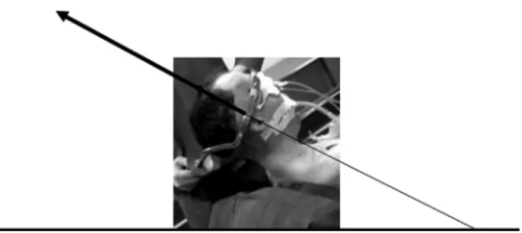
Once a cervical fracture is confirmed, patients with AS can be managed with axial traction. However, the traction direction must be placed in a superior and anterior direction so that the patient is realigned to the pre-existing kyphosis, and minimal weight should be used (Fig. 1). Placing the neck in an extended position may induce neurological complications. Even with prop-erly positioned axial traction, the possibility of further neurological
Fig. 1. Axial traction: in the case of ankylosing spondylitis, the direction of the
traction has to be considered in the context of the cervical kyphosis.
Fig. 2. AP and lateral radiographs of a patient non-operatively treated using a halo
vest. Note that the anterior turnbuckles have to rely on the chest because of the kyphosis.
deterioration exists, as this means of immobilisation still allows for rotational movement. Monitoring of the traction must be quite precise: repeated clinical evaluations must focus on the detection of potential neurological worsening including consciousness alter-ations. Axial traction is an emergency stabilisation procedure that can be used as a transitory stabilisation before surgery. It can be converted to a halo brace if non-operative treatment is ultimately the management choice.
Controversy exists as to whether patients with a frac-ture/dislocation should be treated with external immobilisation alone or with surgical fusion followed by external immobilisation. The halo brace alone has been advocated as a “classical” mode of treatment, but it is not without complications[25]. While the majority of patients placed in a halo brace achieve spinal fusion without difficulty, failures of union or increased neurological deficit have been encountered.
Some details regarding the installation of the halo brace require emphasis: because of the cervical kyphosis, a standard halo vest is usually inappropriate, and the device must be custom fitted. The connectors between the halo and the vest must be positioned so as to resist the tendency for anterior flexion. Therefore, the lateral connectors should not be placed at the projection of the shoul-ders because this placement may induce a side effect tendency for hyperextension, excessive traction on the pins, and risk of pulling out from the skull. Other significant side effects include dramatic translation and overdistraction at the site of the fracture. The ante-rior turnbuckles have to be placed on the anteante-rior valve of the vest because of the kyphosis. An additional posterior turnbuckle can be used posteriorly. In cases of extreme kyphosis or if a progressive correction is planned using the halo vest, another anterior turn-buckle can be used anteriorly (Fig. 2). Our experience is that in the first few days, the specific tools have to be easily available in case of an urgent need to remove the halo vest (e.g., neurologi-cal medullar decompensation with neurogenic cardiovascular or respiratory acute complication).
Fig. 3. Kyphosis-related technical difficulties: endotracheal intubation, installation,
and the surgical approach are demanding.
Among the complications of non-operative treatment, the dis-ruption of both the anterior and posterior supporting vertebral structures may lead to instability of such a degree that halo fixa-tion is not successful with translafixa-tion and rotafixa-tional displacements. This risky situation is not easily detected on x-rays, mainly at the lower cervical levels. In some cases, CT scan is the only solution in severely kyphotic patients. The same problems can arise in the assessment of bone fusion at the end of the theoretical 3 months of immobilisation. Another issue is pressure ulcerations, which are more prone to develop beneath the halo vest in the case of the AS spine because of the extensive thoracic kyphosis. This process might occur especially when patient care is difficult, as may be the case in those patients in a suboptimal general condition and who are potentially poorly compliant for verticalisation and for accept-ing such an uncomfortable brace. This complication can be avoided by scrupulous padding or the use of custom-fitted vests. Cranial fixation may also induce severe complications, such as septic intol-erance or mobilisation of the pins. Providing that the halo is placed properly without any skin contact and sufficiently below the ears to reduce pull-out risk, and providing that the pins are precisely monitored, the halo vest remains a therapeutic option.
2.2. Surgical treatment
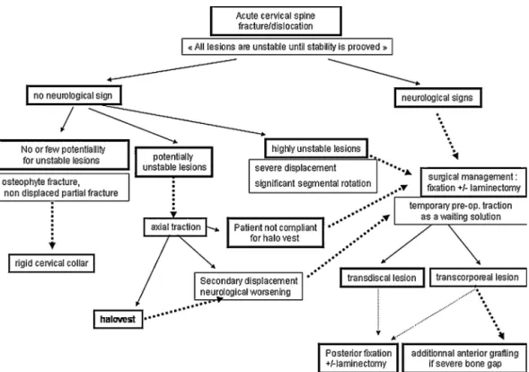
Surgical treatment is indicated in case of neurological deficit, when it can clearly be related to evidence of cord or nerve root compression on imaging (Table 1). Then, a procedure of decom-pressive laminectomy and fusion is performed. In some cases, the cord compression is not obvious because local instability can be the actual cause of a neurological worsening. We have also observed this situation in some cases initially treated with a halo brace in which clinical monitoring identified some neurological worsening resulting from a lack of stabilisation. The main potential advantage of surgical fixation is the possibility of immediate mobilisation of the patient and improvement for nursing care. However, technical difficulties may carry some important limitations because of the kyphosis and instability, including problems with tracheal intu-bation, installation in the prone position, and the surgery itself (Fig. 3). Most of the time, a postoperative immobilisation is manda-tory because of the specificities of those patients (residual kyphosis and sagittal imbalance, poor bone quality, highly unstable lesions). A custom-made cervical collar can be sufficient.
Because the cervical spine is fully stiff or even completely fused in some cases, the usual classification for cervical fractures is not completely appropriate for defining the surgical strategy and technical aspects. Based on therapeutic considerations, one would propose here classification of cervical lesions in the case of AS according to the position of the disruption line: within the for-mer disc space or within the cancellous bone of the vertebral body, namely transdiscal and transosseous lesions (Fig. 4). Transdiscal lesions are located between the former endplates, which are rein-forced structures. We consider that this type of lesion preserves the bone stock of the anterior column, with fair contact between the fragments after reduction. While a case of corpectomy at the site of a C6C7 transdiscal fracture has been reported without compli-cation[4], for most authors, posterior fixation alone seems stable enough, providing that the number of fixations is sufficient with at least two above and two below[3,7,8]. Cervical pedicle screws are reported to be the most reliable but the most challenging fixation
[3]; however, screws in the lateral mass fixations show satisfactory outcomes[7,8]. Of note is the fact that posterior montages should not end at the cervicothoracic junction and must be extended to the thoracic spine if necessary. No authors have proposed lami-nar hook fixation, which we also do not recommend because of the risk of spinal hematoma in the context of AS. Only few authors have suggested anterior fixation alone as an option because of the lack of fixation[6], even if Kouyoumdjian et al.[27]suggested ante-rior plate fixation to provide sufficient stability if the hardware is long enough to avoid significant moment arms such as long bone fractures.
Furthermore, Einsiedel et al.[26]reported about historic series that early implant failures occurred exclusively after single-session anterior stabilization (50%). That’s why they now perform circum-ferential approach in one or two stage.
Combined anterior and posterior instrumentation may be nec-essary when the structural integrity of the vertebral body has been significantly compromised, and kyphotic deformity at the site of the fracture is present. Some authors[4,26]believe that the circumfer-ential fusion should be the suitable method on these reasons: • inelastic nature of all spinal structures, a fracture in ankylosing
spondylitis always extends across all anterior and posterior ele-ments;
• poor bone quality;
• difficulty in localising anatomical land marks.
A purely posterior fixation/fusion seems indicated if the anterior weight-bearing column is well aligned and without fracture gaps
[4,6,8].
Transosseous lesions tend to collapse the cancellous bone of the vertebral body, resulting in a lack of bony contact in extension and a potential increase in instability for flexion, translation, and rota-tion. This problem raises the question of whether or not to perform an additional anterior approach for filling the gap with a cage or bone graft. Major gaps are to be grafted[5]for stability and fusion, specifically at the cervicothoracic junction. However, in our expe-rience and for other authors[28], minor bony gaps at the anterior column are likely to fuse with posterior fixation alone. This idea is supported by the good rate of fusion reported in the literature regarding anterior opening osteotomies at the cervical and lumbar levels[2,9]. While the indication limit is not clear cut, we would like to emphasise that the anterior approach is not systematic to us even for transosseous lesions. In addition, such an anterior approach may be problematic because of the kyphosis deformity and the depth of the grafting site. In some cases, the kyphosis necessitates a partial sternotomy for anterior access to the lower cervical spine.Fig. 5
shows a proposed algorithm for summarising the management of acute cervical lesions.
Table 1
Surgical management of AS cases at the cervical level: bibliography table.
Author Year Journal n Age Neurological
status
Level Management Results Complications
Taggard and Traynelis
[8]
2000 Spine 7 60 y.o. (49–83) 3 tetraplegia 3 C5C6 4 C6C7 Posterior approach ± cervical traction preop rib harvesting 100% fusion at 3/4 month 1 deep venous thrombosis 1 upper gastrointestinal hemorrhage 2 pneumoniae 2 deceased El Masry et al. [4]
2004 Injury 1 82 y.o. Sensory C7 deficit C6C7 Circumferential single session Anatomical reduction and fusion 4 years None Cornefjord et al.[3]
2005 Eur Spine J 19 60 y.o. (32–78) 2 paraplegia 2 motor weakness 4 sensory deficit 5 C5C6 5 C6C7
Posterior fixation No reoperated for loosening of the instrument or healing pb 1 deep wound infection 2 extensive peroperative bleeding Mountney et al.[5]
2005 Eur Spine J 1 36 y.o. Hyperflexia C7 Traction first And anterior approach And posterior fixation 15 days after Good results at 18 months None Payer[6] 2006 J Clin Neurosci 4 77 y.o. 70 y.o. 66 y.o. 52 y.o. C7 motor weakness Tetraplegia Normal Tetraplegia C6C7 C6C7 C6C7 C6C7 Posterior approach Anterior approach Circumferential two-session Circumferential single session Partially recover redislocation Stable fixation, 12 months Stable fixation, no neurological recovery None Reopearted circumferential None None Shen and Samartzis [7]
2006 J Trauma 2 79 y.o. (77–81) 1 tetraplegia 2 C6C7 Posterior approach Fusion and intact instrumentation at 1 year and 3 month
1 pneumoniae with death at 3 months Einsiedel et al.[26] 2006 J Neurosurg 37 65 y.o. (36–82) Two institutions 9 Frankel A 11 Frankel B 6 Frankel C 10 Frankel D 19 C6C7 6 two-segments 10 anterior approaches 11 circumferential single session 13 circumferential two-session 3 posterior approaches 5 early implant failure with anterior approach 3 deceased (RDS and cerebral ischemia) 3 infections 1 deep venous thrombosis Kouyoumdjian et al.[27]
2012 OTSR 19 61 y.o. (33–64) 10 medular deficit 7 radicular pain 9 C5C6 10 C6C7 13 anterior approaches 3 conservative treatment 2 circumferential 1 posterior
Good for bone healing 5 deceased 1 haematoma drained 2 cases of screw brached out 1 pressure sore on minerva Y.O.: year old; pre-op: preoperative.
Fig. 4. Imaging classification of the cervical fractures: (A) transdiscal, and (B) transcorporeal. Transcorporeal lesions correspond to the collapse of the cancellous bone and
Fig. 5. Algorithm for the management of acute cervical lesions in the case of ankylosing spondylitis.
Lesions of the upper cervical spine require a specific strategy: in our experience, the cases are not frequent and are mainly located at C2. Facing these potentially unstable lesions and poor bone qual-ity, an extensive occipitocervical fixation is the safest option despite the limited mobility at follow-up. This stabilisation procedure gen-erally requires a posterior autogenous bone graft to fuse the skull to the C2 or C3 levels. Postoperatively, an additional cervical collar is mandatory, and in some highly unstable lesions, a postoperative halo vest is initially preferred.
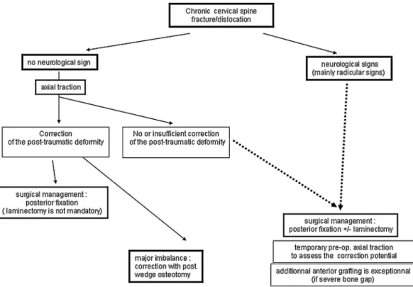
Chronic spinal lesions with non-union constitute another type of patient. Most of the cases correspond to failed or delayed diagnoses. Their management includes an initial stage of cervical traction to assess the possibility of reduction. In our experience, if no correc-tion can be obtained and if the neurological status is normal, a posterior fixation without laminectomy can be proposed. Then, the aim is to stabilise to the point that the sagittal balance is considered acceptable. If some correction can be obtained, the management of the lesions is the same as for acute lesions. We have found that pos-terior fixation alone is sufficient to obtain fusion provided that the fixation is long enough and that postoperative immobilisation is adequate. The indication for additional anterior surgery is rare and is mainly associated with a severe anterior bone defect or highly significant imbalance. As for anterior approaches in acute cases, the kyphosis is a significant technical difficulty. In some cases, the imbalance is unacceptable in a stiff spine without any significant improvement after the traction stage; in these extreme situations, a correction using a wedge osteotomy can be considered despite the risks of the procedure.Fig. 6provides a proposed algorithm summarising the management of chronic cervical lesions.
Spinal epidural hematoma is a severe potential complication, with a risk rate that is higher than in the non-AS population. AS affects the cancellous bone in such a way that, with a fracture, there is persistent bleeding that may predispose to formation of an epidural hematoma in the rigid cervical canal. This potential complication raises the question of whether or not to perform laminectomy in cases of posterior fixation. Should this choice be considered standard? The potential advantage is that it would
create a passageway for the postoperative bleeding. The disadvan-tage is the related increase in local instability after the removal of the spinous processes, and a reduction of the key zone for avive-ment and grafting. Septic complications constitute a significant issue because of the specific anatomical features of the skin and deep soft tissues (e.g., postoperative bleeding, fatty involution of the muscles). In addition, the general status of these patients is frequently debilitated. The septic risk is significantly increased in patients initially treated with a halo brace and then operated as a secondary measure because of local displacement or neurological worsening. Severe respiratory complications may also be observed as the lungs are frequently fibrotic and the ribs ankylosed, thus fixing the rib cage. These complications may necessitate making difficult decisions regarding postoperative immobilisation to avoid chest compressions and significantly interfere with the surgical strategy.
3. Correction of sagittal deformity
Because the cervical spine is kyphotic, the occurrence of the fracture could be an opportunity to increase lordosis at the site of the fracture. Such a case has been reported in a patient whose surgery was delayed from the time of the fracture[6], but most published series aim only at restoring the former kyphosis. In our experience, another option can be the progressive correction of kyphosis in patients treated with a halo vest. Progressive ante-rior distraction can be applied on the anteante-rior turnbuckles (Fig. 2) to gradually increase the chin-to-chest distance. The rhythm and amount of correction must be adapted to the patient’s tolerance and to the radiological evolution. This procedure is demanding and necessitates daily monitoring of the patient. It is clear that the risk of translation and overdistraction is major during the lordosis manoeuvres because of the instability. The anterior opening must be limited, specifically above C6, for avoiding the additional cere-bral risk of stretching the vertecere-bral arteries. The risk of posterior impingement of the medulla in extension would require associated laminectomy, such as for a correction osteotomy (see below). As
Fig. 6. Algorithm for the management of chronic cervical lesions in the case of ankylosing spondylitis.
Fig. 7. Different types for the impairment of the chin–brow angle in ankylosing
spondylitis. A. Upper kyphosis indicating cervical osteotomy. B. Lower kyphosis indicating lumbar osteotomy.
for deformation surgery at the medullar levels, electrophysiological monitoring of the spinal tracks is relevant[13].
Outside the context of trauma, the loss of horizontal gaze and onset of breathing and eating difficulties arising from stiff kypho-sis are the main indications for cervical extension osteotomy. The site of osteotomy is C7 for all studies, with the following technique initially described by Simmons, inspired by the Smith Petersen procedure at the lumbar spine. C7 is the best choice for several reasons: the vertebral arteries are not involved, the spinal canal is sufficiently wide, and the lever arm for extension is maximised.
Technically, the removal of the articular joint at C7/T1 is performed to avoid impingement of the T1 roots. A partial decancellation of the vertebral body has been proposed for avoiding the uncontrolled anterior opening, which is quite a dangerous step[15]. Neuromon-itoring (or the wake-up test) is helpful in preserving the medulla from irreversible lesions[13]. The gain in lordosis may reach 30◦to 40◦. The results in the literature yield a contrast with two clear-cut types of outcomes: a pretty satisfied patient with good outcomes, no complications, and good fusion, and rare but major neurological complications or even death.
The measurement of the kyphotic deformity is usually done using the chin–brow angle (CBA). The 0◦value indicates the nor-mal and horizontal gaze. However, the CBA integrates the sagittal balance of the whole spine. We emphasise here that one needs to analyse the kyphosis separately at the cervicothoracic spine, the thoracolumbar spine, and even the hip joints. The chin-on-chest (CC) distance is the portion of the deformation that is related only to the cervical spine. A normal CC distance categorises the indica-tions into two types: local kyphosis of the upper spine, based on CC distance, which may require cervical extension osteotomy, and global kyphosis that could be treated at the lumbar level (Fig. 7).
4. Discussion and conclusion
This review is a level IV of evidence combining experts’ opinion and series of cases reported in the literature. The analysis is limited by the number of references in the literature regarding cervical surgery in the context of AS. Most series are retrospective, con-cern a limited number of patients, and are not fully documented. The large period of inclusion of the published series accounts for the low frequency of cases and introduces some heterogeneity into the reported studies. In elderly patients and those with a severe cord lesion, the presence of AS leads to an especially poor progno-sis with a high mortality rate despite appropriate management of trauma cases[18,21,29,30]. Based on the literature and our expe-rience with AS at the cervical level in our department, we cannot address all the questions that remain. However, our main point is
that these cervical lesions are specific. Further points to empha-sise are as follows: emergency management should respect the kyphotic posture. While limited, non-operative treatment using a halo vest remains a therapeutic option, as long as its use is rigor-ously monitored. Classifying the fractures according to the anterior bony gap could be a way of selecting the most appropriate tech-niques for the surgical procedure. In the case of surgical treatment, fixations have to be long but not systematically circumferential. Regarding kyphosis correction surgery, the technical aspects and the neurological complications have been reported previously. To us, the strategy is always to consider the lumbar osteotomy first when possible, with a clear distinction of the CC distance from the CBA.
Disclosure of interest
The authors declare that they have no conflicts of interest con-cerning this article.
Funding
None.
References
[1]Feldtkeller E, Vosse D, Geusens P, et al. Prevalence and annual incidence of vertebral fractures in patients with ankylosing spondylitis. Rheumatol Int 2006;26:234–9.
[2]Etame AB, Than KD, Wang AC, et al. Surgical management of symptomatic cer-vical or cervicothoracic kyphosis due to ankylosing spondylitis. Spine (Phila Pa 1976) 2008;33:E559–64.
[3]Cornefjord M, Alemany M, Olerud C. Posterior fixation of subaxial cervical spine fractures in patients with ankylosing spondylitis. Eur Spine J 2005;14:401–8.
[4]El Masry MA, Badawy WS, Chan D. Combined anterior and posterior stabili-sation for treating an unstable cervical spine fracture in a patient with long standing ankylosing spondylitis. Injury 2004;35:1064–7.
[5]Mountney J, Murphy AJ, Fowler JL. Lessons learned from cervical pseudoarthro-sis in ankylosing spondylitis. Eur Spine J 2005;14:689–93.
[6]Payer M. Surgical management of cervical fractures in ankylosing spondylitis using a combined posterior-anterior approach. J Clin Neurosci 2006;13:73–7.
[7]Shen FH, Samartzis D. Surgical management of lower cervical spine fracture in ankylosing spondylitis. J Trauma 2006;61:1005–9.
[8]Taggard DA, Traynelis VC. Management of cervical spinal fractures in ankylos-ing spondylitis with posterior fixation. Spine (Phila Pa 1976) 2000;25:2035–9.
[9]Belanger TA, Milam RAt, Roh JS, et al. Cervicothoracic extension osteotomy for chin-on-chest deformity in ankylosing spondylitis. J Bone Joint Surg Am 2005;87:1732–8.
[10]Chin KR, Ahn J. Controlled cervical extension osteotomy for ankylosing spondylitis utilizing the Jackson operating table: technical note. Spine (Phila Pa 1976) 2007;32:1926–9.
[11]Hoh DJ, Khoueir P, Wang MY. Management of cervical deformity in ankylosing spondylitis. Neurosurg Focus 2008;24:E9.
[12]Khoueir P, Hoh DJ, Wang MY. Use of hinged rods for controlled osteoclastic correction of a fixed cervical kyphotic deformity in ankylosing spondylitis. J Neurosurg Spine 2008;8:579–83.
[13]Langeloo DD, Journee HL, Pavlov PW, et al. Cervical osteotomy in anky-losing spondylitis: evaluation of new developments. Eur Spine J 2006;15: 493–500.
[14]Pavlov PW. Correction and stabilisation in ankylosing spondylitis of the cervi-cothoracic spine. Eur Spine J 2009;18:1243–4.
[15]Tokala DP, Lam KS, Freeman BJ, et al. C7 decancellisation closing wedge osteotomy for the correction of fixed cervicothoracic kyphosis. Eur Spine J 2007;16:1471–8.
[16]Willems KF, Slot GH, Anderson PG, et al. Spinal osteotomy in patients with ankylosing spondylitis: complications during first postoperative year. Spine (Phila Pa 1976) 2005;30:101–7.
[17]Argenson C, Lovet J, Sanouiller JL, et al. Traumatic rotatory displacement of the lower cervical spine. Spine (Phila Pa 1976) 1988;13:767–73.
[18]Foo D, Sarkarati M, Marcelino V. Cervical spinal cord injury complicating anky-losing spondylitis. Paraplegia 1985;23:358–63.
[19]Grisolia A, Bell RL, Peltier LF. Fractures and dislocations of the spine com-plicating ankylosing spondylitis. A report of six cases. J Bone Joint Surg Am 1967;49:339–44.
[20]Podolsky SM, Hoffman JR, Pietrafesa CA. Neurologic complications following immobilization of cervical spine fracture in a patient with ankylosing spondyli-tis. Ann Emerg Med 1983;12:578–80.
[21]Weinstein PR, Karpman RR, Gall EP, et al. Spinal cord injury, spinal fracture, and spinal stenosis in ankylosing spondylitis. J Neurosurg 1982;57:609–16.
[22]Smith MD, Scott JM, Murali R, et al. Minor neck trauma in chronic ankylosing spondylitis: a potentially fatal combination. J Clin Rheumatol 2007;13:81–4.
[23]Harrop JS, Sharan A, Anderson G, et al. Failure of standard imaging to detect a cervical fracture in a patient with ankylosing spondylitis. Spine (Phila Pa 1976) 2005;30:E417–9.
[24]Wang YF, Teng MM, Chang CY, et al. Imaging manifestations of spinal fractures in ankylosing spondylitis. AJNR Am J Neuroradiol 2005;26:2067–76.
[25]Surin VV. Fractures of the cervical spine in patients with ankylosing spondylitis. Acta Orthop Scand 1980;51:79–84.
[26]Einsiedel T, Schmelz A, Schultheiss M, et al. Injuries of the cervical spine in patients with ankylosing spondylitis: experience at two trauma centers. J Neu-rosurg Spine 2006;5:33–451.
[27]Kouyoumdjian P, Guerin P, Schaelderle C, Asencioa G, Gille O. Fracture of the lower cervical spine in patients with ankylosing spondylitis: Retrospective study of 19 cases. Orthop Traumatol Surg Res 2012;98:543–55.
[28]Chang KW, Tu MY, Huang HH, et al. Posterior correction and fixation with-out anterior fusion for pseudoarthrosis with kyphotic deformity in ankylosing spondylitis. Spine (Phila Pa 1976) 2006;31:E408–13.
[29]Hunter T, Dubo HI. Spinal fractures complicating ankylosing spondylitis. A long-term follow-up study. Arthritis Rheum 1983;26:751–9.
[30]Kiwerski J, Wieclawek H, Garwacka I. Fractures of the cervical spine in anky-losing spondylitis. Int Orthop 1985;8:243–6.