Raman Hyperspectral Imaging: An essential tool in the pharmaceutical field
Texte intégral
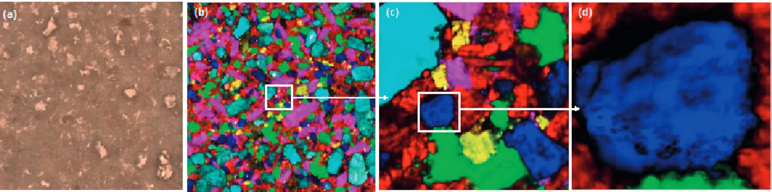
Figure
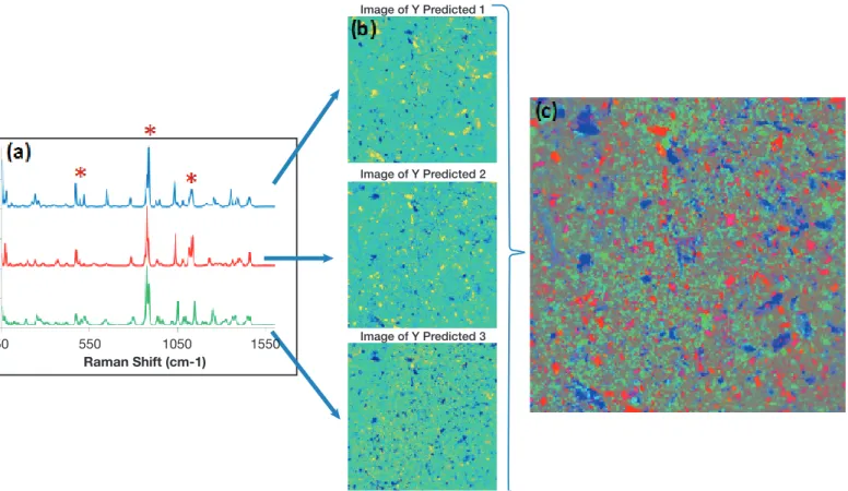
![Figure 4: Concentration maps of 4-AP in a paracetamol tablet from 0.025% to 0.2% obtained by SERS chemical imaging [11].](https://thumb-eu.123doks.com/thumbv2/123doknet/6572797.177841/4.892.72.838.912.1058/figure-concentration-paracetamol-tablet-obtained-sers-chemical-imaging.webp)
Documents relatifs
En outre le Tableau 3 nous montre que 70% des patients internés font plus d’un mois à l’hôpital et parmi ces patients la majorité est dénutrie nous explique le Tableau numéro
b) ligament interosseux. b) ligament commun interne très large, formé de deux plans de fibres superposés. Il se prolonge par la bride carpienne du perforé. la
The second disclosure profile concerns press releases issued by targets in hostile takeover bids and/or during the offer period (class 2), primarily used by the issuing
La pension moyenne des femmes faisant valoir un premier droit à la retraite dans l’année, tous régimes confondus (y compris la majoration de pension pour enfants), est inférieure
Electronic and Magnetic Communication in Mixed- Valent and Homovalent Ruthenium Complexes Containing Phenylcyanamide type Bridging Ligands.. Muriel Fabre and Jacques
Toutefois, ces pratiques idéales étant illusoires, seule la vaccination a permis le contrôle de la bronchite infectieuse dans les élevages intensifs de poulets de chair, de
Ainsi, le développement durable fait ressortir la perspective long terme ou court terme des acteurs « Si on fait du bio pour faire du blé à mon avis, cela peut marcher mais
Given a constraint mixing the rounding operator with non-linear real arithmetic terms, one can reduce it to an extension of non-linear real arithmetic with floor and ceiling
