Université de Montréal
Psychiatric symptoms in idiopathic rapid-eye-movement sleep behaviour
disorder
by
Maria Ţuineag
Department of Psychiatry Faculty of Medicine
Master’s Thesis Presented to the Faculty of Superior Studies
In Partial Fulfillment of the Requirements for the Degree
Master of Science (M.Sc.) in Biomedical Sciences
May 2012
Université de Montréal Faculty of Superior Studies
This thesis, entitled
Psychiatric symptoms in idiopathic rapid-eye-movement sleep behaviour
disorder
Presented by Maria Ţuineag
Was evaluated by a jury composed by Dr. Marie Dumont, PhD President Dr. Jean-François Gagnon, PhD Research director Dr. Jacques Montplaisir, MD, PhD Research co-director Dr. Roger Godbout, PhD Jury member
Sommaire
Le trouble comportemental en sommeil paradoxal (TCSP) idiopathique est caractérisé par une activité motrice indésirable et souvent violente au cours du sommeil paradoxal. Le TCSP idiopathique est considéré comme un facteur de risque de certaines maladies neurodégénératives, particulièrement la maladie de Parkinson (MP) et la démence à corps de Lewy (DCL). La dépression et les troubles anxieux sont fréquents dans la MP et la DCL. L’objectif de cette étude est d’évaluer la sévérité des symptômes dépressifs et anxieux dans le TCSP idiopathique.
Cinquante-cinq patients avec un TCSP idiopathique sans démence ni maladie neurologique et 63 sujets contrôles ont complété la seconde édition du Beck Depression
Inventory (BDI-II) et le Beck Anxiety Inventory (BAI). Nous avons aussi utilisé le BDI for Primary Care (BDI-PC) afin de minimiser la contribution des facteurs confondant
dans les symptômes dépressifs.
Les patients avec un TCSP idiopathique ont obtenu des scores plus élevés que les sujets contrôles au BDI-II (9.63 ± 6.61 vs. 4.32 ± 4.58; P < 0.001), au BDI-PC (2.20 ± 2.29 vs. 0.98 ± 1.53; P = 0.001) et au BAI (8.37 ± 7.30 vs. 3.92 ± 5.26; P < 0.001). Nous avons également trouvé une proportion plus élevée des sujets ayant des symptômes dépressifs (4/63 ou 6% vs. 12/55 ou 22%; P = 0.03) ou anxieux (9/50 or 18% vs. 21/43 ou 49%; P = 0.003) cliniquement significatifs. La proportion des sujets ayant des symptômes dépressifs cliniquement significatifs ne change pas en utilisant le BDI-PC (11/55 or 20%)
Les symptômes dépressifs et anxieux sont fréquents dans le TCSP idiopathique. L’examen de routine des patients avec un TCSP idiopathique devrait inclure un dépistage systématique des symptômes dépressifs et anxieux afin de les prévenir ou les traiter.
Mots clé: trouble comportemental en sommeil paradoxal, dépression, anxiété, maladie de Parkinson, démence à corps de Lewy
Summary
Idiopathic rapid-eye-movement sleep behaviour (iRBD) disorder can be a premotor feature of Parkinson’s disease (PD) or dementia with Lewy bodies (DLB). Depressive and anxiety symptoms are frequent nonmotor features in PD or DLB. We assessed the frequency and severity of depressive and anxiety symptoms in patients with iRBD compared to healthy control subjects. Fifty-five iRBD patients and 63 age and sex-matched healthy subjects were studied. Participants completed the Beck Depression Inventory – Second Edition (BDI-II) and Beck Anxiety Inventory (BAI). We assessed the depressive and anxiety symptoms and compared the proportion of participants with clinically significant depressive or anxiety symptoms. We also used the BDI for Primary Care (BDI-PC) to minimize confounding factors that could overestimate depressive symptoms. iRBD patients scored higher than controls on the BDI-II (9.63 ± 6.61 vs. 4.32 ± 4.58; P < 0.001)), BDI-PC (2.20 ± 2.29 vs. 0.98 ± 1.53; P = 0.001) and BAI (8.37 ± 7.30 vs. 3.92 ± 5.26; P < 0.001). Compared to controls, we found a higher proportion of patients with iRBD with either clinically significant depressive (4/63 or 6% vs. 12/55 or 22% P = 0.03) or anxiety symptoms (9/50 or 18% vs. 21/43 or 49%; P = 0.003). The proportion of iRBD patients with clinically significant depressive symptoms remains unchanged using the BDI-PC (11/55 or 20%). Depressive and anxiety symptoms are frequent features in iRBD. Routine examination of patients with iRBD disorder should include an assessment of depressive and anxiety symptoms in order to prevent or treat them.
Key words: REM sleep behaviour disorder, depression, anxiety, Parkinson’s disease, dementia with Lewy bodies
List of tables
Table 1. Between-subgroups comparisons for the Beck Depression Inventory Second Edition
Table 2. Between-subgroups comparisons for the Beck Anxiety Inventory
List of figures
Figure 1 Beck Depression Inventory Second Edition (BDI-II) scores distribution.
Abbreviations list
AD (Alzheimer’s disease) BAI (Beck Anxiety Inventory)
BDI-II (Beck Depression Inventory, 2nd Edition) BDI-PC (Beck Depression Inventory, Primary Care) dDpMe (dorsal Deep Mesencephalic reticular nucleus) DLB (Dementia with Lewy Bodies)
DPGi (Dorsal Paragigantocellular reticular nucleus)
DSM-IV (Diagnostic and Statistical Manual of psychiatric disorders, 4th Edition) EEG (Electroencephalography)
EOG (Electrooculography) EMG (Electromyography)
GABA (Gamma-Amino-Butyric-Acid) Giv (Gigantocellular reticular nucleus)
ICSD-II (International Classification of Sleep disorders, 2nd Edition) iRBD (idiopathic Rapid-Eye-Movement sleep behaviour disorder) Ltd (Laterodorsal tegmental nucleus)
MAO (Mono-Amine-Oxidase) MDD (Major Depressive Disorder)
MCRF (Medullary magnocellular reticular Formation) MSA (Multiple System Atrophy)
NREM (Non-Rapid-Eye-Movement) OSA (Obstructive Sleep Apnea) PD (Parkinson’s Disease)
PPN (Pedunculopontine nucleus) PSG (Polysomnography)
REM (Rapid-Eye-Movement)
RBD (Rapid-Eye-Movement sleep behaviour disorder) SLD (Sublateral tegmental Dorsal nucleus)
SN (Substantia Nigra)
SNpc (Substantia nigra, pars compacta) SWS (Slow Wave Sleep)
Acknowledgements
The present work is the product of the collective effort made by many people. First of all I would like to thank Jean-François Gagnon - my mentor, my supervisor and my great friend, who helped me go through all the challenging situations I have faced the last three years. His enthusiasm, expertise, understanding and patience added considerably to my experience and consolidation of my scientific preparation.
I would also like to express my gratitude to Professor Jacques Montplaisir who, some time ago, responded favourably to my email coming from the other side of the ocean. Professor Montplaisir helped me accomplish my dream in studying the fascinating field of sleep. His imposing and elegant presence has inspired me all along.
A big “Merci!” to all my colleagues and friends at the Sleep Centre for their availability, patience and generosity: Jessica, Véronique, Shady, Dominique, Mireille and Jean.
Last but not least, thank you to all my family scattered all around the world: my parents back home, my brother and my relatives here in Montreal.
TABLE OF CONTENTS
1. GENERAL INTRODUCTION...1
1.1 Wakefulness ...2
1.2 Non-rapid-eye-movement (NREM) sleep ...2
1.3 Rapid-eye-movement (REM) sleep ...3
1.3.1 The relation between dreaming and sleep stages ...4
1.3.2 The mechanism of REM sleep ...6
1.4 Sleep disorders ...8
2. REM SLEEP BEHAVIOUR DISORDER (RBD)...10
2.1 Definition and clinical features ...10
2.2 Diagnostic criteria, differential diagnosis, and treatment of RBD ...12
2.3 Pathophysiology of RBD ...15
2.4 Idiopathic versus secondary RBD: association with neurodegenerative disorders ...17
3. PSYCHIATRIC DISORDERS IN RBD ...19
4. STUDY OBJECTIVES AND HYPOTHESIS ...21
5. METHODS AND RESULTS ...23
5.1 Article ...24
5.2 Additional results ...52
6. DISCUSSIONS AND PERSPECTIVES ...53
6.1 Psychiatric symptoms in iRBD ...53
6.2. Risk factors for psychiatric symptoms in iRBD ...55
6.3 Psychiatric symptoms inventories ...56
6.4 Pathophysiology of cluster symptoms ...57
6.5 Limitations of the study ...62
7. REFERENCES ...64
8. ADDENDUM ...74
8.1 Contribution of authors ...74
1. GENERAL INTRODUCTION
Throughout the history of medicine, defining sleep has been a daunting challenge due to its complex cyclical generation pattern, its importance for individual functioning, and its involvement in pathology. According to a behavioural definition, sleep is a reversible behavioural state of perceptual disengagement from and unresponsiveness to the environment (Principles and Practice of Sleep Medicine, Meir H. Kryger, Thomas Roth and William C. Dement, 5th Edition, 2011). From a neurobiological standpoint, human sleep is defined in terms of a series of physiological changes revealed by a sleep recording procedure called polysomnography (PSG). PSG is a multiparametric test that records electroencephalographical activity (EEG), ocular movements using an electrooculogram (EOG), and muscle tone using an electromyogram (EMG).
Based on these three parameters (EEG, EOG, and EMG), three distinct states of consciousness can be identified: wakefulness, non-rapid-eye-movement (NREM) sleep, and rapid-eye-movement (REM) sleep. NREM sleep is further subdivided into four stages: 1, 2, 3, and 4. Stages 3 and 4 are collectively referred to as slow-wave sleep (SWS) (Rechtschaffen and Kales, 1968).
1.1 Wakefulness
In basal conditions (supine position, eyes closed, in a dark, quiet room) EEG activity during wakefulness is predominated by alpha-rhythm, which is more prominent in occipital leads. Alpha waves have a frequency range of 8 to 13 Hz and average amplitude of about 50 µV. This rhythm reflects a relaxation state and is abruptly replaced by the beta rhythm—a more rapid and desynchronized activity—when subjects are visually or cognitively stimulated.
1.2 Non-rapid-eye-movement (NREM) sleep
Stage 1 sleep appears in the transition from wakefulness to other sleep stages, or following partial arousals during sleep. Alpha activity decreases or disappears and is replaced by a recording characterized mostly by low-amplitude theta waves (4–8 Hz). At this time, EMG activity decreases and EOG shows slow rolling eye movements. As sleep deepens, stage 2 occurs, accounting for approximately 50% of the total sleep time. The hallmark of stage 2 is the presence of sleep spindles and complexes. K-complexes are relatively high amplitude biphasic waves with an initial negative component and a total duration of at least 0.5 seconds. They are believed to occur spontaneously, but can be associated with sensory stimuli and often precede arousal. Spindles are commonly known as a group of rhythmic waves characterized by progressively increasing, then gradually decreasing, amplitude. They may occur at different frequencies of 10 to 16 Hz, but Rechtschaffen and Kales specifically define
spindles associated with the onset of stage 2 as heaving a frequency of 12 to 14 Hz and duration of 0.5 to 1.5 seconds (Harris, 2005).
SWS (stages 3 and 4) is characterized by high-amplitude (> 75 µV) delta waves (0.5–4 Hz). In 1968, Rechtschaffen and Kales set the criteria for stages 3 and 4 SWS. The appearance of high-amplitude delta activity for more than 20% of a 30-second epoch is scored as stage 3 sleep, and >50% as stage 4. Although K-complexes and spindles may appear during SWS, they may be masked by high-amplitude delta activity.
1.3 Rapid-eye-movement (REM) sleep
REM sleep accounts for about 25% of sleep time. As the sleep cycles unfold during the night, the tendency shifts from an abundance of SWS during the first part of the night to a preponderance of REM sleep during the second part. REM sleep may be divided into a phasic and tonic phase. The tonic phase is characterized by an EEG desynchronization and striated muscle atonia (except for the diaphragm). In addition to EEG desynchronization, the phasic phase features rapid eye movements and a series of other phenomena such as contractions of the muscles of the medium ear, autonomic fluctuations (irregular pulse and breathing), erections, vasodilatation of the pelvic organs, and clonic twitches of the face and extremities. The rapid eye movements are saccadic, predominantly horizontal and occur in bursts or alone. On the EEG, saw-tooth waves (in the theta range, 4-7 Hz) may appear in conjunction with eye movements.
Waking EEG is also characterized by desynchronization, a diffuse pattern of relatively low-amplitude, mixed-frequency activity that includes higher frequency beta activity. Comparatively to REM sleep, EOG and EMG activity during wakefulness is very variable and is strictly related to the activity the subject is engaged in. When the subject is cognitively active, eye movements are rapid and variable in direction, with intermixed blinking. On eye closure, eye movements may be absent with occasional blinks in response to stimuli. EMG also varies widely in wakefulness, ranging from gross body movements that may obscure EEG and EOG activity to a diminished activity with constant tonus during relaxed wakefulness.
REM appears to be a state in which the awakened brain is trapped in a paralyzed body, hence the term “paradoxical sleep,” where an activated brain operates in a seemingly unresponsive organism. REM sleep is associated with vivid dreaming, although more and more reports indicate mental activity in NREM sleep as well.
1.3.1 The relation between dreaming and sleep stages
The initial perspective on the relationship between dreams and sleep stage was referred to by many as the “REM sleep = dreaming” perspective from which dreaming was viewed as a characteristic exclusive to REM sleep. Mentation reported from NREM sleep was attributed to confounding factors, for example recall of mentation from previous REM episodes or subjects’ waking confabulations. However, subsequent studies cast doubt on this perspective (Foulkes, 1962) primarily by demonstrating elevated levels of mentation recalled from NREM sleep stages. Although the “REM
sleep = dreaming” remains a widespread theory, a debate over whether the quality of NREM and REM sleep mentation reports differ, largely overshadowed it. Initially, qualitative differences in REM and NREM reports suggested that a different form of mentation occurs in NREM sleep. From these developments, two relatively distinct points of view concerning REM/NREM mentation emerged and continue to influence the field. These points of view differ as to whether they consider NREM sleep mentation to originate from imagery processes that are fundamentally the same as or different from those that produce REM sleep mentation. Nielsen (2000) refers to these as the 1-generator and 2-generator models. Although dreams can be produced in SWS (Foulkes, 1962), dreams during REM sleep are the most typical with a rich and varied content involving emotion and motor behavior. Animal research shows that when the regions responsible for the muscle atonia in RBD are injured, fight-or-flight behaviors appear while other signs of REM sleep (EEG, visual system) are still present (Hendricks et al, 1982). Mentation differences originate primarily from differences in memory activation. When such activation is high and diffuse, during most REM but some NREM sleep, then organization is more intensely stimulated and conscious interpretation more probable and coherent, giving rise to the more vivid and memorable dreams associated with REM sleep. When memory activation is low and less diffuse, during most NREM but some REM sleep, then organization is less intensely stimulated and conscious interpretation less probable and coherent. It is thus the diffuseness or availability of diverse memory elements and not sleep stage physiology that determines the occurrence and form of sleep mentation (Nielsen, 2000).
1.3.2 The mechanism of REM sleep
Luppi et al. (2010) proposed in their rat model study an updated theory of REM sleep onset and maintenance. According to Luppi, REM sleep is induced by the activation of glutamatergic neurons found in the sublateral tegmental dorsal nucleus (SLD) in the caudal pons. These findings support earlier studies according to which neurons in SLD that trigger REM sleep are indeed glutamatergic (Lu et al., 2006; Clement et al., 2009). These REM-on neurons (neurons that fire during REM sleep) are actively inhibited during wakefulness and SWS by GABA-ergic REM-off neurons (neurons that cease fire during REM sleep) localized in the ventral part of a neuronal complex that comprises the vlPAG (ventrolateral periaqueductal grey) and dDpMe (dorsal deep mesencephalic reticular nucleus). These REM-off neurons are activated during wakefulness by hypocretin and aminergic neurons projecting from the hypothalamus. Two other REM sleep generators have been localized in the hypothalamus (melanin/GABA-ergic neurons), in the dorsal part of the vlPAG – dDpMe complex and in the paragigantocellular reticular nucleus (DPGi) These REM-on neurons have an intrinsic “clock-like” mechanism that allow periodic initiation of the REM sleep. They also inhibit the wakefulness-promoting hypocretin, aminergic, and GABA-ergic neurons, thus creating a reciprocal inhibition-activation network.
Escaping from the inhibition of the waking-promoting neurons (REM-off), the REM-on neurons localized in the ventral part of the SLD project in an ascending way inducing cortical activation via intralaminar thalamic relay neurons. This activation occurs in
collaboration with wakefulness/REM-on cholinergic neurons and glutamatergic neurons from the laterodorsal tegmental nucleus (Ltd), the pedunculopontine (PPN), mesencephalic, and pontine reticular nuclei and the basal forebrain. At the same time, glutamatergic REM-on neurons in the caudal part of the SLD induce muscle atonia and sensory inhibition projecting to anterior spinal cord alpha motor neurons via excitatory projections to the glycinergic nuclei located in the medulla a (ventral – Giv and alpha - Gia gigantocellular nuclei). This network also blocks impulses from the motor cortex which are responsible with movement associated to the dream content, so motor control in REM sleep is acquired by means of two systems: tonus abolition through a descending way (SLD – GiV/GiA – motors neurons in the spinal cord) and inhibition of dream enactment movements from the motor cortex.
A major discovery regarding REM inhibition in rats was that serotonergic neurons from the raphe nuclei and noradrenergic neurons from the locus coeruleus cease firing specifically during REM sleep, i.e., they show a REM-off firing activity, reciprocal to that of on neurons. Luppi proposes that rather than inhibiting directly SLD REM-on neurREM-ons, noradrenaline and serotREM-onin might inhibit REM by means of a tREM-onic excitation of the dDPMe and vlPAG REM-off GABAergic neurons. Partly supporting this hypothesis, noradrenaline but not serotonin application in the dDPMe was shown to increase wakefulness and decrease SWS and REM sleep (Luppi et al, 2010).
Similar to the studies in rats, the main nucleus responsible for triggering REM sleep (REM-on cells) has been identified in cats as the noradrenergic complex locus coeruleus/ subcoeruleus nucleus (Siegel, 2006), the equivalent of the SLD in rats. The
lack of coherence regarding the neurotransmitter involved in the REM-trigger centers between studies performed on rat and cat is regarded as an inter-species variability (Luppi et al, 2011). Other regions identified from lesion studies in cats are the medullary magnocellular reticular formation (MCRF)—which is the final common pathway of spinal motor neuron inhibition, the PPN (pedunculopontine nucleus) and the Ldt.
The dopaminergic substantia nigra (SN) has been proposed as a component of this REM sleep system due to its apparent role in lesion studies, but there is a paucity of direct evidence to implicate this nucleus. (Boeve et al, 2007).
1.4 Sleep disorders
The International Classification of Sleep Disorders, Second Edition (ICSD-II, 2005)
categorizes sleep disorders into four categories: dysomnias, parasomnias, sleep disorders associated with mental, neurologic, or other medical disorders, and proposed sleep disorders (disorders for which there is insufficient information available to confirm the unequivocal existence of the disorder, e.g., subwakefulness syndrome).
According to Principles and Practice of Sleep Medicine, 5th Edition (2011),
parasomnias are defined as “unpleasant or undesirable behavioural or experiential phenomena that occur predominantly or exclusively during the sleep period.” Primary parasomnias (i.e., sleep disorders per se) can be categorized by the sleep state in which
they occur, such as NREM arousal parasomnias, REM parasomnias, and miscellaneous parasomnias (irrespective of the sleep state).
NREM arousal parasomnias are characterized by a state of dissociation in which the brain is partially awake and partially in NREM sleep. The brain is awake enough for performing very complex motor and verbal tasks, but is asleep enough not to have conscious awareness of or responsibility for the actions. This category of parasomnias include somnambulism (sleepwalking), confusional arousals and sleep terrors. Sleepwalking episodes are partial arousals typically from slow wave sleep during the first half of the sleep period. These events can be minor or elaborate behaviours such as dressing, unlocking locks and even driving. The patients do not exhibit significant autonomic features (tachycardia, sweating, etc) or expression of fear. These elements help differentiate somnambulism from sleep terrors which are a more intense form of arousal disorders with a predominance of autonomic expression. The patient arises suddenly with a piercing scream or cry, autonomic output and behavioural manifestation of intense fear. The onset of the episode is abrupt and the patient displays tachycardia, flushing, diaphoresis and mydriasis. They typically appear in the first third of the night and patients have no memory of the event. As with somnambulism, patients have a normal neurological examination during wakefulness. Both sleepwalking and night terrors are childhood-onset disorders and they usually are self-limited, symptoms disappearing by adulthood. Confusional arousals can occur from any arousal from slow wave sleep. They consist of disorientation, slow speech and mentation or inappropriate behaviour. Unlike somnambulism and sleep terrors, patients with confusional arousal
have partial memory impairment of the event (Principles and Practice of Sleep
Medicine, 5th Edition (Chapter 60 - Classification of sleep disorders, Michael J.
Thorpy).
REM parasomnias usually reflect a dissociated sleep state in which not all the elements leading to the initiation and maintenance of REM sleep appear to function. This is very well illustrated by REM sleep behaviour disorder (RBD), a sleep parasomnia in which normal muscle atonia is missing. Other REM sleep parasomnias include nightmares, REM-related painful penile erections and REM-related sinus arrest.
2. REM SLEEP BEHAVIOUR DISORDER (RBD) 2.1 Definition and clinical features
RBD was first described as a distinct clinical condition by Schenck and Mahowald (Schenck et al., 1986). According to the ICSD-II, RBD is characterized by the intermittent loss of REM sleep EMG atonia and the appearance of elaborate motor activity associated with dream mentation (dream enactment). In patients with RBD, both the tonic and phasic component of REM sleep are altered. Partial or complete loss of tonic chin EMG atonia (REM sleep without atonia) and excessive chin and limb phasic EMG activity are characteristic features in patients with RBD (Schenck et al., 1993).
The three core clinical aspects of RBD are abnormal vocalization, abnormal motor behaviour, and altered dream mentation. Although vocalization is often encountered in the general population, vocalization in RBD patients tends to be loud, suggesting unpleasant dreams. The abnormal vocalization ranges from shouting to screaming and swearing (Boeve et al., 2010). Excessive muscular activity during REM sleep often leads to undesirable motor behaviours such as kicking, punching, falling out of bed, or even running (Schenck et al., 2002). These abnormal behaviours may cause injuries to the patient or bed partner, ranging from ecchymoses to subdural hematoma (Olson et al., 2000).
Most RBD patients report vivid nightmares, with a surprisingly consistent content involving insects, animals, or people chasing or attacking them, their relatives, or their friends. The patient is almost always the defender and not the attacker (Boeve, 2010). In addition, the aggressiveness displayed during dreaming is in flagrant contrast to the somewhat calm and mild-mannered temperament of the patient during wakefulness (Schenck et al., 2002; Olson et al., 2000, Fantini et al., 2005). Moreover, sexual dream content was not reported by RBD patients compared to the dream content of normal subjects (Fantini et al., 2005). RBD usually appears in the fifth decade of life, although symptoms can appear as early as adolescence. Based on a large series study with 4,972 participants ranging in age from 15 to 100 years, RBD prevalence was estimated at 0.5% in the general population (Ohayon et al., 1997). Traditionally, RBD is described as a strikingly male-predominant parasomnia, with 80% of patients being men (Wing et
al., 2008). This difference might have a hormonal basis, or alternatively, because women display milder symptoms, they would be less likely to seek medical help (Schenck et al., 1993).
2.2 Diagnostic criteria, differential diagnosis, and treatment of RBD The ICSD-II lists four major criteria as the diagnosis of RBD:
A. Presence of REM sleep without atonia: the EMG finding of excessive amounts of sustained or intermittent elevation of submental EMG tone or excessive phasic submental or (upper or lower) limb EMG twitching;
B. At least one of the following is present:
1. Sleep-related, injurious, potentially injurious or disruptive behaviour by history 2. Abnormal REM sleep behaviour documented during PSG monitoring;
C. Absence of EEG epileptiform activity during REM sleep unless RBD can be clearly distinguished from any concurrent REM sleep related seizure disorder;
D. The symptoms are not better explained by another sleep disorder, medical or neurological disorder, mental disorder, medication or substance use disorder.
REM sleep without atonia (RSWA) refers to the electrophysiological finding of loss of complete or partial EMG atonia during REM sleep. It is one of the features sought in PSG when evaluating a patient with suspected RBD. According to Montplaisir et al. (2010), the polysomnographic cut-offs for excessive muscular activity are tonic chin EMG density ≥30% of total REM sleep time, phasic chin EMG density ≥15% of total REM sleep time, and ≥24 leg movements per hour of REM sleep. Each 20-second
epoch on the PSG was scored as tonic or atonic depending if EMG activity was present for more or less than 50% of the epoch duration. Excessive EMG activity was defined by amplitude of chin EMG signal of at least twice that of the background or greater than 10 µV. Phasic chin EMG density was scored from the submental EMG activity and represented the percentage of 2-seconds mini-epochs containing EMG events lasting 0.1 to 10 seconds, with an amplitude exceeding four times the amplitude of background EMG activity (Montplaisir et al, 2010).
The differential diagnosis of RBD includes the NREM parasomnias (somnambulism, night terrors, confusional arousals), nocturnal panic attacks, nocturnal seizures, nightmares, nocturnal wandering associated with dementia, and obstructive sleep apnea (OSA). The history usually allows differentiating these disorders from RBD. As stated above, both sleepwalking and night terrors are childhood-onset disorders and they usually are self-limited, symptoms disappearing by adulthood, whereas RBD has an onset typically around the age of 50. The RBD episodes usually occur in the second part of the night, which is prevalent in REM sleep, with non-stereotyped movements and violent verbalization, eyes closed and clear recollection of the dream upon awakening (Leu-Semenescu and Arnulf, 2010). Sleepwalking arises mainly in the first third of the night, when there is an abundance of slow wave sleep. However, when diagnostic clarification is necessary, particularly when the risk for injury is high, the behaviours occur at any time of the night or when symptoms suggestive of OSA are observed, PSG with simultaneous video monitoring should be used (Boeve et al., 2010).
To date, no double-blind, placebo-controlled clinical trials have assessed the efficacy of any treatment for RBD. The mainstay treatment for RBD remains clonazepam in doses ranging from 0.5 mg to 2 mg at bedtime (Gagnon et al., 2006). Clonazepam is a benzodiazepine that completely inhibits excessive motor behaviour during RBD episodes in approximately 80% of patients: another 10% of patients have partial benefit (Schenck et al., 1993) and 10% don’t benefit at all from the drug. Clonazepam slightly decreases phasic activity in the EMG, but does not restore the normal muscular atonia in patients under treatment (Lapierre and Montplaisir, 1992) (Sforza,1997). The mechanism of action of clonazepam in RBD is unknown, but the fact that it results in striking clinical improvement without PSG changes on the tonic EMG activity raises the possibility that it preferentially acts on locomotor systems (namely the inhibition of the motor cortex projection) rather than on those that control REM muscle atonia (Watanabe and Sugita, 1998). When clonazepam is ineffective, melatonin (3–12 mg) has been shown to have some effect used alone or in conjunction with clonazepam (Kunz, et al., 1999; Boeve et al., 2003). Unlike clonazepam, melatonin restores muscle atonia in RBD patients (Iranzo et al., 2009).
There have been numerous reports of RBD onset associated with some medications. The most often cited are selective serotonin reuptake inhibitors such as fluoxetine, paroxetine, sertraline, and citalopram (Shenck et al 1992; Winkelman et al, 2004); the tetracyclic antidepressive mirtazapine (Onofrj et al., 2003; Nash et al., 2003), and the MAO inhibitors phenelzine and selegiline (Akindele et al., 1970) (Gagnon et al., 2006).
RBD symptoms have also been well documented after alcohol withdrawal. However, idiopathic RBD can be differentiated from drug-induced RBD by thorough documentation of the onset of symptoms in relation to the time of drug use or by removing the offending drug, moment that usually corresponds to a the ceasing of RBD symptoms.
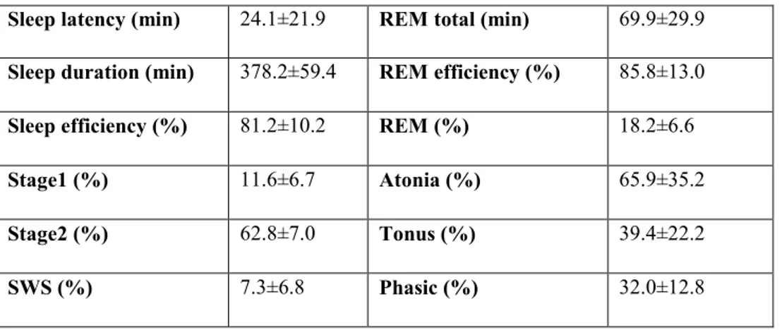
Sleep architecture is preserved in idiopathic RBD, with comparable values to healthy age-matched subjects for total sleep time, sleep efficiency, and REM sleep percent and efficiency (Montplaisir et al, 2010; Fantini et al, 2003). Some quantitative EEG studies found a slowing of the EEG during REM sleep in idiopathic RBD (Fantini et al., 2003; Iranzo et al., 2010). However, a recent study performed in our laboratory found no slow-wave anomalies in idiopathic RBD compared to control subjects (Latreille et al, 2011). These results suggest a specific neuropathologic damage to centers controlling REM sleep preferentially, sparing other sleep components.
2.3 Pathophysiology of RBD
The pathophysiology of RBD has been hypothesized based on studies in animals, including cats and rats, from pharmacological manipulations and imaging studies of brainstem lesions in humans.
As explained above in the rat model proposed by Luppi (Luppi et al., 2011), the SLD nucleus involved in the mechanism of REM sleep has two distinct subpopulations of neurons: an ascending group that induces cortical activation and a descending group
that is responsible for muscle atonia during REM sleep. Thus, the absence of atonia during an RBD episode could be due to the degeneration of either the descending subpopulation of glutamatergic neurons in the SLD or the nuclei in the lower part of the network (i.e., the GABA/glycinergic GiA and GiV) that control muscle tone via the alpha motoneurons in the anterior horns of the bone marrow. If this latter theory is true, this would explain why the neurons in the SLD degenerate preferentially to abolish atonia only but preserve the other components of REM sleep such as dreaming which supposedly is induced by the ascending projection of the ventral part of the SLD to the cortex.. Although it remains to be confirmed whether these two SLD neuron subpopulations are present in rats, they have been identified in cats (Lai and Siegel, 1997).
Although RBD has been strongly associated to PD, several studies indicate that it is unlikely its pathophysiology is due mainly to the dysfunction of the dopaminergic nigrostriatal system (Kim et al, 2010) Arguments supporting this assertion are the fact that about half of RBD patients do not develop PD, that the treatment with dopaminergic agents usually does not improve RBD and that medication such as selective serotonin-reuptake inhibitors can exacerbate or even induce RBD. However, an imagery study by Iranzo et al (2010) found changes in subtantia nigra of iRBD patients similar to those in patients with Parkinson’s disease, which might suggest that at least a part of the iRBD subjects have a substantia nigra dopaminergic related dysfunction. Lesions in the subcoeruleus/coeruleus complex (and/or possibly in the
PPN as well) cause REM sleep without atonia, and lesion size determines the complexity of motor behaviour (Hendricks et al., 1982).
Only a few cases of RBD associated with brainstem lesions have been reported in humans, using single-case imaging studies. Reports range from patients with new-onset RBD after a presumably ischemic lesion in the left upper pons (Kimura et al., 2000) to multiple sclerosis lesions in pontine white matter (Plazi and Montagna, 2002) and tumours located in the brainstem (Zambelis et al., 2002). Moreover, data from two RBD patients who underwent post-mortem neuropathological analysis showed brainstem-predominant Lewy bodies disease with degeneration of the substantia nigra and coeruleus/subcoeruleus complex (Uchiyama et al., 1995; Boeve et al., 2007). In conclusion, although the neuronal structures involved in RBD have not been definitively established, it is widely acknowledged that they are located in the brainstem.
2.4 Idiopathic versus secondary RBD: association with neurodegenerative disorders RBD can be a self-standing entity (termed idiopathic) (iRBD), without any apparent
concomitant neurological disease. It can also be found concomitantly with neurodegenerative diseases, especially synucleinopathies such as Parkinson’s disease (PD), dementia with Lewy bodies (DLB), and multisystem atrophy (MSA), and it is then termed secondary RBD (Gagnon et al., 2006). Moreover, iRBD has been
recognized as an early sign of synucleinopathies, being part of a cluster of premotor signs of these diseases. Premotor signs are considered to be the result of the earliest pathological changes. It is now widely accepted (Braak et al, 2003) that the pathology of PD begins in a variety of central and peripheral system regions (notably the nucleus of vagus nerve in the medulla oblongata, or even in the peripheral autonomic system) before the involvement of the substantia nigra and the appearance of the motor symptoms. Features arising from this involvement have been described with terms such as “preclinical”, “prodromal or “premotor” (Lang, 2010). Lately, several synucleinopathy markers deemed “premotor”, have been identified in iRBD patients, such as cognitive and olfactory impairment, subtle motor abnormalities, cardiac autonomic dysregulation, and other autonomic manifestations such as constipation, erectile dysfunction, and urinary retention (Gagnon et al., 2009; Postuma et al., 2009; Lanfranchi, 2007; Postuma and Montplaisir, 2006). These findings indicate that RBD is an early manifestation of a neurodegenerative process, thus challenging the notion of idiopathic RBD.
Several series of iRBD patients have been followed prospectively, showing that a majority have converted to some type of neurodegenerative disease, particularly synucleinopathies such as PD, DLB, and MSA. In their follow-up study, Schenck et al. showed that 38% of iRBD patients developed a parkinsonian syndrome 3.7 years after diagnosis and after a mean interval of 12.5 years from the onset of symptoms (Schenck et al., 1996). Iranzo and colleagues reported on a series of 44 patients with iRBD with at least 2 years of clinical follow-up, of whom 45% developed a neurological disorder
after a mean of 11.5 years from the reported onset of RBD. A majority of the converted patients (9/20 or 45%) developed PD (Iranzo et al., 2006). Another major prospective study (Postuma et al., 2009) performed on 93 iRBD patients found the estimated 5-year risk for neurodegenerative disease at 17.7%, the 10-year risk at 40.6%, and the 12-year risk at 52.4%. Twenty-six patients developed a neurodegenerative disorder: PD in 14, DLB in 7, probable Alzheimer’s disease (AD) in 4 and MSA in 1.
Although it is difficult to explain why not all iRBD patients convert to some neurodegenerative disease, and why they seem to remain in a ‘frozen’ neuropathological state, a step has been made towards identifying a marker for patients who will eventually convert. Postuma et al. (2010) found that in patients with iRBD who were initially free of neurodegenerative disease, the severity of REM atonia loss on baseline PSG (Postuma et al., 2010) as well as olfaction and colour vision impairment (Postuma et al., 2011) predict the development of synucleinopathies.
3. PSYCHIATRIC DISORDERS IN RBD
At least two other potential pre-motor symptoms concomitant with iRBD have been suggested: depression and anxiety. Psychiatric symptoms such as depression and anxiety are frequent non-motor features of PD, DLB, and MSA, and they can considerably complicate the course of the disease (Aarsland et al., 2007; Benrud-Larson et al., 2005; Dodel et al., 2008). Although it has been suggested that depression and
anxiety are associated with difficulty in accepting the diagnosis of an incurable evolving disease, PD patients tend to have more severe depressive symptoms than patients with other chronically disabling diseases (Ehmann et al., 1990). Moreover, depression and anxiety often develop before the onset of parkinsonism in PD and DLB (Weisskopf et al., 2003; Remy et al., 2005). Depression and anxiety can be difficult to assess in patients with PD due to overlapping symptoms and the difficulty of assessing depression in cognitively impaired patients. A variety of scales are available to assess these two psychiatric disorders in PD patients. The Beck Depression Inventory (BDI) is one of the most commonly used self-rated instruments for major depression in clinical practice (Watkins et al., 1995). The BDI is suitable for screening if an appropriate cutoff is used. It is also suitable for assessing the severity of depressive symptoms and for monitoring changes during treatment. In addition, it can be used in phenomenological studies of depression in PD (Schrag et al., 2007). The Beck Anxiety Inventory (BAI) assesses anxiety symptoms. It consists of a questionnaire containing 21 items to measure the severity of somatic, affective, and cognitive symptoms associated with panic attacks and generalized anxiety. The BAI has not been validated in PD. However, it meets the criteria for the suggested scale to screen for symptoms of panic attacks in PD patients. It is probably less suitable to screen for other anxiety disorders. It also meets the criteria for the recommended scale for determining the epidemiology and markers of anxiety symptoms, and for monitoring changes in symptom severity after treatment (Leentjens et al., 2008).
Only a small body of literature addresses psychiatric disorders in iRBD. In Schenck’s initial cohort, a psychiatric disorder and/or its treatment was causally associated with RBD onset in 9.4% of patients, but seven of nine cases had a definite organic origin: cessation of ethanol, amphetamine use, cocaine abuse, fluoxetine treatment of obsessive-compulsive disorder, and rapid imipramine withdrawal (Schenk et al., 2002). Wing et al. (2008) reported a lifetime prevalence of 33% (27/82) of psychiatric disorders in RBD, with a predominance of depression diagnosis (22%, 18/82) (Wing et al., 2008). Olson (Olson et al, 2000) reported a similar prevalence (25.8%). In their cohort of 93 RBD patients, Olson et al. (2000) found that psychiatric disorders included affective disorders (10), substance abuse (9), and personality disorders (3).
Only one study to date has specifically addressed the issue of psychiatric disorders in iRBD. Teman et al., 2009 found that both early-onset and late-onset iRBD patients (age cut-off at 50 years) had significantly more past and present psychiatric diagnoses, mainly affective or anxiety disorders, than non-RBD controls (Teman et al., 2009). However, no studies have used validated scales to assess depressive or anxiety symptoms in iRBD.
4. STUDY OBJECTIVES AND HYPOTHESIS STUDY OBJECTIVES
1. To assess the severity of depressive symptoms in patients with iRBD compared to healthy controls.
2. To assess the severity of anxiety symptoms in patients with iRBD compared to healthy controls.
3. To determine the proportion of individuals in each group with clinically significant depressive symptoms.
4. To determine the proportion of individuals in each group with clinically significant anxiety symptoms.
SCIENTIFIC HYPOTHESES
1. Patients suffering from iRBD will have more severe depressive symptoms than sex- and age-matched healthy controls.
2. Patients suffering from iRBD will have more severe anxiety symptoms than sex- and age-matched healthy controls.
3. The proportion of individuals with significant clinical depressive symptoms will be higher in the iRBD group than in the control group.
4. The proportion of individuals with significant clinical anxiety symptoms will be higher in the iRBD group than in the control group.
5.1 Article
Patients with idiopathic REM sleep behavior disorder are at risk for depressive and anxiety symptoms
Maria Ţuineag,1,2 MD,Jacques Montplaisir,1,2 MD, PhD,Ronald B. Postuma1,3 MD, MSc, Véronique Latreille,1,4 BSc, Isabelle Godin,1,4 BSc, Julie Carrier,1,4 PhD and Jean-François
Gagnon,1,5 PhD
Running Head: Depression and anxiety in iRBD
1
Centre d’Études Avancées en Médecine du Sommeil, Hôpital du Sacré-Cœur de Montréal, Québec, Canada ; 2Département de psychiatrie, Université de Montréal, Québec, Canada; 3
Department of Neurology, Montreal General Hospital, Quebec, Canada; 4Département de psychologie, Université de Montréal, Québec, Canada; 5Département de psychologie,
Université du Québec à Montréal, Québec, Canada
Dr. Montplaisir serves on scientific advisory boards for Servier, Jazz Pharma, Valeant,
IMPAX Laboratories. He serves as an Associate Editor of Sleep Medicine and Sleep Medicine
Reviews. He receives research support from GlaxoSmithKline, and Merck Pharma. MT, RBP,
VL, IG, JC and JFG have nothing to disclose.
Funding/Support: This study was supported by grants from the Fonds de la Recherche en
Santé du Québec (FRSQ 11834) and the Canadian Institutes of Health Research (MOP-62955
and MOP-84482) to JM, JC, and JFG.
Key words: REM sleep behavior disorder, depression, anxiety, Parkinson’s disease, dementia with Lewy bodies
ABSTRACT
Objectives: To assess the frequency and severity of depressive and anxiety symptoms in
idiopathic REM sleep behavior disorder (iRBD), a risk factor for Parkinson’s disease (PD) or dementia with Lewy bodies (DLB).
Design: Cross sectional case-control study.
Setting: University hospital-based sleep research clinic.
Participants: Fifty-five iRBD patients and 63 age and sex-matched healthy subjects.
Measurements: Participants completed the Beck Depression Inventory – Second Edition
(BDI-II) and Beck Anxiety Inventory (BAI). We assessed the depressive and anxiety symptoms and compared the proportion of participants with clinically significant depressive or anxiety symptoms. We also used the BDI for Primary Care (BDI-PC) to minimize confounding factors that could overestimate depressive symptoms.
Results: iRBD patients scored higher than controls on the BDI-II (9.63 ± 6.61 vs. 4.32 ± 4.58;
P < 0.001), BDI-PC (2.20 ± 2.29 vs. 0.98 ± 1.53; P = 0.001) and BAI (8.37 ± 7.30 vs. 3.92 ±
5.26; P < 0.001). Compared to controls, we found a higher proportion of iRBD patients with past psychiatric diagnoses (6/63 or 9.5% vs. 17/55 or 31%; P = 0.007), clinically significant depressive (4/63 or 6% vs. 12/55 or 22% P = 0.03) or anxiety symptoms (9/50 or 18% vs. 21/43 or 49%; P = 0.003). Proportion of iRBD patients with clinically significant depressive symptoms remains unchanged using the BDI-PC (11/55 or 20%).
Conclusions: Depressive and anxiety symptoms are frequent features in iRBD. Routine
examination of patients with iRBD should include an assessment of depressive and anxiety symptoms in order to prevent or treat them.
INTRODUCTION
Idiopathic REM sleep behavior disorder (iRBD) is a parasomnia characterized by excessive muscular activity during REM sleep. This often leads to undesirable verbal or motor
behaviors which may cause injuries to the patient or bed partner (1-3). RBD prevalence has been estimated at 0.5% in the general population (4), predominantly affecting older males (5). Clonazepam is the first line of treatment for RBD (6).
iRBD is an early harbinger for the development of some neurodegenerative diseases,
especially synucleinopathies such as Parkinson’s disease (PD) or dementia with Lewy bodies (DLB). Studies have reported a risk of 40% or more of developing a synucleinopathy 10 years after an iRBD diagnosis (7-9). Sleep architecture remains well preserved in iRBD (10).
However, many markers of PD or DLB have been reported in iRBD, including cognitive, olfactory, motor, and autonomic abnormalities (11,12).
Although psychiatric symptoms such as depression and anxiety are frequents in PD and DLB (13-15), there is only a small body of literature that addresses depression and anxiety in iRBD. The past psychiatric diagnoses in iRBD has been estimated from 26 to 33% (2,5). Moreover, one study found that both early-onset and late-onset iRBD patients had significantly more past and present psychiatric diagnoses than non-RBD controls (16). However, no study to date has assessed depressive or anxiety symptoms in iRBD using validated scales.
The objectives of the present study are: 1) to assess the severity of depressive and anxiety symptoms in iRBD compared to healthy controls and to identify risk factors; 2) to determine the proportion of subjects with clinically significant depressive or anxiety symptoms; 3) to determine the proportion of iRBD patients with past psychiatric diagnoses.
METHODS
Participants
All subjects gave their written informed consent prior to the experiment, and the study was approved by the Ethics Committee of Sacré-Coeur Hospital, Montreal. One hundred and thirty-four subjects, including 70 patients with iRBD and 64 healthy control subjects
participated to this study. iRBD patients were recruited during their initial polysomnography (PSG) recording for RBD (N=30) or during their annual follow-up without PSG (N=40). All iRBD patients were referred to the sleep clinic by their general practitioner for a complaint of violent behavior during sleep. Control subjects were recruited in the community, via
newspaper or word-of-mouth, to participate in a project of sleep and aging. All participants resided in the province of Quebec, Canada and completed at least a primary degree of education (none was illiterate). Inclusion criteria were the presence of video-PSG-confirmed RBD for patients with iRBD and the absence of RBD confirmed by one night video-PSG for control subjects (10). Participants were excluded for the following criteria: aged over 90 years old, dementia according to the DSM-IV criteria and a neuropsychological evaluation when available (17),parkinsonism, narcolepsy, drug-induced RBD or epilepsy. All iRBD patients underwent magnetic resonance imaging to exclude brain tumor and stroke. A comprehensive neurological evaluation was performed by a movement disorders specialist to exclude
sleep disorders specialist based on clinical history and overnight video-PSG recording using the criteria of the International Classification of Sleep Disorders, Second Edition (19). Forty-four of iRBD patients were free of psychiatric medication at the time of the PSG, but 26 were taking antidepressant or anxiolytic medication. However, for all patients (except one) on medication at the moment of the PSG, RBD symptoms onset predated the taking of
psychiatric medication. Fifty-five patients met the inclusion criteria (42 men; mean age 64.80 ± 9.53 years; age range from 36 to 81 years; mean RBD duration from symptoms onset 10.38 ± 8.47 years). Twelve patients were excluded for dementia, one for PD, one for drug-induced RBD and one for age over 90 years. Sixty-three control subjects met the inclusion criteria (47 men; mean age 63.79 ± 9.45 years; age range from 42 to 86 years). One control subjects were excluded for age over 90 years old.
For comparison purposes, the iRBD group was later divided into subgroups according to gender, age of RBD symptoms onset (early-onset = <50 years/late-onset = >50 years; 16), use of psychiatric medication (with/without), and past psychiatric diagnoses (with/without). We considered “with” psychiatric medication subjects taking a mood stabilizer, antidepressant, or anxiolytic medication at the time of the study. Forty percent (22/55) of iRBD patients were taking psychiatric medication. Fifteen were taking antidepressants, nine were taking anxiolytics, and three were taking a mood stabilizer. Two control subjects were taking an anxiolytic. An additional 13 iRBD patients were taking clonazepam solely to treat RBD symptoms. They were included in the “without” psychiatric medication group when assessed for depressive symptoms, due to the low dose, time of administration (before bedtime) and clonazepam is not an antidepressant medication. However, these patients were included in the “with” psychiatric medication group when assessed for anxiety symptoms. Past psychiatric
diagnoses was determined by a structured interview in controls and by reviewing the medical records in iRBD, the same method used in previous studies (2,5,16). At their visit at the sleep clinic for the evaluation or the follow-up of their RBD condition, each patient was evaluated by a psychiatrist who conducted a structured clinical interview.
Assessment of mood symptoms
Mood symptoms were assessed with the French version of the Beck Depression Inventory – Second Edition (BDI-II; 20)and the Beck Anxiety Inventory (BAI; 21). A research assistant reviewed the validity of the answers after the administration of the questionnaires. The BDI-II is a self-administered depression screening inventory composed of 21 items rated on a 4-point Likert scale ranging from 0 to 3, higher scores indicating higher levels of depressive
symptoms. According to the interpretation guidelines (20),the score in BDI-II ranges from 0 to 13 in patients with minimal depression, 14 to 19 with mild, 20 to 28 with moderate and 29 to 63 with severe depression. A score >13 on the BDI-II was used to determine the presence of clinically significant depressive symptoms (20). In order to exclude confounding factors that could overestimate depressive symptoms in some patients, we also applied the BDI for Primary Care (BDI-PC; 22).The BDI-PC includes seven items from the original BDI-II (sadness, pessimism, past failure, loss of pleasure, self dislike, self criticism, and suicidal ideation), specifically chosen for better association with a depression diagnosis. A score >3 on the BDI-PC was used to determine the presence of clinically significant depressive symptoms (22).
Of the 55 iRBD patients who completed the BDI-II, 43 also completed the BAI (33 men; mean age, 64.41 ± 8.75 years; age range from 36 to 80 years; mean RBD duration from symptoms onset 8.88 ± 7.46 years). Scores were compared to those of 50 control subjects (34 men; mean age, 66.34 ± 8.67 years; age range from 45 to 86 years). The BAI is a 21-item self-report questionnaire rated on a 4-point Likert scale ranging from 0 to 3, higher scores
indicating higher levels of anxiety symptoms. According to the guidelines (21), a BAI score ranging from 0 to 7 reflects a minimal level of anxiety, 8 to 15 indicates mild, 16 to 25
indicates moderate, and 26 to 63 indicates severe anxiety. A score >7 on the BAI indicates the presence of clinically significant anxiety symptoms (21).
Statistical analysis
Normally distributed continuous data were compared between groups using bilateral student t-tests for independent samples. For variables that were not distributed normally, between-group differences were assessed using the nonparametric Mann-Whitney U test. Chi-square test (Yates corrected) was used to compare the proportion of participants. Correlations between measured values were determined by Pearson correlation analysis for normally distributed data or Spearman’s test for non-normally distributed data. Results are expressed as means ± standard deviation and a statistical significance was considered at p < 0.05.
RESULTS
No between-group difference was found for age. The proportion of participants with past psychiatric diagnoses was higher (χ2 test = 7.25; 1 degree of freedom [df]; p = 0.007) in the iRBD group (17/55 or 31%; affective disorder = 6, anxiety disorder = 4 and subjects with
more than one psychiatric diagnosis = 7) than the control group (6/63 or 9.5%; affective disorder = 4, subjects with more than one psychiatric diagnosis = 2).
Depressive symptoms
iRBD patients scored significantly higher on the BDI-II than controls (9.63 ± 6.61 vs. 4.32 ± 4.58; p < 0.001) (Figure 1). Moreover, iRBD patients scored significantly higher than controls on the BDI-PC (2.20 ± 2.29 vs. 0.98 ± 1.53; p = 0.001). The proportion of subjects with clinically significant depressive symptoms was higher in the iRBD than control group (12/55 or 22% vs. 4/63 or 6%, X2 = 4.75; 1 df; p = 0.03). Seven subjects in the iRBD group (11%) had mild depressive symptoms, four (7%) had moderate depressive symptoms, and one had severe depressive symptoms. All subjects in the control group with clinically significant depressive symptoms were classified as mildly depressive. Moreover, the proportion of iRBD patients with clinically significant depressive symptoms remains similar using the BDI-PC (11/55 or 20%), but only a trend was found compared to control subjects (6/63 or 10%; X2 = 2.42; 1 df; p = 0.18).
____________________________
Insert Figure 1 approximately here ____________________________
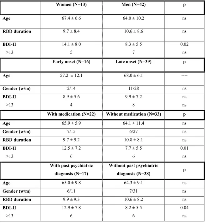
BDI-II scores were significantly higher for women than men, for patients taking psychiatric medication than those free of psychiatric medication, and for patients with past psychiatric diagnosis than those without past psychiatric diagnosis (Table 1). However, men with iRBD also scored higher than control men (8.26 ± 5.53 vs. 4.13 ± 4.59; p < 0.001) as well as women
with iRBD than control women (14.07 ± 8.00 vs. 4.88 ± 4.62; p = 0.001). Moreover, iRBD patients not taking psychiatric medication scored higher than similar controls (7.69 ± 5.22 vs. 4.04 ± 4.38; p < 0.001) and iRBD patients without past psychiatric diagnosis scored higher than similar controls (8.18 ± 5.55 vs. 3.89 ± 4.17; p < 0.001).
____________________________
Insert Table 1 approximately here ____________________________
Anxiety symptoms
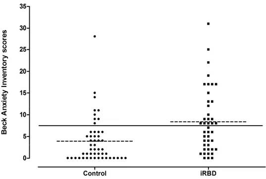
iRBD patients scored significantly higher on the BAI than controls (8.37 ± 7.30 vs. 3.92 ± 5.26; p < 0.001) (Figure 2). A higher proportion of iRBD patients had clinically significant anxiety symptoms than controls (21/43 or 49% vs. 9/50 or 18%, X2 = 8.70; 1 df; p = 0.003). In the iRBD group, 13 subjects (30%) had mild anxiety symptoms, seven (16%) had moderate anxiety symptoms, and one had severe anxiety symptoms (2%). In the control group, eight subjects (16%) had mild anxiety symptoms and one (2%) had moderate symptoms.
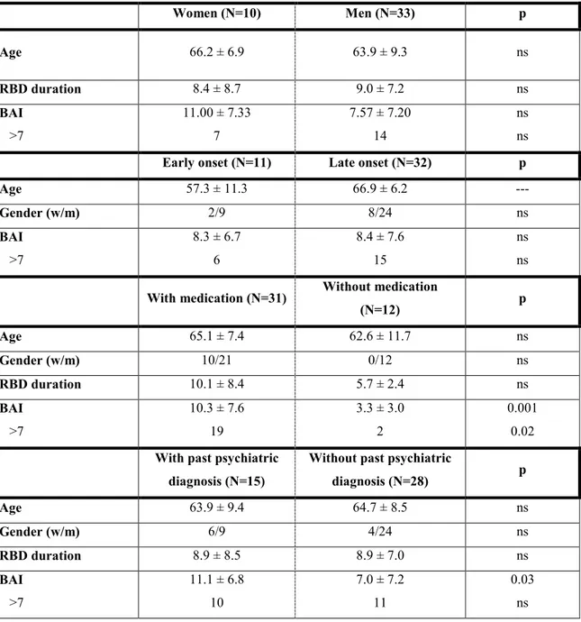
iRBD patients taking psychiatric medication scored significantly higher on the BAI than those without psychiatric medication (Table 2). This is consistent with the higher proportion of subjects taking psychiatric medication who present with clinically significant anxiety
symptoms. No significant difference was in iRBD patients not taking psychiatric medication compared to similar controls (3.33 ± 2.93 vs. 3.69 ± 5.15; p = 0.60). Moreover, iRBD patients
with past psychiatric diagnosis scored significantly higher on the BAI than those without past psychiatric diagnosis. Similar results were observed when participants with past psychiatric diagnosis were excluded in both groups (6.99 ± 7.24 vs. 3.31 ± 3.87; p = 0.01).
____________________________
Insert Figure 2 approximately here ____________________________
____________________________ Insert Table 2 approximately here ____________________________
In iRBD, a positive correlation was found between BDI-II and BAI scores (r=0.621; p < 0.001). No correlation was found between BDI-II or BAI scores and age or RBD duration.
DISCUSSION
Our results show that patients with iRBD exhibit more severe depressive and anxiety symptoms than control subjects. Moreover, a higher proportion of iRBD patients than controls reported clinically significant depressive or anxiety symptoms. Similar to another study (16),we found that more iRBD patients than controls had past psychiatric diagnosis, mainly involving affective and anxiety disorders. In fact, 31% of our iRBD patients had a lifetime history of psychiatric disorders which is similar to the
Rochester’s series (26%; 2) or the Hong Kong Chinese’s cohort (33%; 5).A positive correlation was found between BDI-II and BAI scores in the iRBD group. Thus, the results of the present study indicate that routine examination of patients with iRBD should include an assessment of psychiatric symptoms to prevent their appearance and offer therapeutic alternatives.
We found that 22% of individuals in the iRBD group had clinically significant
depressive symptoms, compared to 6% of controls. A limitation of our study might be that our control group is not representative of the general population. However, in Quebec’s elderly population, the prevalence of depression has been estimated at 6.8% (4.6% in men and 8.3% in women; 23), which is similar to the prevalence reported in other countries (24). Our results also show that 49% of iRBD subjects and 18% of controls had clinically significant anxiety symptoms. The prevalence of anxiety
disorders in Quebec’s elderly population has been estimated at 5.6% (3.6% in men and 6.9% in women; 23),with an even higher prevalence of anxiety symptoms (>15% in the
community samples; 25). This is similar to the prevalence observed in other countries (25).Therefore, our estimated prevalence of depression and anxiety is higher in iRBD subjects and approximately similar in controls to the estimates for Quebec’s general population or for other countries. However, differences in the proportion of men to women limit direct comparison.
Our study identifies female gender as a risk factor for more severe depressive symptoms in iRBD. This is similar to the general population, where women have a higher prevalence of depression than men (23,24,25). However, our iRBD and control groups were matched for gender, and the severity of depressive symptoms was higher in males with iRBD compared to male controls. Other risk factors for more severe
depressive or anxiety symptoms identified in our study are the use of psychiatric medication and the presence of past psychiatric diagnosis. This is consistent with the results reported in the literature (24).
We found that iRBD patients taking psychiatric medication, mainly antidepressants or anxiolytics, reported more severe depressive or anxiety symptoms. Different
explanations may be proposed for this. First, the psychiatric medication may actually lower inventory scores that would be even higher without psychiatric medication. Second, the efficacy of the psychiatric medication may be questioned. Antidepressants such as selective serotonin reuptake inhibitors and tricyclic are largely used in PD. However, their efficacy in treating depression in PD is in dispute (27-29). Moreover,
the efficacy of pharmacologic treatment of anxiety in PD has not been well
demonstrated (30). Given the association between iRBD and the development of PD, which suggests some similarities in their pathophysiology, it is also possible that iRBD patients treated for depression or anxiety may not benefit from their medications. However, further studies are needed to better determine the efficacy of psychiatric medications in iRBD.
In the present study, we used two widely employed scales to assess the severity of depressive and anxiety symptoms in iRBD. The BDI-II is a modified version of the original BDI, designed to better correspond to the DSM-IV criteria for major depression (20). The BDI-II appears to have strong validity as a screening measure for depression in older adults in the general population (31). Although the BDI-II does not have an equal number of items for each criterion of the DSM-IV, it covers all the cognitive and somatic-affective dimensions of major depression. Although the validity of the BDI-II to assess depressive symptoms in iRBD has never been established, it has been
demonstrated valid for screening and measuring depressive symptom severity in PD (32).
BAI measures the severity of somatic, affective, and cognitive symptoms associated with anxiety (21). It has good validity, especially in assessing panic attack symptoms, as it covers 10 of the 13 criteria listed in the DSM-IV. It is probably more suited to screening panic attacks than other anxiety dimensions such as generalized anxiety
disorder (33). This makes it a reliable tool for assessing anxiety in PD, panic disorder being one of the most frequent dimensions of anxiety in this neurodegenerative disease (34). Although the validity of the BAI for assessing anxiety symptoms in iRBD has never been established, its use has been suggested to measure anxiety in PD (33).
Two hypotheses may explain the high frequency of depressive and anxiety symptoms in iRBD. First, iRBD and depression/anxiety may share common neuronal and
neurotransmitter deficiencies. The physiopathology of RBD involves brain regions that regulate REM sleep (35),including the noradrenergic locus coeruleus/subcoeruleus and the serotoninergic raphe systems. These structures have connections with the limbic system, basal ganglia, and hypothalamus, areas involved in the regulation of emotions that were also shown to play a role in depression and anxiety (36). Moreover, RBD represents an early stage in the development of synucleinopathies such as PD or DLB. A high prevalence of depression and anxiety was previously reported in these
conditions (13-15). Some studies also reported that depressive and anxiety symptoms often develop before the onset of parkinsonism in PD and DLB (37-39). Follow-up studies comparing the risk of developing a neurodegenerative disease in iRBD
according to their psychiatric status are needed to better understand this possible link.
The second hypothesis is that the presence of comorbidities in iRBD directly results in depression and anxiety. Indeed, iRBD patients often present olfactive loss (12),
impairment (11,40), or subtle motor impairment (12), that might reduce their quality of life and increase the risk of developing depressive or anxiety symptoms. Sleep
disruption and fear of hurting themselves or the bed partner, which are very frequent in iRBD (2,3),and the stress of a higher risk of developing a neurodegenerative disease in the future may also be psychological burdens that could trigger and maintain a mood disorder. However, the higher score for the iRBD group and the unchanged proportion of iRBD patients with clinically significant depressive symptoms on the BDI-PC, which was developed to minimize the contribution of confounding factors, suggests that comorbidities might not be the primary causes of psychiatric symptoms in iRBD. Further studies are needed to better understand the pathophysiology of psychiatric symptoms in iRBD and the role of comorbidities.
REFERENCES
1. Schenck CH, Mahowald MW: REM sleep behavior disorder: clinical, developmental, and neuroscience perspectives 16 years after its formal identification in SLEEP. Sleep 2002;25:120-138
2. Olson EJ, Boeve BF, Silber MH: Rapid eye movement sleep behavior disorder: demographic, clinical and laboratory findings in 93 cases. Brain
2000;123(Pt2):331-339
3. Schenck CH, Hurwitz TD, Mahowald MW: REM sleep behaviour disorder: an update on a series of 96 patients and a review of the world literature. J Sleep Res1993;2(4):224-231
4. Ohayon MM, Caulet M, Priest RG: Violent behavior during sleep. J Clin Psychiatry 1997;58(8):369-376
5. Wing YK, Lam SP, Li SX, et al: REM sleep behaviour disorder in Hong Kong Chinese: clinical outcome and gender comparison. J Neurol Neurosurg
Psychiatry 2008;799(12):1415-1416
6. Gagnon JF, Postuma RB, Montplaisir J: Update on the pharmacology of REM sleep behavior disorder. Neurology 2006;67(5):742-747
7. Postuma RB, Gagnon JF, Vendette M, et al: Quantifying the risk of neurodegenerative disease in idiopathic REM sleep behavior disorder. Neurology 2009;72(15):1296-130
8. Iranzo A, Molinuevo JL, Santamaria J, et al: Rapid-eye-movement sleep behaviour disorder as an early marker for a neurodegenerative disorder: a descriptive study. Lancet Neurol 2006;5(7):572-577
9. Schenck CH, Bundlie SR, Mahowald MW: Delayed emergence of a parkinsonian disorder in 38% of 29 older men initially diagnosed with idiopathic rapid eye movement sleep behaviour disorder. Neurology 1996;46(2):388-393
10. Montplaisir J, Gagnon JF, Fantini ML, et al: Polysomnographic diagnosis of idiopathic REM sleep behavior disorder. Mov Disord 2010 Oct 15;25(13):2044-2051
11. Gagnon JF, Vendette M, Postuma RB, et al: Mild cognitive impairment in rapid eye movement sleep behavior disorder and Parkinson's disease. Ann Neurol 2009;66(1):39-47
12. Postuma RB, Gagnon JF, Vendette M. Monplaisir JY: Markers of
neurodegeneration in idiopathic rapid eye movement sleep behaviour disorder and Parkinson's disease. Brain 2009;132(Pt 12):3298-3307
13. Aarsland D, Bronnick K, Ehrt U, et al: Neuropsychiatric symptoms in patients with Parkinson's disease and dementia: frequency, profile and associated care giver stress. J Neurol Neurosurg Psychiatry 2007;78(1):36-42
14. Benrud-Larson LM, Sandroni P, Schrag A, Low PA: Depressive symptoms and life satisfaction in patients with multiple system atrophy. Mov Disord
2005;20(8):951-957
15. Dodel R, Csoti I, Ebersbach G, et al: Lewy body dementia and Parkinson's disease with dementia. J Neurol 2008;255(Suppl 5):39-47
16. Teman PT, Tippmann-Peikert M, Silber MH, et al: Idiopathic rapid-eye-movement sleep disorder: associations with antidepressants, psychiatric diagnoses, and other factors, in relation to age of onset. Sleep Med