EXPOSITION DE LA MOULE BLEUE (MYTILUS EDULIS) À DEUX SOUCHES BACTÉRIENNES D'ORIGINE FÉCALE (ESCHER/CHIA COLI ET
ENTEROCOCCUS FAECALIS) ET À UNE SOUCHE ENDÉMIQUE (VIBRIO SPLENDIDUS): EFFETS SUR LE SYSTÈME IMMUNITAIRE
Mémoire présenté
dans Je cadre du programme de maîtrise en Océanographie en vue de l'obtention du grade maître ès Sciences
PAR
© ISABELLE BOILY
Avertissement
La diffusion de ce mémoire ou de cette thèse se fait dans le respect des droits de son auteur, qui a signé le formulaire
«
Autorisation de reproduire et de diffuser un rapport, un mémoire ou une thèse». En signant ce formulaire, l'auteur concède à l'Université du Québec à Rimouski une licence non exclusive d'utilisation et de publication de la totalité ou d'une partie importante de son travail de recherche pour des fins pédagogiques et non commerciales. Plus précisément, l'auteur autorise l'Université du Québec à Rimouski à reproduire, diffuser, prêter, distribuer ou vendre des copies de son travail de recherche à des fins non commerciales sur quelque support que ce soit, y compris l'Internet. Cette licence et cette autorisation n'entraînent pas une renonciation de la part de l'auteurà ses
droits moraux nià
ses droits de propriété intellectuelle. Sauf entente contraire, l'auteur conserve la liberté de diffuser et de commercialiser ou non ce travail dont il possède un exemplaire.Composition du jury:
Émilien Pelletier, président du jury, Université du Québec à Rimouski
Sophie Gauthier-Clerc, directrice de recherche, Université du Québec à Rimouski
Karine Lemarchand, codirectrice de recherche, Université du Québec à Rimouski
Ahmed Siah, examinateur externe, BC Center for aquatic health sciences
REMERCIEMENTS
Tout d'abord, je tiens à remercier ma directrice Sophie Gauthier-Clerc pour sa grande disponibilité, sa compréhension et pour tous les conseils qu'elle m'a donnés afin de m'aider lors de ma maîtrise et aussi dans ma carrière future. Elle m'a rendue plus critique, plus autonome et m'a donnée un bagage de connaissances qui m'aidera tout au long de ma carrière. Merci. l'aimerais également remercier ma codirectrice Karine Lemarchand pour sa disponibilité et pour m'avoir maintenue sur la bonne voie tout au long de ma maîtrise. Je la remercie aussi d'avoir accepté de manipuler des organismes de plus d'une cellule afin de m'aider durant mes échantillonnages. Ce fut grandement apprécié.
Je souhaite également remercier Émilien Pelletier et Ahmed Siah d'avoir accepté de faire partie de mon jury. Je remercie également le CRSNG pour son soutien financier.
Je tiens aussi à remercier tous ceux qui m'ont aidée durant mon projet, Karine Lemarchand, Amélie Jauvin, Romy Ricci, et Geneviève Côté. Merci à Michel Founier et Nicolas Toupoint grâce à qui j'ai pu avoir des moules. Merci à Ahmed Siah de m'avoir fourni une souche de V splendidus et à l'hôpital de Rimouski pour la souche de E.coli. Merci aussi à Jocelyne Pellerin et Réjean Tremblay pour leurs conseils et pour m'avoir permis de travailler dans leur laboratoire et à tous les autres qui ont contribué à mon projet, Bertrand Genard, Pierre Rioux, le personnel de Nutrocéan et de la station aquacole de Pointe-au-Père.
Je remercie finalement ma famille et mon chum, Rémi, pour m'avoir soutenue tout au long ma maîtrise et ce, autant durant les hauts que durant les bas.
RÉSUMÉ
Les environnements aquatiques sont naturellement habités par plusieurs espèces bactériennes. Cependant, l'augmentation de l'urbanisation des côtes estuariennes mène à l'introduction de bactéries exogènes (e.g. bactéries fécales Escherichia coli et Enterococcus faecalis) dans les eaux côtières. En tant qu'organismes filtreurs, les bivalves vivant dans
ces milieux côtiers peuvent accumuler une grande quantité de bactéries, incluant des bactéries indigènes et exogènes. Présentement, la plupart des recherches effectuées sur l'interaction des bactéries avec les bivalves se concentrent sur les effets des bactéries pathogènes en présence ou non d'autres stresseurs (par exemple, les polluants chimiques) sur la physiologie ou l'immunité des espèces commerciales. Mytilus edulis, est fréquemment utilisée comme espèce sentinelle en écotoxicologie mais est également une espèce commerciale importante au Québec. Le but de cette étude est de mesurer les effets d'une exposition ponctuelle à Escherichia coli et à Enterococcus faecalis sur son immunocompétence puisque la présence de ces bactéries exogènes pourrait moduler la réponse immunitaire de la moule bleue parfois affectée lorsqu'il y a présence de stresseurs immunosuppresseurs (contaminants chimiques, etc.). Nous avons comparé les effets de ces challenges bactériens aux effets d'une exposition à une espèce bactérienne indigène aux eaux côtières, Vibrio splendidus, qui est reconnue comme n'ayant aucun effet sur l'immunité de la moule bleue. Les trois challenges bactériens ont été réalisés in vivo en utilisant des concentrations bactériennes représentatives des concentrations retrouvées dans les environnements côtiers (de 103 à 105 bactéries mC!). Ils ont été menés en septembre et décembre, afin d'avoir des conditions de température différentes et une condition physiologique différente pour les moules. La réponse immunitaire de Mytilus edulis a été déterminée 24 h et 48 h après les challenges bactériens en se basant sur des paramètres de l'immunité cellulaire (abondance des hémocytes, viabilité des cellules, production de NOx et de ROS et phagocytose) et humorale (enzymes hydrolytiques). Nos résultats démontrent qu'à des concentrations bactériennes représentatives de celles qui sont retrouvées dans l'environnement de manière périodique suite à un déversement d'eaux usées, les bactéries exogènes
E.
coli et Ejaecalis n'ont pas d'effets sur l'immunocompétence de M edulis etce, peu importe la période durant laquelle le challenge a eu lieu. Le fait qu'aucune variation de l'immunité n'ait été observée 24 et 48 heures après les infections suggère que les moules ont été capables de se défendre contre ces bactéries sans que cela n'affecte à long terme leur système immunitaire. En conclusion, la contamination ponctuelle des eaux côtières par des bactéries fécales ne représente pas une menace additionnelle pour l'immunocompétence de M edulis en comparaison avec d'autres stresseurs anthropiques tels que les contaminants chimiques.
ABSTRACT
Many bacterial species arc natural inhabitants of aquatic environments. However, in estuarine coastal watersheds, the rapid increase of urbanization and the intensification of anthropogenic use of the watershed could lead to the introduction of exogenous bacteria (e.g. fecal bacteria su ch as Escherichia coli and Enterococcus faecalis) in coastal waters. Filter feeding bivalves living in these coastal areas can accumulate a high number of bacteria, including indigenous and exogenous species. Presently, most investigations dealing with the interaction of bacteria and bivalves focus on the effects of bacterial pathogens alone or their interactive effects with other stressors, like chemical pollutants, on the physiology or the immune capacity of commercial species (e.g. oyster). Since Mytilus edulis is widely used as a sentinel species in ecotoxicology, the aim of this study was to assess the effect of its potentials exposure toward Escherichia coli and Enterococcus faecalis on its immune defenses. For this purpose, we have compared the effect of such a bacterial challenge with the effect of an exposure toward Vibrio splendidus which is considered as an indigenous Mytilus edulis species of coastal estuarine waters. The three bacterial challenges were realized in vivo, using bacterial concentrations that are representative of the environment loads (103 to 105 cells mr') and conducted in September and in December. The immune response of Mytilus edulis was determined after 24 h and 48 h of post-challenge on the basis of both cellular (total haemocytes count, cell viability, phagocytosis, production of NOx and ROS) and humoral (hydrolytic enzymes) parameters. Our results demonstrated no inhibitory nor inductive effect of the bacterial challenges on the mussels' immune responses, whatever the bacterial strain, its concentration and the period of the challenge. In our experimental conditions, the presence of faecal bacteria in coastal waters does not represent an additional threat for immune competency. Those results permit also a better understanding of the immune reaction of M edulis in response to environmental stresses in experimental conditions as representative as possible of environmental conditions.
TABLE
DES MATIÈRES
REMERCIEMENTS ...
...
...
VII
RÉSUMÉ ... IX
ABSTRACT ... XI
TABLE
DES MATIÈRES
... XIII
LISTE
DES
TABLEAUX
...
... XV
LISTE
DES
FIGURES ... XIX
LISTE
DES
ABRÉVIATIONS
ET DES ACCRONYMES
... XXI
INTRODUCTION
GÉNÉRALE
... 1
CHAPITRE
1.EXPOSURE
OF A SENTINEL
SPECIES
(MYTILUS EDULIS)TO FECAL
BACTERIA IN COASTAL WATERS:
EFFECT
ON
THE
IMMUNE SYSTEM
...
...
11
ABSTRACT ... 11
1.1 INTRODUCTION ... 13
1.2 MATERIAL AND METHODS ...
16
1.3 RESUL
TS ... 21
1.4 DISCUSSION ... 38
1.5 CONCLUSION ... 41
DISCUSSION GÉNÉRALE ET
CONCLUSION
... 43
l
u
STE DES TABLEAUX:[Bcl]Table 1: Four ways ANOVAs of the effect oftime (24 h and 48 h), treatment (non-injected, unchallenged, 102, 103 and 104 cfu injected), month (September and December) and the strain (V splendidus,
E.
coli andE.
faecalis) and their interaction on cellular imtnune parameters ......25
Table 2: Three ways ANOV As of the effect of time (24 h and 48 h), treatement (non-injected, unchallenged, 102, 103 and 104 cfu injected) and the strain (V splendidus,E.
coli and
E.
faecalis) and their interaction on the ROS production in September and DeCelTI ber ... 26Table 3: Scheirer-Ray-Hare test and Four ways ANOVAs of the effect time (24 h and 48 h), treatment (non-injected, unchallenged, 102, 103 and 104 cfu injected), month
(September and December) and the strain (V splendidus,
E.
coli andE.
faecalis) and their interaction on humoral immune parameters ... 27Table 4: Parameters of cellular immunity: viability of haemocytes, abundance of haemocytes, phagocytic capacity, ROS production and NOx production 24 hours after the bacterial challenge (cfu injected) performed in September with Vibrio splendidus, Enterococcusfaecalis and Escherichia coli. Values are mean ± SD (N=3) ... 28
Table 5. Parameters of cellular immunity: viability of haemocytes, abundance of haemocytes, phagocytic capacity, ROS production and NOx production 24 ho urs after
the bacterial challenge (cfu injected) performed in December with Vibrio splendidus, EnterococcusfaecaUs and Escherichia coli. Values are mean ± SD (N=3) ... 29
Table 6. Parameters of cellular immunity: viability of haemocytes, abundance of
haemocytes, phagocytic capacity, ROS production and NOx production 48 hours after
the bacterial challenge (cfu injected) performed in September with Vibrio splendidus,
Enterococcusfaecalis and Escherichia coli. Values are mean ± SD (N=3) ... 30
Table 7. Parameters of cellular immunity: viability of haemocytes, abundance of
haemocytes, phagocytic capacity, ROS production and NOx production 48 hours after
the bacterial challenge (cfu injected) performed in December with Vibrio splendidus,
Enterococcus faecalis and Escherichia coli. Values are mean ± SD (N=3) ... 31
Table 8. Parameters of humoral immunity: L-Ieucine-aminopeptidase, proPhenoloxydase -like, acid phosphatase and total proteins 24 hours after the bacterial challenge (cfu
injected) performed in September with Vibrio splendidus, Enterococcus faecalis and,
Eschericha coli. Values are means ± SD (N=3) ... 32
Table 9. Parameters of humoral immunity: L-leucine-aminopeptidase, proPhenoloxydase -like, acid phosphatase and total proteins 24 hours after the baeterial challenge (cfu injected) performed in December with Vibrio splendidus, Enterococcus faecalis and Escherichi a coli. Values are means ± SD (N=3) ... 33
Table 10. Parameters of humoral immunity: L-leucine-aminopeptidase,
proPhenoloxydase-like, acid phosphatase and total proteins 48 hours after the bacterial challenge (cfu
injected) performed in September with Vibrio splendidus, Enterococcus faecalis and
Escherichia coli. Values are means ± SD (N=3) ... 34
Table 11. Parameters of humoral immunity: L-Ieucine-aminopeptidase, proPhenoloxydase-like, acid phosphatase and total proteins 48 hours after the bacterial challenge (cfu
injected) performed in December with Vibrio splendidus, Enterococcus faecalis and
/LISTE
DES
FIGURES
V
B
c
2]
Figure 1. Différents types d'hémocytes chez M edulis. Images tirées de Le Foll et al.,
(201 0)
..
...
...
..
....
...
...
..
...
..
...
...
...
...
...
... 3
Figure 2. Membrane cellulaire d'une bactérie a) Gram-négative et b) Gram- positive ... .4
LISTE DES ABREVIATIONS ET DES ACCRONYMES
AMPs: Peptides anti-microbiens
AP: Acid Phosphatase
A.D.: Arbitrary Unit
DAF-FM: 4-amino-5-methylamino-2', 7' Difluofluorescein diacetate
PAMPs: Pathogen-associated molecular patterns
DeF -DA: 5 (6) carboxy-2' 7' -dichlorofl uorescin-diacetate
LAP: L-Ieucine-aminopeptidase
LPS: Lipopolysaccharide
L TA:
lipoteichoic acidNADPH: Nicotinamide adénine di nucléotide phosphate réduit
NO: Monoxyde d'azote N02-: Nitrites
N03: Nitrates
NOx: Oxydes d'azote
PNPP: 4-nitrophenylphosphate
ProPO: proPhenoloxydase
INTRODUCTION GÉNÉRALE La moule bleue
La moule est présente sur toutes les zones côtières du monde, spécialement dans les zones intertidales. Elle est sessile et s'accroche sur des substrats durs. La moule a un mode d'alimentation suspensivore par filtration. À 8°C, elle peut filtrer jusqu'à cinquante litres d'eau par jour (Tremblay et al., 2004). Elle se nourrit de phytoplancton, de détritus organiques et de bactéries. Sa sédentarité et son mode d'alimentation par filtration font en sorte que la moule accumule les différents contaminants et les bactéries qui sont présents dans l'eau ou sur les particules filtrées. Ces particularités à accumuler les contaminants et les bactéries présents dans son environnement en font un organisme sentinelle largement utilisé lors d'études écotoxicologiques (Bayne, 1976; ROPME, 2010;Roslev et al., 2009).
Les bactéries des eaux côtières
Les parcs mytilicoles sont situés dans les zones côtières, ce qui peut entraîner les moules à être exposées à des bactéries dont la source peut être naturelle ou anthropique, comme la décharge d'eaux usées dans l'environnement par manque d'installations d'épuration ou suite au lessivage du bassin versant lors des fortes précipitations. La concentration bactérienne totale dans les milieux côtiers peut varier entre 105 mL-1 et 106 mL-1 bactéries (Jacquet et al., 1998;Li & Dickie, 2003). Les rejets d'eaux usées dans l'environnement marin introduisent des bactéries non indigènes au milieu marin (Garrido-Pérez et al., 2008; Wells, 2003). Ces bactéries sont généralement associées au tube digestif des animaux et des humains (e.g.Escherichia coli, Salmonella spp.). Les plus fréquemment présentes sont les entérobactéries telles qu'Escherichia coli et les entérocoques du groupe D, aussi appelés entérocoques fécaux, tels qu'Enterococcus faecalis. Ces bactéries sont d'ailleurs utilisées comme indicateurs de pollution des eaux par des matières d'origine fécale (Roslev et al., 2009; Poté et al., 2009).
Parmi les bactéries indigènes et exogènes présentes en zones côtières, certaines sont potentiellement virulentes pour les bivalves et peuvent présenter un risque pour la santé
humaine, comme par exemple celtaines souches du genre Vibrio (ASPGC-PHAc., 2011). La plupart des bactéries du genre Vibrio sont opportunistes, c'est-à-dire qu'elles seront non pathogènes pour un organisme jusqu'à ce que leur nombre soit suffisant pour contrer ses défenses immunitaires ou jusqu'à ce que celles-ci soient diminuées. C'est alors qu'el1es expriment leur potentiel pathogène. Chez les bivalves, ces bactéries sont associées à 20% des maladies causées par des bactéries (Garay et al., 1985; Lane & Birkbeck, 2000;
Potasman et al., 2002; Pruzzo et al., 2005). Les bactéries du genre Vibrio sont des bactéries largement répandues dans les environnements marins côtiers où leur concentration peut atteindre 103 mL-I. Alors que le potentiel pathogène de certaines souches de bactéries indigènes aux environnements côtiers est bien connu, les effets des bactéries exogènes sur la santé des' moules sont moins bien documentés.
Le système immunitaire des bivalves
Le système immunitaire de la moule bleue est constitué de défenses innées,
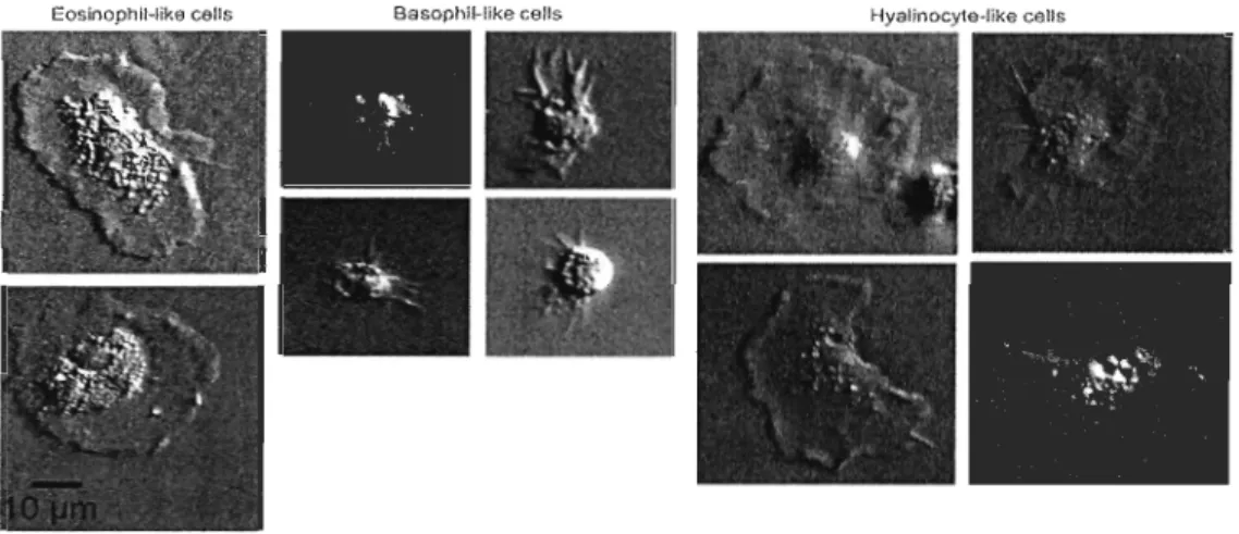
comprenant les défenses cellulaires et humorales (Seo et al., 2005). L'immunité cellulaire est assurée par les hémocytes. Chez les bivalves, les hémocytes sont constitués de deux principaux types de cel1ules : les granulocytes et les hyalinocytes, identifiés selon leur taille, leur forme et leur contenu en granules. Les granulocytes sont des cellules au cytoplasme bien développé contenant de nombreuses granules qui ont une tail1e d'environ 10 !-lm (Le Foll et al. 2010). Les hyalinocytes sont des cellules sans granules qui sont généralement plus petites que les granulocytes (Auffret, 1988), comme il est possible de l'observer à la figure l. Leur taille fait entre 7 !-lm et 10 !-lm (Le Foll et al. 2010). Les hyalinocytes, bien que morphologiquement hétérogènes, se divisent également en trois catégories: les cel1ules «blast-like» ayant un noyau central sphérique ou ovoïde entouré d'un cytoplasme basophile contenant peu d'organelles, les hyalinocytes «macrophage-like» contenant un cytoplasme basophile et les hyalinocytes, ayant un noyau ovoïde plus gros et de forme irrégulière entouré d'un cytoplasme contenant plusieurs organelles (Hi ne, 1999).
Les granulocytes sont eux aussi hétérogènes selon la présence ou non d'enzymes Iysosomales, de phenoloxydases, d'enzymes antioxydantes, de lectines et de leur capacité
phagocytaire. Ils peuvent aussi être divisés en deux groupes, les granulocytes eosinophiles et les granulocytes basophiles qui assureraient, en majeure partie, la phagocytose (Hi ne, 1999; Le Foll et al., 2010; Pipe, 1990).
EosinophlHike œlis Basophll-like celis Hyalinocyte-like ceUs
Figure 1. Différents types d'hémocytes chez M edulis. Images tirées de Le Fol! et al., (2010).
Les hémocytes sont impliqués lors du processus inflammatoire et ont un rôle dans le processus de réparation des tissus. La réponse immunitaire comprend quatre évènements: l'adhérence, l'absorption, la destruction et l'élimination des particules étrangères (Feng, 1988). L'adhérence et la reconnaissance du non-soi sont essentielles pour initier la réponse immunitaire, car c'est l'interaction entre ces protéines et les motifs de reconnaissance de la paroi bactérienne qui déclenche la réaction immunitaire. Cette reconnaissance est possible grâce aux PAMP (pathogen-associated mo/ecu/ar patterns). Il s'agit d'un motif de reconnaissance présent sur la surface des micro-organismes tel que les lipopolysaccharides (LPS, dans lesquels sont contenus les lipides A qui sont à l'origine de la toxicité de plusieurs bactéries Gram-négatives), les acides teichoïques qui sont présents seulement chez les bactéries Gram-positives et le peptidoglycane (Beutler, 2004; Perry et al., 2004; Wedenmaier & Peschel, 2008). Tel que le montre la figure 2, les bactéries Gram-négatives (figure 2 a) et Gram-positives (figure 2 b) ne présentent pas la même paroi cellulaire. La membrane externe des bactéries Gram-positives est caractérisée par plusieurs couches de
peptidoglycanes qui représentent de 40 à 80 % de la matière sèche de la paroi et elle
contient des acides tels que les acides teichoïques. Les bactéries Gram-négatives
comportent également du peptidoglycane, mais en proportion moindre (une seule couche),
recouvert d'une membrane composée de molécules lipidiques, de polysaccharides et de protéines. Les complexes lipides-polysaccharides sont appelés lipopolysaccharides ou LPS.
a) Extérieur de ln cellule Cytoplasme b) Cytoplasme mcrnbran.JirC$ La structure de
l'enveloppe externe est analogue c\ celle d'une membrane
cellulaire. Différentes protéines. dont des pennes, y sont
insérées. la membrane cellulaire est constituée de deux feuillet' phosp ho-lipidiques maintenus par des liaisons hydrophobes.
Figure 2. Membrane cellulaire d'une bactérie a) Gram-négative et b) Gram-positive.
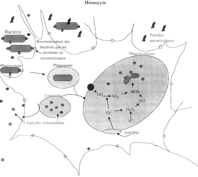
Chez l'huitre Crassostrea gigas, plusieurs protéines de reconnaissance du non-soi ont été identifiées: les lectines (Gueguen et al., 2003), les protéines de liaison au LPS qui permettent la reconnaissance des bactéries Gram -négatives (Gonzalez et al., 2005), les protéines de reconnaissance du peptidoglycane qui permettent la reconnaissance des bactéries Gram-positives (Itoh & Takahashi, 2008) et les protéines de liaison au p-glucane (1toh & Takahashi, 2009). Après le processus de reconnaissance, l'absorption, est caractérisée par la phagocytose des débris tissulaires et des bactéries qui auraient pu pénétrer dans les tissus des bivalves. Lors de la phagocytose, durant laquelle il y a formation de pseudopodes qui permettent à la cellule d'envelopper les particules étrangères, entraînant leur internalisation dans une vésicule appelée phagosome. La destruction de ces particules étrangères relève de deux procédés qui agissent simultanément ou séparément dans la cellule: les enzymes lysosomales et la flambée oxydative. La flambée oxydative est catalysée par l'assemblage du complexe enzymatique NADPH (nicotinamide adénine dinucléotide phosphate) oxydase et induite par la phagocytose. L'oxygène est réduit en métabolites bactéricides, incluant le peroxyde d'hydrogène (H202), les groupements hydroxyl (OH) et les anions superoxydes (02-), ce qui crée un environnement microbicide. Ces composés oxygénés réactifs dégradent et tuent les micro-organismes (Pipe, 1992; Perry et al., 2004; Lichtman & Abbas, 2005). D'autres métabolites pouvant avoir une action biocide sont également produits par l'activation de la NADPH oxydase, comme les NOx, soit l'oxyde nitrique (NO), les nitrites (N02-) et les nitrates (N03), qui sont des métabolites stables du NO générés par la flambée oxydative (Gourdon et al., 2001).
L'autre composante du système immunitaire est l'immunité humorale. Les facteurs humoraux sont les enzymes lysosomiales, les lectines et les agglutinines qui permettent une dégradation extracellulaire des particules (Chu, 1988). Après la phagocytose et l'internalisation des particules dans le phagosome, le lysosome se lie au phagosome pour former le phagolysosome où les différentes enzymes lysosomiales vont digérer les particules (Seo et al., 2005). Mais les enzymes lysosomiales sont également larguées dans le plasma durant de la phagocytose, ce qui fait de l'activité de ces enzymes un indicateur de l'activité du système immunitaire contre des agents pathogènes (Oubella et al., 1994). La
présence d'agglutinines et de lectines est également importante pour limiter l'action des bactéries en les agglutinant et en favorisant ainsi leur lyse et leur phagocytose. De plus, elles servent également de récepteur aux hémocytes et de médiateur pour la phagocytose (Chu, 1988). Les lectines font partie des protéines reconnaissantes du non-soi et se lient aux oligosaccharides présents à la surface des micro-organismes. Une défense humorale importante est la mélanisation des pathogènes et des tissus endommagés. Cette mélanisation est effectuée à l'aide de la phenoloxydase, un enzyme contenant du cuivre et present dans l'hémolymphe sous une forme inactive: la prophenoloxydase. La prophenoloxydase est clivée en phenoloxydase qui catalyse la synthèse d'o-diphénol. La polymérisation non-enzymatique des o-diphénols mène à la production de mélanine. L'activité de la phenoloxydase a été démontrée chez plusieurs espèces de bivalves, dont
Mytilus edulis (Hellio et al., 2007; Munoz et al., 2006; S6derhtill & Cerenius, 1998).
Il y a aussi d'autres effecteurs de l'immunité humorale comme les peptides antimicrobiens (AMPs). Les AMPs sont sécrétés par les hémocytes dans l'hémolymphe et ont un rôle multifonctionnel dans l'immunité innée. Outre leurs propriétés antimicrobiennes, ils interviennent également dans la stimulation de réponses de l'immunité et peuvent exercer des effets immunomodulateurs (Nicolas, 2009). Les AMPs présentent de larges spectres d'activité antimicrobienne vis-à-vis de bactéries à Gram-positif et à Gram-négatif, de champignons, de protozoaires et de virus enveloppés. Leur diversité structurale est grande. La majorité sont cationiques et sont à l'origine de trois grandes familles : les peptides linéaires formant des hélices, les peptides riches en cystéine avec un ou plusieurs ponts disulfure et les peptides contenant un pourcentage élevé d'un type d'acide aminé (Andrès & Dimarcq, 2004). Le mode d'action des peptides antimicrobiens est principalement la perméabilisation des membranes. Cette perméabilisation entraîne la libération des produits intracellulaires bactériens dans le milieu extérieur entraînant la mort de la bactérie.
Hémocyte
1
1 1
1
Peptides Enzymes Iysosomiales•
Stress et immunité
Le système immunitaire des moules leur permet de répondre aux infections que peuvent causer les bactéries présentes dans leur environnement. Cependant, une exposition à différents stress peut avoir des effets sur l'immunité des bivalves. Les stress tels que le brassage mécanique, la réduction de la salinité et l'exposition à l'air ont comme effets de diminuer la concentration d'hémocytes, l'activité phagocytaire et la flambée oxydative ainsi que d'augmenter l'activité de la phosphatase acide. (Blaise et al., 2002;Bussell et a!., 2008; De Donno et a!., 2008; Kuchel et a!., 2010; Oweson & Hernroth, 2009). L'état physiologique des moules est également un paramètre qui peut influencer leur réponse immunitaire. Les coûts énergétiques qu'engendrent la gamétogénèse et la ponte durant une période où les conditions environnementales sont stressantes (e.g. températures élevées) et le stockage de réserves énergétiques sont des facteurs qui peuvent affecter la concentration des hémocytes et l'activité phagocytaire (Delaporte et a!., 2006).
Les souches pathogènes des bactéries du genre Vibrio interagissent avec les hémocytes en interférant sur plusieurs paramètres de leur système immunitaire, comme le nombre d'hémocytes en circulation dans l'hémolymphe, leur propriété d'adhésion et l'activité phagocytaire, en augmentant la production de ROS et en régulant l'expression de certains gènes reliés
à
la réponse immunitaire. (Lambert el a!., 2003; Labreuche et a!., 2006; Pari si et a!., 2008). Le genre Vibrio est d'ailleurs à l'origine de divers épisodes de mortalité chez les bivalves. Par exemple, en France, depuis 1991, des bactéries du genre Vibrio, conjointement à d'autres facteurs environnementaux tels qu'une augmentation de la température de l'eau et une diminution de la salinité, sont responsables d'un taux de mortalité allant jusqu'à 90% en période estivale dans les productions d'huîtres creusesCrassostrea gigas (IFREMER, 2012; Lambert et a!., 2007; Munari et a!., 2011; Matozzo et
a!.,2007).
Les moules sont également exposées à des bactéries indigènes non-pathogènes et de façon sporadique
à
des bactéries exogènes du milieu marin provenant du rejet des eaux usées (Roslev et a!., 2009). Bien que différentes études mentionnées aient déjà été réaliséesafin de connaître la réaction du système immunitaire des bivalves face à une exposition à des bactéries, elles ont majoritairement été effectuées in vitro, pour étudier la réponse des hémocytes exposés majoritairement avec des bactéries pathogènes ou avec des quantités supérieures de bactéries (pathogènes ou non) que celles qui se retrouvent généralement dans le milieu marin, soit entre 106 et 109 bactéries mL-J (Araya et al., 2010; Costa et al., 2009; Blaise et al., 2002;Lambert et al., 2003). De plus en plus d'études sur l'interaction entre le système immunitaire des bivalves et les bactéries sont effectuées in vivo. Certaines études ont démontré qu'une infection avec Vibrio spp. chez plusieurs espèces de bivalves pouvait provoquer une surexpression du gène Tg-sHSP, relié aux «small heat shock proteins» (Bao et al., 2011), une sous-expression des gènes codant pour certains peptides antimicrobiens (Li et al., 2010 b), une diminution de la capacité phagocytaire et de l'adhérence des hémocytes ainsi qu'une augmentation de la production de ROS (Labreuche et al., 2006). Mais encore fois, ces études ont été réalisées majoritairement avec des bactéries pathogènes ou avec une quantité de bactéries supérieure à celles qui sont rapportées dans les environnements côtiers. Il y a donc un manque de connaissances sur les effets de ces bactéries non-pathogènes exogènes à l'environnement marins sur la réponse immunitaire des bivalves aux concentrations auxquelles elles sont retrouvées dans l'environnement.
Objectifs et hypothèses
Au Québec, la production de moules est passée de 66 tonnes en 1996 à 540 tonnes en 2009, allant même jusqu'à atteindre 753 tonnes en 2005 (Anonyme, 2007). Bien que des mortalités élevées chez les moules causées par des particularités phénotypiques aient été rapportées dans les années 90 (Tremblay et al., 1998; Myrand et al., 2000), aucune mortalité causée par des maladies infectieuses n'a cependant été constatée chez Mytilus edulis sauvage ou d'élevage. Une production mytilicole plus intensive et une détérioration de la qualité de l'eau pourraient augmenter le risque de propagation de maladies et mener à des mortalités massives ou imposer d'autres contraintes sur l'expansion des cultures de moules (FAO, 2011). Le contact avec des bactéries non-indigènes pourrait mobiliser le
système immunitaire de la moule bleue et la rendre plus vulnérable aux conditions de stress soumises par son environnement.
L'objectif général de ce projet a été d'évaluer la réactivité de l'immunocompétence cellulaire et humorale de la moule bleue (Mytilus edulis) face à une exposition in vivo à différentes souches bactériennes susceptibles de se retrouver en zone côtière à proximité des élevages de moules. D'une part, une souche bactérienne indigène au milieu marin,
Vibrio splendidus a été utilisée. D'une autre part, les souches Escherichia coli et
Enterococcus faecalis ont été utilisées puisqu'elles ne sont pas pathogènes mais sont
exogènes à l'environnement des moules et se retrouvent en grande quantité dans les eaux marines côtières. Les infections réalisées avec
E.
coli etE.
jàecalis avaient pour but demieux comprendre l'effet de bactéries Gram-négatives et Gram-positives provenant de
rejets d'origine anthropique ou agricole sur le système immunitaire de la moule bleue, alors que celles réalisées avec Vibrio splendidus avaient pour but d'évaluer l'effet d'une souche indigène sur l'immunité de la moule avec une quantité classiquement retrouvée dans le milieu marin. Ainsi trois challenges avcc 0, 102, 103 ct 104 bactéries (plus un groupe non-injecté appelé contrôle) ont successivement été réalisés avec les souches d' Escherichia coli,
Enterococcus faecalis et Vibrio splendidus durant les mois de septembre et de décembre
afin de considérer l'état physiologique des moules en fonction de leur cycle reproducteur.
Les hypothèses sont que l' immunocompétence est modifiée par une infection
bactérienne par rapport à un témoin non infecté (Hl), que l'immunocompétence varie selon la souche bactérienne utilisée pour l'infection (H2), que l'immunocompétence varie selon
la durée de l'infection (H3) et que l'immunocompétence varie selon la concentration de bactéries injectées lors de l'infection (H4).
CHAPITRE 1
EXPOSURE OF A SENTINEL SPECIES (MYTILUS EDULIS) TO FECAL BACTERIA IN COAST AL WATERS: EFFECT ON THE IMMUNE SYSTEM
Abstract
Many bacterial species are natural inhabitants of aquatic environments. However, in estuarine coastal watersheds, the rapid increase of urbanization and the intensification of anthropogenic use of the watershed could lead to the introduction of exogenous bacteria (e.g. fecal bacteria such as Escherichia coli and Enterococcus faecalis) in coastal waters. Filter feeding bivalves living in the se coastal areas can accumulate a high number of bacteria, including indigenous and exogenous species. Presently, most investigations dealing with the interaction of bacteria and bivalves focus on the effects of bacterial pathogens alone or their interactive effects with other stressors, like chemical pollutants, on the physiology or the immune capacity of commercial species (e.g. oyster). Since Mytilus edulis is widely used as a sentinel species in ecotoxicology, the aim of this study was to assess the effect of its potentials exposure toward Escherichia coli and Enterococcus faecalis on its immune defenses. For this purpose, we have compared the effect of such a bacterial challenge with the effect of an exposure toward Vibrio splendidus which is considered as an indigenous Mytilus edulis species of coastal estuarine waters. The three bacterial challenges were realized in vivo, using bacterial concentrations that are representative of the environment loads (103 to 105 cells mrl) and conducted in September and in December ta get distinct thermal conditions (16°C and 3°C respectively) that are representative of fall in Québec (Canada), a period characterised by high rainfall events
allowing important bacterial runoff. The immune response of Mytilus edulis was
determined after 24 h and 48 h of post-challenge on the basis of both cellular (total haemocytes count, cell viability, phagocytosis, production of NOx and ROS) and humoral (hydrolytic enzymes) parameters. Our results demonstrated no inhibitory nor inductive effect of the bacterial challenges on the mussels' immune responses, whatever the bacterial
strain, its concentration and the period of the challenge.
In
conclusion, the presence of faecal bacteria in coastal waters does not represent an additional threat for immune competency.1.1 INTRODUCTION
Blue mussel (Mytilus edulis) is a major species in shellfish farming in eastern Canada (DFO, 2011) and is weil recognised as a sentinel species in ecotoxicology. In coastal areas,
mussels can be exposed to various abiotic factors that may act as physiological stressors or
immunomodulators, leading exceptionally to large mass mortality events in cultivating
areas (FAO, 2011). Temperature (Munari et al. 2011), salinity, mechanical stress (Bussell
et al. 2008, Kuchel et al. 2010) and contaminants (Gagnaire et al., 2007; Wootton et al.,
2003) are typically abiotic factors that can induce changes in the abundance or in the
specific functions of immune cells (Bussell et al., 2008; Hannam et al., 2010; Kuchel et al., 2010; Munari et al., 2011; Park et al., 2009; Gagnaire et al., 2007; Paul-Pont et al., 2010).
Bivalves are also subjected to constant microbial threats in the natural environment which may represent additional stressors. Their total concentration can reach 105 to 106 per mL in water of coastal areas (Jacquet el al., 1998; Li & Dickie, 2003; DFO, 2011) and increase by 100 fold in bivalves flesh (Wright et al., 1996) as a result of the filtration
activity of the latter. Among these bacteria, some are indigenous species of the marine
environment (e.g. Vibrio spp.) but some others like fecal bacteria indicators are exogenous
species originating from anthropized watersheds or direct sewage discharges. Vibrio spp.
are widespread indigenous strains and their concentration can reach 103 bacteria per mL.
They can be pathogenic or not, or opportunistic for bivalves but they are reported as the causing agent in 20% of shellfish bacterial diseases (Garay et al., 1985; Lane & Birkbeck,
2000; Potasman et al., 2002; Pruzzo et al., 2005). Escherichia coli and Enterococcus
faecalis are also common species of coastal areas but reported as exogenous and related to fecal contamination. The life span of the se exogenous bacteria is relatively short in the
marine environment, around 24 hours for E.coli (Roslev et al., 2009; Hurst et al., 2002).
However, their bioconcentration was reported in the flesh of mussels after only one hour of in vivo exposure (Marino et al., 2005). They may also represent a real threat for bivalve health in targeting their immune system. Akaishi el al., (2007) reported that blue mussel
exposed in vivo to municipal wastewater lost their ability to resist to pathogens. An in vivo challenge of Plactinopecten vessoensis to E. coli and E. faecalis led to a modulation of the activity of several enzymes from the hemolymph and the haemocytes of the scallop (Li et
al., 2010 a).
The immune system of bivalves comprises cellular and humoral defences. Immune cells called haemocytes and granulocyte are recognized as the main cellu lar effectors. During an infection, haemocytes react to the inflammation by a panel of activities such as phagocytosis and production of reactive oxygen species (ROS) or nitrogen oxides (NOx) to engulf and neutralized infectious agents respectively. In addition to these cellular defences, the humoral defences located in the plasma ine/ude several Iysosomal enzymes and several proteins like agglutinins, lectins and antimicrobial peptides. The humoral factors have a role of defence and recognition the foreign (non-self) micro-organisms. For example, agglutinins and lectins attach the haemocytes to non-self partie/es as a cell surface recognition factor (Chu, 1988). This system is typically able to recognize microorganisms by a common structure present on the membranes of the latter. Those structures, known as pathogen-associated molecular patterns (P AMPs), are receptors as lipoteichoic acid (LTA) in Gram-positive bacteria and lipopolysaccharide (LPS) in Gram-negative bacteria (Beutler, 2004; Feng, 1988; Perry et al., 2004; Pipe, 1992; Wedenmaier & Pesche l , 2008).
During bacterial infections, studies demonstrated an increase of the mortality of bivalves, an increase of the number of haemocytes and a decrease of their adherence (Costa et al., 2009; Labreuche et al., 2006). In addition, some in vitro studies with haemocytes have demonstrated that sorne bacterial infection (Vibro spp., Micrococcus lysodeikticus) can lead to a decrease of the phagocytic activity and an increase of the production of reactive oxygen species (ROS) and nitrogen species (NOx) (Lambett et al., 2003; Costa eL
a!., 2009). However, the major drawback of these studies cited above using in vivo challenges is that bacterial concentrations used are very h igh (around 107 bacteria and more) and do not represent the actual bacterial concentrations to which bivalves are exposed naturally. It is important to learn more about the effects of these bacteria, at
environmentally representative concentrations and at different temperatures representative of seasonal variations of water temperature, on the immune system of the blue mussel to determine .if the contact with exogenous bacteria can make mussels more vulnerable to pathogens through an alteration oftheir the immune system.
High rainfall events that generally occurred at fall in East Canada. In the bay of Gaspé (Quebec), it leads periodically to the closure of mussel cultivation areas because of the contamination of the water (Girard et al., 2008). Then the objective of this project was to document the immune response of Mytilus edulis toward in vivo infections with two bacterial fecal indicators, E. coli and E. faecalis were especially selected to assess the effect of a Gram-negative and a Gram-positive bacteria respectively, on the reactivity of the immune system of the mussel in response to the challenge. These two infections were compared to an in vivo infection with an indigenous V splendidus strain, known to generate no immune response in Medulis during in vitro exposition (Tanguy et
al.
,
2011). The exposure times to bacteria were 24 h and 48 h. Both durations were chosen in accordance to the life span of E.coli and Efaecalis in the field and in order to gather relevant information about potential "chronic and immunosuppressive effects" of such bacterial challenge in addition to multiple abiotic stressors mussels will also have to face with. In addition, an unchallenged group of mussels was use to assess the effect of the injection since muscle tissues may have been injured by the infection during the challenge and an inflammatory response may have been inducing to repair damaged tissue, rem ove the broken cells (by phagocytosis) and to control the microorganisms that have may entered by the wound. The injection may enhance an increase of the number of haemocytes in the muscle and induce their phagocytic activity (Col es & Pipes, 1994; Feng, 1988; Perry et al., 2004). That is why we firstly to make sure that immune parameters of non-injected mussels were different than unchallenged mussels (infected with sea water).The experiments were done in September and 111 December 2010 to compare the immune response of mussels during two contrasted period of thermal conditions during the fall as weil as different physiological status of mussels.
Hence this study allowed us to investigate the potential impact of exogenous bacteria on the immune response of Mytilus edulis, by testing these following hypotheses: immunocompetency is changed by a bacterial infection from an uninfected control (Hl), immunocompetency varies depending on the bacterial strain used for infection (m), immunocompetency varies depending on the duration of infection (H3) and immunocompetency varies depending on the concentration of bacteria injected during the infection (H4).
1.1.2 MATERIAL AND METHODS
1.2.1 Mussels
Collection and acclimation of mussels was done in September and in December 2010.
Two-year old animais (mean length
= 67
± 4 mm) were collected on a commercial shellfish farm from Magdalen Islands (Québec, Canada). The gonado somatic index was 12 ± 4 % in September and 22 ± 5 % in December. Upon their arrivai at the laboratory, mussels were maintained for 10 days in c10sed circuit ftItrated seawater tanks with aeration. Temperature in the tanks was 10°C. Every two days, half the water was changed, dissolved oxygen concentration and temperature were monitored, and mussels were fed with an al gal concentrate composed of lsochrysis galbana, Pavlova lutheriei and Nannochloropsis sp.(30000 cells. mL-l). After 10 days of acclimation, a small notch was carved in the dorsal side of the shell for further in vivo bacterial challenges. Then, the m ussels were maintained in acclimation for three additional days prior to bacterial challenges.
1.2.2 Bacterial strains
Ail bacterial strains were grown in Iryptic Soy Broth (ISB) at room temperature for
(E. coli, GRAM-, ATee 25922) and Enterococcus faecalis (Efaecalis 2724, GRAM+) for
a 24 h period. The growth curve of each strain was realised by measuring the optical
density at 600 nm (OD600) every 2 h in parallel with enumeration of total cells by flow
cytometry, according to the protocol proposed by Lebaron et al. (2002). The following
equations represent the relation between OD600 and their corresponding cell number for
each strain:
E. coli: Number ofbacterial cells = 6.108x OD6oo
-
7.10
7•.••....•... (1)E. faecalis: Number of bacterial cells = 3E+08 x OD600 - 6E+06... (2)
V. splendidus: Number ofbacterial cells = 4E+07 x OD600 + 2E+06... (3)
1.2.3 Bacterial Challenges
Three groups of 15 mussels were inoculated within their adductor muscle with 100 /lI
of a sterile seawater solution inoculated with 0 (unchallenged), 102,
10
3 or 104 livingbacterial cells. Ali solutions were plated on Tryptic Soy Agar (TSA) to test the viability of
the cells inoculated. The inoculated mussels were maintained out of the water for 30
minutes after the injection to promote bacteria diffusion in their tissues. To test the effect of injection on immune parameters, an additional group of 15 mussels (non-injected) without
any treatment was subjected to the sa me handling conditions including the 30 minutes of
emersion mentioned above. For each as say, the five groups were triplicated in individual aerated tanks filled with 0.2 /lm filtrated seawater and maintained for a total period of 48 ho urs at 15 De in September and at IODe in December. Due to technical constraints, the
three challenges realized in September and December could not be performed
simultaneously on the same mussel batch, nevertheless ail treatments (non-injected,
1.2.4 Collection of hemolymph
Hemolymph was collected within the adductor muscle 24 h and 48 h after injection
using a disposable syringe equipped with a 23g needle. For each group, the hemolymph of
5 individuals [rom the same tank was pooled. Hemolymph was filtered through an 80 /lm
filter before analysis to avoid c\ogging of cytometer by aggregates.
1.2.5 Cellular immune parameters analyses by flow cytometry
Ali cellular immune parameters of the hemocyte population were determined with a
FACScan flow cytometer (Becton Dickinson). To discriminate hemocytes from debris
during the cellular analyses, hemocytes were labeJed with SYBR ® Green 1 (final
concentration: 10 X, Invitrogen) in the dark for 10 minutes. The labeled hemocytes were
discriminated from background on a FLl (530 nm) versus Forward scatter height (FSC-H,
related to cell size) cytogram due to their high fluorescence intensity relative to their high
nucleic acid content. This hemocyte population was then gated on a FSC-H versus Side
Scatter height (SSC-H, related to cell complexity) cytogram and the settings (FSC Linear
E-OO; SSC 227 Log) were saved for further analyses on 10000 events.
Viability
Hemocyte viability was determined using the propidium iodide dye (Sigma P4170;
final concentration 10 /lg.mL-') as reported in Duchemin et al. (2008).
Abundance of hemocytes
Hemocytes counting protocol was adapted from Duchemin et al. (2008). Results are
expressed as the abundance ofhemocytes per mL ofhemolymph. ROS production
In September, hemocytes were incubated in a 1 /lM final concentration DCF -DA dye
solution (5 (6) carboxy-2'7'-dichlorofluorescin-diacetate from Sigma #21884) during 10
quantified on the FLl detector. In Oecember, hemocytes were incubated in a 10 /lM final
concentration dye solution (5 (6) carboxy-2'T-dichlorodihydrofluorescin-diacetate from
Invitrogen #C-400) during 2 hours in the dark at room temperature. The cel! fluorescence
was also quantified on the FU detector. Results were expressed as the mean fluorescence
in arbitrary unit (a.u.) detected in cells.
Phagocytic capacity
Phagocytic capacity of hemocytes was determined according to Duchemin
et al.
(2008). Fluoresbrite YG latex beads of 1.716 /lm diameter (Analychem Corp. #17687) were
added to 200 /lI of hemolymph with a ratio of 30 beads per hemocyte into a 96-well microplate. The microplate was gently centrifuged 5 min at 300 g and incubated in the dark
at room temperature for 4 h. Then, hemocytes were resuspended in 200 /lI of an
anticoagulant solution (Auffret
et
al.
,
2006) and analyzed on the FU detector. The recorded cell fluorescence is propoliional to the amount of fluorescent beads that are engulfed and/or sticked on cells. Therefore, to assess the percentage of hemocytes with adhered beads, 3% sodium azide (phagocytic activity inhibitor) was added during the assayfor three additional samples. The phagocytic capacity, also called phagocytic index, was
determined, on the basis of the FL1-fluorescence, as the percentage of hemocytes having
engulfed three beads and more corrected by the percentage of hemocytes with three and
more adhesive beads.
Nitrogen oxides (NOx2 production
Hemocytes were incubated in a 10 /lM final concentration DAF-FM dye solution (4-amino-5-methylamino-2', 7' Difluofluorescein diacetate from Invitrogen #D23844) during
10 minutes in the dark at room temperature. Cellular fluorescence was quantified using the
FL1 detector. NOx production was expressed as the mean fluorescence in arbitrary unit
1.2.6 Humoral immune parameters
Hemolymph was centrifuged at 1800 g 10 minutes at 4°C. The supernatant (plasma)
was collected and stored at - 80°C until further analyses.
Protein determination
The protein concentration of plasma was determined using the method of Bradford
(1976). Bovine serum albumin was used as standard and absorbance was measured at 595
mTI.
Prophenoloxidase activity-like (proPO)
The method was adapted from Asokan et al. (1997). Brietly, sodium dodecyl sulfate
(SOS) 1 mg.mL-1 final concentration was added to the plasma in a 96-well microplate as an
enzymatic activator. Samples were incubated for 30 min at 37°C under agitation. After
incubation, L-DOPA (3, 4-dihydroxy-L-phenylalanine, 3 mg.mL-1 final concentration) was
added as substrate. The absorbance was measured at 492 nm each minute during 15 min
with a SpectraMax 190 spectrophotometer (Molecular devices). One activity unit was
defined as an increase of the absorbance of 0.001 per minute and the activity of the
prophenoloxidase enzyme was normalized as unit.mg protein-I.
Acid phosphatase activity CAP)
The method was modified from Andersch and Szcypinski (l947) and Cheng (1992).
The 4-nitrophenylphosphate (pNPP) was used as substrate and incubated (12 mM final
concentration) with the plasma for 60 min at 3TC under agitation. The reaction product, p-nitrophenol, was measured at 405 nm. The AP activity was expressed as nmol of nitrophenol.mg protein -1.
L-Ieucine-aminopeptidase activity CLAP)
According to Travers et al. (2008), the substrate used for the reaction was the L-leucine-p-nitroanilide and was added to plasma (10 mM final concentration in Tris buffer,
calculated using a standard curve ranging from 0 to 100 mU. mL-I. The concentration of LAP was expressed as mU.mg protein-I.
1.2.7 Statistical analyses
Significant differences between non-injected, unchallenged and injected were tested by four-ways analysis of variance (ANOVA) using JMP 7.0. Tuckey's HSD followed to identify differences between groups. The Scheirer-Rey-Hare test was used in case of the variances was heterogenic or not normal. For ail tests, the dependant variables were the immunological parameters (NOx production, phagocytic capacity, viability, abundance of hémocytes, proteins concentration, LAP activity, AP activity and proPO activity) and the factors were the sampling time, the month, the bacterial strain and the treatment (bacterial concentration). The Significant differences between non-injected, unchallenged and injected of the de pendant variable «ROS production» were tested by three-ways analysis of variance (ANOVA) in September and in December and followed by Tuckey's HSD. Results were deemed significant at
P
< 0,05.1.3 RESULTS
1.3.1 Non-injected group vs unchallenged group
No significant differences have been observed between the non-injected group and the unchallenged group (Table 1). The treatments had no effects on the viability and the abundance of haemocytes. The mean viability is al ways higher th an 87 % for al! groups during both trials. The values of the abundance of haemocytes per mL after 24 h ranged from 2.74 x 10⁵ to 4.73 x 10⁵ haemocytes per mL for the non-in jected group and from 2.13 x 10⁵ to 2.91 x 10⁵ haemocytes per mL for the unchallenged group in September (Table 4) and from 2.85 x 10⁵ per mL to 8.19 x 10⁵ per mL for the unchallenged group and from 2.73
X 105 haemocytes per mL ta 3.82 x 10⁵ haemocytes per mL for the non-injected group in December (Table 5). After 48 h, it range from 1.31 x 10⁵ ta 3.09 x 10⁵ haemocytes per mL for the non-injected group and from 2.88 x 10⁵ ta 5.32 x 10⁵ haemocytes per mL for the unchallenged group in September (Table 6) and from 2.40 x 10⁵ ta 4.50 x 10⁵ haemocytes per mL for the non-injected group and from 4.40 x 10⁵ ta 5.44 x 10⁵ haemocytes per mL for the unchallenged group in December (Table 7).
No effect of the treatments on ail tested cellular parameters has been observed (Table 1 and 2). The Tuckey's HSD showed that the significant differences observed (P < 0,05) are between different groups of mussels and cannat be considered as effects of infections (data not shawn). The mean values of the phagocytic capacity after 24 h for the non-injected group ranged from 11% ta 35 % in September and from 19% ta 42% in December while after 48 h, they ranged from 23% ta 39% in September and from 56% ta 57% in December. For the unchailenged group, values ranged from 20% ta 40 % in September (Table 4) and from 36% ta 44% in December after 24 h (Table 5) and from 33% ta 44% in September (Table 6) and from 61 % ta 77% in Oecember after 48 h (Table 7). Results for ROS and NOx production are presented in Table 4 ta 7. In September, the mean ROS production (values in arbitrary units) after 24 h for the non-injected group ranged from 770 a.u. ta 1316 a.u. and the values for the unchallenged group ranged from 847 a.u. ta 1334 a.u. After 48 h, they ranged from 1031 a.u. ta 1303 a.u. for the non-injected group and from 1012 a.u. ta 1572 a.u. for the unchallenged group. In December, the mean values of the ROS production were 587 a.u. and 540 a.u. after 24 h and 430 a.u. and 791 a.u. after 48 h for the non-injected group and were 483 a.u. and 612 a.u. after 24 h and 325 a.Ll. and 566 a.u. after 48 h for the unchallenged group. For the NOx production after 24 h in September, the values ofthe non-injected group range from 133 a.u. ta 146 a.u. and from 130 a.u. ta 163 a.u. for the Llnchallenged group and in December, the values ranged from 120 a.u. ta 162 'a.u. for the non-injected group and from 113 a.u. ta 138 a.u. for the unchallenged group. After 48 h, the NOx production values in September range from 130 a.u. ta 155 a.u. for the non-injected group and from 110 a.u. ta 145 a.Ll. for the unchallenged group and in December, it range from 114 a.u. ta 127 a.u. for the non-injected group and from III a.u.
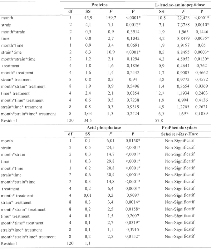
to 130 a.u. for the unchallenged group. Whatever the sampling time or the month, no significant differences had been observed between the non-injected and the unchallenged groups and this, for ail tested humoral parameters (Table 3). As for cellular parameters, the Tuckey's HSD showed that the significant differences observed (P < 0,05) are between different groups of mussels and cannot be considered as effects of infections (data not shown). Results for the humoral parameters after 24 h are presented in Tables 8 and 9 after 48 h in Tables 10 and Il. After 24 h, a strong variability had been observed in September and in December for the acid phosphatase activity (AP) ranging from 8 to 128 nmol.mg-1 proteins for the non-in jected group and from lOto 90 nmol.mg-1 proteins for the unchallenged group. After 48 h, AP of the non-injected group range from 46 to 67 nmol.mg-1 proteins in September and from 8 to 78 nmol.mg-1 proteins in Oecember. For the unchallenged group AP ranges from 39 to 88 nmol.mg-1 proteins in September and from 21 to 69 nmol.mg-1 protein in December. The prophenoloxydase-like activity after 24 h ranged from 5 to 16 U.mg-I proteins for the unchallenged group and from 4 to 17 U.mg-I proteins for the challenged group. After 48 h, ProPO-like ranged from 3 to 17 U.mg-1 for the non-injected group and from 4 to 18 u.mg-I for the unchallenged group. The L-Ieucine-aminopeptidase activity, ranged from 25 to 131 mU.mg-1 proteins for the unchallenged group and from 14 to 139 mUmg-I proteins for the non-injected group after 24 h. After 48 h, LAP ranged from 14 to 59 mUmg-I proteins for the non-injected group and from 13 to 51 mUmg-1 proteins for the unchallenged group.
1.3.2 Challenged vs unchallenged groups Viability and abundance of haemocytes
No significant differences were observed between the challenged and the unchallenged groups (Table 1). The results showed that the treatment had no effects on the viability of the haemocytes. The percentage of viable haemocytes was never less th an 85 % 24 h after the challenge for both trials as reported in Tables 4 and 5 and 48 h after the
challenge as reported in Table 6 and 7, respectively for September and December. However, after 24 h the abundance of haemocytes presented a high variability between
replicates in each group, ranging from 2.13 x 10⁵ to 8.19 X 10⁵ cells.mL-'. None dose
response was demonstrated, neither in September nor in December, 24 h and 48 h after the
challenge. 48 h after the challenge, the values range from l.95 x 105 to 3.90 x 10⁵ cells. mL-' in September and from 2.25 x 10⁵ to 5.89 ~ 105 cells.mr' in December between the
Table 1: Four ways ANOY As of the effect of time (24 h and 48 h), treatment (non-injected,
unchallenged, 102, 103 and 104 cfu injected), month (September and December) and the strain (V.
splendidus, E. coli and E. faecalis) and their interaction on cellular immune parameters
Phagocytic capa city NO, production
df SS F P df SS F P month 1 2151,9 15,6 0,0001 * 1 5028,6 10,2 0,0018* strain 2 5377,8 19,5 <,0001* 2 5328,9 5,4 0,0057* time 2773,4 20,2 <,0001* 1032,7 2,1 0,1505 treatment 4 2433,8 4,4 0,0023* 4 991,3 0,5 0,734 month*strain 2 4259,4 15,5 <,0001 * 2 1568,4 1,6 0,2082 month*time 1883,1 13,7 0,0003* 1 0,3 0,0006 0,9806 strain*time 2 3153,6 11,5 <,0001* 2 6358,2 6,4 0,0022*
month*strain*time 2 2457,1 8,9 0,0002* 2 5198,3 5,3 0,0064*
month* treatment 4 179,2 0,3 0,8602 4 2325,7 1,2 0,3237
strain* treatment 8 1661,1 1,5 0,1606 8 3053,8 0,8 0,6265
month*strain* treatment 8 482,1 0,4 0,8959 8 2375,6 0,6 0,7747
time* treatment 4 104,4 0,2 0,9433 4 3373,2 1,7 0,1523 month*time* treatment 4 110,6 0,2 0,9373 4 3055,9 1,5 0,1925 strain*time* treatment 8 836,8 0,7 0,6379 8 5096,8 1,3 0,2543 month*strain*time* 8 592,8 0,5 0,8252 8 5301,9 1,3 0,2287 lrealmenl Residual 120 16499,8 120 59195,1 Viability Abundance df SS F P df SS F P Month 0,5 1,1 0,2888 0,5 1,1 0,2888 Strain 2 6,6 7,8 0,0006* 2 6,6 7,8 0,0006* Time 1 0,001 0,002 0,9661 1 0,001 0,002 0,9661 treatment 4 4,3 2,5 0,0428* 4 4,3 2,5 0,0428* month*strain 2 4,4 5,2 0,0064* 2 4,4 5,3 0,0064* month*time 0,4 1,01 0,317 1 0,4 1,01 0,317 strain*time 2 2,5 3 0,0547 2 2,5 3 0,0547 month*strain*time 2 0,4 0,5 0,5853 2 0,4 0,5 0,5853 month* treatment 4 2,7 1,6 0,1708 4 2,7 1,6 0,1708 strain* treatment 8 2,1 0,6 0,7562 8 2,1 0,6 0,7562
month*strain* treatment 8 6,6 1,9 0,0561 8 6,6 2 0,0561
time* treatment 4 2,9 1,7 0,153 4 2,9 1,7 0,153
month*time* treatment 4 0,2 0,1 0,9828 4 0,2 0,1 0,9828
strain*time* treatment 8 2,5 0,7 0,6547 8 2,5 0,7 0,6547
month*stra in*tÎllle* 8 3,5 1,04 0,4061 8 3,5 1,04 0,4061
treatment
Table 2: Three ways ANOV As of the effect of time (24 h and 48 h), treatement (non-injected,
unchallenged, 102, 103 and 104 cfu injected) and the strain (V splendidus, E. coli and E. faecalis)
and their interaction on the ROS production in September and Oecember
ROS production in September ROS production in December
df SS F P df SS F P Strain 2 519834,7 5,6 0,0059* 1 165790 4,6 0,0373* Time 249375,2 5,4 0,0240* 184956 5,2 0,0284* strain * time 2 174364,3 1,9 0,1622 1 32362,6 0,9 0,3470 treatment 4 416435 2,2 0,0754 4 468594,8 3,3 0,0205* strain * treatment 8 1193489,7 3,2 0,0042* 4 68127,4 0,5 0,7527 time * treatment 4 14916,8 0,1 0,9881 4 279269,2 1,9 0,1204
strain * time
*
treatment 8 87898,1 0,2 0,9824 4 123581,8 0,9 0,4936Table 3: Scheirer-Ray-Hare test and Four ways ANOVAs of the effect time (24 h and 48 h), treatment
(non-injected, unchallenged, 102, 103 and 104 cfu injected), month (September and December) and the strain (V splendidus, E. coli and E. faecalis) and their interaction on humoral immune parameters
Proteins L-Ieucine-aminopeptidase df SS F P SS F P month 1 45,9 159,7 <,0001 * 10,8 22,423 <,0001* strain 2 4,1 7,1 0,0012* 7,1 7,3758 0,0010* mo nth * strain 2 0,5 0,9 0,3914 1,9 1,965 0,1446 time 0,8 2,7 0,1042 4,2 8,8479 0,0035* month*time 0,9 3,4 0,0691 1,9 3,9197 0,05 strain*time 2 6,3 10,9 <,0001 * 8,5 8,8495 0,0003* month*strain*time 2 1,2 2,1 0,1294 4,3 4,5052 0,0130* treatment 4 1,8 1,6 0,1856 0,9 0,4641 0,762 month* treatment 4 1,6 1,4 0,2442 1,7 0,9003 0,4662 strain* treatment 8 0,8 0,3 0,94 3,8 0,9772 0,4572 month*strain* treatment 8 1,9 0,9 0,5496 1,4 0,3654 0,9369 time* treatment 4 2,4 2,1 0,0854 ' 2,7 1,3934 0,2403 month*time* treatment 4 0,6 0,5 0,7238 1,9 0,994 0,4136 strain*time* treatment 8 0,8 0,3 0,9519 4,9 1,2765 0,2621 month*strain*time* treatment 8 3,03 1,3 0,2424 6,5 1,697 0,1059 Residual 120 34,5 57,8
Acid phosphatase ProPhenoloxydase
df SS F P Scheirer-Ray-Hare
month 0,1 6,01 0,0158* Non-Significatif
strain 2 0,5 24,5 <,0001 * Non-Significatif
month*strain 2 0,3 14,7 <,0001 * Non-Significatif
time 0,3 29,8 <,0001 * Non-Significatif
month*time 0,2 20,8 <,0001 * Non-Significatif
strain*time 2 0,6 30,4 <,0001 * Non-S ignificatif
month*strain*time 2 0,3 14,8 <,0001 * Non-Significatif
treatment 4 0,2 6,4 0,0001 * Non-Significatif
month* treatment 4 0,01 0,2 0,9097 Non-Significatif
strain* treatment 8 0,3 3,4 0,0014* Non-Significatif
month*strain* treatment 8 0,2 2,5 0,0158* Non-Significatif
time* treatment 4 0,1 1,5 0,2007 Non-Significatif
month*time* treatment 4 0,1 2,7 0,0319* Non-Significatif
strain*time* treatment 8 0,1 1,1 0,3915 Non-Significatif
month*strain*time* treatment 8 0,2 2,5 0,0152* Non-Significatif
Values are mean ± SD (N=3).
Vibrio splendidus Viability (%)
Abundance (105 haemocytes per mL)
Phagocytic capacity (%)
ROS production (relative fluorescence)
NOx production (relative fluorescence) Enterococcus faecalis
Viability (%)
Abundance (105 haemocytes per mL)
Phagocytic capacity (%)
ROS production (relative fluorescence) NOx production (relative fluorescence) Escherichia coli
Viability (%)
Abundance (105 haemocytes per mL)
Phagocytic capacity (%)
ROS production (relative fluorescence) NOx production (relative fluorescence)
Non-injected Unchallenged 102 103 104
93 ± 1 93 ± 1 92± 1 93 ± 3 93 ± 1 3,16±1,01 2,67±1,21 2,88±1,032,81±1,02 2,77±0,7 30± 9 28 ± 5 27 ±21 28 ±9 39 ±7 912 ± 135 847 ± 205 863 ± 246 938 ± 165 1166 ± 302 146 ± 7 163 ± 15 163± 10 135±34 136± 16 96 ±1 93 ± 2 94 ± 1 96± 1 93 ± 1 4,73±2,14 2,13±0,5 3,32±2,45 3,34±0,9 3,13±0,06 Il ± 14 20 ± 12 24± II 17 ± 3 20 ± 13 770± 179 1334±319 1266±186 1021±168 1129±150 135 ± 39 159 ± 29 150 ± 35 113 ± 3 130 ± 27 95±0,8 97±0,7 95±0,9 96±0,5 95±1,7 2,74 ± 0,4 2,91 ± 0,9 2,42 ± 0,6 3,40 ± 0,2 3,08 ± 0,9 35±7 40±5 27± 16 32±12 36±6 1316± 232 1151 ± 136 1069± 130 989± 90 1176± 214 133±14 130± 12 136±5 120±33 118±17