CHARACTERIZATION ANI) REGULATION 0F ENZYMES RESPONSIBLE FOR STEROD ACTIVATION, INACTIVATION ANT) BIOAVAILABILITY DDR1NG
THE OVULATORY PROCESS IN THE MARE
par
KRISTY ANGELA BRowN
Département de biomédecine vétérinaire
Faculté de médecine vétérinaire
Thèse présentée à la Faculté des études supérieures en vue de l’obtention du grade
Phi1osophi Doctor (Ph.D.) en sciences vétérinaires option reproduction 2Dt6 j Avril, 2006 ©Kristy A. Brown, 2006
n
Direction des bibliothèques
AVIS
Lauteur a autorisé l’Université de Montréal à reproduire et diffuser, en totalité
ou en partie, par quelque moyen que ce soit et sur quelque support que ce soit, et exclusivement à des fins non lucratives d’enseignement et de
recherche, des copies de ce mémoire ou de cette thèse.
L’auteur et les coauteurs le cas échéant conservent la propriété du droit
d’auteur et des droits moraux qui protègent ce document. Ni la thèse ou le mémoire, ni des extraits substantiels de ce document, ne doivent être
imprimés ou autrement reproduits sans l’autorisation de l’auteur.
Afin de se conformer à la Loi canadienne sur la protection des renseignements personnels, quelques formulaires secondaires, coordonnées
ou signatures intégrées au texte ont pu être enlevés de ce document. Bien
que cela ait pu affecter la pagination, il n’y a aucun contenu manquant.
NOTICE
The author of this thesis or dissertation has granted a nonexclusive license allowing Université de Montréal to reproduce and publish the document, in
part or in whole, and in any format, solely for noncommercial educational and research purposes.
Ihe author and co-authors if applicable retain copyright ownership and moral rights in this document. Neither the whole thesis or dissertation, nor
substantial extracts from it, may be printed or otherwise reproduced without the author’s permission.
In compliance with the Canadian Privacy Act some supporting forms, contact
information or signatures may have been removed from the document. While this may affect the document page count, it does not represent any loss of
Université de Montréal faculté des études supérieures
Cette thèse intitulée
CHARACTERIZATIONANI)REGULATION 0F ENZYMES RESPONS1BLE FOR STEROD ACTIVATION, INACTWATION AND BIOAVAILABILITY DURIG
THE OVULATORY PROCESS 1N THEMARE
présentée par
KRIsTY ANGELA BRowN
a été évaluée par un jury composé des personnes suivantes
BruceD. Murphy, président-rapporteur
Jean Sirois, directeur de recherche Jacques G. Lussier, codirecteur
Alan K. Goff, membre du jury Van Luu-The, examinateur externe
RÉSUMÉ
La montée préovulatoire d’hormone lutéinisante (LH) est responsable du déclenchement des changements morphologiques et biochimiques qui vont ultimement être responsables de la relâche de l’ovocyte et de la formation du corps jaune (CL). Lors de cette période, la machinerie stéroïdogène interrompt sa production d’oestrogènes en faveur de la biosynthèse de progestérone, ce qui résulte en une baisse d’œstradiol tard en estrus. Cette baisse en oestrogènes actifs a longtemps été attribuée au manque en potentiel biosynthétique, en particulier à la perte de la cytochrome P450aromatase (CYP19A1), enzyme limitante responsable de l’aromatisation des androgènes. Le traitement de juments avec la gonadotrophine chorionique humaine (hCG) provoque la lutéinisation du follicule préovulatoire et l’ovulation, qui aura alors lieu entre 39 et 4$ h après injection. Les échantillons utilisés pour la présente étude ont été isolés à différents temps après le traitement d’hCG, notamment à 0 h, représentant un follicule isolé avant hCG, ainsi qu’à 12, 24, 30, 33, 36 et 39 h post-hCG. Comme référence, un CL au jour 2 du cycle oestral ainsi que différents tissus ont été obtenus en guise de comparaison. L’hypothèse générale de cette thèse est que le changement des concentrations de stéroïdes observé lors du processus ovulatoire est le résultat de la modulation de l’expression de gènes spécifiques, responsables de l’activation, l’inactivation et la bio-disponibilité des stéroïdes.
Le deux premiers papiers examinent la régulation transcriptionnelle de la 17F3-hydroxystéroïde déshydrogénase de type 1 (173HSD1), une protéine responsable de la réduction de l’oestrone en sa version plus active, l’oestradiol, du membre 2B1 de la famille de peptides transporteurs d’anion organiques (OATP2B1), encodé par le gène SLCO2B1 et qui est responsable de l’influx de stéroïdes sulfonés, ainsi que la sulfatase de stéroïdes (ST$) qui catalyse l’enlèvement du groupe sulfonate pour en faire des stéroïdes libres. La structure primaire du gène, de l’ADN complémentaire (ADNc) et de la protéine de la 17f3HSD1 ont été caractérisées. La structure du gène de la 173HSD1 était hautement homologue à celle des autres espèces. La structure primaire de l’ADNc et de la protéine de la SLCO2B1 ont aussi été caractérisées. Des analyses RT
PCRlSouthern ont démontré des changements significatifs dans les concentrations de transcrits 17HSDi, SLCO2B1 et STS dans les follicules préovulatoires lors de l’induction gonadotropine-dépendante de la lutéinisation folliculaire. Des concentrations élevées d’ARN messager de la Y7f3HSD1, SLCO2B1 et STS étaient présents dans les follicules préovulatoires avant hCG. Une diminution rapide et dramatique des transcrits 173HSDi et SLCO2B1 a été détectée dans des préparations de cellules granulosa isolées de follicules obtenus 12 h post-hCG et les concentrations sont demeurées basses, presque indétectables, par la suite. Le transcrit du SLCO2B1 était aussi exprimé et réprimé dans la thèque interne, mais sa diminution était plus graduelle que dans les cellules de la granulosa. La régulation de la STS était quelque peu différente, car son transcrit était hautement exprimé dans les deux types cellulaires et qu’il était significativement réprimé dans les cellules granulosa entre 30 et 39 h post-hCG. Le promoteur de la 17HSD1 a été séquencé et plusieurs éléments cis potentiels ont été identifiés, incluant les séquences liant les facteurs de transcription Spi, NFKB et GATA. La diminution de liaison des extraits de noyaux de cellules granulosa observée après le traitement de gonadotrophine pourrait expliquer la diminution d’expression du transcrit 17HSDl observée dans les follicules préovulatoires lors du processus ovulatoire. La co-localisation du SLCO2B1 et de la STS dans les follicules préovulatoires avant hCG suggère une voie alternative de biosynthèse d’oestrogènes qui impliquerait l’importation et l’utilisation de stéroïdes sulfonés, et que la répression de cette voie ainsi que la voie traditionnelle utilisant le cholestérol comme point de départ, constituerait une base moléculaire pour la diminution de biosynthèse d’oestrogènes observée lors de cette période.
Les troisième et quatrième papiers examinent la régulation de gènes impliqués dans l’inactivation et l’exportation d’oestrogènes lors du processus ovulatoire et catalysés par la 17r3HSD4, une enzyme responsable de l’oxydation de l’oestradiol en oestrone, ainsi que la sulfotransférase d’oestrogènes (EST), qui catalyse la sulfoconjugaison des oestrogènes, et la protéine «multidrug resistance-l » (MRP1), encodée par le gène ABCC1 et qui est impliquée dans l’exportation des stéroïdes sulfonés. Les structures
primaires des ADNc et des protéines de la 17HSD4, EST et ABCC1 ont été caractérisées. Les analyses par RT-PCRiSouthern, d’immunobuvardage et immunohistochimiques, ont démontré des changements significatifs dans les concentrations de 17pHSD4, EST et ABCC1 lors du processus ovulatoire. De basses concentrations présentes avant traitement ont été augmentées après hCG et sont demeurées élevées par la suite dans les cellules granulosa. Ces résultats démontrent que la diminution d’œstradiol observée avant l’ovulation n’est pas seulement due à la diminution en potentiel biosynthétique, mais aussi à l’augmentation de l’expression de protéines responsables de l’inactivation et de l’exportation d’oestrogènes. De plus, ces résultats démontrent qu’il est peut-être nécessaire d’éliminer les oestrogènes actifs lors de ce processus.
Le dernier papier visait à caractériser une autre l73-hydroxystéroide déshydrogénase, 1 7f3HSD5, faisant partie de la famille des aldo-kéto réductases et qui s’est avérée posséder une activité 20Œ-HSD, soit la AKR1C23. Les structures primaires de l’ADNc et de la protéine de la AKR1C23 ont été déterminées et des analyses RT PCR1Southern, d’immunobuvardage et d’irnrnunohistochimie ont été utilisées pour examiner la régulation de la AKR1C23 lors du processus ovulatoire. Les concentrations de AKR1C23 étaient basses dans les follicules préovulatoires avant hCG, puis une augmentation significative a été détectée dans les follicules obtenus entre 12 et 39 h post-hCG. Considérant son activité 2OaHSD, qui corrèle avec les concentrations de 20Œ-dihydroprogestérone dans la liqueur folliculaire, ces résultats suggèrent un rôle pour une enzyme qui métabolise la progestérone dans les cellules granulosa du follicule préovulatoire lors du processus ovulatoire.
Globalement, ces résultats démontrent que lors du processus de lutéinisation!ovulation folliculaire chez la jument, la modulation des gènes impliqués dans l’activation, l’inactivation et la bio-disponibilité des stéroïdes sexuels favorise un environnement cellulaire sans oestrogènes actifs, ainsi que la présence d’une enzyme
ayant la capacité de métaboliser la progestérone et ce, surtout dans les cellules de la granulosa.
Mots-clés: Cheval, ovaire, follicule préovulatoire, hCG, stéroïdes, 1713-hydroxystéroïde déshydrogénase, sulfoconjugaison, transport de stéroïdes sulfonés, sulfatase de stéroïdes, aldo-kéto réductase.
SUMMARY
The preovulatory rise in luteinizing hormone (LH) is responsible for triggering the morphological and biochemical changes that ultimately lead to the release of the oocyte and the formation of the corpus luteum (CL). During this period, the steroidogenic rnachinery interrupts production of estrogens in favor of biosynthesis of progesterone, resulting in a late-estrus drop in plasma 1 7-estradiol. This decrease in active estrogens has long been believed to be due to a lack of estrogen biosynthetic potential, in particular due to the loss of cytochrome P45Oarornatase (CYP19A1), the rate-limiting enzyme that aromatizes androgens. Treatment of mares with human chorionic gonadotropin (hCG) causes preovulatory follicles to undergo luteinization, with ovulation usually occurring 39 to 48 h later. Samples used in this study were obtained at different times relative to hCG treatrnent, namely at O h, which represents a follicle isolated prior to hCG treatment, and at 12, 24, 30, 33, 36, and 39 h afler hCG. For reference, a CL sample at day 8 of the cycle along with various other equine tissues were used for comparison. The overali hypothesis of this thesis is that the change in steroid concentrations observed during the ovulatory process is a resuit of the programmed modulation of the expression of specific genes responsible for the activation, inactivation and bioavailability ofsteroids.
The first and second papers examine the transcriptional regulation of 1713-hydroxysteroid dehydrogenase type 1 (Ï7I3HSD1), a protein responsible for the reduction of estrone to the more biologically potent 17f3-estradiol; member 231 of the organic anion transporting polypeptide family (OATP2B1), encoded by the SLCO2B1 gene and responsible for the importation of sulfoconjugated steroids; as well as steroid sulfatase (STS), an enzyme responsible for the hydrolysis of sulfoconjugated steroids. The primary structures of the equine 1713HSD1 gene, cDNA and protein were characterized. The 1713HSD1 gene structure was highly homologous to that of other species. The primary structures of the equine SLCO2B 1 cDNA and protein were also characterized. RT-PCR/Southern blot analyses demonstrated significant changes in
steady-state levels of 17f3HSD1, SLCO2B1 and STS transcripts in preovulatory follicles during the gonadotropin-dependent induction of follicular luteinizationlovulation. Elevated concentrations of 17I3HSD1, SLCO2B1 and STS mRNA were present in equine preovulatory follicles prior to hCG treatment. A rapid and dramatic decrease in 1 7HSD I and SLCO2B 1 transcripts was detected in isolated preparations of granulosa ceils obtained 12 h post-hCG and rernained low, almost undetectable, thereafter. Levels of SLCO2B 1 mRNA were also expressed and downregulated in the theca interna, in a pattem more graduai than what was obsewed in the granulosa ceil layer. The regulation of STS was slightly different in that levels were high in both celi types, but STS mRNA was downregulated significantly 30-39 h post-hCG in granulosa celis. The equine 17J3HSDY promoter was sequenced and several putative cis-acting elernents were identified, including sequences that bind the transcription factors Spi, NFicB and GATA. The decrease in granulosa celi nuclear extract binding observed after gonadotropin treatment may account for the downregulation in 17I3HSD1 transcript observed in preovulatory follicles during the ovulatory process. The co-localization of SLCO2B 1 and STS in preovulatory follicies prior to hCG treatment suggest an alternate pathway of estrogen biosynthesis and that the gonadotropin-dependent downregulation of both pathways during the ovulatory process provides an additional molecular basis for the decreased 17r3-estradioi biosynthesis observed during that period.
The third and fourth papers examine regulation of genes involved in estrogen inactivation and export during the ovulatory process as catalyzed by 1 7f3HSD4, which is responsible for the oxidation of I73-estradio1 to estrone; as well as estrogen sulfotransferase (EST), which catalyzes the suifoconjugation of estrogens; and multidrug resistance protein-1 (MRP1), encoded by the ABCCI gene and involved in the efflux ofsulfoconjugated steroids. The primary structures ofthe 17HSD4, EST and
ABCC1 cDNA and proteins were characterized. RT-PCPiSouthern blot analyses in
combination with immunoblot and immunohistochemistry demonstrated a significant change in steady-state levels of 173HSD4, EST and ABCC1 during the ovuiatory process. Low concentrations present prior to hCG treatment were upregulated after hCG
treatment and remained high thereafter, with the upregulation occurring mainly in the granulosa celi layer. These resuits indicate that the decrease in Ï7-estradiol observed prior to ovulation is not only due to the decrease in biosynthetic potential, but also to upregulation of proteins involved in estrogen inactivation and export. Moreover, active estrogens may need to 5e eliminated during this process.
The last paper was aimed at characterizing the regulation of another 1 7H$D, 17I3HSD5, an aldo-keto reductase which was later dernonstrated to possess progesterone-metabolizing activity, AKR1 C23. The primary structures of the AKR1 C23 cDNA and protein were determined and RT-PCR/Southem blot analyses as well as immunoblot and immunohistochemistry were used to examine the regulation of AKR1C23 during the ovulatory process. While low levels ofAKR1C23 were present in preovulatory follicles prior to hCG treatment, a significant increase in AKR1C23 was observed in granulosa cell samples isolated from preovulatory follicles obtained 12-39 h post-hCG. Considering its 2OŒHSD activity, which correlates with follicular fluid concentrations of 20Œ-DHP, these resuits suggest the need for a progesterone metabolizing enzyme in granulosa ceils of preovulatory follicles during the ovulatory process.
Overali, these resuits demonstrate that, during the process of follicular luteinizationlovulation in the mare, the modulation of genes involved in steroid activation, inactivation and bioavailability favors a cellular enviroment without active estrogens and the presence of a progesterone-metabolizing protein occurring mainly in the granulosa celi layer.
Keywords: Horse, ovary, preovulatory follicle, hCG, steroids, 1 7-hydroxysteroid dehydrogenase, sulfoconj ugation, sulfoconj ugated steroid transport, steroid sulfatase, aldo-keto reductase.
TABLE 0F CONTENTS TITLE PAGE j IDENTIFICATION 0FJURY ii RÉSUMÉ iii SUMMARY TABLE 0F CONTENTS x
LIST 0F FIGURES xiv
LIST 0F ABBREVIATIONS xvii
AKNOWLEDGEMENTS xix
INTRODUCTION 1
1. Overview ofthe equine estrous cycle 1
1 .1 Morphological and hormonal changes during the equine estrous cycle 2
1.2 Cellular aspects of luteinization 6
1.3 Luteinization-associated modulation of gene expression 7
2. Steroid hormones 9
2.1 Role of steroid honriones inphysiologicalprocesses 10
2.1.1 Estrogens and celi cycle progression 12
2.2 Molecular biology of ovarian steroid hormone biosynthesis 14 2.2.1 Steroidogenesis from cholesterol to estrone 16 2.2.1.1 Steroidogenic acute regulator (StAR) 16 2.2.1.2 Cytochrome P450 side-chain cleavage (CYP1 lAi) 17 2.2.1.3 CytochromeP450 1 7a-hydroxylase/C 17-20 lyase
(CYP17A1) 18
2.2.1.4 3 -hydroxysteroid dehydrogenase/ ketosteroid isomerase
(313H5D/KSI) 18
2.2.1.5 Cytochrome P450 aromatase (CYP19A1) 19
2.2.2 1 73-hydroxysteroid dehydrogenases 20
2.2.2.1 17-hydroxysteroid dehydrogenase type 1 (173HSD1) 22 2.2.2.1.1 Cloning and characterization of 17HSD1 23
2.2.2.1.2 Biochemistry and enzymology of17I3HSD1 24 2.2.2.1.3 Transcriptional regulation of 17f3HSD1 25 2.2.2.2 17Ç3-hydroxysteroid dehydrogenase type 2 (173HSD2) 25 2.2.2.3 17-hydroxysteroid dehydrogenase type 3 (1713HSD3) 26 2.2.2.4 173-hydroxysteroid dehydrogenase type 4 (173HSD4) 26 2.2.2.4.1 Cloning and characterization of 17HSD4 27 2.2.2.4.2 Biochemistry and enzymology of 17f3HSD4 2$ 2.2.2.4.3 Transcnptional regulation of 17f3HSD4 29 2.2.2.5 173-hydroxysteroid dehydrogenase type 5 (17HSD5) 29 2.2.2.6 17-hydroxysteroid dehydrogenase type 6 (17HSD6) 30 2.2.2.7 17-hydroxysteroid dehydrogenase type 7 (17f3HSD7) 30 2.2.2.8 173-hydroxysteroid dehydrogenase type 8 (173HSD8) 31 2.2.2.9 17f3-hydroxysteroid dehydrogenase type 9 (17HSD9) 32 2.2.2.10 173-hydroxysteroid dehydrogenase type 10 (173HSD10) 32 2.2.2.11 17-hydroxysteroid dehydrogenase type 11 (17HSD1 1) 32 2.2.2.12 173-hydroxysteroid dehydrogenase type 12 (17HSD12) 33 2.2.3 20Œ-hydroxysteroid dehydrogenase (2OŒHSD) 34 2.2.3.1 Cloning and characterization of2OaHSD 34 2.2.3.2 Biochemistry and enzymology of2OŒHSD 35 2.2.3.3 Transcriptional regulation of 2OŒHSD 35
3. Sulfoconjugation 36
3.1 Role of sulfoconjugation inphysiologicalprocesses 36
3.2 Properties of sulfoconjugated steroids 37
3.3 Sulfotransferases 38
3.3.1 Hydroxysteroid sulfotransferase 39
3.3.2 Estrogen sulfotransferase (EST) 40
3.3.2.1 Cloning and characterization of EST 41 3.3.2.2 Biochemistry and enzymology of EST 41
3.3.2.3 Transcriptional regulation of EST 42
3.4.1 Steroidsulfatase(STS) 43 3.4.1.1 Cloning and characterization ofSTS 43
3.4.1.2 Biochemistry and enzymology ofSTS 44
3.4.1.3 Transcriptional regulation cf STS 44
3.5 Transporters ofsuffoconjugated steroids 45
3.5.1 Influxtransporters and organic anion transporting polypeptides 45 3.5.1.1 Cloning and characterization ofSLCO2BY 47 3.5.1.2 Bicchemistry and enzymology cf OATP2B1 47 3.5.1.3 Transcriptional regulation cf SLCO2B1 48 3.5.2 Efflux transporters and the ATP-binding cassette family 48 3.5.2.1 Ooning and charactenzation cf ABCC1 50 3.5.2.2 Biochemistry and enzymology cfMRP1 51 3.5.2.3 Transcriptional regulation ofABCCI 51
HYPOTHESIS AI\D OBJECTIVES 52
SUMMARY 0F ARTICLE ONE 53
ARTICLE ONE: Molecular cloning cf equine 1 7f3-hydroxysteroid
dehydrogenasetype I and its downregulation during follicular
luteinization in vivo. 55
SUMMARY 0F ARTICLE TWO 87
ARTICLE TWO: Down-regulation cf messenger ribonucleic acid encoding an importer cf sulfoconjugated steroids during human chorionic
gcnadotropin-induced follicular luteinization in vivo. 89
SUMMARY 0F ARTICLE THREE 119
ARTICLE THREE: Human chorionic gonadotropin-dependent regulation cf 1 713-hydroxysteroid dehydrogenase type 4 in preovulatcry follicles and its potential role in follicular luteinization. 121
SUMMARY 0F ARTICLE FOUR 153
ARTICLE FOUR: Human chorionic gonadctropin-dependent upregulation cf genes responsible for estrogen sulfoccnjugation and export in
granulosa celis cf luteinizing preovulatory follicles. 155
ARTICLE FIVE: Human chorionic gonadotropin-dependent induction of an equine aldo-keto reductase (AKR1C23) with
20Œ-hydroxysteroid dehydrogenase activity during follicular
luteinization in vivo. 193
GENERAL DISCUSSION 229
The gonadotropin-dependent downregulation of 1 7I3HSD 1 mRNA provides an additional molecular basis for the decrease in 1 713 -estradiol biosynthesis
observed during the process ofluteinization. 229
The high level of SLCO2B 1 and STS mRNA expression and their gonadotropin-dependent downregulation in preovulatory fotticles is
suggestive of a putative alternative pathway for 1713-estradiol biosynthesis. 232 The induction of 1713HSD4 involves oxidative 1713HSDs in the luteinization
process and raises the possibility that estrogens may need to be inactivated
during this process. 235
The gonadotropin-dependent induction of EST and ABCC I provides new insights on the role of phase II conjugation enzymes and their contribution
towards the inactivation of estrogens during follicular luteinizationlovulation. 236 The induction ofAKR1C23 suggests a potential role for local progesterone
metabolism during gonadotropin-dependent follicular luteinization. 239
CONCLUSION 241
LIST 0F FIGURES
1NTRODUCTION
Figure 1. Estrous cycle in the mare depicting a major follicular wave. 1 Figure 2. Hormonal changes during the equine estrous cycle. 5 Figure 3. Structure ofthe steroid hormone backbone and parent molecules. 10
Figure 4. Simplified celi cycle. 13
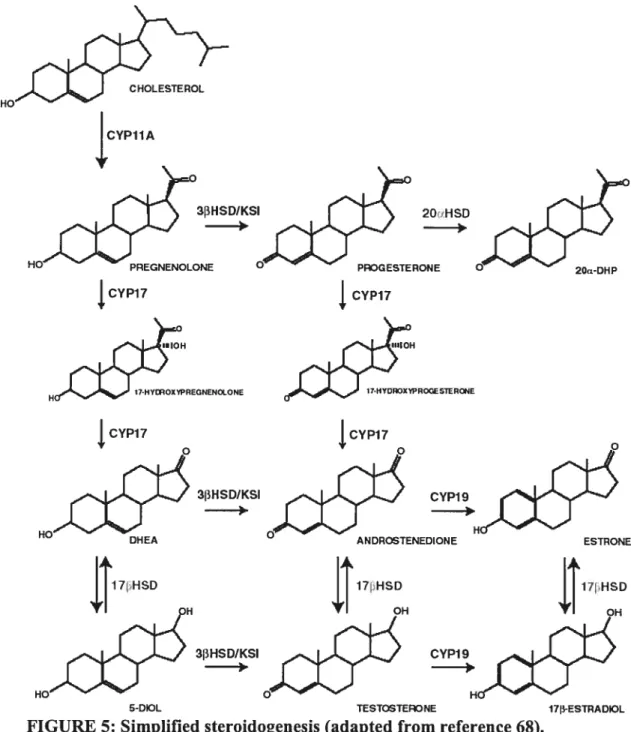
Figure 5. Simplified steroidogenesis. 15
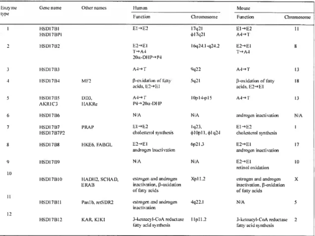
Figure 6. The different 17J3HSDs. 21
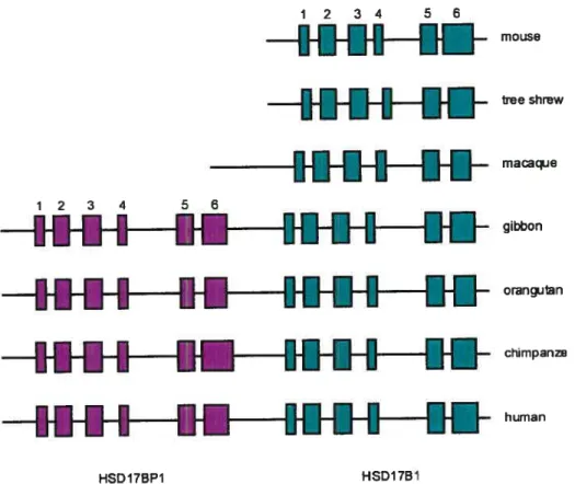
Figure 7. Comparison ofthe genomic structure ofHSD17B1 genes. 24
Figure 8. Human 1713HSD4 gene structure. 27
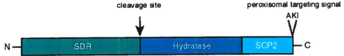
Figure 9. Structure ofthe 17f3HSD4 protein. 28
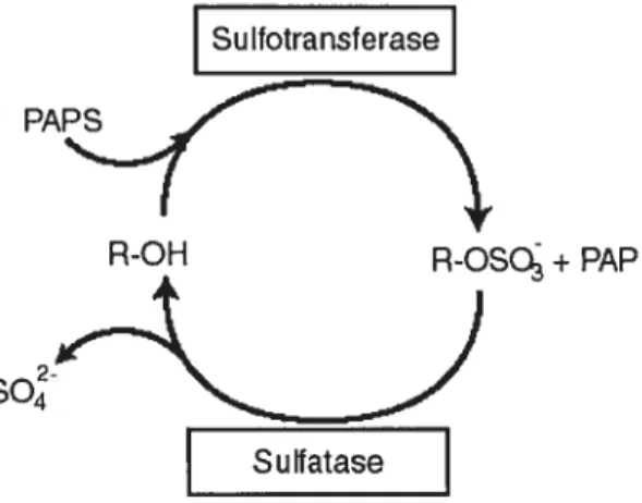
Figure 10. Sulfoconjugation. 36
Figure 11. Steroid sulfoconjugates. 37
Figure 12. Crystal structure ofhuman STS. 44
Figure 13. Typical transmembrane domain structure of OATPs. 46 Figure 14. Predicted transmembrane structure ofthree classes of ABC
transporters. 49
ARTICLE ONE
Figure 1. Cloning strategy for equine 17I3HSD1. 79
Figure 2. Equine 17[3HSD1 gene structure. 80
Figure 3. Equine Y7F3HSD1 predicted amino acid sequence. 81 Figure 4. Expression of 17HSD transcript in equine tissues. 82 Figure 5. Downregulation of17HSD1 mRNA byhCG in equine
preovulatory follicles. 83
Figure 6. Ceil type-dependent expression and regulation of 17f3HSD1
mRNA in equine preovulatory follicles. 84
Figure 7. Isolation and characterization ofthe equine 17f3HSD1 promoter. 85 Figure 8. Gonadotropin-dependent regulation ofDNA binding activities in
ARTICLE TWO
Figure 1. Cloning of equine SLCO2B 1. 113
Figure 2. Deduced nucleotide sequences for equine SLCO2B1. 114 Figure 3. Deduced primary structure of the equine OATP2B 1 protein and
comparison with known OATP2B1 homologues. 115 Figure 4. Expression ofSLCO2B1 and STS mRNA in equine tissues. 116 Figure 5. Regulation of SLCO2B 1 and SIS transcript by hCG in equine
preovulatory follicles. 117
Figure 6. Regulation ofSLCO2B1 (A) and STS (B) mRNA in equine
granulosa and theca interna celis. 112
ARTICLE THREE
Figure 1. Nucleotide sequence ofequine 1713HSD4. 146 Figure 2. Predicted amino acid sequence ofequine 1713HSD4 and
comparisons with other mammalian homologues. 147 Figure 3. Expression of 17HSD4 mRNA in equine tissues. 14$ Figure 4. Regulation of 17f3HSD4 transcript by hCG in equine
preovulatory follicles. 149
Figure 5. Regulation of 1713HSD4 mRNA in equine granulosa and theca
interna ceils. 150
Figure 6. Regulation of 17HSD4 protein by hCG in equine preovulatory
follicles. 151
Figure 7. Immunohistochemical localization of 17HSD4 in equine
preovulatory follicles. 152
ARTICLE FOUR
Figure 1. Cloning of equine EST and ABCC1. 183
Figure 2. Deduced primary structure ofthe equine EST and MRP1
proteins. 184
Figure 3. Expression of EST and ABCC1 mRNA in equine tissues. 185 Figure 4. Regulation of EST and ABCC1 transcript by hCG in equine
figure 5. Regulation of EST and ABCC1 mRNA in equine granulosa and
theca interna ceils. 187
Figure 6. Immunohistochernical localization of EST in equine
preovulatory follicles. 188
Figure 7. Immunohistochemical localization ofMRP1 in equine
preovulatory follicles. 189
Figure 8. Concentrations of sulfoconjugated and free estrogens in follicular
fluid. 190
ARTICLE FIVE
Figure 1. Cloning of equine AKR1C23. 221
Figure 2. Deduced primary structure of the equine AKRI C23 protein and
comparison withknown 20Œ-HSDs and bovine PGfS. 222
Figure 3. Expression ofAKR1C23 rnRNA in equine tissues. 223 Figure 4. Regulation ofAKR1C23 transcript by hCG in equine
preovulatory follicles. 224
Figure 5. Regulation ofAKR1C23 mRNA in equine granulosa and theca
interna cells. 225
Figure 6. AKRIC23 primary antibody specificity and regulation of
AKR1 C23 protein by hCG in equine granulosa and theca interna. 226 Figure 7. Immunohistochemical localization ofAKR1C23 in equine
preovulatory follicles. 227
Figure 8. Enzyme kinetics ofAXRIC23 and steroid levels in follicular
fluid. 228
CONCLUSION
Figure 1. Model describing the regulation of steroid activation,
inactivation and bioavailabiÏity during the ovulatrny process in
LIST 0F ARBRE VIATIONS
1 7f3HSD 1 7f3-hydroxysteroid DHT dihydrotestosterone
dehydrogenase DNA deoxyribonucleic acid
20a-DHP 20a-dihydroprogesterone El estrone 2OŒHSD 20u-hydroxysteroid E2 1713 -estradiol
dehydrogenase eCG equine chorionic
3 13HSD/KS I 313 -hydroxysteroid gt1pin
dehydrogenase/ketosteroi EMSA electromobility shift assay
d isomerase ER estrogen receptor
3 ‘-UTR 3 ‘-untranslated region EST estrogen sulfotransferase 5’-UTR 5’-untranslated region F$H follicle-stimulating hormone
A4 androstenedione GnRH gonadotropin-releasing
ABC ATP-binding cassette hormone
aldo-keto reductase GRE glucocorticoid response
APS adenosine-5’- element
phosphosulfate GSH glutathione
ATP adenosine triphosphate hCG human chorionic
BCRP breast cancer resistance gonadotropin
protein HEK293 human embryonal kidney
bp base pair 293
cAMP cyclic adenosine IL interleukin
monophosphate kb kilobase
CDK cyclin-dependent kinase kDa kilodalton
cDNA complementary DNA LDL low-density lipoprotein
CL corpus luteum LH luteinizing hormone
CHO Chinese hamster ovary MDR multidrug resistance
CRE cAMP response element mRNA messenger RNA
CREB CRE binding protein MRP MDR-associated protein
CREM CRE modulator NAD nicotinamide adenine
NADP nicotinamide adenine PR progesterone receptor dinucleotide phosphate recFSH recombinant FSH NBD nucleotide-binding domain RNA ribonucleic acid
NFiB nuclear factor-içB RT reverse transcriptase
nt nucleotide $CP2 sterol carrier protein 2
OATP organic anion transporting SDR short-chain dehydrogenase/
polypeptide reductase
P4 progesterone SF-1 steroidogenic factor-1
PAPS 3 ‘-phosphoadenosine-5‘- SLCO solute carrier/OATP
phosphosulfate SREBP sterol regulatory element PCR polymerase chain reaction binding protein
PGF2Πprostaglandin StAR steroid acute regulator
PKA protein kinase A STS steroid sulfatase
PPAR peroxisome proliferator- T testosterone
ACKNOWLEDGEMENTS
This work is a testament to my thesis supervisor, Dr Jean Sirois, who unknowingly changed my perception of research by showing me that success does not have to be at the expense of living a balanced life. Not only did he guide me through the elaboration ofthe project, but he gave me the freedom to explore new ideas and concepts, as well as the much needed moral support. For that I will be etemally grateful.
I would also like to express my gratitude to my co-director, Dr Jacques Lussier, who has provided important help in technical and procedural aspects during the course ofrny
Ph.D..
I am also thankful for the guidance of Dr Bruce Murphy who has played a pivotal role inmy career, from the time he recommended joining Jean’s laboratory to the much needed advice given in the past months. Also, to ail the members of my thesis committec, I greatly appreciate the investment ofyour valuable time and energy into this work. I am also indebted to the members of our shrinking laboratory. To Kham and Derek for their scientific and not so scientific dialogue. To Angelica, for showing me that it is possible to do it ail, and mostly to Nadine, who was an indispensable source of technical knowledge but aiso a good friend. I would also like to express my appreciation to Dre Monique Doré for her understanding, and of course, her great knowledge of what is really a signal in immunohistochemistry, as weii as to all her laboratory. Also, to Micheline, for putting a calm face on last minute fellowship applications as well as all her help the rest ofthe time.
I am grateful to my family, for their love and support, and this includes my newly “acquired” famiiy. To my mother for teaching me that I could do anything and to my father for his analytical mmd and always having an answer to even the most difficult question. Most of all, I wouid like to express my love and gratitude to my husband Keith, for sticking with me through my wiid panic attacks and for making me laugh even when I didn’t want to. Above all else, for being himself
INTRODUCTION
1. Overview of the equine estrous cycle
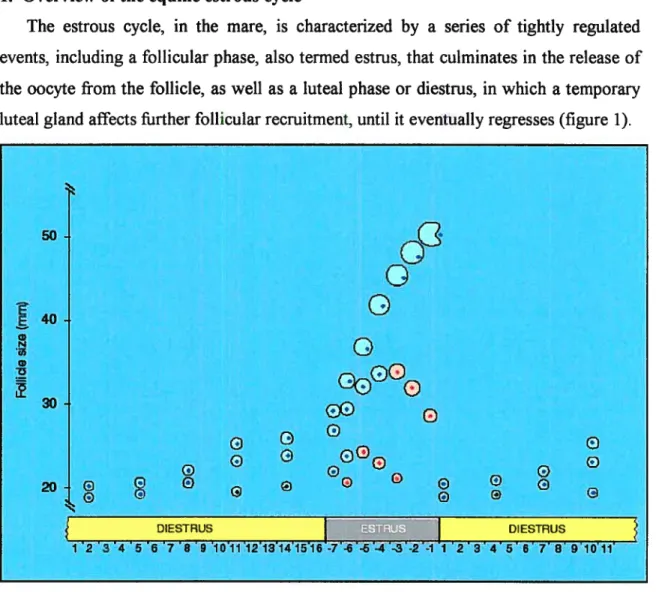
The estrous cycle, in the mare, is characterized by a series of tightly regulated events, including a follicular phase, also termed estrus, that culminates in the release of the oocyte from the follicle, as well as a luteal phase or diestrus, in whïch a temporary luteal gland affects further follicular recruitment, until it eventually regresses (figure 1).
FIGURE 1: Estrous cycle in the mare depicting a major follicular wave (adapted from reference 1).
The mare provides an interesting model for the study of gonadotropin-dependent luteinizationlovulation for many reasons. Being a seasonal polyestrous animal, the mare goes through an ovulatory season which is accompanied by several estrous cycles, typically occurring during spring and summer, followed by an anovulatory season (1). Daylight is a key factor in the initiation of the ovulatory season, as melatonin has been shown to affect gonadotropin releasing hormone (GnRH) secretion (2, 3). Also, this
50 40. E C) N u, o 30 20
G
G G g G Ge
G ge
e
gÇ2
G DIESTRUS 3 4 6 6 7 B 9 10111213141_ g OE Ge
e
G g DIESTRUS 23 4567891O11animal develops relatively large preovulatory follicles, reaching 40 to 45 mm in diameter. Therefore, follicular development can be precisely monitored using ultrasound imagery (4).
The estrous cycle (figure 1), defined as the duration between two ovulatory events, lasts on average 22 days, which includes a relatively long follicular phase (estrus of about 6.5 days) and a luteal phase (diestrus) averaging about 15 days (1). These events are iargely dictated by gonadotropin secretion from the anterior pituitary, namely foilicle-stimulating hormone (FSH) and luteinizing hormone (LH). Gonadotropins are heterodimeric peptide hormones, composed of a common a-subunit and a hormone specific -subunit that are non-covalently bound (5). Typical anterior pituitary gonadotropins, such as FSH and LH, are found in ail mammals and their secretion is mediated by the pulsatile release of GnRH by the hypothalamus (6, 7). The growth of ovarian follicles and the differentiation of ovarian granulosa cells are complex processes that depend on the sequential stimulation by FSH and LH. Primates and equids are particular in that they will also produce their own placental gonadotropins, referred to as human chorionic gonadotropin (hCG) and equine chorionic gonadotropin (eCG), respectively. While hCG is necessary for the early pregnancy to continue, the biological function of eCG remains a matter of speculation.
1.1 Morphological and hormonal changes during the equine estrous cycle
Day O of the estrous cycle is defined as the day of ovulation, which has been shown to occur less than 48 h before the end of estrus (3). The rupture of the ovarian follicle and the expulsion of the oocyte result in morphological and biochemical changes leading to the formation of a transient endocrine organ called the corpus luteum (CL). In most species, both the granulosa cells and the theca interna contribute to this organ, however, this is flot the case in the mare (8). The theca interna of the equine preovulatory follicle has been shown to degenerate prior to ovulation, resulting in a CL that is uniquely composed of granulosa cells (9). By day 3 ofthe estrous cycle, the CL is completely formed and progesterone production is at its maximum. LH appears to be responsible for maintaining the CL, as well as stimulating the biosynthesis of
progesterone (3). If a conceptus is present, it will release a yet undefined factor that will maintain the CL beyond its normal cyclicaf lifespan of 15-16 days and suppress the upregulation ofoxytocin receptors in the endometrium, until day 3$-40 where the CL is thought to be maintained by eCG secretion from the chorion resulting in the sustained progesterone biosynthesis required for the maintenance of gestation (10). However, in the event that gestation does not occur, the pituitary-released oxytocin will cause the production of prostaglandin F2 (PGf2) from the uterus, resulting in CL regression. This occurs approximately 14 days after ovulation.
During the estrous cycle, a pool of follicles of five different classes is present (reviewed by 11). These are classified on their dependency and sensitivity to gonadotropins, and the transition from one class to the next is tightly regulated via growth factors, peptide hormones, and steroid hormones. The first class of follicles, the primordial follicles, consist of one primary oocyte surrounded by a layer of flattened cells. These follicles remain dormant until development is initiated and the granulosa cells begin to proliferate. The second class of follicles, the pnmary follicles, are now surrounded by a single layer of granulosa ceils and enveloped by a basal lamina that separates the granulosa celis from the surrounding strornal/thecal elements. The mechanisrns involved in the reinitiation of growth of primordial follicles are flot well understood, yet it is believed that follicles leave the primordial pooi in an ordered fashion (11). The third class of follicles comprises preantral or secondary follicles. These have on average two layers of granulosa cells, expressing FSH-receptors, as well as a layer of cubic ceils surrounding the granulosa ceils called the theca interna. Preantral follicles are exposed to the blood supply, with a network of vessels forming just outside the basal lamina, hence, they are exposed to various factors in circulation,
including gonadotropins (1, 11).
Around the time of ovulation, levels of FSH start to rise (figure 2), resulting in the recruitment of secondary follicles and the ernergence ofthe first follicular wave (12). In mares, two types of follicular waves have been described, minor waves where the largest follicle does not attain the diameter of the dominant follicle and major waves
characterized by the presence of both dominant and subordinate follicles (13). The first wave of follicles can be characterized as minor and is typically flot sustained, resulting in atresia (3). The ovulatory follicle is part ofthe fourth class offollicles and most often emerges from the second wave of follicular growth, which is usuaÏÏy initiated at mid diestrus (figure 1) (3). After the preantral follicle develops six or seven granulosa celÏ layers, as well as a more pronounced thecal layer with accompanying LH-receptors, the formation of small fluid-fihled cavities begins. These cavities eventually aggregate and resuit in the formation of a single fluid-filled space, termed the antrum. At this stage, the newly formed antral or tertiary follicles have a more complex morphology. They are composed of multiple granulosa and theca interna layers, as well as a new type of fibroblastic ceil, called the theca extema. The granulosa cells surrounding the oocyte are now referred to as the cumulus oophorus, or cumulus celis.
Several days after the second peak in FSH and emergence of the second follicular wave, the two largest follicles enlarge to mean diameters of 22 and 19 mm (12). This marks the begiirning of continued growth of the largest follicle to become dominant and reduced or terrninated growth of the remaining follicles to becorne subordinate follicles (12). Even thougli the difference in diameter between the two Iargest follicles remains constant, the surface area of the largest follicle increases relative to the next-largest follicle. During this tirne, secretory products of the largest follicle suppress circulating FSH concentrations thereby starving the smaller follicles of gonadotropin support, which causes their regression; these atretic follicles comprise the fifth class of follicles (12). The dominant follicle-produced factors are primarily steroids and proteins of the TGf3 family.
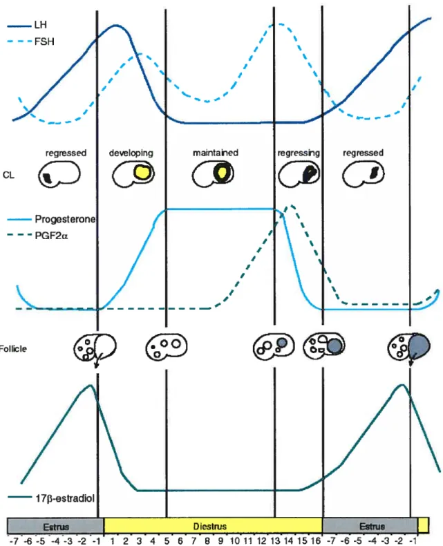
In rnost mammals, the onset of follicular luteinization is triggered by a sharp rise and fall in LH, characterized as an LH surge. This is not the case in mares (figure 2). As it continues to develop, the dominant follicle also responds to the rising LH concentrations by producing sufficient 17f3-estradiol to exert positive feedback stimulation on LH secretion. This resuits in a graduai increase in LH, begiirning 6 or 7 days before ovulation and peaking approximateiy one day after ovulation (figure 2) (3, 14).
FIGURE 2: Hormonal changes during the equine estrous cycle (adapted from t LH - --FSH ‘ CL 1 t I / t t regressed maintahed
60
— — -PGF2u reg ressed 1 F I I t t I jt FoIIicIe/
I 73-estradioI 7 8 g -4 -3 -2 -1 reference 1).This suggests that in horses, the ovulatory process is initiated when a threshold concentration of gonadotropins is reached. During the peri-ovulatory period, the preovulatory follicle responds to LH by markedly increasing its antrnm size and blood flow (9). Granulosa ceils begin secreting progesterone and decrease their secretion of estrogen, which accounts for the late-estrus drop in plasma estrogen concentration and the small rise in plasma progesterone just before ovulation. A cascade of events whicb include prostaglandin synthesis (15), progesterone action on the follicle (16), and consequent expression ofproteolytic enzymes such as ADAMTS (16) expression resuits in the break-down ofthe follicular-ovarian membranes and expulsion ofthe oocyte.
1.2 Cellutar aspects of luteinization
Luteinization is characterized as being a differentiation process by which the diverse celis ofthe preovulatory follicle acquire the functional and morphological characteristics of luteal ceils. This process is initiated by LH in the mare, and begins approximately 2 days prior to ovulation (17). It has been shown that ovulation in mares can be induced during the follicular phase by the administration of hCG when the preovulatory follicle reaches approximately 35 mm, and the duration ofthe ovulation, defined as the interval from hCG injection to follicular rupture, lias been demonstrated to be approximately 36-4$ h (1$). Before hCG, the granulosa ceIl layer is composed of 4-9 compact layers of small elongated cells with some evidence of mitosis, whereas, the theca interna layer is characterized by many polyhedral cells that have a plump appearance and high level of vascularization indicative of steroidogenesis (9). The administration of hCG resuits in a dramatic increase in intracellular spacing ofthe granulosa cell layer, more than doubling its thickness after 39 h, with the maximum expansion of the granulosa ccli layer being reached after 24 h. Mitotic figures can no longer be observed and there is a significant increase in mucoid substances, the best known of which is hyaluronan, is detected between the granulosa cells (9). The theca interna bas been shown to undergo significant thinning, with fewer ceils present and the presence of occasional pyknotic nuclei. A dramatic incrcase in edema, hemorrhage and hyperemia, as well as in the number of blood vessels is also observed, reaching a maxima at 36 and 39 h post-hCG (9). These
data, particularly the incipient degeneration of the theca, support the hypothesis that the granulosa celis are the sole contributor to the CL.
The differentiation process from granulosa to granulosa-lutein celis involves many changes at the cellular level. Granulosa-lutein celis become hypertrophied, becoming the largest steroidogenic ce!! type in the body (17). Gap junctions that allowed rapid communication between granulosa ce!!s during follicular development are !ost (17), but are reestab!ished later on in the process (19). The smooth endoplasmic reticulum and golgi apparatus become more prominent, and the size and comp!exity of mitochondria increase (17). The cytoske!eton is also affected by the process of !uteinization and is believed to play an important role in steroidogenesis (8). Changes inc!ude the acquisition of smooth muscle actin by the granulosa celis, as well as desmin, cytokeratin and vimentin (8). Although it is flot the case in ail species (17, 20, 21), the absence of mitotic figures observed during hCG-induced luteinization in the mare (9) may reveal the occurrence of granulosa ccli exit from the ceil cycle and complete cessation of division resulting in their termina! differentiation afier the LH surge, as seen in the rat (22).
1.3 Luteinization-associated modulation of gene expression
The mechanisms involved in signal transduction following LH are complex and their study is stili in relative infancy. However, some generalizations can be made about what is known thus far. The protein kinase A (PKA) pathway has been linked to the luteinization process. The binding of LH to its receptor, a seven-transmembrane domain G-protein-coupled receptor, leads to its activation and the subsequent production of cyclic adenosine monophosphate (cAMP) from adenylate cyclase and adenosine triphosphate (ATP) (7). The intracellular messenger, cAMP, then binds to the regulatory subunits of the inactive PKA hoioenzyme and activates PKÀ’s catalytic subunits by causing their dissociation from the holoenzyme complex. They are then free to catalyze the phosphorylation of serine and threonine residues of various proteins, including cAMP response element modulator (CREM), nuclear factor-icB (NFiB), cAMP response elernent binding protein (CREB), as we!l as many other nuc!ear receptors (7).
The activation of CREB the via its phosphorylation by PKA resuits in the activation of many genes, such as steroid acute regulator (StAR) (23), CYP liAi (8), as well as the Niemmann-Pick Cl protein involved in the low density lipoprotein (LDL) pathway (24). Numcrous other transcription factors, including steroidogenic factor-1 (SF-1), liver receptor homolog-1, sterol regulatory element binding proteins (SREBPs), and the GATAs, have been shown to be involved in the luteinization process (8). The complexity of the regulation of expression of this multiplicity of factors is beyond the scope ofthe present review.
As previously mentioned, the follicle wall undergoes dramatic changes during luteinization, as seen in its vascularization, as well as in its structural integrity. Many vasoactive and inflammatory agents have been shown to be involved in the ovulatory process (25). Vascular endothelial growth factor has been linked to the increased vascular permeability and shown to stimulate angiogenesis in response to hypoxia in the inner follicular compartments (26), whereas P-selectin is known to play a critical role in the initial steps of leukocyte recruitment ftorn the bloodstream during inflammation and has been shown to be upregulated during luteinization (27). These effects may be secondary to prostaglandin synthesis, which lias also been shown to play a key role in the ovulatory process (15). The structural remodeling of the follicle during luteinization has been investigated over the past decade. Many proteolytic activities have been identified and shown to be regulated. They include the plasminogen activators, which have been shown to activate collagenases (26), the metalloproteinases (16, 26), as well as the metalloproteinase inhibitors (26). The rodent model, in which luteal cells undergo terminal differentiation during the process of luteinization, have been shown to lose cyclin D2 expression after LH, with a resulting increase in ccli cycle inhibitors (22). The progesterone receptor (PR) lias also been shown to be upregulated, supporting the hypothesis that progesterone action is required for luteinization to occur (28). Indeed, the PR knockout mouse is incapable of ovulation (29).
In section 2.2, the molecular biology of ovarian steroid biosynthesis will be addressed, it is nonetheless noteworthy to mention certain key aspects of the regulation
of steroidogenic enzymes during the process of luteinization. The most notable functional change observed during luteinization is the shift in ovarian steroid biosynthesis, from 17-estradio1 to allow the large-scale synthesis ofprogesterone. In a simplified view of the regulation of steroidogenic enzymes, and this holds true for many species including the mare, the LH surge results in the upregulation of enzymes involved in progesterone biosynthesis, such as StAR and CYP liAi (30-4 1), whereas those downstream of progesterone, the expression of CYPI7A1 and CYP19A1 are turned off (36, 42-47).
2. Steroid hormones
Steroids are a subclass of a large farnily of chernical compounds known as terpenoids (48). They are ail derivatives of a perhydrocyclopentanophenanthrene ring
system, with a skeleton fonned of four interconnected rings of 17 carbon atoms (Figure 3). A few polar hydroxyl groups may be attached to this ring structure, but they are not numerous enough to render a steroid water-soluble. Thus, as with oxygen, carbon dioxide and fatty acids, steroid hormones diffuse rapidly through the lipid portions of membranes.
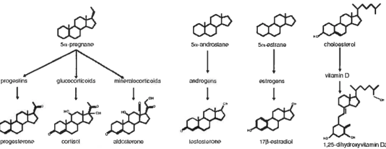
Steroid compounds exhibiting hormonal activity have been classified into six families, namely progestins, glucocorticoids, mineralocorticoids, androgens, estrogens and vitamin D, having one of four parent molecules (figure 3, reviewed in 48). Progestins ail consist of 21 carbons, having two keto groups on carbons 3 and 20. Glucocorticoids and mineralocorticoids are similar to progestins, however, they have an additional hydroxyl group on carbon 21, as well as a side-chain composed of two
carbons at position 17 and the unsaturation of the bond between carbons 4 and 5. Androgens are composed of 19 carbons with a keto group present at position 3 and a hydroxyl group present at position 17. Estrogens are different as their A-ring bas been aromatized. They also have two hydroxyl groups present at carbons 3 and 17 (48).
cycIojnIanotiydroinanthrn
c6
&t-prçJmnpiojstins gIucocortIDcId rnIn9ruacorhDoIds
FIGURE 3: Structure of the steroid hormone backbone and parent molecules (adapted from reference 48).
Specific compounds are named according to their substituent’s suffixes and their position on the parent molecule. Also, the Greek letters Œ and f3 are added depending on
whether the substituent is above or below the plane of the ring, respectively. For example, the addition of a hydroxyl group at position 17 and below the plane of an androstane parent molecule, as well as a keto group at position 3 and the unsaturation of the A ring at position 4, will resuit in a compound systematically named 17f3-hydroxyandrost-4-en-3 -one or testosterone (48).
2.1 Rote of steroid hormones in physiological processes
The majority of steroids are produced by specialized ceils in specialized tissues, such as the adrenal and gonads, and their actions can be local as well as have an effect in distant tissues (reviewed in 49). Peripheral tissues have also been shown to produce or inactivate steroids, however, the action of these steroids is usually restricted to the site of expression. The adrenal cortex has been shown to express significant amounts of
:rogstion choIosloI
L
vitrnin D 5-ifldrœIanI.
rndrog.nsI
lsIosIrcr CH coliLsol 5estrancI
I[oçjnsI
17-osIiodiDI i!dostorono 1 25-diI1t1toxyviIomin D2many different steroids, including aldosterone, cortisol, corticosterone, dehydroepiandrosterone (DHEA), and androstenedione. The zona glomerulosa is responsible for the secretion of the rnineralocorticoid, aldosterone, and its effects include the regulation of electrolyte metabolisrn, namely sodium, potassium, and hydrogen ions, and the way they are handled by the kidneys. The zona fasciculata and zona reticularis of the cortex are responsible for the production of other adrenal steroids. Cortisol is a glucocorticoid and has important effects on the metabolism of glucose and other organic nutrients, it has also been shown to facilitate the body’s response to stress, regulate the immune system, and exert mineralocorticoid effects at high concentrations. DHEA and androstenedione are androgens that are much less potent than testosterone, but are involved in several physiological functions in females (49).
The gonads strongly express the enzymes involved in sex steroid biosynthesis (reviewed in 49). The testes are capable of producing large amounts of androgens, such as testosterone, which play an important rote in the differentiation and growth of the reproductive tract, extemal genitalia, as well as certain regions of the brain. After puberty, they direct the development and maintenance of secondary sex characteristics, as well as sexual behavior (49). The ovaries are the main site of estrogen and progestin biosynthesis. During development, estrogens stimulate the growth of female extemal genitalia, as well as mammary gland development and the formation of mammary ducts and fat deposition. They are also responsible for stimulating bone growth and for the ultimate cessation of bone growth (49). After sexual maturation, and as noted above, during the follicular phase ofthe cycle, 17f3-estradiol is secreted by the granulosa celis of the preovulatory follicle. As described earlier, these steroids have both positive and negative feedback effects on gonadotropin secretion. Estrogens also modulate the growth of smooth muscle and proliferation of the epithelial linings of the reproductive tract. They increase contractions and ciliary activity in the uterine tubes, myometrial contractions and responsiveness to oxytocin in the uterus, as well as stimulate the secretion of abundant and clear cervical mucus and prepare of the uterine endometrium for progesterone actions by increasing the number ofprogesterone receptors (49).
Progesterone, on the other hand, is mainly produced by the CL and placenta, although its biosynthesis begins in the preovulatory follicle prior to ovulation. Physiologically, progesterone is necessary for gestation, as it decreases contractions of the uterine tubes and myometrium. In humans, it also stirnulates secretions from the endometrial glands, induces thick and sticky mucus from the cervix, and decreases proliferation of vaginal epithelial cells. At the level of the hypothalamus, high concentrations of progesterone inhibit GnRH secretion, which results in a negative feedback inhibition of FSH and LH secretion thereby preventing LH surges during the luteal phase and gestation (49).
2.1.1 Estrogens and celi cycle progression
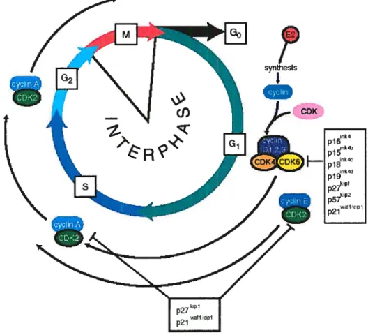
The cdl cycle is composed of four major phases (figure 4) (reviewed in 49, 50). The interphase is considered to be the interval between the end of one division and the appearance of the structural and gene expression changes that indicate the beginning of the next division. In a dividing cell population, interphase is the longest phase of the cycle. The S phase (synthesis) occurs after the first interval, G1, and is characterized by extensive DNA replication. Following DNA synthesis, there is a second interval (G2) before cytokinesis begins. The final phase, the M phase, includes the mitotic and cytokinetic events that resuit in two daughter cells. There are numerous control points in the cell cycle and at each of these, certain events must occur in order for the cell to proceed to the next phase. One of particular significance is the transition between G1 and S while another is that between G2 and M. The phosphorylation of cyclin-dependent kinases and consequent phosphorylation of specific substrates have been shown to dictate the progress through the cdl cycle. When celis undergo terminal differentiation, they exit the cell cycle and enter a phase known as G0.
Estrogens have been shown to increase DNA synthesis in the rodent mammary gland and uterus by recruiting non-cycling cells to the ceil cycle, whereas cells that were already cycling had a reduced G1 phase length (51). Breast cancer cells have been extensively used to study the regulation of the ceil cycle by sex steroids. They were
shown to be most responsive to estrogens, through the estrogen receptors, early during the G1 phase, right afier mitosis (51-53).
The regulation of the immediate-early gene c-myc is one of the earliest effects detectable after estrogen treatment and this holds truc in many ccli systems, including rat uteri (54, 55), normal breast epithelial ceils and breast cancer celis (56, 57). Mitogens, such as estrogens, have been shown to be responsible for mediating the association of cyclin Dl with cyclin-dependent kinases (CDKs) and inducing their kinase activity. This resuits in the hyperphosphorylation, with the help of the cyclin E CDK2 complex, of tumour suppressor proteins, such as retinoblastoma (58). This hyperphosphorylation has been shown to cause the release of the E2F transcription factors, responsible for the transcription of many proteins involved in ccii cycle
progression (59). Recent work demonstrates the positive effects of estrogens at the G2/M phase ofthe celi cycle in ovarian granulosa and breast cancer cells (60).
2.2 Molecular biology of ovarian steroid hormone biosynthesis
Steroidogenic tissues, including the adrenals, placenta, brain and gonads, ail reiy on cholesterol as a precursor for the biosynthesis of a range of steroid hormones (48). Aithough de novo cholesterol synthesis is possible in steroidogenic celis, the principal sources of cholesterol appear to be the plasma lipoproteins (61). The initiation of steroid biosynthesis has been shown to begin in the mitochondria. It is believed that cholesterol destined for steroid conversion is transported to these organelles in vesicies that travei along the cytoskeletal fibers, and that proteins like sterol carrier protein 2 (SCP2) may then promote the transfer of cholesterol from the vesicles to the mitochondria (62-64).
The biosynthesis of ovarian steroid hormones requires the involvement of several enzymes sequestered in specific subcellular compartments. These can be classified into two major classes of proteins, the cytochrome P450 heme-containing proteins and the hydroxysteroid dehydrogenases (65). The accepted nomenclature for the cytochrome P450 enzymes is CYP, followed by an Arabic number representing the P450 family and a letter indicating the subfamily (66).
Eariy on in follicular development, the principal ovarian steroidogenic cells, the theca interna and granulosa cells, contribute to the production of active estrogens, as first described in Armstrong and Dorrington’s two-cell theory (67). Although the two celi theory appiies to the mare, the role of each cdl type is somewhat different. It can be hypothesized, however, that the characteristics of the essential steroidogenic enzymes, such as activity and subcellular localization, are conserved. The first and rate-limiting step in the production of ail steroid hormones is the transfer of cholesterol from the outer to the muer mitochondrial membrane via the actions of StAR, and its subsequent conversion to pregnenolone by CYP liAi (figure 5) (65).
CYP11A 3I3HSD/KSI HO PREGNENOLONE CYP17 CYP17 3I3HSDIKSI CYP19j3 O ANDROSTENEDIONE ESTRONE t17IHsD 3I3HSD/KSI CYP19_0f:5
5-DLOL TESTOSTERONE 17j-ESTRADlOL
FIGURE 5: Simplified steroidogenesis (adapted from reference 68).
Although it is well established that StAR is involved in the import of cholesterol, some evidence supports the use ofmitochondrial contact sites in this process (69). lii the mare, preovulatory levels of StAR and CYP1 lAi are highest in the theca interna, whereas luteinization resuits in a switch in celi-type expression to granulosa celis (30, 41) as tue theea eelts degenerate. The produet of CYPI1A1, pregnenolone, can then either be acted upon by the enzyme 3f3-hydroxysteroid dehydrogenase/ketosteroid
C HOLESTEROL
J,
“10H 17-HYDT4DXWRVGCSTtR4E “10H 17HYLPOXWREGNENaONE H 2Ori-DHP CYP17isomerase (3f3HSD/KSI) catalyzing its conversion to progesterone or be converted to DHEA via the actions of the CYP17A1 protein (65). The equine preovulatory follicle expresses these enzymes in the granulosa ceils and theca interna, respectively. Therefore, pregnenolone from the theca may either traverse the basement membrane of the follicle and further modified by 3HSD/KSI in granulosa celis (30) or may remain in the thecal layer and undergo catalysis by CYP17A1 (43). DHEA then diffuses to the granulosa celi layer where enzymes like 3[3HSD, CYP19A1 and the 1713-hydroxysteroid dehydrogenases (17HSD) catalyze its conversion to active estrogens (65).
2.2.1 Steroidogenesïs from cholesterol to estrone
Many enzymes in the steroidogenic pathway are important for the production of estrogens. However, since the focus of this work is aimed at characterizing the final steps in estrogen biosynthesis as well as its inactivation, only a brief overview of the molecular characterization of the steroidogenic enzymes involved in the multi-step process of cholesterol conversion to estrone will be described.
2.2.1.1 Steroidogenic acute regulator (StAR)
StAR is responsible for the transiocation of cholesterol from the outer to the inner mitochondrial membrane (70). It lias been cloned and characterized in many species, including the mare (41), mouse (71), rat (72), human (73), cow (74), sheep (31), pig (75), and hamster (76). Two isoforms of the equine cDNA were identified and found to measure 1599- and 2918 base pairs (bp) in length (41). Both transcripts had open reading frames of 855 nucleotides, but were variable in their 5’- and 3’-untranslated regions (UTRs) (41). Once translated, the equine protein was composed of 285 amino acids, $6-90% identical to the StAR proteins from the species mentioned above (41).
StAR is believed to be active in its 37-kDa precursor form. However, this cytosolic phospho-protein exhibits a relatively short haif-life. Once translocated across the mitochondrial membrane, along with cholesterol, it is truncated to a 30-kDa form that is more stable than its full-length counterpart, yet its role remains unclear (77-79). The role
of StAR in steroid biosynthesis bas been extensively demonstrated in vivo (78), as well as in celi cultures (73, 80).
Many studies have addressed the transcriptional regulation of StAR in the ovary. In the mare, the effects of gonadotropins on StAR transcript expression were examined in follicles isolated at different times after an ovulatory dose of hCG and visualized by Northern blot analysis (41). In that study, no significant effect of gonadotropin treatment was discernable in intact follicle wall samples. Nonetheless, when individual celi compartments were examined, a significant increase in StAR transcript was observed 30-39 h post-hCG in granulosa celis, while the high levels present in theca interna prior to hCG significantly decreased 36 h after treatment. In vitro studies demonstrated that gonadotropins and activators of the PKA pathway upregulated StAR expression in granulosa ceils from rat (33, 72, 81), porcine (82, 83), bovine luteal cells (40), and human (32) and PGF2u repressed StAR expression (40). Other in vivo studies demonstrated that StAR is regulated in a gonadotropin-dependent and stage-specific manner in developing follicles (32, 33, 38, 84). High levels of StAR mRNA have been detected in the corpus luteum of many species, including the mare (41) and previous studies have also shown a decrease in StAR expression in the corpus luteum during luteal regression (31, 32, 38, 40, 74, 75, $4-86).
2.2.1.2 Cytochrome P450 sïde-chain cleavage (CYP11A1)
The first rate-limiting and hormonally regulated enzymatic conversion in the biosynthesis of steroids from cholesterol is dictated by cytochrome P450 side-chain cleavage (CYP liAi) in tandem with its associated electron-transport chain (61, 87). CYP liAi bas been shown to be localized to the matrix side of the iimer mitochondrial membrane and to catalyze the conversion of cholesterol to pregnenolone, a common precursor to all steroid hormones (61, 87). Its cDNA has been cloned and characterized in a number of different species, including the mare (30). The equine transcript measures 1837 nucleotides (nt), with an open reading frame of 1560 nt and encoding a 520-amino acid protein that is highly homologous to other mammalian orthologues.
The transcriptional regulation of the CYP liAi gene has been shown to be under gonadotropin control. In the mare, studies using Northern blot analyses have demonstrated that the theca interna is the main site of CYP liAi transcript expression prior to hCG treatment, with a downregulation occurring 30-39 h afier hCG. In granulosa ceils, an increase in CYP1 lAi mRNA is observed 39 h post-hCG (30). A high level ofCYP1 lAi expression was also noticeable in equine corpora lutea (30).
2.2.1.3 Cytochrome P450 17Œ-hydroxylase/C17-20 Iyase (CYP17A1)
CYP 17A1, previously known as cytochrome P450 1 7Œ-hydroxylase/C 17-20 lyase, is responsible for the conversion of pregnenolone to DHEA and has also been shown to convert progesterone to androstenedione, in a two-step reaction. It has been cloned in various species, including the mare (43). When expression of the equine CYP17A1 transcript was examined, a single 2.4-kb mRNA hand was detectable by Northern blot. The regulation of CYP17A1 mRNA in preovulatory follicles afier hCG treatment revealed that high levels were present prior to gonadotropin treatment and that these significantly decreased 36-39 h post-hCG. The signal was restricted to theca interna, and was undetectable in both granulosa ceils and corpora lutea, indicating that the thecal layer is responsible for CYP17A1 activity.
2.2.1.4 33-Hydroxysteroid dehydrogenase! ketosteroid isomerase (3llSDIKSI) The dually functional enzyme 3f3HSD/KSI converts A5-3t3-hydroxysteroids to ketosteroids, first through catalyzing the dehydrogenation of hydroxysteroids, and by subsequent isomerization of the A5-ketosteroid product to yield the Œ,13-unsaturated ketones. This enzyme has been shown to catalyze the conversion of pregnenolone to progesterone, as well as DHEA to androstenedione. It is therefore clear that 3HSD/K$I is essential for the biosynthesis of all steroid hormones, including glucocorticoids, mineralocorticoids, progesterone, androgens, and estrogens ($8-91). It has been localized to the endoplasmic reticulum as well as the mitochondrial membrane and is expressed in many tissues, including the gonads, adrenal cortex, placenta, and in certain peripheral tissues that are not traditionally recognized as steroidogenic (88-93). In other species, such as the human and rat, many isoforms of the enzyme have been isolated
(94-96). In the horse, only a single 1612-bp 3HSD/K$I cDNA has been cloned comprising an open reading frame of 1119 nt, that translates to a 373 amino acid protein (30). In the rat, the adrenal 3f3H$D/KSI transcript was shown to be upregulated by ACTH and downregulated by corticosterone (97), whereas the ovarian 3I3HSD/KSI appeared to be upregulated by hCG ami downregulated by prolactin (98). The expression of equine 3I3HSD/K$I mRNA, however, does flot appear to be under LH control (30). It was demonstrated by Northern blot that 33HSD was highly expressed in the granulosa ceils of the equine preovulatory follicle, as well as in corpora lutea, and that no significant difference in 3-HSD/KSI mRNA expression was detectable afier hCG treatment. No signal was detected in the theca interna.
2.2.1.5 Cytochrome P450 aromatase (CYPJ 9A1)
CYP19A1 catalyzes one of the final steps in estrogen biosynthesis, via the conversion of androstenedione and testosterone to estrone and 17-estradio1, respectively. The CYP19A1 gene is interesting in that it has at least nine different untranslated first exons (99). They are referred to as exons 1.1, 1.2, 1.3, 1.4, 1.5, 1.6, PlI, 2a, and 1f, and are alternatively spliced into a common 5’-splice acceptor site located 38 bp upstream ofthe translation start site in exon 2. The CYP19A1 gene has been cloned in a number of species, including the human (99) and pig (100), and cDNAs including promoters 1f and PlI have been cloned and characterized in the mare (43). The equine 26$2-bp promoter 1f transcript has been shown to encode a 503 amino acid-protein that is highly conserved compared to other species. The predicted equine protein has many conserved features, including a membrane-spanning region, an I helix, a heme-binding region and a putative cAMP-dependent protein kinase phosphorylation site.
The transcriptional regulation of CYP19A1 has also been characterized in various species. In the mare, a biphasic pattem of expression was detected in granulosa celis of preovulatory follicles by Northern blot analysis following hCG treatment (43). When individual promoters were examined, it was demonstrated that activation of Pli was responsible for CYP19A1 expression prior to the ovulatory stimulus and in corpora
lutea, whereas the increase in transcript observed 30-39 h post-gonadotropin treatment was attributable to promoter 1f activation. No expression was detected in theca interna. NFKB-specific inhibitors were shown to suppress basal promoter II activity in cultured human granulosa celis, indicating a potential role for NFiB in ovarian aromatase expression (101). Also, activation of peroxisome proiiferator-activated receptor (PPAR) y and retinoid X receptor, resuited in decreased NFKB/promoter II interactions, and resulted in a downregulation ofaromatase expression (101).
The activity of different promoters has been used to explain the tissue-specific distribution ofthe CYP19A1 transcripts. It also accounts for differences in the time and site of expression, as demonstrated by the differential expression in early versus mid pregnancy porcine placentas, as well as foetal versus adult human liver (100, 102). CYP19A1 expression has been detected in the gonads (103), brain (104), adipose tissue (105, 106), and skin (107). The targeted disniption ofthe CYP19A1 gene in mice results in arrested folliculogenesis, no corpora lutea, elevated levels of LH, FSH aiid testosterone, and infertility (108).
2.2.2 173-Hydroxysteroid dehydrogenases
The biological potency of androgens and estrogens is rnodulated by the family of 17f3-HSDs. They are responsible for the oxidation or reduction of steroids at the C17 position; the keto-forms being inactive and the hydroxy-forms being active and able to activate their cognate receptors (109). Twelve 17I3HSD5 have been identified thus far, differing in their substrate specificities, cofactor preference, subcellular localization, and tissue distribution (figure 6). Ail 17I3HSDs are coded by different genes, with distinct amino acid sequences, therefore, they shouid not be referred to as isozymes. Although 1 7HSDs have been shown to be capable of both oxidation and reduction reactions in
vitro, they are unidirectional enzymes in vivo (110, 111). It is well recognized that
NAD and NADPH are the most abundant forms of intracellular nicotinamide adenine
dinucleotide and nicotinamide adenine dinucleotide phosphate cofactors, respectively, thereby dictating the direction of 173HSD activity. Indeed, certain 17HSDs prefer a
phosphorylated cofactor (NADP(H)), whule enzymes responsible for the oxidative reactions prefer NAD(H) (110, 111).
ltiizvme Gene naine OLlier naines I lurnan Mn tue
t3I’
Function Chromosome Fonction Chromosome
l1SD17l3I El—E2 17q2I EI—E2 II
HSD17BI’I rI7q21 A4I
2 HSDI7B2 E2—E1 16q21.I-c24.2 E2—*EI 6
i-’A1 T-A4
2Ote-Dl-l-4P4
3 l-lSD17B3 A41 q22 A3—tT I)
4 I ISDI 7fl4 M F2 l—osid;iiiist of frite 5g2 I —oxidatiunoftiitly 18
aci]s,li24EI acids,E2—’El
5 FISDI7[15 DDJ. A4— I lOpl4-p15 A4—I 13
A KRI(‘3 tIAKRe l’1- 20a-Dl Il’
6 HSDI 71M; NA N;A audmiten inactivation NiA
7 HSDI7B7 ppp EI—’E2 lq23. El—E2
I-ISDI 7B7I’2 cholrtstrtol Svitthesis l0pI I. 4rI g24 cholesterolsynthesis
8 HSDI7B% HKE6.FABGL E2—El 6p2I3 E2—’El 17
androgen inacti yahoo androgeninacti ssttion
C) FISDI 7 t NrA NsA E2-El 10
retinol oxidation 10
IISDI7BI0 IIADII2. SC’HAD, esirogenandandrngen Xpl 1.2 estrogen tord androgen X ERAB inactivation, 3oxidation inactivation. [3oxidation
offtittyacids ofOrtieacids
I-ISDI7HII Panlb, retSDR2 estrogenmdtuidrogen 4q22.I NIA 5 inactivatiOfl
12
HSDI7BI2 KAR, KIKI 3-ketoacyl-CnA reductase 1 IpI 1.2 3-ketoacvl-CoA reductase 2 Ortieacid synthesis Oittv ucidsynthesis
FIGURE 6: The different l7f3HSDs. El: estrone; E2: 17f3-estradiol; A4: androstenedïone; T: testosterone; P4: progesterone; 20Œ-DHP: 20Œ-dihydroprogesterone; N/A: flot avaïlable (adapted from reference 109).
The 17HSDs are readily grouped into two families based on protein architecture: the short-chain dehydrogenase/reductase (SDR) family (112) and the aldo-keto reductase (AKR) family (113). Members of the SDR family are often multimeric and share several amino acid sequence motifs, such as the Rossman fold motif (TGxxxGxG) involved in cofactor binding, a NAG domain involved in structural stabilization and located between the cofactor binding site and the active site, an active centre (YxxSK), and a site that dictates the direction of the reaction (PGxxxT) that is located C-terminal to the active site (112). Even though these domains are highly conserved among SDR