Université Toulouse 3 Paul Sabatier (UT3 Paul Sabatier)
Directeurs de Thèse : Unité de recherche :
Titre :
Présentée et soutenue par :
le : 28 mai 2014
Jury :Président:
Rapporteur:
Rapporteur:
Directeur de thèse:
Directeur de thèse:
Examinateur:
Dr. Malte Drescher
Dr. Julie Gehl
Prof. Dr. Karin Hauser
Dr. Marie-Pierre Rols
Prof. Dr. Andreas Zumbusch
Prof. Laurent Paquereau
THÈSE
En vue de l’obtention du
DOCTORAT DE L’UNIVERSITÉ DE TOULOUSE
Délivré par :
Cotutelle internationale avec l'Université de Constance, Allemagne
Christelle ROSAZZA
Internalisation et trafic intracellulaire de l'ADN plasmidique délivré
par electroporation in vitro
École doctorale et discipline ou spécialité :
ED BSB : Biologie cellulaire
Institut de pharmacologie et biologie structurale
Dr. Marie-Pierre ROLS
Prof. Dr. Andreas ZUMBUSCH
University of Konstanz, Germany
Copenhagen Hospital Herlev, Denmark
University of Konstanz, Germany
CNRS Toulouse, France
University of Konstanz, Germany
CNRS Toulouse, France
Abstracts
1. French
L’électroporation est une méthode physique de délivrance de molécules dans les cellules ou tissus. Le transfert des petites molécules se déroule vraisemblablement par diffusion à travers de présumés electropores alors que le transfert d’ADN plasmidique est plus compliqué et reste à élucider. Lors de l’application des impulsions électriques, la membrane devient perméable et l’ADN est électrophorétiquement poussé vers celle-ci où il est inséré en agrégats distincts pendant une dizaine de minutes. Ensuite, l’ADN est internalisé, navigue à travers le cytoplasme jusqu’à atteindre le noyau où l’expression de l’ADN est initiée. Mon travail de thèse se concentre sur l’internalisation et le trafic intracellulaire de l’ADN, deux étapes assez peu décrites dans le cadre de l’électroporation.
L’internalisation de l’ADN semble se faire majoritairement par endocytose. L’utilisation d’inhibiteurs pharmacologiques de l’endocytose et de marqueurs fluorescents permet de conclure que l’endocytose médiée par la clathrine et les rafts lipidiques ainsi que la macropinocytose sont impliquées (respectivement 25%, 50% et 30%). L’endocytose semble être la principale (sinon la seule) voie par laquelle les agrégats d’ADN entrent dans les cellules. De plus amples recherches doivent être menées pour dépeindre précisément le mécanisme d’internalisation de l’ADN d’autant plus que l’endocytose médiée par les rafts lipidiques représente un large panel de possibilités (caveoline, flotilline, GEEC…). Nos résultats sont en accord avec la distribution spatiotemporelle de l’ADN observée à la membrane.
Une fois dans le cytoplasme, l’ADN semble suivre le trafic endosomal classique. La colocalization en dynamique effectuée dans des cellules exprimant séparément plusieurs marqueurs d’endosomes (Rab5, Rab11, Rab9 et Lamp1) démontre que l’ADN est présent dans les endosomes précoces, tardifs et de recyclages dans des proportions calculées pour être respectivement 70%, 50% et 30%, valeurs moyennées sur l’heure qui suit l’électrotransfert. Entre 1-2 h après la délivrance, 60% de l’ADN est situé dans des structures marquées Lamp1 avec une prédominance probable des lysosomes. Ces résultats sont en accord avec l’observation de l’ADN toujours présent en agrégats dans le cytoplasme et ils renforcent l’observation d’un mécanisme d’endocytose mentionné précédemment.
L’ADN dans le cytoplasme est transporté activement par les filaments d’actine et les microtubules. L’utilisation d’inhibiteurs pharmacologiques combinée au suivi de particules uniques effectué sur un grand nombre d’agrégats d’ADN clairement démontre que le cytosquelette contrôle la migration de l’ADN dans le cytoplasme. L’ADN exhibe le mouvement typique des endosomes avec des phases intermittentes de transport actif et de diffusion. Les phases de transport actif ont en moyenne une vitesse de 250 nm/s, une persistance de 6 s et engendre un déplacement de 1,3 µm. Cependant, les distributions de ces paramètres sont larges avec des vitesses allant de 50 nm/s à 3400 nm/s, des déplacements de 0,1 µm à 12 µm et des durées de 2 s à 30 s. Ces résultats confirment d’autres travaux qui présentent les microtubules comme un moyen de migration de l’ADN dans les cellules. Cela est aussi en accord avec la présence de l’ADN dans des endosomes vu que leurs membranes contiennent des protéines (probablement Rabs) capables de se lier aux
moteurs moléculaires. De plus, cela explique comment les gros agrégats d’ADN peuvent traverser le cytoplasme dense et atteindre avec succès le noyau, phénomène improbable par simple diffusion.
Le chemin de l’ADN que nous décrivons est efficace étant donné que la perturbation d’une étape intermédiaire entraine une réduction de l’expression génique. Bien que nos découvertes doivent être confirmées et nécessitent de plus amples investigations, la prochaine étape clé à comprendre est l'échappement supposé de l'endosome qui doit forcement se réaliser afin d’obtenir une expression de l’ADN.
2. English
Electroporation is a physical method of delivery of molecules into cells or tissues. The transfer of small molecules most likely occurs via diffusion through putative induced electropores whereas the transfer of plasmid DNA is more complex and remains to be elucidated. During the electric field pulses, the membrane becomes permeable and the DNA is electrophoretically pushed on it where it is inserted as discrete clusters for tens of minutes. After the electric pulses, DNA is internalized, navigates through the cytoplasm until it reaches the nucleus where DNA expression is initiated. This PhD work focuses on the internalization and intracellular trafficking of the DNA, two steps rather enigmatic in the context of electroporation. The relevant cell structures that could participate into these steps are the endocytotic machinery followed by the endosomal trafficking and the cytoskeleton (actin filaments and microtubules).
DNA internalization seems to be mainly performed via endocytosis. The use of pharmacological endocytic inhibitors combined with endocytic markers led us to the conclusion that clathrin- and lipid raft-mediated endocytosis as well as macropinocytosis are involved (25%, 50% and 30%, respectively). Endocytosis seems to be the main (if not the only) path by which DNA aggregates enter the cells. More investigations have to be done to depict precisely the DNA internalization mechanism especially because raft-mediated endocytosis represents a large panel of possibilities (caveolin, flotillin, GEEC…) which are worth describing. These findings are in agreement with the observed spatiotemporal distribution of the DNA aggregates at the membrane (clusters of few hundreds of nm persisting for few min and inaccessible from the extracellular medium).
Once inside the cells, DNA seems to follow the classical endolysosomal trafficking. Dynamic colocalization performed in cells expressing separately several labeled endosomal markers (Rab5, Rab11, Rab9 and Lamp1) allows us to conclude with no ambiguity that DNA is present in early, recycling and late endosomes in proportions calculated to be respectively 70%, 50% and 30% over the hour following the DNA electrotransfer. Between 1-2 h after delivery, 60% of the DNA is located in Lamp1-structures with most probably a predominance of the lysosomes. These results are in agreement with the observed DNA being still in clusters in the cytoplasm and it reinforced the earlier mentioned mechanism of endocytosis.
DNA in the cytoplasm is actively transported by both the actin filaments and the microtubules. The use of pharmacological inhibitors combined with SPT performed on a high number of DNA aggregates clearly shows that cytoskeleton mediates the DNA journey in the cytoplasm. DNA exhibits the typical motion of endosomes with intermittent phases of active transport and diffusion. Under our experimental conditions, active motion phases features on average velocity of 250 nm/s and persistence of 6 s and leads to
a displacement of 1.3 µm. However, distributions of theses parameters are broad with velocities from 50 nm/s to 3400 nm/s, displacements from 0.1 µm to 12 µm and persistence from 2 s to 30 s. These findings confirm previously published article pointing at the microtubules as a means of DNA migration in the cells. It is also in agreement with DNA being in endosomes since their membranes process proteins (probably Rabs) that can bind molecular motors. In addition, it explains how such big DNA clusters can travel through the highly crowded cytoplasm since successful to reach nucleus diffusion is improbable.
The DNA route(s) we described correspond to the efficient one(s) since disturbance of any intermediate step results in a reduced gene expression. Although our findings must be confirmed and further investigated, the next key step to unravel is the putative endosomal escape that has to occur for successful gene expression.
Publications
Articles
1. Rosazza C, Escoffre JM, Zumbusch A, Rols MP. The actin cytoskeleton has an active role in the electrotransfer of plasmid DNA in mammalian cells. Mol Ther. 2011 May;19(5):913-21.
2. Rosazza C, Phez E, Escoffre JM, Cézanne L, Zumbusch A, Rols MP. Cholesterol implications in plasmid DNA electrotransfer: Evidence for the involvement of endocytotic pathways. Int J Pharm. 2012 Feb 14;423(1):134-43.
3. Rosazza C, Buntz A, Rieß T, Wöll D, Zumbusch A, Rols MP. Intracellular tracking of single-plasmid DNA particles after delivery by electroporation. Mol Ther. 2013 Dec;21(12):2217-26.
4. Wiktorowski S, Rosazza C, Winterhalder M J, Daltrozzo E., Zumbusch A. Water-soluble Pyrrolopyrrole Cyanine (PPCy) NIR fluorophores. ChemComm. 2014, accepted for publication
5. Rosazza C, Deschout H, Braeckmans K, Rols MP, Zumbusch A. Internalization and intracellular pathways of plasmid DNA delivered by electroporation, in preparation.
Reviews
1. Escoffre JM, Mauroy C, Portet T, Wasungu L, Rosazza C, Gilbart Y, Mallet L, Bellard E, Golzio M, Rols MP, Teissié J. Gene electrotransfer: From biophysical mechanisms to in vivo applications. Part 1 - Biophysical mechanisms. Biophys. Rev. 2009 Dec; 1:177-184
2. Chabot S, Rosazza C, Golzio M, Zumbusch A, Teissié J, Rols MP. Nucleic acids electro-transfer: from bench to bedside. Curr Drug Metab. 2013 Mar;14(3):300-8.
Table of contents
INTRODUCTION ... 1
GENERAL BACKGROUND ... 9
CHAPTER 1:THE CYTOSKELETON ... 10
I. The actin cytoskeleton... 10
I.1. Actin polymerization ... 10
I.2. Actin-binding proteins ... 11
I.3. Actin structures ... 12
I.4. Actin localizations and functions ... 14
II. The microtubules ... 18
II.1. Tubulin polymerization ... 19
II.2. Microtubule-binding proteins ... 20
II.3. Microtubule structures and functions ... 21
II.4. Motor proteins ... 24
CHAPTER 2:ENDOCYTOSIS ... 27
III. Overview ... 27
III.1. Classification ... 28
III.2. Lipids ... 29
III.3. Coat components ... 30
III.4. GTPases ... 31
III.5. Regulatory proteins ... 32
III.6. Fission mechanism ... 32
III.7. Actin cytoskeleton related proteins ... 34
IV. Clathrin-mediated endocytosis ... 36
V. Caveolae-mediated endocytosis... 37
VI. Clathrin- and caveolin- independent endocytosis ... 39
VI.1. RhoA or IL-2 pathway ... 40
VI.2. Cdc42 or CLIC/GEEC pathway ... 40
VI.3. Flotillin-dependent pathway ... 41
VI.4. Arf6 pathway ... 41
VII. Macropinocytosis ... 42
VIII. Intracellular trafficking ... 44
VIII.1. Early endosomes ... 44
VIII.2. Recycling endosomes ... 45
VIII.3. Maturating and late endosomes ... 46
VIII.4. Lysosomes ... 46
VIII.5. Transport ... 47
VIII.6. Bypass of the endosomal compartments? ... 47
CHAPTER 3:FLUORESCENCE AND MICROSCOPY ... 48
1. Microscopy, an important tool for cell biology ... 48
3. Fluorescent probes ... 50
4. Fluorescence microscopy ... 52
5. Wide-field and confocal microscopes ... 55
6. Single particle tracking ... 56
7. Applications of SPT ... 62
CHAPTER 4:ELECTROPORATION... 64
I. Electropulsation ... 64
I.1. Historic ... 64
I.2. Electric field characteristics ... 65
I.3. Cell in suspension and electric equivalent ... 66
I.4. Induced membrane potential difference ... 66
I.5. Other physicochemical consequences ... 68
II. Electropermeabilization ... 70
II.1. Threshold ... 70
II.2. Asymmetry ... 71
II.3. Reversibility ... 73
II.4. Associated molecule exchange ... 73
II.5. Transient permeabilization structures ... 74
II.6. Cellular response ... 76
II.7. Applications ... 77
III. DNA electrotransfer ... 79
III.1. Threshold ... 79
III.2. Electrophoretic component ... 80
III.3. Asymmetry ... 80
III.4. DNA-membrane interaction ... 81
III.5. DNA Internalization ... 83
III.6. Intracellular migration ... 89
III.7. Crossing the nuclear envelope ... 91
III.8. Associated gene expression ... 92
III.9. Applications ... 92
RESULTS AND DISCUSSIONS ... 97
CHAPTER 1:ACTIN CYTOSKELETON AND DNA ELECTROTRANSFER ... 98
I. Results ... 98
I.1. Determination of the latrunculin B incubation condition ... 98
I.2. Effect of actin disruption on gene expression ... 99
I.3. Effect of actin disruption on DNA/membrane interaction ... 101
I.4. Effect of the electric field on the actin cytoskeleton in the presence of DNA... 102
II. Discussion ... 105
II.1. Actin and DNA coincide at the plasma membrane level ... 105
II.2. Actin is involved in DNA accumulation at the cell membrane ... 105
II.3. Actin is involved in the first step(s) of DNA internalization/trafficking ... 106
II.4. A proposed mechanism ... 107
II.5. Conclusion ... 108
I. Results ... 110
I.1. Effect of the different endocytic inhibiting drugs on cell viability ... 110
I.2. Effect of endocytic inhibition on gene expression ... 112
I.3. Colocalization study between DNA and endocytic markers ... 115
II. Discussion ... 119
II.1. Cholesterol is required for the DNA internalization ... 119
II.2. Clathrin-mediated endocytosis of DNA ... 120
II.3. Caveolin/raft-mediated endocytosis of DNA ... 121
II.4. Macropinocytosis of DNA... 123
II.5. DNA internalization and trafficking ... 125
II.6. Conclusion ... 126
CHAPTER 3:DNA ACTIVE TRANSPORT... 127
I. Results ... 127
I.1. Determination of the conditions of incubation for each drug ... 127
I.2. Effect of cytoskeleton alteration on gene expression ... 129
I.3. DNA mode of motions ... 130
I.4. Means of intracellular DNA transport... 133
II. Discussion ... 140
II.1. DNA aggregates are actively transported along actin filaments ... 140
II.2. Fast and long-range transport of DNA aggregates is microtubule-dependent ... 141
II.3. DNA aggregates are transported by simultaneous action of several motors ... 142
II.4. Diffusion behavior of DNA aggregates ... 144
II.5. Conclusion ... 145
CHAPTER 4:DNA INTRACELLULAR TRAFFICKING ... 146
I. Results ... 146
I.1. About the method ... 146
I.2. DNA and endo-lysosomal trafficking ... 148
II. Discussion ... 151
II.1. DNA aggregates are routed to the endosomal trafficking ... 151
II.2. Conclusion ... 152
MATERIALS AND METHODS ... 153
1. Cells culture ... 154
2. Plasmids ... 154
3. Chemicals ... 154
4. Electropulsation ... 155
5. DNA fluorescence staining ... 156
6. Immunofluorescence of the cytoskeleton ... 156
7. EGFP-protein transient expression ... 157
8. Cell viability ... 157
9. Single particle tracking procedure ... 158
10. Colocalization DNA/endocytic ligand procedure ... 159
11. Fluorescence microscopy ... 161
CONCLUSION AND OUTLOOK ... 164 REFERENCES ... 167
Introduction
Introduction
Gene therapy was first introduced in the 80’s, and relies on the delivery of genetic material (mainly DNA) to the nucleus of cells, so that its product is expressed and generates a therapeutic effect. Different approaches are possible:
- Correcting a defective gene by providing a functional one to the cells - Introducing a gene encoding a therapeutic protein
- Transferring a gene provoking cell death
- Delivering antisense DNA that inhibits the synthesis of a protein or the replication of a virus. Therefore, gene therapy leads to applications with a scope far beyond healing genetic diseases, since DNA can, in principle, replace any medication controlling protein synthesis. Gene therapy seems to be an excellent alternative to fight diseases currently not curable or only partially treated with classical pharmaceutical approaches. Moreover, gene therapy offers many advantages compared to the employment of recombinant proteins. Recombinant proteins are expensive to produce and are rapidly cleared from the blood flow, while gene therapy provides to a long-term and eventually regulated production of a therapeutic protein. In gene therapy, gene expression is local, therefore side effects due to exogenous proteins in the bloodstream are prevented.
The main drawback for the utilization of gene therapy as a medical treatment is the lack of effectiveness of the current methods. Several biological barriers (extracellular medium, plasma membrane, cytoplasm, nuclear envelope) hinder gene targeting into the nucleus. Regardless of the method of delivery in vivo (intramuscular or intravascular injection, inhalation, ect.), it will inevitably come into contact with the extracellular environment. Most likely due to nucleases, intravenously injected naked DNA has a half-life within serum in the range of 1.2 min to 21 min, depending on the topology of the DNA (Houk et al., 1999). DNA that escapes nucleases can also bind to non-targeted proteins and cells which can inhibit its activity, or segregate it for degradation and/or removal. The immune system is another obstacle to consider. While mostly associated with viral approaches, immune activation can occur for some non-viral methods as well. This is thought to be due to unmethylated CpG (cytosine-phosphate-guanine) motifs on the plasmid DNA and their recognition by Toll-like receptors (Zhao et al., 2004). When the vector reaches the target cell, it then faces a second important barrier which is the plasma membrane. Naked DNA alone does not associate with the plasma membrane due to the high amount of negative charges that both the DNA and the cell surface bear. Gene transfer methods provoke or facilitate DNA endocytosis or direct entry into the cytoplasm. Vectors that access the cytoplasm via endocytosis are directed to the endosomal trafficking. In that case, an additional barrier for DNA to overcome is the escape from the endosomes before its degradation in the lysosomes. Once in the cytosol, DNA has to traffic to the nucleus for gene expression to occur. Intracellular migration can represent a significant barrier for successful DNA delivery as well. First, nucleases present in the cytoplasm can degrade free DNA. Second, the cytoplasm is a viscous crowded environment that decreases the mobility of macromolecules (Luby-Phelps, 2000; Lukacs et al., 2000). Thus, if DNA is released, from the plasma membrane or even from an endosomal membrane, at a site distant from the nucleus, its diffusion is not likely to be an efficient means of reaching the nucleus. DNA that has successfully traversed the barriers previously mentioned finally encounters the nuclear envelope. Depending
Introduction
on cell types, DNA doses and detection methods, only 1% to 5% of the plasmids locate within the nucleus after their delivery (Cohen et al., 2009). The majority of DNA that has entered the cell will, in fact, never reach the nucleus. While DNA transfer is higher within dividing cells, even the rupture of the nuclear envelope does not completely abolish nuclear import as a barrier to delivery (Ludtke et al., 2002). Therefore, the “ideal vehicle for gene transfer must have the following properties: specificity to target cells, efficient gene delivery, resistance to metabolic degradation and/or attack by the immune system, minimum side effects, and eventually controlled temporal gene expression” (Burgain-Chain et al., 2013).
There are numerous gene transfer methods in vivo that are usually separated into two categories: viral and non-viral. Viruses are the most efficient vehicles because they have innate mechanisms to target cells, to overcome cell membranes and to reach the nucleus. However, viral methods have some disadvantages such as high costs for their production and more importantly they can induce undesired effect like immune response, pathogen reversion, mutagenesis and carcinogenesis. Due to these limitations, the delivery of genetic material by non-viral methods may be of great value for the development of gene therapy especially when repeated administration is needed. Non-viral methods are classified into two groups according to the vector employed: chemical and physical. Chemical approaches include cationic lipid-DNA complexes (lipoplex), cationic polymer-DNA complexes (polyplex) or both lipids and polymers in complexes with DNA (lipopolyplex). The lipids or polymers encapsulate and condense DNA via electrostatic interactions between the positively charged lipids or polymers and the negatively charged DNA. DNA is consequently more protected against nuclease activity, especially if PEGylation is performed, and the reduced particle size, in addition to the net positive charge, favor plasma membrane crossing. The complexes enter the cell via endocytosis or fusion with the plasma membrane. For lipoplexes, endosomal escape is mediated by fusion between the lipids of the lipoplexes and those of the endosomal membranes. For the polyplexes, the polymers act as a proton sponge that first prevents endosomal acidification and second induces a higher influx of ions that swells and lyses the endosomal compartment. One advantage of the chemical methods is that the cell targeting can be enhanced by linking specific ligand to the lipids or polymers. One disadvantage is the toxicity and loss of the vectors when injected in vivo. Due to their positive charge, the particles can interact with many proteins of the serum (albumin, glycoproteins, immunoglobulins, fibronectin …) or blood cells (Cullis et al., 1998). Physical approaches are mostly based on a mechanical or physical destabilization of the plasma membrane allowing access to the cytoplasm. The simplest method consists in hydrodynamic injection of naked DNA into the target tissue via the bloodstream. The rapid injection of a large volume increases the vascular pressure, opens the vascular pores and allows DNA to overcome the plasma membrane of surrounding cells. This method is efficient only in highly vascular tissues (liver, kidney, muscle). However, because of the rapid degradation of the DNA, it is more efficient to combine it with another physical method. Microinjection of DNA directly into the nucleus is very efficient since there are no biological barriers to cross but the cost, the difficulty and the low number of cells that can be treated this way represent serious disadvantages for clinical applications. Lasers can be used to destabilize cell membrane (putative pores) and introduce molecules into the cytoplasm. This method is very efficient in vitro and can be used in many tissues but suffers from low accessibility in vivo since laser light
Introduction
penetration depths are low. The issue of superficial penetration is encountered also with gene gun methods. The latter utilizes metal nanoparticles coated with DNA that are accelerated and bombarded onto the tissue of interest. Particles traverse membranes and DNA is released in the nucleus. Another physical method is sonoporation, where the application of ultrasound in the presence of microbubbles alters nearby membranes. Upon absorption of ultrasound waves, air-filled microbubbles cavitate, oscillate, collapse and release local shock waves that can induce local disruption of the plasma membrane in the form of transient pores allowing the passage of DNA. Finally, electroporation, which is the method of interest for this PhD work, relies on the application of electric fields that destabilize membrane permeability and molecules such as DNA can enter cells via a mechanism that is presumed to be based on the formation of pores. It is one of the more efficient techniques among non-viral methods but it does not reach the efficiency observed for viral approaches. For all mentioned non-viral methods, the general issue is the lack of gene transfer efficiency and the lack of knowledge about the detailed mechanisms by which plasmid DNA gets over the numerous biological barriers to finally reach the nucleus where it can be read by the cell machinery.
Figure 1: Schematic mechanism of plasmid DNA electrotransfer. During the application of the electric pulses, the plasma membrane is permeabilized (step 1), the negatively charged DNA electrophoretically migrates toward the positive electrode (step 2) and interacts with the electropermeabilized membrane by forming aggregates (step 3). The mechanism of electrophoretically driven insertion and the translocation (step 4) of the DNA through the cell membrane are not yet characterized but two models can be proposed. The DNA could pull the membrane during its insertion and initiate the formation of an endosome-like vesicle, or the DNA could be inserted where an electropore is formed, pull and pass through it. Once inside the cell, DNA migrates in the cytoplasm (step 5) where two possibilities exist. Either DNA uses the cytoskeleton to be actively transported or DNA diffuses toward the highly crowded cytoplasm. Subsequent steps are the crossing of the nuclear envelope (step 6) and the expression in proteins of the DNA (step 7). Using fluorescence microscopy, several steps of the DNA electrotransfer could be observed such as (a) the DNA aggregate formation, (b) the DNA remaining as aggregates at the nucleus vicinity and (c) the successful DNA
expression visible as EGFP fluorescence signal. From (Chabot et al., 2013)
The objective of my PhD work was to improve the understanding of the mechanisms involved in gene delivery by electroporation. This approach is interesting for many reasons. It is easy to perform, cheap, safe and promises a great future for the delivery not only of plasmid DNA but all kinds of molecules from small drugs to proteins and other nucleic acids (Neumann et al., 1972; Gehl, 2003; Escoffre et al., 2007). This method is exploited to potentiate the therapeutic effects of genes (electrogenetherapy, genetic
Introduction
vaccination) in clinical trials (Daud et al., 2008; Low et al., 2009; Sardesai et al., 2011) and those of anticancer drugs in clinics (electrochemotherapy) (Mir et al., 2006). In addition, all types of tissues can be treated by electroporation with, however, pronounced differences in efficiency (Andre et al., 2004). One of the most extensively used tissues for electrotransfer is skeletal muscle since there, electrotransfer is very efficient. It offers promising treatment for muscle disorders, but one can also use skeletal muscles as endocrine organs that produce and release the proteins into the blood circulation to exert a therapeutic effect at distance. Numerous studies have proved that gene electrotransfer can induce long-term therapeutic effects in various diseases such as cancer, muscle and blood disorders, arthritis, cardiac diseases (Burgain-Chain et al., 2013). Although, electroporation has been discovered some decades ago (Neumann et al., 1972), little is known about the exact way electric fields disturb membranes and how molecules take advantage of this disturbance to access the cytoplasm.
The application of an external electric field alters the resting transmembrane potential difference which exists along all cell membranes and is uniform. When the induced potential difference overcomes a critical value, the membrane becomes permeable to otherwise non-permeant molecules (Fig. 1, step 1). Under well controlled conditions, the electropermeabilization is reversible and the plasma membrane can return into its initial state after a progressive resealing. The physicochemical structures driving the permeability are thought to result from the reorganization of lipids into pores, hence the term electroporation. Although pores on cell membrane were never observed, simulations predict the formation of hundreds of thousands of them among which a large part (98%) are small pores (1 nm radius) and a very small part (2%) are macropores with an average radius of 20 nm (Krassowska et al., 2007). However, in the absence of experimental evidences of their existence, one prefers to speak about membrane defects or transient permeable structures. The passage of small molecules, below 4 kDa, seems to follow a diffusion-like process according to the gradient of concentration with a rapid and direct access to the cytoplasm (Golzio et al., 2004). The latter phenomenon supports the standard theory of pore formation. However, this model does not apply to all molecule sizes. Instead, there are several fundamental differences about the way small molecules and DNA cross the plasma membrane. While small molecules diffuse during and after the application of the electric field, up to the end of the resealing, DNA stays at the membrane for several minutes (Escoffre et al., 2011) (Fig. 1, step 3). Although membrane defects are stable for several minutes and simulations show pores to be large enough to allow its passage, DNA stops its progression at the membrane and stays there for about ten minutes (Golzio et al., 2002b). Therefore, the DNA internalization is achieved after membrane resealing, i.e. after the disappearance of the membrane defects responsible for the passage of the small molecules. This hints at a more complex mechanism for DNA electrotransfer. Another fundamental difference, compared to small molecules, is that the DNA must be present during the electric field to have any chance to be internalized (Golzio et al., 2002b). Small molecules, if added after the pulses, can still diffuse through the membrane whereas DNA, even if added immediately after the end of the electric pulses, can not pass. This fact is explained by the necessity of an electrophoresis force to bring the DNA into contact with the membrane (Fig. 1, step 2). Short durations of electric fields (~µs) are sufficient to permeabilize membranes, but not for DNA electrotransfer, because DNA electrophoresis is absent. The consequence is that DNA
Introduction
accumulates only on one side of the cell. One interesting aspect is, although permeabilization alone is not sufficient for DNA transfer to occur, it is necessary since DNA electrophoresis applied alone does not lead to gene delivery as well. The membrane defects are therefore not directly responsible for the DNA internalization but they definitely play a role. The distribution of the DNA is not uniform but instead plasmid DNA clusters along the permeabilized area of the plasma membrane (Fig. 1a). Clusters are visible as patches the size of which has been estimated to lie between 100 nm and 500 nm. Particles of such size cannot easily cross the plasma membrane, which might be a reason why it takes about 10 minutes to reach the cytoplasm. DNA is not only clustered but inserted and immobile at the membrane for about 10 minutes. Evidences hint at the “existence of two classes of plasmid DNA/membrane complexes: i) complexes of low stability from which plasmid DNA can leave and return to the pulsation buffer, and ii) complexes of high stability, where plasmid DNA cannot be removed even by applying electric pulses of reversed polarity. Only plasmid DNA belonging to the second class leads to effective gene expression” (Faurie et al., 2010). Fortunately, the second class is the most abundant, under standard frequencies for the electric fields. Very shortly after the end of the application of the electric field, the DNA clusters are protected against degradation from extracellular nucleases meaning that they are inserted and enclosed into the membrane (Klenchin et al., 1991) (Fig. 1, step 4). One possibility is that an endocytosis-like process occurs as it is the case for many viral or chemical vectors. The shape, lifetime, size, stability, and resistance from degradation of the DNA complexes at the membrane are consistent with the formation of a vesicle around it. An endocytosis mechanism is the only available way cells possess to internalize big size particles and consists in a well orchestrated machinery deforming the membrane around a cargo. To perform such membrane invagination and cut the bud form the plasma membrane, several minutes are necessary. The hypothesis of endocytosis has been suggested and some articles have been published on that topic. However in these examples, electroporation was used for the delivery of proteins or the conditions of the electric field were drastically different from the one used in the context of the delivery of plasmid DNA (Glogauer et al., 1993; Rols et al., 1995; Antov et al., 2004; Antov et al., 2005). When plasmids enter the cytoplasm by means such as electroporation, they need to travel distances ranging from 1 µm in small cells to hundreds of micrometers in larger cells (Fig. 1, step 5). DNA clusters observed at the membrane, once in the cytoplasm, keep the patch-like structures up to their arrival at the vicinity of the nucleus (Fig. 1b). Unpatch-like small molecules which need a few seconds to reach the nucleus, DNA needs much longer time. About 30 minutes after the application of the electric field, DNA is distributed randomly in the cytoplasm with some aggregates still close to the plasma membrane and some that seem to be closer to the nuclear envelope (Golzio et al., 2002b). Gene expression is detected at the earliest 3 h after the application of the electric field (Fig. 1, step 7 and 1c) (Rols et al., 1998b). The way DNA navigates in the highly crowded cytoplasm is not determined yet. The sole involvement of diffusion is improbable, since even single DNA fragments of 2000 bp are immobile (Lukacs et al., 2000). However, DNA reaches the nucleus, since expression is detected. The involvement of the cytoskeleton is possible. The research group of Dean showed that stabilization of the microtubules, using taxol, enhances DNA expression after delivery by electroporation (Vaughan et al., 2006). In addition, they showed that DNA could interact with the microtubules in the presence of cell extracts, meaning that DNA
Introduction
can interact with some proteins, which themselves bind to microtubules. It is therefore possible that electrotransferred DNA uses the cytoskeleton as means of transport in the cytoplasm. This would explain how the observed big DNA aggregates can be found in the perinuclear region.
In this PhD work, the involvement of the cytoskeleton in the internalization and transport of electrotransferred DNA, as well as the different possible endocytic pathways have been investigated. The aim was to better understand how DNA penetrates the plasma membrane and travels inside the cell to be able later to control and optimize the electroporation method. Understanding intracellular trafficking is crucial to improve the ability of plasmids to reach the nucleus following not only electroporation, but all methods of gene delivery. For example, it has been observed that lipoplexes accumulate in the perinuclear region after endocytosis, but following endosomal escape, the DNA must still travel some distance to enter the nucleus (Zerial et al., 2001; Fattakhova et al., 2006). Therefore, independently of the manner by which DNA is transferred into a cell, at some point it has to navigate in the cytosol towards the nucleus. For safety reasons, it is also necessary to precisely identify the delivery mechanisms to avoid undesired side effects in clinical applications which have been observed in virus-based clinical trials (Brakebusch et al., 2003; Escoffre et al., 2012a).
Figure 2: Overview of the fluorescence-based methods performed during this PhD work. DNA interaction with the membrane and internalization were investigated using single-cell level analysis using fluorescence microscopy. Colocalization studies were performed between DNA and the actin cytoskeleton and some endocytic markers. At the cytoplasm level, mainly single particle tracking methods were conducted, as a first step, with DNA alone and, as a second step, in a two-color manner with endosomal markers. As a complement of the microscopy methods, investigations at the cell population level were performed in which the overall DNA electrotransfer efficiency was evaluated using flow cytometry.
To address the above described hypotheses, the use of fluorescence-based techniques is of pivotal importance. They are powerful methods of investigation because of their specificity, sensitivity and high spatiotemporal resolution. Fluorescence microscopy is the technique of choice for the detection of signal at the subcellular level in living cells and for the subsequent localization, spatiotemporal tracking and
Introduction
identification of interacting partners of the object of interest. There is a large panel of microscopy methods and one can select the suitable system according to the question to be addressed. Flow cytometry experiments can shed light on the efficacy of DNA delivery at the scale of a cell population. In this PhD work, several methods were employed from simple one color wide-field or confocal microscopy to two-color single particle tracking (Fig. 2). Fluorescence-based techniques were used to detect DNA and partners at the different stages of the electroporation process (Fig. 1 steps 3-7 and Fig. 2). At the membrane level, the formation of fluorescent DNA aggregates has been followed via intensity measurements and the involvement of the actin cytoskeleton has been verified using the disrupting drug latrunculin B and flow cytometry. Complementary observations were performed by imaging the organization of the actin cytoskeleton during the first steps of the DNA delivery. These results are described in chapter 1 of the results and discussion part. DNA internalization has been investigated through colocalization studies with known endocytic markers (CTB, TF, and dextran) and gene expression studies using numerous pharmacological inhibitors of endocytosis (chapter 2 of the results and discussion part). Afterwards, DNA migration in the cytoplasm has been quantitatively described using single particle tracking experiments (chapter 3 the results and discussion part). The localization over time, with suitable time scale and spatial resolution, of a fluorescent particle highlights precisely the different modes of motion that particle can follow inside a cell. It can reveal the participation of different cellular components in the movement of the particle. It is actually the only way to access such type of information. As a first step, DNA aggregates were tracked in intact cells. The information obtained served to describe the DNA aggregate kinetics in the cytoplasm. Afterwards, DNA was tracked in cells treated with drugs altering either the actin or the tubulin filaments to discriminate the contribution of each transport system. Finally, in chapter 4 of the results and discussion part, further characterization of the DNA intracellular trafficking was performed using simultaneous particle tracking of DNA and some endo-lysosomal proteins such as some Rabs and the Lamp1 proteins. Dynamic colocalization identifies without any ambiguity molecules that interact with each other or at least share subcellular compartments. The combination of analyses at the single-cell level using microscopy and measurements at the cell population level using flow cytometry enables an overview of the local mechanisms and their relevance in the DNA transfer to the cell nucleus. Before the description of the results obtained during my PhD work, the general background part of this manuscript reports the different biological and physical aspects implied in the topic. In a first chapter, details about the cytoskeleton composition and functions are given. Chapter 2 reviews the different mechanisms of endocytosis and the intracellular trafficking routes of incoming cargos in cells. In chapter 3, the principle of fluorescence and microscopy tools are described with a focus on single particle tracking. Finally, chapter 4 provides the state of art of electroporation with a description of the electropermeabilization and DNA electrotransfer processes.
General background
General background - Chapter 1: The cytoskeleton - I. The actin cytoskeleton
Chapter 1: The cytoskeleton
The cytoskeleton is a fiber network present in the cytoplasm of all cells. It has first been discovered to be responsible for the cell shape, hence its name. Eukaryotic cells have three types of cytoskeleton, actin filaments or microfilaments, tubulin filaments or microtubules and intermediate filaments (Alberts et al., 2002) (Fig. 3). The filaments are polymers of proteins with specific organization, localization and functions. Even though they are called skeleton, they do not have a rigid and stable structure. Instead, they have a highly dynamic behavior and undergo permanent polymerization and depolymerization. The filaments can be organized in branched networks, cables, bundles according to the function they have to fulfill. This high level of organization is regulated by a hundreds of proteins (linking, cross-linking, anchoring, capping, severing proteins …). Most of the mechanic properties of the cells come from the cytoskeleton, and they have role in many other processes. A non-exhaustive list of functions includes cell shape regulation, cell motility, cell division (mitotic ring formation and contraction), internal structure maintenance e.g. cell compartments, anchoring to neighbor cells, separation of the chromosomes during mitosis, contraction of muscle cells, formation of protrusions and membrane involutions, intracellular transport of organelles and proteins …
Figure 3: The cytoskeleton of eukaryotic cells. It is composed of three types: the actin filaments, the intermediate filaments and the microtubules from the thinner to the thicker. Images were recorded using transmission electron microscopy (Alberts et al., 2002).
I. The actin cytoskeleton
Actin is a 43 kDa protein of 375 amino-acids. It is one of the most abundant proteins in eukaryotic cells. It can represent 15% of the protein mass in muscle cells. The monomer form, globular or G-actin, has several subunits and a central cleft where a nucleotide (ATP or ADP) and a divalent cation, like magnesium or calcium, bind (Fig. 4a). G-actin spontaneously assembles in filaments (F-actin) of 7 nm diameter by a polymerization-like process (Fig. 4b). Microfilaments have a double strand helical conformation of 37 nm turn (13 G-actins). All actin units point in the same direction called the pointed end, the other extremity being called barded end, which gives a structural polarity to the filament.
I.1. Actin polymerization
Assembly starts by a slow phase of nucleation because actin dimers or trimers are unstable (Pollard, 1986). Spontaneous nucleation occurs only when the actin concentration is higher than a critical value.
General background - Chapter 1: The cytoskeleton - I. The actin cytoskeleton
Afterwards, G-actin polymerizes very fast from the preformed nuclei. This is called the elongation step or polymerization step even though there are no covalent bonds between actin monomers. Once formed, filaments are in equilibrium between dissociation of the filament at one end and association of the monomers at the other end (steady-state). In addition to the structural polarity, actin filaments have a kinetic polarity due to differences in polymerization rates (Pollard et al., 2003). Monomers associate 10 times faster at the (+) or barbed end than at the (-) or pointed end (Fig. 4c). Moreover, ATP-actin associates better than ADP-actin. When polymerized, ATP-actin hydrolyzes rather fast in ADP-Pi-actin (half-life 2 s), the Pi dissociation is slower with a half-life of 6 min (Pollard et al., 2003). The latter properties together with the polarity of the filaments create the so called treadmilling phenomenon: the (+) end of the filaments tends to capture most of the ATP-actin monomers which hydrolyze afterwards in ADP-actins, therefore, at the (-) end, which is less active, actin units are mainly actin previously incorporated at the (+) end. The ADP-actin, being less stable, shifts the equilibrium to the depolymerization. Consequently, at one extremity, the actin filament permanently grows and at the other end it permanently dissociates, the released ADP-actin is recycled in ATP-actin that can bind again at the (+) end. This generates a molecular motor capable of mechanical work on which are based cell mobility and membrane trafficking as two examples. The stall force for a single polymerizing filament can reach 10 pN (Theriot, 2000; Ladoux et al., 2012).
Figure 4: Actin structure and polymerization. (a) Actin monomer structure (ribbon and space-filling models). It has a bi-globular shape with a central cleft where a nucleotide and a cation bind. (b) Actin polymerization steps. The spontaneous nucleation of unstable dimers and trimers of actin is followed by a rapid elongation phase. Actin units point all in the same direction, the pointed end, the opposite end being called barded end. (c) Elongation rate constants are higher at the barded end and with ATP-actin (T) compared to ADP-actin (D). Once ATP-actin incorporated, it rather rapidly hydrolyzes in APD-Pi, the dissociation of the Pi is a slower process. (a) and (b) are taken from (Pollard et al., 2009), (c) is from (Pollard et al., 2003).
I.2. Actin-binding proteins
There are more than hundred actin-binding proteins, i.e. proteins that interact with actin monomers or filaments, to maintain a pool of actin monomers, initiate polymerization, restrict filaments length, regulate the assembly and turnover of the filaments, control the spatial organization, or connect to other intracellular structures (Winder et al., 2005) (Fig. 5). Among actin-binding proteins are profilin which promotes nucleotide exchange and subsequently accelerates the regeneration of ATP-actin, and ADF/cofilin that severs
General background - Chapter 1: The cytoskeleton - I. The actin cytoskeleton
and depolymerizes ADP-actin but also favors actin turnover. Because the nucleation step is unfavorable, Arp2/3 complex, formin or spire proteins interact with actin (G or F) to generate faster new filaments. To control and limit the fast actin elongation step, capping proteins bind to the barded end of the filaments and prevent further addition of actin units. In addition to proteins that assist the polymerization process, some proteins participate in the building of the cytoskeleton network by connecting the filaments in organized structures like bundles and branched meshes (-actinin, fascin, fimbrin). To assist intracellular processes, actin-binding proteins serve to connect the microfilaments to other elements of the cell such as membranes or other cytoskeleton components. In vitro studies demonstrated that actin-based motility requires a little number of actin-associated proteins which are ADF/cofilin, profilin, Arp2/3 complex, and capping protein (Loisel et al., 1999). Nevertheless, the majority of processes based on the treadmilling property of the actin filaments involve a considerably larger number of actin-binding proteins including regulators. Actin-binding proteins are regulated by numerous signaling pathways in order to modulate the spatiotemporal activity of actin dynamics. Their best described regulators are the Rho-family small GTPases.
Figure 5: Actin-binding proteins. There are numerous actin-binding proteins taking part in several aspects of the cytoskeleton functions. They regulate the actin polymerization and structural organization and serve as intermediates connecting actin filaments to other components such as other filaments and membranes. From (Winder et al., 2005)
I.3. Actin structures
Actin can organize in two types of superstructures, bundles and branched networks, and localize at different sites in the cell. Higher order organizations are necessary to generate powerful and diverse mechanical properties.
General background - Chapter 1: The cytoskeleton - I. The actin cytoskeleton
I.3.a. Bundles
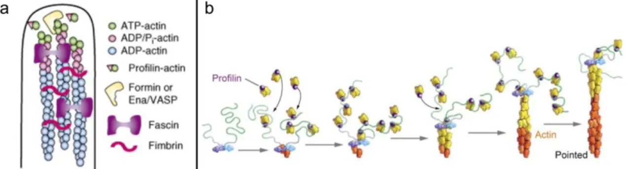
The actin network is organized as parallel or anti-parallel alignment of microfilaments creating linear patterns (Fig. 6a). It is generally achieved either by proteins having two actin-binding domains or by complexes of proteins having each one actin-binding site (Winder et al., 2005). The architecture of such proteins or complexes determines the stiffness of the bundles. Tight actin bundles are obtained through fascin or fimbrin proteins found in microvilli, filopodia, or invapodia (Revenu et al., 2004), looser structure can be achieved via the -actinin present in stress fibers (Pellegrin et al., 2007). Such organization is supported by formins (Faix et al., 2006; Pollard, 2007) (Fig. 6b). The protein consists of FH2 donut-shaped dimer domains interacting with the barded end of a filament and FH1 domains that tether multiple profilin-actin complexes near the barbed end. It considerably accelerates monomer incorporation such that more than hundred actin units can be polymerized in 1s. Because FH2 domains remain attached to the barded end, it also prevents the filament from capping.
Figure 6: Bundle of actin. (a) Representation of filament bundles in filopodia. Formins, present at the tips, drive filament elongation. Filaments are bundled by fimbrin and fascin. From (Lambrechts et al., 2008) (b) Formins initiate the nucleation and assists the elongation of the actin filament at the barbed end. They bind profilin-actin monomer such that polymerization occurs and remain associated to the growing (+) end of the filament. From (Pollard, 2007).
I.3.b. Branched networks
Filaments are arranged in Y or X-junctions that create a meshed structure usually consisting of short filaments (Revenu et al., 2004) (Fig. 7a). It is mediated by proteins or complexes containing multiple actin-binding domains that are in general separated by a long and/or flexible spacer region which allows a more perpendicular arrangement of the filaments (Winder et al., 2005). A frequently involved protein is the filamin (X-junctions) but the main crosslinker is the Arp2/3 complex which is in addition an actin nucleation complex (Pollard, 2007). Lamellipodia, lamella, ruffles and the actin cortex are organized in branched networks (Chhabra et al., 2007). The Arp2/3 complex consists of 7 subunits and binds to already existing actin filaments and initiates the polymerization of another filament (Pollard, 2007). It anchors the pointed end of the daughter filament to the mother filament and the free barded end of the daughter filament grows away from the complex (Fig. 7b). The regulation of this network construction is taken in charge by the WASP (Wiskott–Aldrich syndrome protein) or WAVE (WASP-family verprolin-homologous protein) proteins by bringing together an actin monomer and the Arp2/3 complex which mimic the actin nuclei
General background - Chapter 1: The cytoskeleton - I. The actin cytoskeleton
necessary for the elongation to occur. A high capping activity is associated with this process to prevent extensive elongation (Schafer, 2004a).
Figure 7: Branched network of actin. (a) Representation of branched elongating actin filaments in a lamellipodium. The Arp2/3-complex initiates the formation of new branches on existing filaments. Cofilin increases filament turnover by severing existing filaments and by inducing depolymerization or by creating new free barbed-ends. From (Lambrechts et al., 2008) (b) Actin monomer first bind to the WASP protein, which is a member of the nucleation-promoting factors. WASP recruits then the Arp2/3 complex which gets activated by linking to the side of an actin filament. The complex mimics an actin nucleus and a new filament grows. From (Pollard et al., 2009).
I.4. Actin localizations and functions
Although actin filaments are dispersed throughout the cell, they are most highly concentrated at the periphery of the cell, just beneath the plasma membrane. The spatiotemporal control over the polymerization state of the microfilaments together with interactions with appropriate molecular structures, establish the base of the so called motility. In response to signaling, the actin cytoskeleton is engaged in many cellular processes such as cell adhesions to the extracellular matrix or to other cells, cell morphology, cell migration, endocytosis, intracellular trafficking (Pollard et al., 2009; Olson et al., 2010)… (Fig. 8). The contribution of the actin network in these cell processes can be as structural elements or as scaffolds for signal transduction.
Figure 8: Actin localization and functions. (a) Actin found at the plasma membrane take part in many mechanisms such as cell migration, connection to substrates or other cells, internalization of particles via endocytosis, intracellular trafficking… It is also used for structural functions to maintain organelles at the appropriate positions. From (Olson et al., 2010) (b) Cell migration process. From (Ladoux et al., 2012)
General background - Chapter 1: The cytoskeleton - I. The actin cytoskeleton
I.4.a. Cell adhesion to the substrate
The connection of cells to the extracellular matrix is mediated by a complex assembly of proteins involving the integrin receptors (Fig. 9) (Brakebusch et al., 2003; Ladoux et al., 2012). Ligand (collagen, fibronectin) binding to integrins leads to its clustering and the recruitment of actin filaments and signaling proteins that generates focal adhesions (FAs). Actin filaments are organized into stress fibers which, due to the myosin II, possess contractile properties (Pellegrin et al., 2007). The combination of the contractile machinery and mechanosensitive proteins present at several level of the cell (signaling, membrane, cytoskeleton and associated proteins) creates a mechanoresponsive network. This network participates in cell shape, cell polarity, and cell migration.
Figure 9: Schematic representation of a focal adhesion. Mediated by integrins, actin filaments are organized into contractile fibers that anchor the cell on the substrate. From (Ladoux et al., 2012)
I.4.b. Cell migration
“Cell migration is a highly orchestrated multistep process” (Ridley, 2006; Ladoux et al., 2012). Before cell migration occurs, a polarized morphology must be created. At the cell front, substantial protrusions of the membrane are generated via the polymerization of actin (lamellipodia, filopodia). Lamellipodium provides a tight brush-like structure able to push along a broad length of plasma membrane whereas the filopodium serves more as sensor and explores the local environment. At the leading edge of the lamellipodium, the cell generates focal adhesions to anchor the protrusion and drag the cell body. To move forward, the cell retracts its trailing edge by combining acto-myosin contractility of the stress fibers and disassembly of adhesions at the cell back (Fig. 8b).
I.4.c. Cell-cell adhesion
The junction between cells is organized by assemblies of proteins among which are cadherins (Kobielak et al., 2004; Cavey et al., 2009). The initial cell contact is an opportunistic event consequent to the
General background - Chapter 1: The cytoskeleton - I. The actin cytoskeleton
exploratory activities of branched actin protrusions (lamellipodia, ruffles). It induces cadherin clustering which triggers actin rearrangement through the recruitment of signaling proteins (Fig. 10). On the one hand, new branched networks drive contact expansion and on the other hand -actinin linked cables formation stabilizes the junctions by connecting actin networks of both cells.
Figure 10: Model of cell-cell adhesion formation. Initial cell-cell contacts are formed by exploratory filopodia or lamellipodia. Cadherin is recruited to anchor actin filaments of both cells. From (Kobielak et al., 2004)
I.4.d. Actin and endocytosis
Rather than being a passive barrier that must be disassembled for vesicle internalization, actin can be considered to play an active role in assisting and even driving certain stages of the endocytic process. The finding that actin cytoskeleton contribute to the endocytic process derived from the use of pharmacological drugs altering actin turnover. In mammalian cells, actin disruption inhibits clathrin-mediated uptake and the formation of coated vesicles (Lamaze et al., 1997; Fujimoto et al., 2000). However, the blocking of endocytosis seems to be partial in mammalian cells whereas it is complete in yeast (Ayscough et al., 1997; Ayscough, 2000). Actin polymerization produces the mechanical forces required to invaginate membrane regions, mediate vesicle scission, or push newly formed vesicles away from the plasma membrane (Qualmann et al., 2002). The detailed functions of the actin cytoskeleton in endocytosis are described in chapter 2 of the general background part.
I.4.e. Actin rocketing
Actin rocketing is form of actin-related motility used by organelles. It was first discovered while studying the intracellular motions of the parasitic bacteria Listeria where a similar coupling of bacteria movement and actin polymerization was observed (Theriot et al., 1992). A burst of actin polymerization in the form of comet tail at the edge of intracellular particles pushes them in one direction. Listeria recruits the motility machinery from the cytoplasm of infected cells to facilitate the infection of surrounding cells and hijack the immune surveillance. The actin polymerization-based transport gradually propel the particles at 0.1-0.5 µm/s (Pantaloni et al., 2001). Comet tails have been also observed at the surface of viruses (Frischknecht et al., 1999), endosomes (Merrifield et al., 1999), and endogenous vesicles (Rozelle et al., 2000).
General background - Chapter 1: The cytoskeleton - I. The actin cytoskeleton
I.4.f. Intracellular transport
Actin filaments can be used as rails to transport diverse cargos or they can be used as frame to anchor organelles. A class of proteins called myosins can interact with both the filament and the cargo. Some have in addition the ability to travel along the filaments in one direction. The myosin superfamily comprises 18 classes (Berg et al., 2001). Humans possess about 40 myosin genes, among which about 25 are unconventional (greatly differ from myosin II) and spread over at least 11 classes. Myosins are composed of three modules, the head, neck and tail domains. They associate with the cargo through the tail region and interact with the microfilament via the head regions, or motor domains, which use ATP to drive movement along actin filaments. During ATP hydrolysis cycles, the motor domain undergoes strong filament binding (ADP-bound or no nucleotide), conformational change, filament unbinding (ATP- or ADP-Pi-bound), conformational relaxation, filament rebinding and so forth. Coordinated ATPase cycles alternately on each motor domain induce the protein to mechanically move step-wise in one direction. The motor head define the direction of the movement while the tail determine the identity of cargo with which it interacts and therefore the function and localization of the protein. Myosins differ in the time spent in the strongly bound state and in the rate at which they consume ATP. The neck or middle domain acts as a lever arm that amplify the conformational changes of the motor domain (Geeves, 2002). Myosins step sizes directly depend on the length of the lever arm and can generate large movements such as the 36-nm step taken by myosin V and myosin VI. In most myosins, calmodulin (or calmodulin-like) light chains bind at the region of the lever arm and control its rigidity (Wolenski, 1995).
While conventional myosin II is almost exclusively involved in producing muscle contraction, unconventional myosins have intense activities at the cell cortex, mediate organelle transport, and regulate signal transduction (Hartman et al., 2012) (Fig. 11a). Myosins I is the second largest class after myosin II, and seem to be key partner for the functions such as motility, endocytosis, and exocytosis (Mermall et al., 1998). Relevant classes for organelles transport are the myosins classes V and VI. They share several properties such as homodimerization providing two coordinated motor heads and therefore processivity. Motors alternate binding and release from actin in a hand-over-hand fashion and both myosin classes have step size of on average 36 nm. Even though myosin VI has a short level arm that should span over about 10 nm, it has a larger angle swing (Spudich et al., 2010) (Fig. 11b). This particularity is a unique feature of myosin VI, but what is the most exceptional property of myosin VI is that it produces force that acts toward the minus end of actin filaments, which is the opposite direction to all other characterized myosins (Wells et al., 1999). Myosin VI is found associated with endocytic vesicles, the Golgi apparatus and membrane ruffles (Fig. 11c) (Sweeney et al., 2007). It contributes to several phases of the endocytic process including the formation and movement of vesicles. It interacts with coated vesicles via adapters such as Dab2 and Sap97 and with uncoated vesicles through for instance GipC.
General background - Chapter 1: The cytoskeleton - II. The microtubules
Figure 11: Myosin motors. (a) Myosins can bind diverse cargo such as lipids, proteins embedded in membrane, actin, protein complexes or RNA complexes. (b) Myosin V and myosin VI structures and their respective walking direction. (c) Membranes and myosins. Myosin can be associated with nearly all cellular membranes where it act as anchor or motor for transport. (a) and (c) from (Hartman et al., 2012) and (b) from (Sweeney et al., 2010)
II. The microtubules
Tubulin is a 55 kDa globular protein of 450 amino-acids. The common members of the tubulin family are -tubulin and -tubulin which have about 50% sequence identity (Burns, 1991). Both have a nucleotide (GTP) binding site but the GTP bound to the -tubulin monomer is physically trapped and never hydrolyzed or exchanged. - and -tubulin assemble to form a heterodimer subunit of 8 nm that is the base of the microtubules (Fig. 12a). Tubulin heterodimer polymerizes in linear protofilaments that associate laterally (- and - interactions) and close to form a 25 nm diameter hollow cylindrical lattice (Fig. 12b). In vivo, predominately 13 protofilaments are necessary to build the microtubule wall. The lateral bonds between monomers in adjacent protofilaments deviate from the horizontal with a 10° pitch, thereby forming a helical path that travels up the microtubule lattice. This path is called a 3-start helix because each turn of the helix spans three tubulin monomers, and three such parallel helices must be started to cover the entire surface of the microtubule lattice. This results in the microtubule having a seam where monomers are laterally adjacent to monomers. Due to the head-to-tail association of heterodimers in the
General background - Chapter 1: The cytoskeleton - II. The microtubules
protofilaments, microtubules have a structural polarity. The (+) end is the side where -tubulin are exposed and the (-) end is the side where -tubulin are exposed (Amos et al., 1974).
Figure 12: Microtubule structure and polymerization. (a) - and -tubulin bind in heterodimers which assemble to form protofilaments. (b) The hollow cylindrical microtubule comprises typically 13 parallel protofilaments that generate a lattice seam (red dashed line). (c) Dynamic instability of microtubules. Assembly and disassembly of microtubules is driven by the binding, hydrolysis and exchange of GTP on the -tubulin. GTP-tubulin adds on the growing microtubule. Its hydrolysis generates GDP-tubulin curved structure that, when occurring at the microtubule end, induces shrinkage of the microtubule by depolymerization (microtubule catastrophe). The presence of GTP-tubulin (by polymerization or from remnant GTP-tubulin the lattice) maintains a straight structure favoring GTP-growing or rescue. From (Akhmanova et al., 2008).
II.1. Tubulin polymerization
Microtubule polymerization occurs in two phases, nucleation and elongation. The nucleation starts by the spontaneous assembly of - and -tubulin monomers in very stable heterodimers. The assembly of 6 to 12 heterodimers is necessary to start a microtubule. The nucleation is a rather slow step and depends on the concentration of GTP-tubulin (Job et al., 2003). Although the microtubule lattice can be formally described as helical, it does not assemble by a classical helical polymerization. Microtubules appear to grow as a bidimensional sheet of interacting protofilaments that later close into a tube (Fig. 12c). Like the actin polymerization, the microtubule assembly has treadmilling dynamics. The structural polarity of the microtubules induces kinetics polarity where the (+) end grows faster than the (-) end (Desai et al., 1997). During polymerization, GTP bound to -tubulin (at the exchangeable or E-site) is hydrolyzed. The resulting GDP does not exchange while -tubulin remains in the polymer. Upon depolymerization, the released tubulin subunits can exchange GDP for GTP and undergo another round of polymerization. GTP hydrolysis