HAL Id: tel-02274693
https://tel.archives-ouvertes.fr/tel-02274693
Submitted on 30 Aug 2019HAL is a multi-disciplinary open access
archive for the deposit and dissemination of sci-entific research documents, whether they are pub-lished or not. The documents may come from teaching and research institutions in France or abroad, or from public or private research centers.
L’archive ouverte pluridisciplinaire HAL, est destinée au dépôt et à la diffusion de documents scientifiques de niveau recherche, publiés ou non, émanant des établissements d’enseignement et de recherche français ou étrangers, des laboratoires publics ou privés.
Opto-Electrochemical Methods for Imaging the
Reactivity of Individual Nanoparticles
Vitor Brasiliense
A thesis presented for the degree of Doctor of Philosophy Supervised by Fréderic Kanoufi and Gilles Tessier
Speciality: Electrochemistry
Thesis presented and publicly defended in Paris on the 11th of December 2017
President of the Jury Pr. Isabelle Ledoux ENS Paris Saclay
Reviewers Pr. Julie Macpherson University of Warwick
Dr. Sébastien Bidault Institut Langevin ESPCI
Examiners Pr. Michel Orrit Leiden University
Pr. Marc Robert Université Paris Diderot
Supervisor Dr. Frédéric Kanoufi Université Paris Diderot
Co-supervisor Pr. Gilles Tessier Université Paris Descartes
ITODYS - Interfaces, Traitments, Organisation et Dynamique des Systèmes ED388 - Ecole Doctorale de Chimie Physique et Chimie Analytique de Paris Centre
Contents
Acknowledgements vii Abstract ix General Introduction 1 1 Introduction 3 1.1 One vs many . . . 41.1.1 Seeing the very Small : Looking for Resolution . . . 4
1.1.2 Electrochemical Detection of Individual Objects . . . 6
1.1.3 Nano-Optics and Plasmonics . . . 14
1.1.4 Fluorescence . . . 15
1.1.5 Return to Microscopy : Super-localization and Super-resolution . . 16
1.2 EC Imaging and Optical EC . . . 18
1.3 Optics-EC for Individual Objects . . . 19
1.3.1 Using optics to replace electrochemical detection . . . 20
1.3.2 Electrochemically assisted optics . . . 23
1.3.3 EC generated optical SP phenomena . . . 25
1.4 What this thesis is all about. . . 27
2 Single Ag NPs Electrochemistry 29 2.1 Why silver? . . . 30
2.1.1 Silver NP reactivity through electrochemical impact experiments . . 31
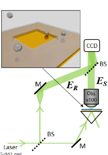
2.2 Coupling 3D Microscopy to Electrochemical impact Experiments . . . 33
2.2.1 Holography . . . 33
2.2.2 Transparent electrodes in microfluidic cells . . . 36
2.2.3 Prep. to Holo+EC . . . 37
2.3 Correlated Impacts . . . 39
2.4 Adding Complexity: precipitating agents . . . 42
2.4.1 Monitoring Electrochemical Impacts in KSCN . . . 43
2.4.2 Spectroscopic insight . . . 45
2.4.3 Quantitative Analysis of the dissolution rates . . . 49
3.2.2 Nanoparticle building block synthesis . . . 62
3.2.3 Laser Assisted Supracrystal Formation . . . 64
3.2.4 Large Scale dynamics: Characterizing the trapping forces . . . 68
3.2.5 Small Scale Dynamics: Measuring D and v NAgg by NAgg . . . 69
3.2.6 Manipulating the Crystal . . . 71
3.2.7 Gravitational Forces (or why does the particles motion seems 2D?) 71 3.2.8 Optical Forces . . . 73
3.2.9 Thermal Effects . . . 74
3.2.10 Mechanism Discussion . . . 78
3.2.11 Conclusion: All-in-one Thermoresponsive plasmonic substrates . . 79
3.3 Another Phoretic Phenomena: Diffusiophoresis . . . 81
3.3.1 Experiment Description . . . 81
3.3.2 Conclusion: SPT in migration . . . 83
3.4 Monitoring Particle Growth through Single particle Tracking . . . 85
3.4.1 Electrosynthesis Principle . . . 85
3.4.2 Extracting r(t) from MSD curves . . . 87
3.4.3 Growth Model . . . 90
3.4.4 Monte Carlo Simulations . . . 90
3.4.5 Results . . . 91
3.5 Conclusion . . . 92
4 Single Particles on Nanoelectrodes 95 4.1 Transition Metal Oxides for Water Splitting . . . 96
4.2 Carbon nanoelectrodes preparation . . . 97
4.3 Monitoring Setup . . . 99
4.4 Single NP Electrosynthesis and Electrochemistry . . . 100
4.5 Particle Deposition routes . . . 101
4.6 Particle Optical Detection and Sizing . . . 103
4.6.1 Using Spectroscopic information for particle sizing . . . 104
4.7 Translating optical signal into electrochemical information . . . 108
CONTENTS v
4.7.2 Application for the Co(II) → Co(III) Transformation . . . 109
4.8 Cathodic and Anodic Electrodeposition and catalysis . . . 109
4.8.1 Cathodic Phenomena . . . 112
4.8.2 Anodic Phenomena . . . 114
4.8.3 Limitations of the Optical approach . . . 116
4.8.4 Scaling Laws and the influence of Rp . . . 118
4.9 More on stochasticty: Morphology and Catalytical Activity . . . 119
4.10 Co-Ni oxide NPs . . . 121
4.11 Conclusion . . . 123
Conclusion 125 A Experimental Methods 129 A.1 Holography . . . 129
A.1.1 Principles and Earlier Versions . . . 131
A.1.2 Further Developments and Digital Holography . . . 133
A.1.3 Optical Setup: Dark Field Digital Off-axis holographic microscopy . 135 A.1.4 From a hologram to a reconstructed volume: The holographic pro-cedure . . . 137
A.2 Superlocalization in the context of holography . . . 142
A.2.1 Superlocalization Principle . . . 142
A.2.2 Localization Limits . . . 142
A.3 Plasmonics and Nanoparticles Spectroscopy . . . 143
A.3.1 Spectral Acquisition Setup . . . 144
A.3.2 Processing of the spectra . . . 144
A.4 MSD and size estimation from Brownian Motion . . . 145
A.4.1 Time Average MSD and precision . . . 147
A.5 Monte Carlo simulation for Diffusion Models . . . 148
A.6 Lithography and Electrode Fabrication . . . 149
A.6.1 Gold Microelectrode Array . . . 150
A.6.2 ITO microelectrodes . . . 151
A.7 Carbon Nanoelectrodes fabrication procedure . . . 154
A.7.1 Pulling the capillaries . . . 154
A.7.2 Pyrolysis . . . 155
Acknowledgements
I always struggle to write Acknowledgments. I wouldn’t dare to say it is the hardest part of the thesis, but sure as hell it isn’t the easiest. Just like an ensemble response is the collection of individual expressions, this thesis would not have been possible without countless interactions with the people around me over not only these last three years, but over my whole life. Although it would be impossible to mention everyone, I will start with the people that first encouraged me to study, my family: Mom, Lucas, you are amazing examples on how to be a great human being. It would not been possible to have come this far without you. I was extremely happy to see you at my defense - It was an important moment for me, and you are at least partially responsible for me being there as well. Muito Obrigado.
I’d also like to thank all my friends from the ESPCI, with whom not only have I learned how to speak French, but who were also my first interface with France when I first arrived in Paris, seven years ago. After all those travels, weekends, soirées with terrible music... Thomas Barrès, Oriane Tapparo, Gabriel Cournelle, Edouard Lees, François Bargain, Aurélien Duval, Mélanie Jacquet, Guillaume Chatté, Marc Gesnik, Sarah Christoph, un grand merci. Talking about my first years in France, I also would like to take the op-portunity to thank Victor Hugo and Catarina Macedo da Silva (ou da Silva Macedo). Besides the friendship we shared over these six years, I would never be able to prepare the Feijoadas I served for my thesis reception without all those cooking evenings in each others houses!
I would also like to sincerely thank the wall color designer of Paris Diderot, that gave me matter to complain over these three years (who thought fluorescent-green was a good idea!?? ). Seriously speaking, I am grateful for all the dear friends I made in Paris VII (now all doctors - we are getting old!), Alexandra Tibaldi, Andrés Lombana, Jonathan Fouineau, Yong Ai, Célia Achaibou (Félicitations pour ton mariage! ). All the good moments shared in and outside the lab, Bootlager’s evenings, Asian Restaurants, "Rhum Arrangés", poorly organized camping trips made the PhD period more than fun and will not be forgotten!
Last, but not the least, I would like to thank my dear Lydia Merakeb, whom I only met at the end of my thesis, and that constitutes the biggest and happiest surprise of this year. Meeting you was la cerise sur le gâteau of a great year!
Angele, Jane Stockmann (livin’ the dream! ), Jean Pinson, Jêrome Médard, Mihn Chau Nguyen, Jean-François Lemineur, merci beaucoup!
In particular I would like to thank two persons who were pivotal for the great atmo-sphere in both labs. Pascal Berto in Paris Descartes, for being a great person to work with, while teaching me so much about practical aspects of optics and lab organization (thanks for putting up with my bordel in the lab :P). Jean-Marc Noël, for the countless discussions about electrochemistry, SECM experiments, tennis matches and especially for the improvised apéros and ebriety stories (Ablon, A-B-L-O-N ).
I would also like to thank Catherine Combellas, who first lured me into the Elec-trochemistry group of the ESPCI, and who constitutes the best example I am aware of on how to manage a research group. Not to mention advices on healthy nutrition and organization skills (which should be read "on how not to eat junk and keep my desk presentable").
Finally, I would like to thank my supervisors, from whom I learned so much not only about Science but about life in research.
Gilles Tessier, whose co-direction was pivotal for the success of my experiments. It was with great competence and optimism that you help me tackle from quotidian problems to big Science discussions. Thank you for the guidance!
Frédéric Kanoufi, from whom have I not only learned electrochemistry, but also the most fundamental aspects of what being a researcher is about. I feel I could not have been better guided over these three years. Not to mention being the nicest person to work with. A big, sincere, thank you!
Opto-Electrochemical Methods for Imaging the
Reactivity of Individual Nanoparticles
Vitor Brasiliense
Abstract
A number of coupled optical and electrochemical single particle techniques are em-ployed for investigating a variety of chemical systems at the level of individual objects. On the optical side, holography and visible spectroscopy are imbued with superlocaliza-tion principles pushing the applicability of these techniques down to sub-diffracsuperlocaliza-tion levels. Nanoelectrochemical techniques such as stochastic impacts and nanoelectrodes are used to complement this information, providing a much more complete characterization of the phenomena.
It is shown that this dual optical and electrochemical single particle characterization is actually crucial to understand complex nano chemical systems in loco. Starting from model reactions, such as Ag oxidation, the complexity of the studied phenomena and systems is progressively increased, as light is shed on transport phenomena, aggregation, as well as redox transformations and catalysis on complicated materials such as ill-defined transition metal (cobalt) oxides.
Keywords: Nanoparticles, Opto-Electrochemical Methods, Holography, Nanoelectro-chemistry, Catalysis
pousser l’applicabilité de ces techniques au-delà de la limité imposée par la diffraction. Des techniques nanoélectrochimiques, comme les impacts stochastiques et l’utilisation de nanoelectrodes, complètent cette étude en renseignant sur la réactivité et sur les étapes de transfert d’électrons. Ces études couplées caractérisent ainsi les phénomènes chimiques de façon bien plus complète.
Il est montré que cette caractérisation à la fois chimique et optique est en fait essen-tielle pour pouvoir comprendre le fonctionnement des systèmes nano chimiques in loco. En démarrant par des réactions modèle, comme l’oxydation de l’argent, la complexité des systèmes étudiés est progressivement augmentée, éclairant des phénomènes de transport, d’agrégation, ainsi que des transformation redox et de catalyse sur des matériaux com-plexes et mal définis tel que les oxydes de métaux de transition (cobalt).
Mots Clés: Nanoparticules, Méthodes Opto-électrochimiques, holographie, Nanoélec-trochimie, catalyse
General Introduction: Coupling
Electrochemistry and Optical
Techniques
Anyone who has ever been to a (decent) rock concert should be familiar with the strange fact that a crowd seems to sing in perfect harmony. Although the vast majority of the concert attendees are not professional singers, the overall melody sounds perfectly in tune. If a recorder was to be placed under each persons mouth, a large number of, out of tune, distorted, slightly hysterical tracks would be heard. Yet when added up, all these voices seem to complement each other, forming an ensemble effect that is beyond any individual performance.
But... What if the information contained in those dissonant and mutually average was the actual key to discovering something new? What if we could select the best voices among the crowd? More than that, one must also come to the realization that there is more in a musician than a mere beautiful voice. Other things must be considered, how strong is his/her personality? How committed is (s)he to actually taking the time and trouble to getting there? And how, from a glance into a crowd could we possibly try to answer any of these questions, without actually getting to know each one of those persons individually.
I am of course no longer talking about people or concerts, but I find the analogy quite useful in describing the goal of my three years work, summarized in this thesis : To individually in situ address small objects reactivity. Instead of looking at the response of chemical entities through a highly convoluted ensemble signal, strategies are developed to give voice to every one of the single particles. This is really what my thesis is all about: describing chemical reactions at the scales of the individual entities involved. Over these pages, I want to tell chemical stories through the lips of the individual actors participating in it. And the actors we chose to focus on are nanoparticles.
But, again, just like people, the behavior of individual particles is hard to grasp through a single point of view. To meet someone only at work or only in leisure time gives us an incomplete understanding of that person’s behavior. Likewise, by scaling down chemical reactions, we need to look at a phenomenon from more than one angle.
The text starts with a brief state-of-the-art review, on which the most relevant ex-amples of interplay between Electrochemistry and Optics are summarized, and the latest efforts towards nanoscale characterizations of individual entities are described.
Chapter 2 starts by concept proving the complementarity of electrochemical and op-tical methods by studying a model system: silver nanoparticles oxidation. It is somehow iconic that the model system chosen was actually one of the first systems to be studied for its photochemistry, as silver photo-(electro)chemistry played a gigantic role on the de-velopment of photography [1]. In spite of a century-long research history, reaction details on the nanoscale are still unresolved. That is where top notch optical techniques come to aid. Super-localization holography and single particle visible spectrometry are used to reveal nanoparticle reactivity dynamics, or what happens when the particle is in the close vicinity of the electrode (a few nanometers apart).
However, as very well argued in the (now classical) P. W. Anderson’s 1972 Science article "More is Different" [2], knowledge of a system’s fundamental laws does not neces-sarily imply the ability to predict the way a system will behave at higher scale. Collective behavior is sometimes entirely unpredictable and the description of a system needs to be addressed with generalizations and laws proper to a given scale. This is the object of Chapter 3. Still using holography, together with analytical and numerical methods, we study aggregation, growth and transport properties of nanoparticles, putting forth phoretic phenomena such as diffusio- and thermophoresis.
Finally, Chapter 4 introduces a new strategy to study particles individually, using nanoelectrodes to (electro)synthesize them in situ. By attaching the particle to the elec-trode it becomes much easier to analyze situations where the particle grows, shrinks, or transforms. Moreover, a still particle can be cycled many times, allowing extensive electro-chemical information to be derived. Further interplay between optics and electrochemistry is introduced, as optical monitoring is used not only to detect and size particles during the deposition, but also to deduce useful chemical information unavailable otherwise, in form of optical cyclic voltammograms.
Detailed technical discussion about the setups, calculation methods and fabrication protocols are separated from the main text and form an extra chapter A.
Chapter 1
Introduction: Coupling
Electrochemistry and Optical
Techniques
Chemistry, often called the central science, is a discipline of multiple scales. From the first chemical courses taught in college, chemistry is presented as a calm ocean seen from a distance. Small waves and fluctuations merge together in patterns impossible to discern, forming an harmonious and smooth pattern. Like tides, reactions happen in a regular, predictable way, perfectly described by well defined rate constants and concentrations. This macroscopic vision - familiar to everyday events and experiments - is in sharp contrast with the microscopic view of chemical systems, where violent collisions deform molecules to the point of ripping pieces apart and fusing others together. Chemical reactions on the microscale are generally the result of imperfect, chance-driven events.
The bridge between these two familiar but contrasted visions is still uncharted ter-ritory, little explored. The apparent calmness and regularity of macroscopic systems ensemble views stems from the fact that our observations are actually averages over many simultaneous events (seriously many... keeping in mind that 1 mole = 1023). As we reduce
the number of simultaneous events, the smooth curves leave place for fluctuations, and chemistry appears less and less tamed behind mean field notions such as rate constants and concentrations.
Over the past few years, advances in analytical techniques allowed a much deeper look into objects in the nano- to meso-scales. As we became able to observe chemical objects on a one-by-one basis, access to a much different world is granted, where events and entities have a much stronger personality. Stochastic effects and fluctuations such as Brownian motion, play an increasingly important role, drawing a world open to imperfection and individuality of behavior.
This chapter describes the different paths into this stormy reality - it briefly reviews the advances made over the past decades, which led towards different ways of achieving
ual nanoparticle often displays enormous spatial variability in reactivity [3, 4]. All these observations relate to the fact that in such small particles, surfaces atoms represent a large proportion of the total material. Defects such as grain boundaries, vacancies, sur-face orientation and roughness are hard to control and seem to have a strong impact on reactivity and catalytic properties [5]. "God made the bulk, the surface was invented by the devil", as very well synthesized by quantum theory pioneer Wolfgang Pauli.
Substantial efforts have therefore been made to clarify the relation between a particle size/shape and activity, but this is a tough quest. Even the most monodisperse synthesis is almost never atomic precise, yielding differences in reactivity from one object to another. For this reason, since the past few decades many analytical techniques have been developed to achieve ultimate spatial resolution: detection of individual objects, such as nanoparticles, molecules and atoms. This section briefly describes how the way towards individual objects studies was paved, and how different routes provide alternative insights into the objects reactivity. Special attention will be given, of course, to the main topics of this text, nanoscale electrochemistry and optics.
1.1.1
Seeing the very Small : Looking for Resolution
If seeing is believing, the refinement of microscopes over the 16th and 17th century con-comitantly with the development of modern Science should be no coincidence. Indeed, the ability to address problems visually was of great importance on the early days of the scientific method[6].
To see very small objects is however a challenging task. Traditional optical microscopy is in general unfit for doing so, since it is fundamentally bound by diffraction, meaning that its resolution is limited by the wavelength of the light (typically 500 nm for visible light). One way around this limitation is to use lower wavelength, more energetic radiation. With this idea came the development of electron microscopy from the 1930’s [7], which indeed lowered the resolution down to a few nanometers and eventually to atomic resolution. Until very recently however, the high energies involved, together with the nonexistence of a good method to prevent electron scattering, relegated electron microscopy methods to a exclusively ex-situ characterization technique status [8, 9].
1.1. ONE VS MANY 5
It was however with the invention of the Scanning Tunneling Microscopy (STM) fifty years later that materials imaging achieved the unprecedented resolution of individual atoms [10]. Indeed, STM represents one of the first examples of direct interactions with matter at the nanoscale, and for that reason some consider STM invention as the founding stone of nanoscience [11]. Although initially limited to very restrictive conditions, requir-ing ultra high vacuum, complicated sample preparation, etc., STM and its many variants (AFM, SNOM, SECM...) quickly evolved and adapted to a wide array of experimental situations. Indeed, today local probe techniques are a standard analytical tool, of daily use for most scientists interested in nanoscale phenomena [11].
Altough impressive, great spatial resolution usually comes at the expense of time resolution (although examples of dynamical local probe studies exist [12, 13, 14]). While able to achieve the ultimate resolution, local probe methods had the inconvenience of being relatively slow, usually unable to properly resolve in time fast events such as chemical reactions. Moreover, the tip is an enormous object (relative to the entities herein studied), therefore it is always possible that it influences the system, changing its properties.
Besides, if a chemical system is to be understood, more chemical-specific information is required. Over the years that followed the invention and popularization of the STM, many other analytical tools were reported to resolve objects individually. To try to extensively describe them all would be a donquichottesque task. Instead we chose to concentrate on the ones more relevant to the subject of this thesis, electrochemistry and optics. Being potentially capable of high temporal resolution, electrochemistry and plasmonics reveal information of chemical nature, thus enabling further insight into a variety of chemical phenomena.
Seeing might be believing, but there is more in a chemical reaction than what meets the eyes.
However, even the best available commercial potentiostats are only able to measure currents of a few fA, corresponding to a few ten thousands electrons per second. Some strategies had then to be developed in order to reliably analyze signals originating from single nano-objects and molecules.
Individual Molecules through Electrochemistry
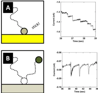
The detection of individual molecules via electrochemical techniques can arise through a number of ways. Although most commonly associated with optical techniques, purely electrochemical techniques also have the potential to detect and analyse single molecules. An electrochemical scanning probe technique, the scanning electrochemical microscope (SECM), for instance, can be used in individual nanoparticles studies, if nanoelectrodes are used to provide enough spatial resolution [15]. Going even further, the SECM has also been used to achieve electrochemical detection of individual molecules [16]. By bringing a slightly recessed nanoelectrode very close to a conductive surface, one can trap a single molecule in a very small confined space between the two electrodes. If then the substrate and electrode are polarized at opposite potentials with respect to the oxidation potential, the trapped molecule will be repeatedly oxidized and reduced, amplifying the signal and allowing single molecule detection. The concept is sketched in Fig.1.1 A. Although making use of a relatively complicated electrode geometry, this example illustrates well the idea of detection by signal amplification. A similar concept was recently addressed using recessed nanoelectrodes built by electrochemical etching of the tip [17].
The same principle of redox cycling amplification was then recently extended to more flexible geometries, using nanolithography techniques to create nanochannels and nanogaps in micro-to-nanofluidic devices [18, 19, 20], shown in Fig.1.1B. In this configu-ration, both the upper and lower electrodes are microelectrodes, therefore the current on both sides can be addressed. The symmetrical response observed when both electrodes are poised at potentials opposite with respect to the redox probe E0 (shown in Fig.1.1C) is then an evidence of the back and forth movement of molecules, and should be propor-tional to the number of molecules between the electrodes. Discrete changes of the current magnitude are then a direct measure of the number of molecules inside the device.
1.1. ONE VS MANY 7
Figure 1.1: Concepts for the electrochemical detection of individual molecules. In A, a recessed electrode approaches an ITO electrode trapping a molecule in a volume of ≈ 1500 nm 3. Ap-plication of a voltage between the electrodes makes the molecule repeatedly oxidize and reduce, generating a detectable current. [16] In (B), a recent revision of the concept, where nanolithog-raphy is used to create a nanochannel of width of a few µm and nanometric height, where the top and bottom walls are conductive surfaces. When the electrodes are poised appropriately, a molecule inside the channel repeatedly oxidizes and reduces, generating symmetric currents on the working and counter electrodes, as respectively shown in red and black (C)[18].
Individual nanoparticles using Electrochemistry
Signal amplification can also be achieved through catalysis, making creative use of the fact that one catalyst molecule generates many analytes, exchanging many electrons. This concept can be used to study nanoparticles individually. Suppose a certain redox reaction, say O + e → R, that for a given potential will take place on a catalytic nanoparticle but not on the electrode. When the NP collides with the inactive electrode, it is polarized at the same potential as the electrode. The catalytic reaction will take place, but it will be limited to the NP surface, as the electrode is inactive. For all practical purposes, the NP will act as a nanoelectrode, with respect to this reaction. Since the reaction is catalytic, the current will be controlled by the reactant transport, rather than by the amount of catalytic material (See Fig.1.2 left). Ultimately the reactant on the particle surface will be completely consumed, and the current will be limited by diffusional transport of the ana-lyte to the NP surface. In this ideal case, a current of magnitude iss= 4π(ln2)n0F DCr0
is to be expected [21], where F is the Faraday constant, D the diffusion coefficient, r0 the
NP radius and C the bulk reactant concentration. The success of the strategy depends on having a clean background current, i.e., an inactive electrode [22]. In order to avoid capacitive current, it is also desirable to shrink the electrode size down to the micro- or
Figure 1.2: Typical expected single nanoparticle electrochemical responses in the case where the particles (A) stick to the electrode and remain active, and (B) eventually deadsorb and lose contact. Current-time traces adapted from [23].
It is also possible for the particle not to stick permanently to the surface, and only weakly adsorb on the electrode surface. In this case a "blip" response is expected. This is sketched in Fig.1.2 B, where each blip corresponds to the arrival of one IrOx NP catalyst (for the oxygen evolution reaction) over a Pt UME.
Blip responses are also observed in the case of deactivation upon collision with the electrode. This could happen for a number of reasons. For example, during catalysis, very reactive intermediates can be generated, which poison the particle surface, stopping the reaction. It could also happen as a result of the interaction of the particle with the electrode material. For instance, the latter was explored in the case of Hg UME to improve the reproductivity of the spikes [24, 25]. This is sketched in Fig.1.3.
1.1. ONE VS MANY 9
This strategy has proved itself quite useful to study a number of catalytic systems, including:
• hydrazine oxidation on Pt [26] in a variety of situations, such as aggregation [27, 28], electrode modification [29], poisoning [25], etc.
• proton reduction on Pt [21]
• oxygen evolution, hydrogen evolution and oxygen reduction on metal and metal oxide nanoparticles [30, 31, 32, 33, 34]
In particular, to address particles individually has helped reveal the profound heterogene-ity taking place in these catalytic systems at the nanoscale [22].
Destructive Individual NP Impacts
Although catalytic reactions do provide large signal amplifications, enhancing the signal-to-noise ratio, modern instrumentation also allows study of NP direct reactivity, in the so called destructive impacts. The concept is similar to catalytic impacts: an inert electrode is poised at a potential where the particles are not stable. The collision of a NP with the electrode then triggers a redox reaction which transforms the particle and yields a measurable electrochemical peak. Since the particles hit the surface one at a time, this transient current contains individual NP information. Instead of reaction promoters, NPs take up the role of main analytes.
This opens up the way to studies of nanoparticles direct oxidation, reduction, disso-lution and phase transformation, leading to an evodisso-lution of the way the technique was perceived by the electrochemical community. More than an asset for fundamental studies at individual particles, impact experiments became an useful chemical-selective charac-terization and diagnosis tool.
Reactivity from peak shape analysis: A lot of information about the particle reac-tivity can be gathered from impact experiments. Analysis of the peak can reveal much relevant information about the NP stability and reactivity. For example, from the dura-tion of the peak, we can draw kinetic informadura-tion: how high is the peak? How long does it last? Likewise, the potential at which the experiment is performed gives insight into the thermodynamics of the system : at which potential do we start to see peaks? Does the shape change with potential? [37]
NP intrinsic properties Intrinsic properties can be derived from a destructive elec-trochemical impact. For instance, if the particle reaction is total, its volume V can be obtained from the application of Faraday’s law to the integration of the peak (exchanged charge, Q), using the particle density ρ and molar mass M (F is the Faraday constant and z is the number of exchanged electrons per reacted molecule):
Figure 1.4: (A) Sketch of the Destructive Individual NP impacts detection strategy, where the particle is oxidized upon contact with the polarized electrode. (B) and (C) : The current time trace reveals oxidative spikes whose current magnitude, duration and overall charge help unveil mechanistic information about the nanoparticle reaction. In particular, the charges can be used to deduce the particle size, and therefore the agglomeration state of the NP solution can be addressed, shown in (D). Adapted from [35, 36]
1.1. ONE VS MANY 11
V = QM zF ρ
Using this idea, the particles size distribution can be obtained, and thus be used to address the aggregation state of a NP colloidal solution [38]. More than simply addressing the size distribution, core-shell geometries can be characterized [39, 40] by setting the potential to first react only the shell material, and later the rest of the particle.
The surface of the particles can also be quantified based on a similar strategy, by tagging the NP with a monolayer of redox reactive compounds, which will react exchanging electrons and leading to spikes whose charge is proportional to the particle surface[41, 42], accordingly to:
A = ATNAQ zF
Where AT is the area occupied by one redox active molecule, and NAAvogadro’s number.
Based on that, more complete experiments can be designed, allowing determination of complex geometries, such as gold nanorods aspect ratios [42].
Concentration from Impact Frequency: By analyzing the frequency of impacts, it is possible to infer the NP concentration. If diffusion transport towards a disk UME of radius a is assumed, the frequency, f , relates to the NP concentration [N P ] and diffusion coefficient DN P by [21, 43, 44] :
f = 4DN P[N P ]a
This expression was validated not only in lab conditions, but in a variety of media, such as seawater [45] and human sweat [46], highlighting the potential utility of the strategy for real field situations.
As far as stochastic impacts are concerned, nanoparticles are perfect prototypes to understand the role of stochasticity in nanosystems. This look at nanoparticles as super molecules, is a recent trend in chemistry and has more or less independently emerged in different fields of nanoscience [22]. This analogy is taken one step forward when plasmon resonances energies are considered, which is the theme of the next section (Sec.1.1.3).
Surface Changing Impacts
For completeness, it should be mentioned that individual nanoparticle impact detection can also be achieved by altering the area of the electrode. For example, the arrival of insulating polystyrene beads over an UME can be detected by reduction of the steady state current. The reaction of a redox mediator is kept in the diffusion limited regime, such that the current is proportional to the electrode area available for the reaction. Arrival
is sketched in (B).
of entities that block the electrode therefore lead to a step-wise reduction of the current, the situation is sketched in Fig.1.5 [47]. These results were later confirmed by optical tracking of fluorescence tagged beads [48].
A similiar concept was recently employed for evaluation of the resistance of single car-bon nanotubes (CNT). A potential difference is applied between two interdigitated comb-like microband electrodes. Since no redox reaction takes place, no meaningful current is recorded. The arrival of conducting nanowires however can short-circuit the electrodes, leading to a discrete enhancement of the current, corresponding to the conductance of one nanowire [49]. From the current/applied voltage relationship, the resistance of single NW can be evaluated.
Other Electrochemical ways of addressing individual entities
As a remainder, it should finally be added that other electrochemical strategies for study-ing sstudy-ingle nanoparticles exist, which are not fully described here due to lack of space.
For example, White’s group recently introduced a Coulter counter setup, allowing not only sizing and counting but also manipulating of individual nanoparticles [50, 51]. Nanoparticles can also be immobilized on nanoelectrodes, either by electrodeposition [52, 14], underpotential deposition [53], or by spearing them [54]. These particle-on-a-stick methods present the advantage of allowing prolonged electrochemical studies, but the data throughput is usually quite low, as only one particle is analyzed per experiment.
Scanning Electrochemical Microscope (SECM) probing can also be used to interrogate nanoobjects individually, either by positioning a nanoelectrode over the object surface and analyzing the reaction products[15], or by analyzing the change in the feedback response [55]. This approach can be taken even further if coupled to other techniques. For instance AFM-SECM force measurements can also be used to probe individual polymer molecules and protein immobilized on the electrode surface [56, 57].
Other forms of electrochemical microscopy, such as the scanning cell electrochemical microscopy (SECCM), can also be used to interrogate very small objects individually.
1.1. ONE VS MANY 13
Figure 1.6: Electrochemical methods for studying individual objects (A) Nanopipettes as Coul-ter CounCoul-ter: the passage of one particle modifies the conductivity of a nanopore leading to a peak, which can be used to size the nanoparticle (Adapted from [50]). (B) AFM-SECM coupled approach for studying the distribution of redox active groups over a single virus (Adapted from [56] (C) SECCM image of the reactivity of a Single Walled carbon nano wire (Adapted from [62]).
Through the use of nanopipettes, the reaction area is reduced to a few hundred squared nanometers, substantially lowering the background current and thus allowing detection of small signals. This setup was used to perform very precise impact experiments [58, 59, 60], as well as to image the reactivity of nanostructures such as carbon nanotubes [61, 62]. Some of these approaches are illustrated in Fig.1.6.
Under certain conditions, the oscillation of the electromagnetic field on specific fre-quencies may trigger oscillations of free or weakly bound electrons (typically those of the conductive band on solids). In a surface, this may activate collective oscillations res-onance, generating a propagating interfacial wave known as Surface Plasmon Polariton Resonance (SPPR). If the conductive surface is small compared to the resonance wave-length - which is the case for nano-sized structures illuminated with visible light, for example - localized surface plasmon resonance (LSPR) modes may be activated. Local-ized plasmons are highly dependent on the local dielectric environment of the particle, as well as on the size and shape of the nanostructure of interest. This allows fine modula-tion of the plasmon band energy, shape and intensity, which makes them specially fit for ultra-sensitive sensors design [67].
Although research on plasmonics was undertaken over the whole 20th century, recent years have witnessed an exponential increase of interest for the field [68], owing to a much more profound understanding of how LSPR modes can be tailored and how LSPR modes of different structures interact with each other.
In great measure, this knowledge was only possible from the development of optical techniques capable of studying nanostructures individually. Precise observation of LSPR shifts and interactions is rarely possible when analyzing NPs ensembles, due to severe broadening of the peaks, blurring the interesting phenomena. Experimental proof of these was only made possible by establishing a structure-LSPR relationship on a particle-by-particle basis.
Other factors crucial for the emergence of the field were : the increase of computational power, in order to perform meaningful simulations; abundant availability of numerical methods (since plasmonics can be well described from Maxwell equations); and the de-velopment of lithographic techniques and chemical strategies to synthesize nanoparticles of varied shapes [69]. Besides, the discovery of powerful surface enhanced spectroscopic methods, fully based on plasmonic effects, was key to motivate an increase of research in the field [70].
Plasmonic nanostructures have a natural ability to act as nano antennas. In doing so they create hotspots that can be used to increase the local intensity of the electromagnetic
1.1. ONE VS MANY 15
field, enhancing spectroscopic signals by orders of magnitude, to the point of allowing single molecules detection [71, 72, 73].
The idea of considering nanoparticles as plasmonic atoms mentioned in Sec.1.1.2 is again found among the plasmonics community, as many analogies exist between atoms and nanoparticles. For instance, the electronic wavefunction of an electron and the electric potential around a metallic nanoparticle vary under similar scaling laws [68]. Moreover, the LSPR energies hybridize in a way analogous to molecular orbitals, allowing analytical prediction of the LSPR energies of complex structures from decomposition in simpler substructures and symmetry considerations [74, 75].
Hotspots can also be used as local sources of heat, and therefore as a tool to finely control temperature at the nanoscale [76]. Modulation of the photo induced thermal response allows design of sensing techniques descending down to the limits of very small single nanoparticles (r ≈ 1nm) and single molecules [77, 78].
Moreover, the signal enhancement around plasmonic structures can also be coupled to scanning probe techniques to assess nano- to molecular scale information about chem-ical systems. This is the principle, for example, of Tip Enhanced Raman Spectroscopy (TERS), which exploit the plasmonic enhancement around an STM/AFM tip and the chemistry specificity of Raman signal to study (electro)chemical systems with molecular resolution [79, 80, 81].
Hot electrons emerging from LSPR are also potentially useful for chemistry, as they are capable of triggering chemical reactions, either by photochemical effects, direct reduction [82, 83], or by local temperature enhancement [84].
1.1.4
Fluorescence
Finally no overview of optical single entity detection methods would be complete without mentioning fluorescence. Fluorescence of single molecules, and specially on cryogenic conditions were among the first single molecules experiments ever performed [85, 86, 87]. I’ll be brief, as the literature on fluorescence methods is too vast to be completely covered here, spanning over many fields such as biophysics studies of diffusion processes, fundamental physics, biochemistry, etc.
Fluorophores allowed the visualization of a myriad of chemical physical processes, especially in biological contexts. Starting from early fluoroscent molecules experiments in cold conditions [85], and particularly intensified since the discovery of green fluorescence protein (GFP), to use individual entities with fluorescent tags is today a common practice in biochemistry and biophysics [88]. With relatively little equipment involved, fluorescence techniques allowed qualitative imaging of intracellular processes, as well as extraction of quantitative data from the tags movement.
Fluorescence was also at the heart of the development of localization and super-resolution techniques, which will be the theme in the next section.
Figure 1.7: Super resolution Principles. (A) Superlocalization Concept, a common step for most superresolution techniques, the PSF is fit by a model function, enhancing the precision on the position estimate. (B) Palm/STORM principles; Stochastic activation of separated emitters that are superlocalized and put together, forming a superresolved image. The inset compares a regular image (a), a PALM image (b) and TEM image of the same structure, superimposed over the PALM image. (C) Principles of STED microscopy, an excitation beam is superimposed to a donut shaped beam that stimulates emission. Only the molecules on the center of the donut emit light by fluorescence. The inset compares the resolution of STED and confocal microscopy. Adapted from [90, 91, 92]
1.1.5
Return to Microscopy : Super-localization and Super-resolution
The ability to individually address single entities was also a key ingredient in pushing the limits of optical imaging further. As in all far-field optical methods, resolution limitations are imposed by diffraction of light. Diffraction and propagation act as a low-pass filter, limiting the resolution to a fraction of the wavelength - typically 0.6λ/N A (NA is the numeric aperture) for incoherent light or twice as much for coherent.
Superresolution microscopy techniques however have recently extended the applicabil-ity of optical techniques down to the nanoscale. Their importance was recognized with the award of the 2014 Chemistry Nobel Prize to super resolution techniques pioneers, Stephan Hell, Eric Betzig and William E. Moerner [89]. Building on these initial seminal works, techniques for stretching visible wavelength optics have been extended and multiplied.
A common ingredient to most superresolution schemes is to identify and localize indi-vidual light sources (emitters or scatterers) with great precision. This is done by fitting the point spread function (PSF) by analytical functions, typically Gaussian or Airy
func-1.1. ONE VS MANY 17
tion profiles and later by using the characteristics of these functions (the Gaussian mean, for example) to evaluate the PSF center, which is a very good estimator of the emit-ter’s position [89, 91, 93, 94]. The details then differ from technique to technique, a few examples are shown in Fig.1.7.
For example, Photo-activated localization (PALM) and Stochastic Optical Reconstruc-tion (STORM) microscopies, consist in using fluorescent activaReconstruc-tion of emitters with low intensity excitation beams and/or particular fluorophore interactions to only activate a few molecules at a time, superlocalize them and by accumulating all the superlocalization events paint a pointillist super-resolved image [95, 94].
In another strategy, Stimulated Emission Depletion (STED) microscopy, one takes advantage of the ability to activate and deactivate the excited state of a molecule with photons of different frequencies. Structured light beams are then used to excite molecules on a diffraction-limited spot, and deactivate the excited state around it, using a donut shaped beam. If the relative intensity of both beams is regulated, it is possible to reduce the actual excitation spot to the size of one molecule. The pattern is then scanned throughout the image and superlocalization of individual emitters yields a superresolved image. [96, 93]
Not only for Biological Samples Superresolution techniques have found many of their most exciting applications in biological systems, but these systems and methodolo-gies can just as likely be used for studying surface reactions, for example [98]. Stratemethodolo-gies using superresolution principles have been developed for intraparticle identification of catalytic reactive sites for reactions involving fluorophores [98]. A few more examples of superlocalization using coherent light to study nanoparticles chemistry will be shown throughout this thesis.
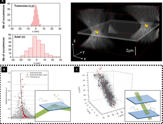
Going Beyond Fluorescence While developed for (and making extensive use of) fluorescence, superresolution principles can just as likely be used with non-fluorescent probes. In this thesis, for instance, we make extensive use of the light scattered by nanoparticles to superlocalize them. Besides imaging, the coherent light scattered by metallic nanoparticles can be used to image optical fields with resolution as good as a few nanometers [99]. This is shown in Fig.1.8 where detection events based on AuNP scattering were used to image evanescent and propagating EM fields.
Mapping Chemical Reactivity in nano Catalysts Superlocalization methods were recently used to map the active sites of many catalysts. If the catalyzed reaction produces fluorescent products, the reaction of one molecule can be detected and superlocalized. The accumulation of many catalysis events leads to the mapping of the active sites on the cat-alyst surface. This method was used to map intraparticle distribution of catalytic activty
Figure 1.8: Superlocalization procedure applied to AuNP of 30 nm, imaged using an holographic setup. (A) A lower limit for the precision of the localization is determined by evaluating the position of two immobile AuNP. A precision of 3 × 3 × 10 nm3 is achieved. Some Examples of applications of nanoparticles superlocalization are shown. The concept is used to image the evanescent EM field resultant of total reflection of the laser at the air-glass surface(B) and a propagative EM wave (laser) (C). Adapted From [99].
on many nanostructures, such as PtNP [100, 101], Au nanoplates[102] and Nanorods [103], etc.
Superlocalization and superresolution methods are therefore an extremely useful tool to analyze nanoscale phenomena optically, and their usefulness also spans over many different fields.
1.2
Electrochemical Imaging and optical (electro)chemistry
Before proceeding to show how these single entity techniques overlap and can be relevant to the context of exploring electrochemical systems, it is worth mentioning already existing bridges between electrochemistry and optics. An exhaustive description of all efforts to couple electrochemistry with optical methods would be just as unbearable as it would be impossible to make. Indeed, most bulk and surface chemistry spectroscopical methods
1.3. OPTICS-EC FOR INDIVIDUAL OBJECTS 19
make use of the interaction of molecules with light. Therefore, any experiment where Raman, IR, XPS, etc. were used to characterize a surface modified by electrochemistry constitute, in a way, an example of opto-electrochemical method. Even more explicitly coupled experiments and setups appear at a pace impossible to keep up [104].
Moreover electrochemical modification - optical diagnosis is not a one way street, as light can also be used to modify surface chemistry, and EC can just as likely be used as an analytical technique to detect these modifications. Lithography, silver-based photog-raphy, and surface modification by polymers or surface photochemical reactions are a few examples [105, 106].
Electrochemistry can just as likely be used to make reactivity-specific images, using a small electrode positioned close to a surface. This is the principle behind the scanning elec-trochemical microscope(SECM), mentioned in Sec.1.1.2. The SECM can also be used to locally modify surfaces [107], and to generate objects with special optical properties[108]. Likewise, spectroelectrochemistry has been extensively used to yield catalytic infor-mation about catalyst, and macro methods remain very relevant to this date [109].
All these methods, however rich in information, probe ensembles of objects. In the wake of single entity methods, and as stressed in the previous sections, there is a richness in responses that can only be appreciated when individual objects are probed one by one. The next section deals with the new step on optical-electrochemical methods development: the efforts to take these coupled strategies to their analytical limits, applying optical and electrochemical coupled methods to study single objects.
1.3
Taking Opto-Electrochemical Methods one Step
Fur-ther: Individual Objects
As seen over Sec.1.1.1, single entity nanoscale studies are intrinsically stochastic and therefore permeated with noise, which generates modest signal to noise ratios. Taken together, these two characteristics make single entity studies prone to artifacts, which calls for coupled alternative characterization methods. Given the concomitant emergence from single entity studies from both plasmonics and single entity electrochemistry, it was more or less natural to try to couple these two fields. Efforts in this direction soon followed, with the appearance of quite a few studies in the recent literature. This section revises the most relevant ones. For a matter of organization, and to emphasize the synergy between electrochemistry and optics, I shall organize them in three subgroups, (i) studies where optical measurements are used to replace electrochemical measurements (ii) studies where electrochemistry is a key ingredient to generate the effects necessary to study individual objects and (iii) study of fields so intrinsically multidisciplinary that both measurements are equally important.
Spectral Monitoring of (Electro)Chemical Reactions The position and intensity of the LSPR band are extremely sensitive to a particle’s surface chemistry and electron density [110, 111]. This makes the LSPR a potentially great measure of these properties, allowing in situ fast tracking of chemical reactions.
In an example from Mulvaney’s group, AuNPs were used to catalyze and monitor ascorbic acid oxidation. As the reaction proceeds, the AuNP acts as an electron buffer, receiving the electrons resulting from the redox reaction. This leads to an increase of electron density, which generates a measurable shift on the LSPR band position. Mon-itoring of the particles spectra thus allows to measure electron injection rates as low as 4600 electrons per second, corresponding to a current of ≈ 0.5f A [112]. This sensitivity is competitive vis-a-vis the best commercially available EC potentiostats.
Long’s group also used changes of plasmonic nanoparticles to monitor electrochemical reactions. For example, they showed that the scattering spectrum of copper nanoparticles can be used to follow surface chemistry and size variations. The electrodeposition and oxidation of plasmonic copper nanoparticles were monitored in this way[113, 114]. These are shown in Fig.1.9.
Exploring Plasmonic properties to design detectors The LSPR spectral shift is not the only way to derive chemical information out of optical data. Plasmonic structures can be designed, for example, to detect a few absorbing molecules adsorbed on their surface based on quenching of the plasmonic band. The idea is to use NPs whose resonance overlaps with the analyte’s absorption spectra, therefore the plasmonic resonance energy transfer (PRET) will quench the plasmonic scattering spectrum [115, 116]. This leads to a decrease of the NP scattered spectrum intensity. On the limit of low concentration with narrow absorption spectra, absorption peaks superposed to the spectrum can be seen, potentially decreasing the detection limit down to single molecule limit [115].
The sensitivity of propagating plasmons to near-field perturbations has also been suc-cessfully used to design detectors with great sensitivity - sometimes called SPP-sensing strategy. This concept was pioneered by Tao’s group to build sensors capable of sizing
1.3. OPTICS-EC FOR INDIVIDUAL OBJECTS 21
Figure 1.9: Monitoring chemical reactions through the spectra of nanoparticles. (up) Mul-vaney’s experiments, where the oxidation of ascorbic acid, catalyzed by the AuNP alters the electronic density of the particles, leading to a blue shift. After longtime (≈ 60 min) relax-ation processes bring the electronic density back to its equilibrium value (Adapted from [112]). (down) monitoring of CuNP oxidation through the spectral intensity and maximum wavelength (Adapted from [113]).
Figure 1.10: Examples of utilisation of plasmonic properties to image electrochemical pro-cesses. In (A) the scattering of surface plasmons imaging is used to detect (a) the arrival and electrooxidation of silver nanoparticles on an electrode surface [117] and (b) reduction of TNT particulates near a fingerprint [118] . (B) Plamonic resonance energy transfer used for detection of individual nanoparticles (in some cases single molecules), either through localized quenching of LSPR by molecules with (a) narrow absorption spectrum[115] or (b) large absorption bands [116], leading to localized quenched LSPR spectrum or to a decrease of the overall intensity.
and imaging single nanoparticles one by one[117, 118, 119].
In these experiments, a thin gold film is used both as electrode material and as a plasmonic substrate to support plasmon-polariton propagation. SPPR is very sensitive to its local dielectric environment, such as inhomogeneities in the optical index, either caused by the presence of small entities in the vicinity of the film or by concentration gradients in the near field (due to a chemical reaction, for example). These heterogeneities are enough to scatter the propagating plasmon, generating propagating electro-magnetic (EM) waves detectable in the far-field.
Besides individual nanoparticles of size down to 35 nm, the method is sensitive enough to allow detection of very small particulates (in the ≈ f g range) of molecules, such as tri-nitro-toluene (TNT) [118]. A similar (but simpler) version of the method later came with the works of Pan and Hill, who used dark field optical detection to correlate particle size to intensity, thus being able to size the deposition of hundreds of AgNPs onto an electrode, as well as their dissolution. The detection limits of their setup was limited to particles of 46 nm diameter [120, 121].
1.3. OPTICS-EC FOR INDIVIDUAL OBJECTS 23
Optical CVs Optically derived EC information can be made quite quantitative. For example, if the optical changes are reported against the potential, optical cyclic voltamo-grams (opCVs) can be recorded. Besides the aforementioned advantages of high through-put and sensitivity, these opCVs have the advantage of providing information specific to the reaction responsible for optical properties changes. All other EC parallel phenomena, such as double layer charging, parasitic reactions, catalysis get filtered out.
The charging and surface reactivity of gold nanoparticles and nanorods were addressed in this way recently. Changes in the spectrum maximum and overall scattered intensity revealed electroadsorption of different anions over the gold surface [122, 123], as well as oxidation reactions [124, 125].
Since the intensity change is proportional to a charging phenomena, its derivative can be assigned to a current. When plotted against the potential, one is able to derive opCV, corresponding specifically to the phenomena changing the optical properties. Besides gold, this strategy was also recently used to investigate lithium ions intercalation on cobalt-based LiCoO2 nanoparticles[126]. These are illustrated in Fig.1.11. Finally, it
is also worth mentioning that any optical property can be appropriated to build opCVs using this methodology. For example, a similar strategy was reported using fluorescence emission intensity [127].
At molecular scale, STM and AFM probes can be used to enhance the Raman scatter-ing of molecules next to a surface to the point of sscatter-ingle molecule detection. Tip enhanced Raman spectroscopy (TERS) setups can then be used to measure electrochemical reac-tions on a single molecule basis, and to build voltammograms from the accumulation of individual redox events [81].
In this section, many examples were shown on how single particle optical signal can be used as channel for chemical information. It was shown that in a few cases, single objects optical measurements can even replace electrochemical detection, allowing high throughput very sensitive monitoring of chemical processes.
By stopping in this section, one might have the impression that optical measurements are superior to electrochemical ones. This is an equivocated point-of-view that we shall try to dissuade in the next sections by showing that a real synergy exists between these two very different ways of observing objects at the nanoscale.
1.3.2
Electrochemically assisted optics
So far it has been shown how optics can be used as an analytical alternative to electron-ics in monitoring electrochemical measurements. However, this is not a one-way path: Electrochemistry can also be an asset in assisting optical studies. Indeed, electrochemi-cal techniques can be extremely handy in creating objects for optielectrochemi-cal-oriented studies on plasmonics.
electrochem-Figure 1.11: CV obtention through processing of optical scattered light. (A) Electroadsorption of anions onto AuNP surface, comparison of the single particle opCV and ensemble electrochem-ical trace (Adapted from [123]) (B) Lithium desintecalation and reintercalation reactions on LiCoO2 particles (Adapted from [126])(C) Single Au Nanowires oxidation reaction (Adapted from [124])
istry in the discovery of surface-enhanced Raman effect (SERS). SERS was first observed and explained having pyridine on a silver electrode as a model system. In these pioneering experiments, it was the anodization of the electrode that triggered etching of the silver sur-face generating plasmonic structures responsible for the signal enhancement [128, 129]. Soon after, SERS studies enabled single molecules studies [130, 131], which attracted lots of attention to surface enhanced techniques[72], ultimately fostering the whole field and shaping plasmonics as we know it today. Taking into account the fast development that followed, one could say that in a way the whole success of the field of plasmonics actually owns a good deal of its hype to electrochemical experiments. Roughening of materials through electrochemistry is still today sometimes used to produce SERS active substrates[132]. Interestingly, with further developments in SERS materials and concepts, surface enhanced tools became quite an asset to study interfacial phenomena. Closing the positive feedback loop, SERS became an extremely useful concept to understand electro-chemical phenomena, providing ultra-low product detection [133, 134], insight into double layer dynamics and adsorption phenomena [135, 136], etc. Electrochemical modulation through redox reactions can also be used as a key component to provide definitive proof of single molecule detection through SERS [137].
Good control of electrochemical depositing and polishing techniques can be crucial to obtain well defined plasmon supporting substrates. In a recent study, extremely precise measurements of light angular momentum were performed using electrochemically grown
1.3. OPTICS-EC FOR INDIVIDUAL OBJECTS 25
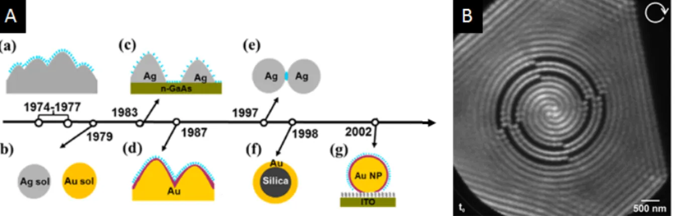
Figure 1.12: Examples where electrochemistry has played an essential role in generating or supporting optical-oriented studies. (A) Brief timeline of SERS important milestones. starting from its (a) first observations on Ag surfaces roughed by electrochemistry, then (b) on Ag and Au colloidal solutions, (c) nanoislands of Ag on conductive surfaces, (d) deposited transition metals on roughed Au surface, (e) Single Molecule SERS, (f) bio-functionallised Au Tags (Adapted from [140]), (g) under potential deposition and redox replacement transition metal on Au NP. (B) Rotating plasmons supported on atomic flat micrometric EC-manufactured Au-flakes [138]
atomic substrates, which were atomic flat over several micrometers [138, 139]. Such extraordinary homogeneity was key in obtaining the well-resolved plasmonic structures, as even the tiniest imperfection would scatter the plasmon, destroying the delicate patterns.
1.3.3
Electrochemically generated optical phenomena in single
particles and photomodification of their properties
Finally, electrochemistry can also sometimes be used to generate optically active single molecules and or modulate their optical response properties. In an example from Orrit’s group, single fluorophore activation was achieved by use of a redox mediator to activate immobilized methylene blue fluorescent probes on a single molecule basis[141]. Similarly, single molecules with high Raman cross-sections have also been generated electrochemi-cally, and used in SERS[137] and TERS[80, 142] experiments.
The electrocatalysis of reactions generating fluorescent probes can also be used to enlighten the spatial distribution of active sites over catalytic nanoparticles, using super-localization methods [98]. Moreover, single molecule fluorescence methods also have the potential to enlighten mechanistic pathways [143] and identify intermediate species [103]. The electrochemical properties of semiconducting NPs can be modified by light, which can be probed at single particle scales. For example, NPs made of light-sensitive semi-conductor anastase T iO2 material have been studied through the electrochemical impact
technique. Depending on whether the particle had been irradiated with light, the direc-tion of the steps can be reversed for a given potential, going from oxidative to reductive steps. What happens is that light can excite electrons to the conduction band and thus change the Fermi level energy. Depending on the electrode potential range with respect
Figure 1.13: (A) Indirect electrochemical generation of a fluorescent molecule through a re-dox mediator, whose luminescence is exalted by the presence of a plasmonic nanorod [141] (B) Switching of electrochemical step impact behavior from anodic to cathodic by action of incident radiation (Adapted from [145])
to the new Fermi Level, electrons will flow on either one way or another [144, 145]. This is illustrated in Fig.1.13.
The emission of light in response to an electrochemical stimulus, electrogenerated chemiluminescence (ECL), has also been used to study molecules and nanostructures individually. For example, Bard’s group used PtNPs to oxidize photoactive Ru(bpy)2+3 and a coreactant, triggering the ECL process, to detect NP collision with photodetectors. Potential dependent changes in the photoluminescence properties of silver nanowires have also been reported[146].
These examples highlight the complexity of optical-chemical small scale studies. On nano to molecular ranges, it is rare that the chemistry or physics of the analytes could be considered alone. Rarely the observation of small entities is with no chemical effects, and even more rarely can we modify a systems chemistry without provoking major alterations of their physical properties. These difficulties are however the price to pay for the detailed knowledge these studies provide. No pain, no gain.
1.4. WHAT THIS THESIS IS ALL ABOUT. 27
1.4
What this thesis is all about.
Such is the context in which the work described in this thesis emerges. Although individual particles studies start to be more and more common, and a few examples of coupling optics and electrochemical methods to understand NP reactivity already exist, it is rare that an equal importance is given to both EC and optical measurements. In general, either EC or optics are used as a support and/or to generate the optical (EC respectively) response, as it was shown throughout this chapter.
If a simplified EC reaction R → O + e− is considered, one could very heuristically say that we have access to the electron flux via electrochemistry, while optical techniques reveal the rate of consumption/generation of R/O. In very simple cases, these two pieces of information may be redundant, but this is certainly not a general rule, and very often the actual mechanism is much more complicated.
The originality of this thesis work relies on not only using optics to derive electrochem-ical information, or to measure electron fluxes precisely. We are really interested in the complementarity of optical and electrochemical signals of individual objects, and in the information that can be extracted by comparing them. The next chapter will particularly stress out this point: starting with a very simple dissolution reaction, the complexity of the situation will be gradually increased, illustrating the emergence of the need for this coupled EC-optical single NP approach.
Chapter 2
Individual Silver Nanoparticles
Electrochemistry. Part I:
Electrochemical Reactivity and
Complexing
The present chapter is heavily based on our publications in J. Am. Chem. Soc. [147], Acc. Chem. Res. [148] and Farad. Disc.[149]. The results herein presented are adapted from these publications.
The importance of the history of a scientific field is often neglected, in spite of its deter-minant role played in further developments [150]. In particular, the choice of first model systems is a crucial step in forging theory and ultimately define a system’s characteris-tic generalizations and simplifications. In demonstrating a new concept, it is important to choose a model complicated enough to prove the utility of new ideas, but not too complicated to the point of obscuring its conclusions.
Take for instance the development of surface probe techniques. Before the invention of the STM, another very similar instrument already performed topographic measurements via tunneling current - the Topografiner [151, 11]. Although it operated by tunnel effect, and had all the elements common to scanning probe techniques, it failed to develop its full potential (at least partially) due to a bad choice of model systems. In contrast, Rohrer and Binning chose to apply the STM to image Silicon 7 × 7 surface, a strongly debated subject among surface scientists. This fortunate choice led to a much more efficient promotion of STM, as their image made the utility of the instrument crystal-clear. The image caused a big impact on the community, leading to further developments and adoption of surface scanning techniques by a wide audience [11]. While we do not compare the importance of our work to the discovery of the STM, careful care was taken to choose an interesting
aesthetically pleasant aspect, Ag has been recognized as a powerful antiseptic agent since old times: registers dating back to Herodotus describe the use of silver in containers to carry water and food, silver cutlery was used in Middle Ages, and silver solutions were routinely prescribed by doctors for a number of diseases until the twentieth century[152]. Although the exact mechanism still remains elusive, and is not likely to be unique, silver antibacterial action seems to be correlated to its ability to oxidize, progressively leaking Ag+ions into the environment [153, 154, 155]. The use of silver nanoparticles (Ag
NPs) was then a natural step, for their high surface-to-bulk area would represent a way to efficiently monitor Ag+ release rate. Moreover, Ag NPs can be more easily internalized,
while surface functionalisation can target specific cellular population intake, which made Ag NPs very interesting candidates for targeted antibiotic delivery [156].
Indeed, market applications of Ag NPs are already numerous and growing, calling for further regulation for their use [154, 155]. In spite of extensive research , with numerous reviews over the last few years, the knowledge about Ag NPs reactivity does not seem to be enough to derive decisive and necessary regulation laws[152, 157].
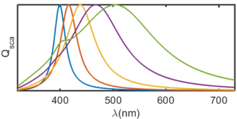
Besides the pharmaceutical industry, plasmonic properties, specially the presence of a LSPR band, have sparked a lot of interest on nano silver research for sensors applications. The LSPR band of Ag can be tuned by size, allowing fine adjustment of the enhancement effect over almost the whole visible spectrum, as shown in Fig. 2.1. Besides, the dielectric constant of silver leads to much lower SP damping in the visible range as compared to most metals including gold. This has made it the metal of choice for LSP or SPP studies whenever its easy chemical transformation, and therefore poor stability under usual conditions, are not a problem.
Moreover, the plasmonic performance - for example the ability to generate SERS effect - is however extremely dependent on the surface state of the NP. Indeed it has been shown that the oxidation of just a few nanometers of Ag NP can lead to a decrease by orders of magnitude of the SERS enhancement factor [158]. All these applications could directly benefit from a more in depth detailed insight into the mechanism of silver oxidation.
Moreover, a considerable body of literature developed over the years, owing to the aforementioned applications. Due to this, there is a considerable amount of data on Ag NPs optical properties, allowing us to focus on the dynamics of the particles chemical