Isolation and culture of protoplasts of Côte d'Ivoire’s
pearl millet (Pennisetum glaucum (L) R) varieties
Kouakou Tiécoura1; Abou Bakari Kouassi1 ; Oulo N’Nan-Alla1 Séry Gonedele Bi1 ; Monique Dinant2et Lucien Ledou2
1- Laboratory of Genetics, UFR Biosciences of University Félix Houphouët Boigny (FHB) of Cocody Abidjan. 22 BP 582 Abidjan 22. Côte d’Ivoire.
2- Laboratory of Génétics Molecular, Department of Botany, University of Liège, Sart Tilman, B22, B-4000 Liege, Belgium
Corresponding Author Oulo N’Nan Alla, Tel: +225 07 16 43 27, E-Mail [email protected] Original submitted in on 2nd July 2015. Published online at www.m.elewa.org on 31st August 2015
http://dx.doi.org/10.4314/jab.v92i1.6 ABSTRACT
Objective: Protoplasts are the ideal material for genetic transformation of plants. This requires that the protoplasts have the ability to regenerate whole plants. The objective of this study is to isolate protoplasts from cell suspensions and test their ability to regenerate embryogenic calli and plants.
Methodology and results: Protoplasts were isolated with different enzyme combinations, from cell suspensions of millet, Pennisetum glaucum. Obtaining callus from protoplasts was carried out on various media at various pH with different glucose concentrations. Approximately 13,106 protoplasts / g were isolated from cell suspensions. Calli were regenerated by culturing the protoplasts at pH 5.8, at a concentration of 0.7 M glucose in either the liquid medium or solid medium. The plating efficiency of protoplast is from 0.012 to 0.013 in solid medium containing 0.6% agarose. No plant has been regenerated from calli provided from protoplasts. All plant regeneration attempts resulted in the formation of globular structures. Cytological studies have shown that the calli derived from protoplasts are formed with 50% of multinucleate cells.
Conclusion and application of results: This study allowed isolating protoplasts, regenerating embryogenic calli from protoplasts of millet varieties of Côte d'Ivoire and highlighting one of the causes of the recalcitrance of the grass crop culture regeneration from provided protoplasts. This study will allow genetic transformation of millet varieties by using protoplasts.
INTRODUCTION
Protoplast culture is a key element in genetic engineering development such as somatic hybridization and gene transfer. However, the application of these techniques is possible if reproducible methods for plants regeneration from protoplasts are available. In cereals, the first calli from protoplasts were obtained with rice (Deka et al., 1976), maize (Chourey et al., 1981), sorghum (Chourey et al., 1985) and wheat (Yasuyuki, et al.,
1988). Plant regeneration from protoplasts was established in Oriza sativa L. (Datta et al., 1990; Baset et al., 1993); in Zea maize (Petersen et al., 1992); in Triticuma estivum L. (Vasil et al., 1990, Chang et al., 1991); in Hordeum vulgare (Ziauddin, 1992); in Sorghum vulgare (Zhi-Ming et al., 1990); in Saccharum officinarum L. (Srinivasan, 1986). Although the majority of these works have used cell suspension as the main source of protoplasts,
Journal of Applied Biosciences 92:8620 – 8629
several other approaches have also been used to regenerate plants. Indeed, protoplasts have been regenerated from rice mesophyll (Gupta and Pattanayak, 1993) and from Tylophoraindica mesophyll (Thomas, 2009). Within the genus Pennisetum, Vasil et al. (1979) obtained calli from cell suspension protoplasts of P. americanum, and they regenerated plantlets. Calli and plantlets were also obtained respectively with protoplasts of P. squamulatum (Gupta et al., 1988) and protoplasts of P. purpureum Schum (Vasil et al., 1983). The conditions for protoplasts isolation of mésophyles of P. glaucum and P. purpureum were reported by
Timbo De Oliveira et al. (2010). Unlike other grasses or cereals such as rice, where the experiments were reproducible, work on Pennisetum has not yet led to reproducible regeneration from protoplasts (Mtili, 1990). Except for the use of isolated protoplasts for the expression of foreign genes (Tiécoura et al., 2001), studies on protoplasts of pearl millet, Pennisetum glaucum, varieties of Côte d'Ivoire are scarce. In this work, we present protoplast culture assays of pearl millet, Pennisetum glaucum, varieties of Côte d'Ivoire, the conditions of isolation, culture and plant regeneration from protoplasts.
MATERIEL AND METHODS
Vegetable material: Protoplasts used, were derived from embryogenic cell suspensions obtained from embryogenic calli of NE (northeast) millet variety (Tiécoura et al., 2014a).
Protoplast isolation: Isolation of protoplasts was performed using the methods of Potrykus (1977) and Shillito (1983) with some modifications: After centrifugation of cell suspension at 160xg / 2-3min, the pellet was weighed to determine the mass of fresh material. The pellet was suspended in protoplast culture medium containing different enzymes: Cellulase "Onozuka" RS (1-4%), R-10 macerozyme (Yakult Honsha Co. Tokyo) (0.5 to 1%), pectolyase Y -23 (SeishinParmaceutical Co. Tokyo) (0.2 to 0.5%), MES (Boehringer) (0.5%), mannitol and CaCl2 (0.25M). The medium at pH 5.6 is sterilized by a filtration on 0.2µm filter. The mixture was digested under stirring (40-50rt / min) in the dark for at least four hours at 25 ° C. After digestion of the skeletal wall, two volumes of a CaCl2 solution (0.25M), MES (0.5%) were added and the whole mixture filtered successively on sieves of 630, 250 and 25 µm. The filtrate was centrifuged for 5min at 160xg and the pellet was recovered in the base solution that was used in the enzyme solution with mannitol (0.6M). Protoplasts were washed 2-3 times with the same solution and taken up in 25ml before being counted. Viable protoplasts, viability according to Larking (1976), were cultured. Protoplast culture: Two media were used: liquid medium (Coulibaly et al., 1986, Xia et al., 1992) and solid
glucose concentration is halved with fresh medium without sugar, or culture is centrifuged and the pellet is taken up in fresh medium with glucose reduced by half. After one week, the sugar concentration was reduced to the concentration of the culture medium of the cell suspension (0.1 M) to allow microcalli to develop. The microcalli that appear were transferred on solid media MS or N6.
• Culture in solid medium (Sea plate, FMC BioProduction, USA): The washed protoplasts were taken up by the culture medium (twice in concentration) at a concentration of 106 / 500µl. To one volume of protoplasts was added a volume of agarose (twice in concentration). The gently homogenized mixture was placed in the Petri dish forming an agarose "pancake" that is grown with or without feeder cells.
• Culture without feeder cells (Potrykus, 1977, Shillito, 1983): The "pancake" was grown without culture medium or immersed in the culture medium and cultivated on a rotating table at 45 rpm / min. After 7 to 10 days, the cakes, dry or not, were immersed in the culture medium with reduced sugar concentration in half. At the second week, the "pancake" was immersed in the medium of normal cell culture. One or two weeks later, the "pancake" was placed on the solid medium. Developing microcalli was transferred on the solid medium.
• Culture with feeder cells: the "nursing" method (Petersen et al., 1992, Kamo et al., 1987): The
used. In the case of the submerged culture, 80% of feeder cells were removed. After one or two weeks of culture, the "pancake" was washed several times with distilled water and then transferred to solid medium MS or N6.
Plants regeneration: For regeneration, the MS medium (0.5 / 2) was used (Tiécoura et al., 2014b). Calli derived from protoplasts grown in liquid or solid media were used
to regenerate plants. Before transferring to the regeneration medium, the calli were cultured for 1-8 weeks on different maintenance media (Table 3). Calli cytogenetic study: DAPI test (Coleman et al., 1985): The test uses two fluorochromes, mithramycin and 4, 6-Diamidino-2-phenylindole. They bind to DNA and used to locate the core in the dividing cells.
RESULTS
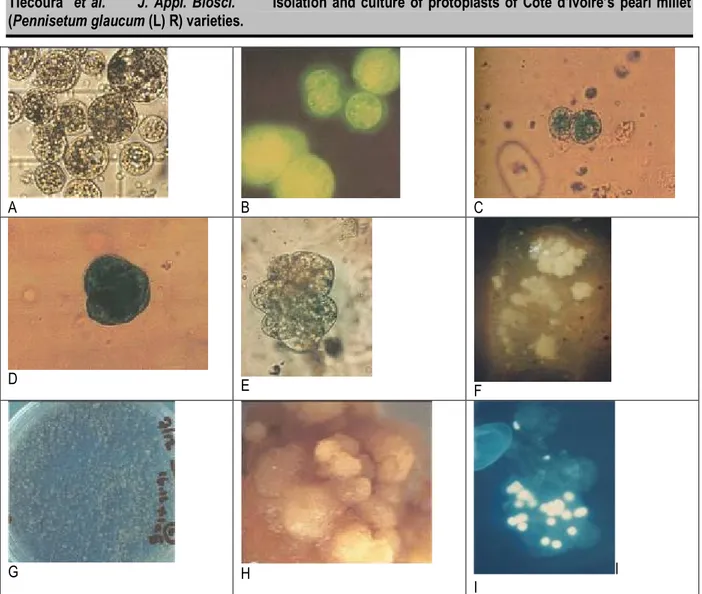
Enzyme combination protoplast isolation: Three days after subculture, the cell suspension was digested with one of three combinations of enzymes for 1 to 6 hours. Table 1 shows the rates of cells having a complete digestion for a given protoplast, with their embryogenic characters, as observed on figure 4A. After one hour of digestion, all the protoplasts isolated (100%) still exhibit
wall pieces. For the enzyme combination (1, 0.5, 0.2), about 6 hours are required to have about 80% protoplasts. For the enzyme combinations (2, 0.5, 0.2) and (4, 1, 0.5), four hours of digestion were required to have about 90% protoplasts. The second enzyme combination (2, 0.5 and 0.2) is used for the further isolation of protoplasts after digestion of at least 4 hours. Table 1: Evolution of completely digested cell rate (%) according to the enzyme combination and the incubation time
Enzyme combination Time of incubation (hours)
Ce % Ma % Pe % 1 2 3 4 5 6
1 0.5 0.2 0 15 30 50 70 80
2 0.5 0.2 0 40 70 90 90 90
4 1 0.5 0 40 80 90 90 90
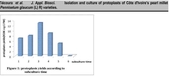
Ce = cellulase; Ma = macerozyme; Pe = pectoliase Age of subculture and protoplasts isolation: The subcultures of 1 to 6 days were used to isolate the protoplasts. Figure 1 shows the yields of protoplasts expressed as X106 per gram of fresh weight (X 106 x g-1 FW). Protoplasts yield varies with subculture times. At the first day, protoplasts yield is of 7.106/g FW. It increases and reaches a peak at the third day (14.106/g FW) and
then decrease to reach its lowest value (0.07.106/g FW) at the 6th day.
Viability of protoplasts: Xia culture media at 0.6M or 0.7M glucose were prepared at different pH. The viability rate and the rate of protoplasts with embryogenic characters were estimated after a week of culture. The results are in Table 2.
Table 2: viability of the protoplasts according to the pH and the glucose concentration pH [glucose] Protoplasts 5.2 5.4 5.6 5.8 6 0.6M viability ± ++ ++ ++ ++ E C - - ± ± ± 0.7M viability ± ++ ++ ++ ++ E C - ± ++ ++ ++ EC = embryogenic characters; - = 0%; ± = 50%; ++ = >80%
0 2 4 6 8 10 12 14 1 2 3 4 5 6 p ro to p la st s y ie ld s( X 1 0 6 x g -1 F W ) subculture time
Figure 1: protoplasts yields according to subculture time
The viability of protoplasts was observed as shown on the figure 4 B. At pH5.2, the viability of protoplasts is 50% whatever the concentration of glucose. For the other pH, it is >80% whatever the concentration of glucose. For embryogenic character of protoplasts at glucose 0.6M, protoplasts are not viable (0% of viability) with pH5.2 and pH5.4. For the other PH, 50% of protoplasts are viable at glucose 0.6M. With glucose 0.7M, 0% of protoplasts retain their embryogenic character at pH5.2. This rate is 50% at pH5.4 to over 80% for pH5.6.
Kinetics of the first cell divisions: Fractions 1.106 protoplasts are cultured in the medium supplemented Xia 2.4-D (2mg / l). The medium is either liquid or solid (agaros 0.6%). The rate of cell divisions based on the culture time is presented in Figure 2. The first divisions of protoplasts are observed until the fourth day (5-15%) (Figure 4C). From 10 to 30% after the first week of culture in liquid or solid respectively, the number of divisions increases rapidly to reach 70 to 80% in the second week of culture in liquid or solid respectively. However, almost 20 to 30% of the protoplasts cultured in solid or liquid
medium respectively do not divided. In the liquid medium, 5-10% of these protoplasts merged and failed. The fusion process is reduced in the solid culture.
Calli from protoplasts culture: Fractions 1.106 protoplasts are cultured in the medium supplemented Xia 2.4-D (2mg / l). The medium is solid with agarose 0.6% or agarose 1.2%. Average of calli developed by culture according to agarose concentration is shown in Figure 3. On media containing 0.6% agarose, MS gives approximately 120 calls per culture and N6 140 callies by culture of 1.106 protoplasts. With 1.2% agarose, the average observed was 40 and 50 calli respectively on MS and N6. The plating efficiency varies from 0.012 to 0.014 protoplasts for 0.6% of agarose and 0.004-0.005 protoplasts for 1.2% agarose.
Plant regeneration from calli derived from protoplasts: Cultivated calli (1 to 8 weeks) are transferred to the MS regeneration medium (0.5, 2). The morphology and the ability of calli to regenerate plants are shown in Table 3.
Table 3: Plant regeneration tests with calli derived from protoplasts
N° Maintenance media Calli morphology Regeneration
1 MS(1-1-2.5) - - - 2 N6(1-100-25) - - - 3 MS(5) - - - 4 MS(2.5)+AgNO3(58.9µM) + - - 5 MS(0)+ABA(0.2) + - - 6 MS(1-1-2.5)+AgNO3 + - -
Nine culture media were used to strengthen the callies before regeneration. The media 1, 2 and 3 gave wet calli regardless globular and non-embryogenic. The media 4 to 8, respectively characterized by the presence of silver nitrate AgNO3 and abscisic acid (ABA) present relatively globular and less wet calli and do not produce plants. The
action of coal, leads to globular and dry embryoids (figure 4H) but do not either give plants. Binocular observations show not-compact and snowy globular embryoids where hairs could be observed. Cytogenetic studies of the calli have shown that several cells are without core and other more cells are with several cores (figure 4I-J).
0 10 20 30 40 50 60 70 80 2 4 6 8 12 ra te o f ce ll s d iv is io n %
culture time (days) Figure 2: Evolution of cells division according
to time and type of culture
liquid culture solid culture 0 20 40 60 80 100 120 140 MS N6 a v e ra g e o f ca ll i b y c u lt u re culture media Figure 3: Calli development according to
media and agarose concentration
0.60% 1.20%
A B C
D E
F
G H I
I
Figure 4: Isolation and protoplasts culture: A= freshly isolated Protoplasts showing dense cytoplasm; B = Viable protoplasts with fluorescein (FDA) staining; C= first division showing wall formation; D-E = successive division gives rise to microcalli; F= calli developed in agarose plating; G = calli developed in liquid plating; H= embryoids structures developed on regeneration medium; I= multineated cells with DAPI staining
DISCUSSION
This study data indicates that enzyme combination that is effective is 2% (w / v) cellulase, 0.5% macerozyme and 0.2 % (w/v) pectoliase. With Musa spp, Assani et al. (2001) used the combination of 2% (w / v) cellulase, 0.5% Macerozyme, 0.25% (w / v) hemicellulase and 0.25% (w / v) Pectolyase. However, with Musa spp, Cavendish Sub-group AAA, Assani et al. (2005) used an enzyme combination composed of 1.5% (w / v) cellulase and 0.15% (w / v) pectolyase. Enzymes are substances that
determined, it is nevertheless important for protoplasts culture. Thus, for P. glaucum, cells were generally left for 12 hours in the enzyme solution (Xia et al., 1992). For Banana, 15 to 17h of digestion or 12 to 14 hours of digestion were requested (Assani et al., 2001; 2005). For Kappaphycus, 4 hours of digestion were requested with 20% abalone enzyme and 12% of cellulose (Zhang Si et al., 2014). Enzyme combinations are also a function of the pectocellulose composition in the walls. This diversity
average value of the protoplast yield, depending on the time of subculture, is due to the quality of the wall and the amount of cells. In the early days of the subculture, the division rate was exponential. From the third day, the divisions diminished given the saturation and the impoverishment of culture medium (Tiécoura et al., 2014a). In these first days of divisions, the cells were young with a primary wall called growth wall (Hongo et al., 2012). The primary wall is simple in its constitution and easy to be digested by enzymes. The amount of young cells increases, there is also an increased yield of protoplasts. In the period of saturation of the culture medium, cells are composed of secondary or tertiary wall (Kim et al., 2012; 2014). This wall with inlays substances is a secondary cell wall that is tough and more difficult to digest by enzymes and lead to the fall of the protoplast yield despite of the long incubation time. The average value of protoplast performance observed in this study is 2-26 times higher than those reported for other grasses: 0,5.106 x g-1 In Sorghum vulgare (Zhi-ming et al., 1990); 1,4.106 x g-1 in Pennisetum glaucum (Mtili, 1990); 6 .106 xg-1 in Oryza sativa (Lee et al., 1989) and 0.65.106 g-1in Kappaphycus (Zhang Si et al., 2014). The highest value was observed in Triticum aestivum with 0.28 to 5 .107x mg-1 fresh weight (He et al., 1992). This variability in the yield of protoplasts confirms the species dependence of cell walls diversity. For the viability of protoplasts, 0.7M concentration of the glucose and a pH of 5.6 seem to be the best for protoplasts viability and embryogenic character conservation. Our parameters determined are similar to those observed in most of the grasses (Mtili 1990, ZhangSi et al., 2014). With Oryza granulata, the rate of protoplast division is about 25% after one week of culture (Baset et al., 1993). Wang et al. (1989) found 5-10% among Japonica varieties of male sterile (CMS) Cell division testifies the vitality of protoplasts and indicates that protoplasts recovered their cell wall. Calcofluor colouration reveals that protoplasts cell wall was restored after three to four days. The protoplasts were progressively obtained, their wall, the first sign of cell totipotency. Approximately 20 to 30% protoplasts witch degenerate would be unable to rebuild their cell wall. The division was done either by budding (Xia et al., 1992) or by segmentation of the cytoplasm (Vasil et al., 1990), or by normal bipartition (Ishak, 1992). In liquid culture, the balance is budding and cytoplasmic partitioning. Segmentation and bipartition were most often observed in solid culture. In both solid and liquid medium, calli were obtained from the protoplasts culture. The production of protoplasts on solid cultures reported in this study presents a great variability in the number of calli obtained.
This was the case with the protoplasts cultures of mesophyll Oryza sativa (Gupta et al., 1993) where 140-250 callies were obtained per culture. For Saccharum officinarum, 20 to 72 callies have been reported (Taylor et al., 1992). The concentration of agarose seemed to influence the efficacy of the spreading protoplasts. A concentration of 0.6% appear to be more appropriate than 1.2%, since the concentration 1.2% seems to reduce the diffusion of the culture medium nutriments or the metabolites arising from the feeder cells; these metabolites promote calli protoplasts. Nevertheless, the plating efficiency of protoplast values obtained in the present study remain low (0.012 to 0.013) compared to these obtained with other species: for Zea mays, with 0.6 or 1.2% agarose, the efficiency is 0.25 (Chourey et al., 1981) and 0.8 with 0.4% agarose (Kamo et al., 1987). For Triticum aestivum L., with 1.2% agarose, the efficiency is 0.02 to 0.11 (Kasem et al., 1993). With 2.5% agarose, the plating efficiency of protoplasts is estimated from 1 to 9.8 with Oryza sativa (Kyozuka et al., 1987). Here, it is established that the agarose effect depends on the species. Plants were no regenerated from calli protoplasts. Despite the action of AgNO3, ABA and activated coal leading to drier callies and observation of hair on the surface of callies, true embryos could not be regenerated; hence the absence of plants. The main action of Abscisic acid was to allow proper maturation of embryos germinating. In our case, there were no true embryos; only snow globular embryoids structures appeared. Factors that may explain the non-regeneration of plants from calli protoplasts are numerous. From the cell suspension to obtained protoplasts calli, it takes at least three months. It is established that after three months, plant regeneration rate from liquid culture, drops to 10% (Tiécoura 2014b). This time could not be reduced and could explain why the calli was unable to develop real embryos despite the use of different media. This recalcitrance of graminea was previously reported with Pennisetum americanum (Mtili, 1990) and with Sorghum bicolor (L) (Ishak, 1992) despite previous reports of the regeneration of protoplasts from these cereals (Vasil et al., 1980; Zhi-Ming et al., 1990). Regenerative capacity can also depend on the species, varieties and genotypes (Vasil et al., 1980). We believe that graminea cell totipotency (see differentiation and dedifferentiation of Poaceae plant cell) is challenging. In the case of Pennisetum glaucum, a mitotic disorder can affect totipotency. The possible multi nucleated cells observed may suggest that the calli from protoplasts is a chimeric cell. The chimeric cell behaviour would lead to a possible carcinogenic response of calli, leading to the lost of their
ability to differentiate into regenerative plants embryos. Finally, genes methylation in cells culture may be a factor
promoting uncontrolled proliferation blocking cell differentiation in graminea.
CONCLUSION
Protoplasts of millet varieties of Côte d’Ivoire can be isolated, cultured and regenerated in calli. In spite of this multitude of culture media of calli, no calli transferred on medium of regeneration can give plants. The non-regenerating of plants reveals that further studies need to be conducted to better understand the totipotency, to
provide solutions to the recalcitrance of graminea relative to plant regeneration from protoplasts. Understanding of this totipotency could provide less expensive ways to improve graminea in general and in particular the Ivorian millet.
REFERENCES
Assani A, Haïcour R, Wenzel G, Côte F, Bakry F, Foroughi-Wehr B, Ducreux G, Aguillar ME, Graphin A, 2000.Plant regeneration from protoplasts of dessert banana CV. Grande Naine (Musa spp. Cavendish sub-group AAA) via somatic embryogenesis. Plant cell Reports 20: 482-488
Assani A, Chabane D, Haïcour R, Bakry F, Wenzel G, Foroughi-Wehr B, 2005.Protoplast fusion in banana (Musa spp.): Comparison of chemical (PEG: polyethylene glycol) and electrical procedure. Plant cell, Tissue and organ culture 83:145-151
Baset A, Cocking EC, Finch RP, 1993. Regeneration of fertile plants from protoplasts of the wild rice species Oryza granulate. Plant Physiology Journal 141: 245-247
Chang YF, Wang WC, Warfield CY, Nguyen HT, Wong JR, 1991. Plant regeneration from protoplasts isolated from long-term cell cultures of wheat (Triticum aestivum L.). Plant cell Reports 9: 611-614
Chourey PS. and Zurawski DB, 1981. Calli formation from protoplasts of a maize cell culture. Theoretical and Applied Genetics 59: 341-344 Chourey PS. and Sarpe DZ, 1985. Calli formation from
protoplasts of Sorghum cell suspension cultures. Plant Science 39: 171-175
Coleman AW. and Goff LJ, 1985. Aplication of flourochromes to pollen biology I: Mithramycin and 4,6-Diamidino – 2 –phenylindole (DAPI) as vital stain and for quantitation of nuclear DNA. Stain Technology 60: 145-154
through plating technique. Molecular Genetics and Genomics 145: 239-243
Gupta HS,. Rech EL, Cocking EC, Davey MR, 1988. Electroporation and heat shock stimulate division of protoplasts of P. squamulatum. Plant Physiology Journal 133: 457-459
Gupta HS and Pattanayak A, 1993. Pant regeneration from mesophyll protoplasts of rice (O. sativa L.). Biotechnology 11: 90-94
He DG, Yang YM, Scott K L, 1992. Plant regeneration from protoplasts of wheat (Triticum aestivum CV. Hartog).Plant Cell Reports 11: 16-19 Hongo S, Sato K, Yokoyama R, Nishitani K, 2012.
Demethylesterification of the primary wall by pectriMethylesterase 35 provides mechanical support to the Arabidopsis stem. Plant Cell 24: 2624-2634
Ishak and M. Jacobs (1992). Plant regeneration through embryogenesis and study of promoter activity using direct gene transfer in sorghum (Sorghum bicolor L.) Moench and indica rice (Oryza sativa L.). Thesis / V. U. B. / pp 155
Kamo KK, Chang KL, Lynn ME, Hodges TK, 1987. Embryogenic calli formation from maize protoplasts. Planta 172: 245-251
Kasem ZA. and Agi F, 1993. Culture of and fertile plant regeneration from regenerable embryogenic suspension cell-derived protoplasts pf wheat (Triticum aestivum L.). Plant cell Reports 12: 175-179
Kim WC, Ko JH, Han KH, 2012. Identification of a cis-acting regulatory motif recognized by MYB46, a master transcriptional regulator of secondary
biosynthesis. Plant Molecular Biology 85: 589-599
Kyozuka J, Yasuyukiand H, Shimamoto K, 1987. High frequency plant regeneration from rice protoplasts by novel nurse culture methods. Molecular Genetics Genomics 206: 408-413 Larking PJ, 1976. Purification and viability determinations
of plant protoplasts. Planta 128: 213-216 Lee L, Schroll RE, Grimes HD, Hodges TK, 1989. Plant
regeneration from indica rice (Oryza sativa L.) protoplasts. Planta 178: 325-333
Mtili EN, (1990). Embryogenèse somatique et culture de cellules et de protoplastes chez le mil (Pennisetum americanum L.). Thèse / Orsay 189.pp 137
Petersen WL, Sulc S, Armstrong CL, 1992. Effect of nurse cultures on the production of macro-calli and fertile plants from maize embryogenic suspension culture protoplasts. Plant Cell Reports 10: 591-594
Mehri-Kamoun R, 2001. Effet de la Pectolyase Y-23 et de la Cellulase RS sur le rendement en protoplastes viables de Prunus cerasus L. « Montmorency ». Biotechnology, Agronomy, Society and Environment. 5 (2): 99-104 Shillito RD, Carswell GK, Johnson C M, Dimaio J, Harms
CT, 1989. Regeneration of fertile plants from protoplasts of elite inbred maize. Biotechnology 7: 581-587
Srinivasan C. and Vasil IK, 1986. Plant regeneration from protoplasts of sugarcane (Saccharum officinarum L.).Plant Physiology Journal 126: 41-48
Taylor PWJ, Ko HL, Adkins SW, 1992. Facteurs affecting protoplast isolation and the regeneration of shoot-like structures from protoplast-derived calli of Sugarcane (Saccharum spp. Hybrids). Australian Journal of Botany 40: 863-876 Thomas T. Dennis, 2009. Isolation, calli formation and
plantlet regeneration from mesophyll protoplasts of Tylophoraindica (Burm.f.) Merrill: an important medical plant. In Vitro Cellular and Developmental Biology Plant 45 (5) : 591-598 Tiécoura K, Ledoux L, Dinant M, 2001. Expression
transitoire du gène de la B-glucuronidase (Gus) dans les protoplastes de mil (Pennisetum glaucum L. R.) : étude comparative de deux promoteurs, CaMV35S et Emu. Science et Technique/Science Naturelle et Agronomie 25 (2) : 87-97
Tiécoura K, Kouassi AB, N’nan-Alla O, Dinant M, Ledoux L, 2014a. Optimisation des conditions d’établissement de suspensions cellulaires embryogènes à partir de cals d’apex caulinairede mil (Pennisetum glaucum L. R.). Agronomie Africaine 26 (3) : 205-215
Tiécoura K, N’nan-Alla O, Kouassi AB, Dinant M, Ledoux L, 2014b. Optimisation des conditions de régénération de plantes fertiles à partir de suspensions cellulaires issues de cals embryogènes d’apex caulinaire de mil (Pennisetum glaucum L. R.). International Journal of Biology and Chemical Science 8 (5): 2222-2231
Timbo de Oliveira AL, Davide LC, Pereira Pinto JEB, Pereira AV, 2010. Protoplast production from Napier grass and Pearl millet triploid hybrids.Ciens.Agrotec.Lavras.34 (5): 1219-1223 Vasil V and Vasil IK, 1979. Isolation and culture of cereal protoplasts I: Calli formation from Pearl Millet (Pennisetum americanum) protoplasts. Pflanzenphiol Z. 92. 379-383
Vasil V and Vasil IK, 1980. Isolation and culture of cereal protoplasts II: Embryogenesis and plantlet formation from protoplasts of Pennisetum americanum. Theoretical Applied Genetics 56: 97-99
Vasil V, Wang DY, Vasil IK, 1983. Plant regeneration from protoplasts of napier grass (Pennisetum purpureum S.). Pflanzenphysiol. Z 111: 233-239 Vasil V, Redway F, Vasil IK, 1990. Regeneration of plants from embryogenic suspension culture protoplasts of wheat (Triticum aestivum L.).Biotechnology 8: 429-434
Wang D, Miller PD, Sondahl M, 1989. Plant regeneration from protoplasts indica type rice and cms rice. Plant Physiology Journal 8: 329-332
Wertz JL, 2011. Les hémicelluloses. Valbiom Doc. Genbloux Agro-Bio tech pp 16
Xia GM, Li Z, Guo GQ, Chen HM, 1992. Direct somatic embryogenesis and plant regeneration from protoplasts of Bupleurum scorzonerifolium W. Plant cell Reports 11: 155-158
Yasuyuki H and Shimamoto K, 1988. Wheat protoplast culture: embryogenic colony formation from protoplasts. Plant Cell Reports 7: 414-417 Zhang S, Liu C, Jin Y, Chi S, Tang X, Chen F, Fang X,
Liu T, 2014. Studies on the isolation and culture of protoplasts from Kappaphycus alvarezii. Acta Oceanologica Sinica 33 (10): 114-123
Zhi-Min W and Zhi-hong X, 1990. Regeneration of fertile plants from embryogenic suspension culture protoplasts of Sorghum vulgare. Plant cell Reports 9: 51-53
Ziauddin A, Marsolais A, Simion E,. Kasha KJ, 1992. Improved plant regeneration from wheat anther
and barley microspore culture using phenylacetic acid (PAA). Plant cell Reports 11: 489-498
![Table 2: viability of the protoplasts according to the pH and the glucose concentration pH [glucose] Protoplasts 5.2 5.4 5.6 5.8 6 0.6M viability ± ++ ++ ++ ++ E C - - ± ± ± 0.7M viability ± ++ ++ ++ ++ E C - ± ++ ++ ++ E](https://thumb-eu.123doks.com/thumbv2/123doknet/2410201.45474/3.892.93.788.500.621/viability-protoplasts-according-glucose-concentration-protoplasts-viability-viability.webp)