R e v i e w open access to scientific and medical research
Open Access Full Text Article
immunological and genetic aspects of asthma
and allergy
Anne-Marie Madore Catherine Laprise
Université du Québec à Chicoutimi, Département des sciences
fondamentales, Saguenay, Canada
Correspondence: Catherine Laprise 555 boulevard de l’Université, Chicoutimi, Québec, G7H 2B1, Canada Tel +1 418 545 5011 (ext. 5659) Fax +1 418 615 1203
email catherine.laprise@uqac.ca
Abstract: Prevalence of allergy and allergic asthma are increasing worldwide. More than half
of the US population has a positive skin prick test and approximately 10% are asthmatics. Many studies have been conducted to define immunological pathways underlying allergy and asthma development and to identify the main genetic determinants. In the effort to find missing pieces of the puzzle, new genomic approaches and more standardized ones, such as the candidate gene approach, have been used collectively. This article proposes an overview of the actual knowledge about immunological and genetic aspects of allergy and asthma. Special attention has been drawn to the challenges linked to genetic research in complex traits such as asthma and to the contribution of new genomic approaches.
Keywords: immune response, allergy, asthma, genetics, genomics
Allergy
Allergy, also called hypersensitivity, is a reaction that occurs when the immune system responds to a harmless antigen.1 According to results published by the National Health
and Nutrition Examination Surveys in 2005, more than half of the US population (54.3%) tests positive to one or more allergens (a common antigen that gives rise to an immediate hypersensitivity response)2 using skin prick tests between 1988 and
1994.3 This survey also underlines the evidence of the growing prevalence of allergy
in industrialized countries.
Allergies have been studied according to the different categories of allergens (indoor, outdoor, food, drug, etc) and according to the different allergic diseases (allergic rhinitis, atopic dermatitis, allergic asthma, etc). Prevalence of allergy to different types of antigen and prevalence of different allergic diseases ranges from 4% to 6% for food allergy,4 to
10% for allergic asthma,5 and up to 49% reported in one study for allergic rhinitis.6
Immunology of allergy
Hypersensitivity reactions have been classified by Coombs and Gell into four different types, characterized by different immunological mechanisms.7 These types have been
well described in a review by Averbeck et al.1 A brief description of each type is as
follows. Type I hypersensitivity refers to immediate hypersensitivity responses against foreign proteins that are common (pollen, grass, animal danders, etc). It can be observed in allergic rhinitis and allergic asthma (see section “immunology of asthma” for more detail).1,8 This type of hypersensitivity is characterized by immunoglobulin (Ig) E
pro-duction during the sensitization phase, which will bind to FcεRI receptors on mast cells and basophils. Upon renewed contact, the allergen will bind cellular IgE. Crosslinking Number of times this article has been viewed
This article was published in the following Dove Press journal: Journal of Asthma and Allergy
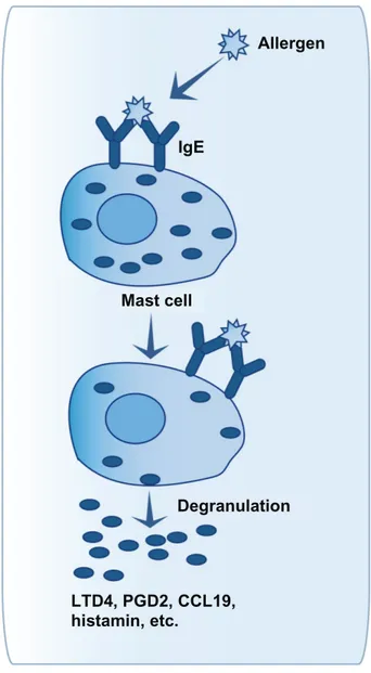
of FcεRI receptors will lead to degranulation of mast cells and basophils (see Figure 1). Mediators released that way will lead to immediate and, sometimes, delayed immune responses.9,10
Type II hypersensitivity is a humoral response mediated by IgG or IgM that are produced against surface antigens on body cells. An example of type II hypersensitivity is drug-induced cytopenia caused by penicillin, cephalosporin, or transfusion reactions.1,11 Immune response occurs within minutes after
antigen contact. In drug allergic subjects, IgG or IgM will be formed in response to drugs or their metabolites which accumulate in membrane structures. This binding will allow
the killing of the target cells by three different ways.1,11 The
first one is the activation of the classic complement pathway, which will lead to cytolysis. The second one is the antibody-dependant cell-mediated cytotoxicity, which will lead to lysis of target cells by natural killer cells. The last one is the recognition of Fc fragments of IgG and IgM by phagocytic cells, which will lead to opsonization of the target cells.
Type III hypersensitivity involves the formation of immune complexes that are not well cleared by innate immune cells as in malaria, rheumatoid arthritis, or farmer’s lung.1,12 This
response can occur within four to six hours. The accumulation of immune complexes (antigens bound to antibodies) in vessels and tissues can be caused by antigen excess for subjects with immunosuppression or insufficient antibody production. It can also be caused by repeated antigen exposure which will lead to excess IgG antibody production. The pres-ence of those persistent immune complexes will give rise to an inflammatory response due to leukocytes’ activation.1,12
Finally, type IV hypersensitivity is a delayed response principally mediated by T cells.1,13 The best known example
of type IV hypersensitivity is contact allergy. The sensitization phase lasts 10–15 days and is asymptomatic. In contact allergy, a hapten, a low molecular weight molecule, will interact with skin cells or proteins. After this contact, Langerhans and den-dritic cells will present antigen to naive T cells to stimulate their differentiation into CD4+ and CD8+ T cells and to induce
production of memory T cells. A renewed contact between the skin and the antigen will stimulate sensitized memory T cells via antigen-presenting cells. Early arrival of CD4+ and CD8+
T cells and activation of keratinocytes, will all contribute to promote inflammatory reaction via cytokine secretion. Finally, this reaction will be controlled by regulatory T cells.1,13
This classification of allergic reactions has been widely accepted but some revised nomenclature was proposed, such as that of Johansson et al14 who are part of the European
Academy of Allergy and Clinical Immunology. This revised nomenclature, based on known immune mechanisms of allergic diseases and hypersensitivity, aimed to classify the hypersensitivity reactions according to the presence of allergy, IgE production, and atopy. This classification gives a more universal definition of hypersensitivity, regardless of the targeted tissue or organ.
Genetics of allergy
Twin studies have demonstrated the strong heritable compo-nent of allergic diseases and atopy, estimated at 33%–76%.15,16
Twin studies with monozygous mice revealed that, even with the same controlled environment, phenotypic variability can
Allergen IgE Mast cell Degranulation LTD4, PGD2, CCL19, histamin, etc.
Figure 1 Mast cell activation after allergen contact.
Notes: when a first contact with allergen has already been made, immunoglobulin (ig) e against this allergen have been produced by B cells and have bound to FcεR1β receptors on mast cells. Another contact with the same allergen will allow its binding on cellular ige on mast cells. This will produce a crosslinking of FcεR1β, leading to activation and degranulation of mast cells within a few minutes. Modulators released by mast cells will promote inflammation by recruitment of new immune cells and activation of structural cells of the airways.
occur in allergic manifestations. These results demonstrated that environment explains about 30% of the phenotypic variability observed in allergy and that 70% is due to other factors, such as epigenetics.17,18
However, search of the genes involved in allergy have been mostly done when studying genetic factors of allergic diseases (Table 1). Indeed, as mentioned by Hong et al,19 it
is often difficult to evaluate the implication of genes on the development of allergy, using this method. However, Ren-konen et al20 have recently published a review of the genetics
of allergic diseases and have found 39 genes associated with allergy.20 In an effort to classify these genes according to
their principal pathway, they concluded that 25 of them are involved in the same interaction pathway with another 70 proteins. Among all of these proteins, 20 are linked to the “host–virus interactions”. When looking to Gene Ontology categories, “response to stress” and “response to various stimuli” were also significantly enriched.20 Another
interest-ing findinterest-ing of this study is the proportion of protein kinases in their results (23%). Indeed, those kinases may be involved in active allergen transport through intact epithelium.20
To give a general idea of the genes identified by genetic studies on allergic diseases, here are some of the general pathways represented: the T helper type 2 (Th2) immune response and IgE switching cytokines and receptors (IL4RA,
IL5, IL13, FCER1A, etc), the chemokines and chemokine
receptors (CX3CR1, CCXCR1, CCR2, etc), the human leu-kocyte antigen (HLA; HLA-DPB1, HLA-DQB1, etc), and the lipoxygenase and cyclooxygenase pathways (CYSLTR1,
LTC4S, etc).6,19–26 Table 1 shows the prevalence of allergy or
some of the most studied allergic diseases and the number of genes already associated with each ones.
Asthma
One of the most studied allergic diseases is asthma. Asthmatic response is provoked by allergy in 75%–80% of all asthmatic cases.29 According to the Global Initiative for Asthma,30 asthma
is defined as a chronic inflammatory disorder of the airways involving many cells and mediators. The principal associated symptoms are airway hyperresponsiveness and usually reversible airflow obstruction. These symptoms lead to recurrent episodes of wheezing, breathlessness, chest tightness, and coughing.
Clinical manifestations of asthma often appear during childhood.31 However, some individuals show a late onset,
sometimes after the age of 40 years.32 According to articles
published between 1987 and 1997, the prevalence of asthma varied between 0.5% and 6% depending on the regions of the world.33–37 Approximately 10 years later, it is estimated that
300 million people suffer from asthma in the world and that this number will reach 400 million people by 2025.5
This represent a world prevalence of approximately 10% and a prevalence of 14.1% for Canada and of 10.9% for the United States.5
Physiopathology and immunology
of asthma
Asthma is a complex trait that is influenced by several genes as well as by the environment, thus it presents heterogeneous clini-cal manifestations always recognized as different subphenotypes. Researchers agree that asthma is not a single disease but rather an array of disorders that share common characteristics.29,38,39
These characteristics are inflammation, intermittent bronchial obstruction, bronchial hyperreactivity, mucus hypersecretion, and hypertrophy and hyperplasia of smooth muscle.8,38
As mentioned above, in 75%–80% of cases40,41 these
phenotypes are caused by an allergic response, which triggers a Th2 immune response.29 It is a type I
hypersen-sitivity reaction, that is an immediate exaggerated or harm-ful immune reaction.8,42 Interestingly, only 7% of allergic
people develop asthma,43 which can lead us to believe that
they present a unique phenotype that distinguishes them from other allergic, but nonasthmatic, individuals. The main immune cells involved in asthma are CD4+ T cells, mast cells,
and eosinophils.44 The different steps of the inflammatory
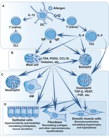
response in asthma, from contact with the allergen to remod-eling, are illustrated in Figure 2 and explained later.
Contact with the allergen
When an allergen penetrates the airways, if not expulsed by the mucociliary barrier, it comes in contact with dendritic cells
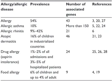
Table 1 Prevalence and number of genes associated with allergy
or most studied allergic diseases27–29
Allergy/allergic disease Prevalence Number of associated genes References Allergy 54% 43 3, 20, 27
Allergic asthma 10% More than 150 5, 22, 24
Allergic rhinitis 9%–42% 21 6 Atopic dermatitis 16% of children in industrialized countries 46 21, 23 Drug allergy (aspirin intolerance) 1%–2% of all admissions and 3%–5% of hospitalized patients 24 25, 26, 28
Food allergy 6% of children and up to 4% of adult
which will internalize and then digest the allergen. Some den-dritic cells then migrate towards the lymph nodes to present the antigen to naive T cells (Figure 2A).45,46 This presentation
hap-pens through the major histocompatibility complex type II con-jugated to the CD80 and CD86 costimulation molecules.47
The introduction of the antigen to naive T cells triggers the differentiation of CD4+ T cells into Th1 or Th2. This
differentiation is modulated by the cytokines present dur-ing the introduction of the allergen, eg, interleukin (IL)-12 (Figure 2A).48 Evidence show, at least for mild to moderate
asthma, that Th2 cells predominate.49 Thus, after the
dif-ferentiation of naive T cells in Th2 cells, the latter produce cytokines, including IL-4 which induces the production of IgE by B lymphocytes (Figure 2A).
immediate response
When an individual has already been in contact with an aller-gen, its presence and the presence of IgE can activate mast cells and stimulate their degranulation just minutes after the contact with the allergen.2,8 The activation happens when the
allergen binds with more than one IgE, which are linked to their high-affinity receptor (FcεRI; Figure 2B).9,10 Mast cells
are key cells in immediate response. In fact, mast cell granules contain proinflammatory molecules such as histamine, tryptase and other proteases, tumor necrosis factor (TNF), and heparin.2
New molecules are also produced and then released. These molecules are leukotrienes and prostaglandins, as well as cytokines, chemokines, and matrix metalloproteinases.2,50–53 The
releasing of these mediators triggers the development of imme-diate response symptoms such as coughing, bronchial spasms, smooth muscle contraction, oedema, mucus secretion, and infil-tration of immune cells.8,54 Mast cells, through these mediators,
contribute to the recruitment and activation of immune cells in the lungs, such as eosinophils, T cells, macrophages, basophils, neutrophils, structure cells (fibroblasts, smooth muscle and epithelial cells), and other mast cells.52,55,56
Basophils, with the presence of FcεRI receptors on their surface and the expression of Th2 cytokines, histamine and granules, can also play a role in immediate response. However, their role in asthma is less documented than that of mast cells.57
Delayed response
Delayed, or late response, is not present in all asthmatics. It is mainly modulated by immune cells recruited by mast cells (Figure 2C).8,29 Among these cells, eosinophils are the
main cells implicated in the development of this response.58
Eosinophils produce Th2 cytokines,59–61 leukotrienes,62–64 and
proteins that cause damage to airway cells, such as major basic protein, eosinophil cationic protein, eosinophil-derived neurotoxin, and eosinophil peroxidase.65,66
Chronic inflammation in airways
Chronic response in asthma is characterized by a persisting inflammation and the presence of structure alterations in the airways (Figure 2D).67
Several immune cells play a role in this response. Mast cells participate in the chronic response through mediators that have an effect on bronchoconstriction and on airway remodeling causing contraction, hypertrophia and hyper-plasia of smooth muscles as well as fibrogenesis.50–53,68,69
Dendritic cells that remain in the airways after coming in contact with the allergen repeatedly present the antigen to CD4+ T cells.70 These cells, as well as the activated T cells,
produce proinflammatory cytokines, maintain the chronicity of the inflammation and the eosinophilia.8,45,46,71 Eosinophils
are indirectly involved in bronchial remodeling regarding collagen deposit, fibrogenesis, angiogenesis, and hyperplasia of smooth muscles in the peripheral airways via mediators secretion and other cells activation.58,72–74
Structural cells also play an important role in maintaining the chronic response. The amount of mucous epithelial cells increases in asthmatics, thus participating in the bronchial obstruction, mainly in peripheral airways.75,76 Furthermore,
epithelial cells, smooth muscle cells, and fibroblasts produce and/or store cytokines, chemokines, and other proinflamma-tory mediators.77–80 In asthmatics, fibroblasts also produce
more collagen and other macromolecules for the extracel-lular matrix, and present an increased differentiation in myofibroblasts.81,82
Phenotypic variability in asthma
To complete this overview of the asthmatic allergic response, the phenotypic heterogeneity has to be taken into consideration. Indeed, apart from allergic asthma, which is the most common form, other types of asthma have been described.8,38,83 One type
is called nonallergic asthma, which develops independently from an allergic component.32 More research still has to be
conducted to better define biological pathways specific to this form of asthma.84,85 However, as with allergic asthma, the
scientific community recognizes that IgE could be implicated in an inflammatory cascade, as well as CD4+ and CD8+ T cells,
eosinophils, and mast cells.86–91
A second type of asthma is characterized by sensitivity to aspirin. It is estimated that approximately 10%–20% of adult asthmatics suffer from this particular type.92 Aspirin
A B C D Epithelial cells Hypersensitivity (permeability) remodeling (metaplasia, mucus secretion) Fibroblast Remodeling (collagen and other macromolecules,
myofibroblast)
Smooth muscle cells
Bronchoconstriction remodeling (hypertrophy and hyperplasia) Neutrophil AM Eosinophil TGF-β, VEGF, FGF, etc. Mast cell LTD4, PGD2, CCL19, histamin, etc. Basophil IL-12 DC IgE IgE B Allergen T (naive) IL-4 Th2 IL-5 IL-12 T (naive) Th1
Figure 2 immune response in asthma.
Notes: The immune response in asthma begins with (A) the contact between antigen-presenting cells (mostly dendritic cells [DC]) and the allergen. These cells stimulate naive T cell differentiation into T helper type 1 (Th1) or Th2 type. interleukin (iL)-4 secretion by Th2 cell leads to the immunoglobulin (ig) E production by B cells. IgE will bind to high-affinity receptors on mast cells and basophils and will allow immediate response to occur when another contact with the allergen will take place (B). After this new contact, mediators released by mast cells will induce recruitment and activation of other immune cell types as eosinophils, neutrophils, basophils, alveolar macrophages (AM), and structural cells (epithelial cells, smooth muscle cells, and fibroblasts). These cell types promote the delayed response that is not present in all asthmatic subjects (C). These cells not only promote the inflammatory response but also provoke hyperreactivity, bronchoconstriction, and remodeling. This will lead to asthma chronicity (D).
sensitization is thought to be non-IgE mediated so could be considered as a nonallergic subphenotype of asthma.92 A
dysfunction of the eicosanoid metabolism is responsible for this type of asthma.92,93 A great number of individuals affected
with this type of asthma will also resist a treatment with corticosteroids.38
Asthmatics who do not respond to glucocorticoid treat-ments represent up to 10% of all patients affected with asthma.94 This type of asthma can be induced by a diminution
of the number of glucocorticoid receptors, by a misrecogni-tion of the ligand by the receptors, by a decreased capacity of the receptors to link with DNA, or by an increase in the expression of certain proinflammatory transcription factors such as nuclear factor (NF)-κB.94
Occupational asthma is exacerbated or induced by irri-tants or particles present in the workplace of the affected.95 It
is estimated that 9%–15% of all cases of asthma in adults are linked to the workplace and that up to 25% of new cases of adult asthma fall in the occupational asthma category.96,97
Finally, exercise-induced asthma affects 7%–15% of the general population and 3%–14% of athletes.98 A study
indicates that up to 39% of college athletes show one of the main symptoms of asthma, that is bronchoconstriction.99
The two major hypotheses that could explain the induction of asthma after an effort are related to the augmentation of ventilation in the lungs.100,101 These two hypotheses are the
osmotic hypothesis (dehydration by evaporation caused by the increase in ventilation) and the thermal hypothesis (airway cooling during exercise and rewarming after exercise).101–103
According to Anderson and Daviskas,101 maybe a mix of these
two hypotheses could better explain the phenotype rather than each one separately.
Asthma can also be classified according to inflamma-tory patterns. The three principal types are eosinophilic asthma, neutrophilic asthma, and paucigranulocytic asthma.39
Paucigranulocytic asthma is defined as an asthma response without eosinophils or neutrophils.29
Another way to classify asthma is according to the severity of the phenotype. This classification is used in the clinical treatment, but is also used in research in order to document the molecular and cellular biology variation related to the severity of the disease. Intermittent, mild, moderate, or severe asthma are part of this classification.104 The definition criteria of these types of asthma
include frequency of day and night symptoms, exacerbation, and maximum expiratory flow in one second (Table 2).30
Changes in principal cells involved in the inflammatory response have been observed according to the severity of asthma. For example, inflammation is located in the main airways in individuals with mild asthma, whereas it is also present in peripheral airways and alveoli in individuals with severe asthma or during exacerbation.105 It is also
interest-ing to note that a recruitment of Th1 and CD8+ T cells
(cytotoxic T cells) has also been observed in the case of severe and chronic asthma and during exacerbation.106–108
Thus, other cell types involved in the Th1 response or in immune response regulation, such as alveolar macrophages (they inactivate dendritic cells and they can be activated or inhibited through different pathways),109–111 could play a role
in the inflammatory response observed in asthma.112,113
Asthma genetics
Several studies demonstrated a strong familial structure in the prevalence of asthma.114–119 Studies conducted on twins on
etc) supported the idea of a genetic component in the devel-opment of asthma.116 Segregation analyses subsequently
demonstrated that asthma does not follow a Mendelian trans-mission model. In fact, aggregation and segregation analyses have demonstrated that asthma is polygenic (several genes implicated), shows genetic heterogeneity (different combina-tions of genes appear in the families that are studied), and pleiotropy (some genes are implicated in the development of more than one phenotype). Moreover, environment plays also a role in the development of the phenotype.116,120 According
to this, asthma is considered as a complex trait.
Many challenges appear in genetic research on asthma, such as (1) finding all the genes associated with asthma and with treatment responses (build a list of asthma genes), (2) defin-ing the mechanisms that trigger the phenotypic heterogeneity observed in asthma (assess the contribution of each gene in the different asthma phenotypes), (3) understanding the biology of the mutated genes in the etiology of the disease (functional role of the associated genes), (4) understanding how the gene–gene and gene–environment interactions work (develop biological and mathematical analysis tools),121and (5) defining the impact of the
epigenetic modulations on the development of asthma.120
The ultimate goal beyond these challenges is to define principal pathways involved in asthma and also pathways specifically involved in asthma subphenotypes to find targets for new treatments.122 According to the great phenotypic
variability observed in asthma, a more precise description of pathways involved in asthma subphenotypes would increase our chance to develop more efficient treatments for people who do not respond to actual therapy. An ideal therapy should be specific enough to be taken orally without affecting the immune response in the whole body.122 Actually, potential
targets identified (for example, mediator antagonists and inhibitors of cytokines, p38 MAP kinase inhibitors, and anti-inflammatory cytokines) are maybe too specific to be very effective.122 In the search for genes and pathways involved
in asthma, association studies between genes or the entire genome and drug response phenotypes could also lead to pharmacogenetic therapies.123 Indeed, characterizing the
genetic profile of an asthmatic patient through genetic tests targeting specific genes of interest could help to select the appropriate treatment or to predict asthma development123,124
and allow improved prevention programs.
Genes associated with asthma
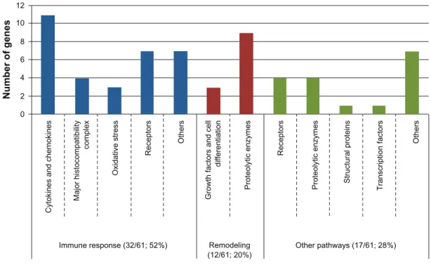
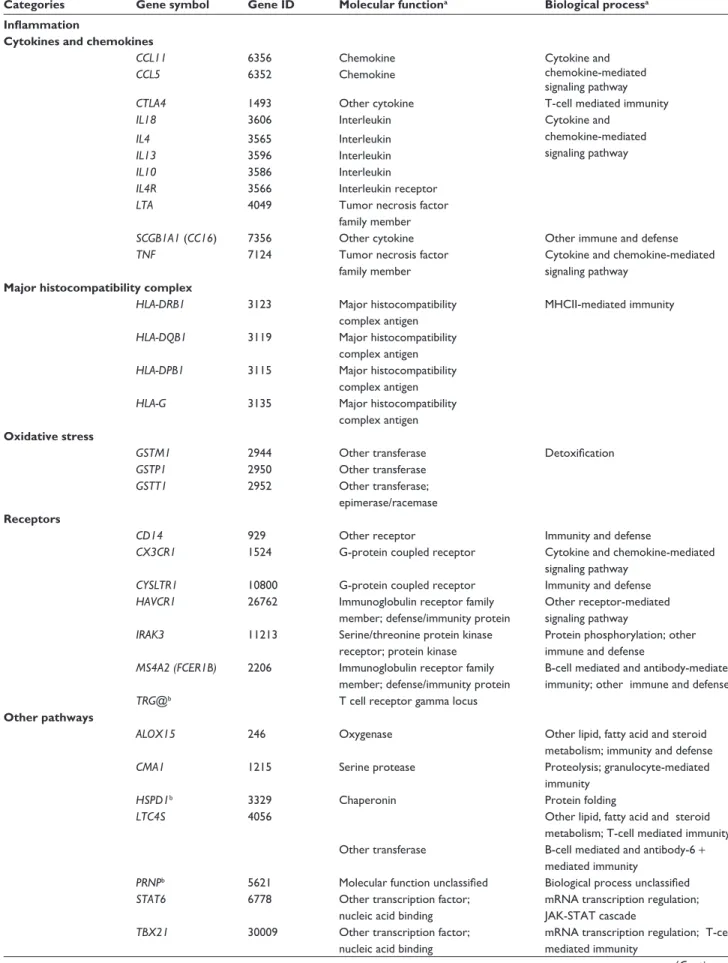
Several reviews have described the knowledge acquired on the genetics of asthma.22,24,121,125 Figure 3 classified the main
genes associated with asthma according to their biological
function into three principal categories: inflammation, remodeling, and other pathways. Most of the associated genes could be classified into the inflammation (32/61; 52%) or remodeling categories (12/61; 20%). Indeed, this classification not only underlines the great proportion of associated genes in the inflammatory response, but also underlines the need to better define the biological func-tion for some of the other genes (other pathways; 17/61, 28%) to better understand their implication in asthma. The complete list of genes for all categories seen in Figure 3 is available in Table 3 with ontology keywords.
Table 4 presents the same 61 genes classified accord-ing to the approach used to target them. As shown in this table, the main approach that has been used in the research of genetic determinants of asthma is the candidate gene approach. At the end of 2005, more than 100 genes had already been associated to asthma and another 54 associa-tions have been performed in 2006 and 2007.22,24 Candidate
gene association studies are based on the hypothesis of an association between the variants of a gene and asthma or one of its associated phenotypes. These phenotypes usually include respiratory capacity (wheezing, bronchial hyperreactivity, and pulmonary function measurements), immunological measures (total or specific blood IgE, allergy) or clinical criteria (atopic dermatitis, eczema, and rhinoconjunctivitis).121 This approach implies a
lit-erature review on genes involved in the development of the trait, or closely related to biological pathways linked with physiopathology of the trait. The choice of candidate genes can be based on several types of information: (1) gene functions, (2) documented associations with the trait in other populations or with closely related phenotypes, (3) difference of expression of the gene for the trait or a closely related phenotype, (4) at least one of these criteria documented for an animal model, or (5) association of another gene in the same biological pathway with the trait or with closely related phenotypes.22,127 Consequently, this
approach is biased according to the literature already pub-lished for the specific trait and the possibility of discovering new potentially interesting genes could be limited.22,127 This
limitation may be even more important considering that the underlying biology of the development of most complex traits remains to be defined.127
In order to go past this candidate gene selection bias, sev-eral approaches have been described. One of these is to select the candidate genes according to their known functions and to crosscheck the results with data obtained from genome-wide linkage, association, or expression studies.127 These genomic
approaches allow targeting of new genes without having to state a hypothesis regarding a specific gene or locus and, therefore, permit to increase the knowledge on the pathology of the trait.22,130
Before going further on the contributions of these techniques on the genetics of asthma, here is a brief descrip-tion of each of them. Linkage analysis and genome-wide association studies (GWAS) are two techniques used to identify chromosomal loci associated in a studied trait. In both cases, analyses are based on two principles: equal transmission of alleles and increase in recombination for increased distances between two markers.131 To identify those
loci, the linkage analysis uses families to compare
microsat-ellite markers transmission with phenotype transmission. Similarities between these two transmissions are translated into a statistical measure, the lod score. The lod score is greater when markers of a locus are more transmitted to sick children in studied families, and it is possible to hypothesize that genes near this locus are involved in the development of this disease.132 For GWAS, familial or case–control
cohorts can be used.133 Contrary to linkage analysis,
differ-ent genetic variant types can be used for GWAS as coding single nucleotide polymorphisms (SNPs), tagging SNPs, or copy number variants. Figure 4 presents a brief descrip-tion of the methodology used for this technique. Linkage analysis has been very efficient to target genes responsible of monogenic trait development but,132,134 in complex traits,
one disadvantage of linkage analysis is its impossibility to detect locus with modest effect on phenotype compared with GWAS.22,135 GWAS also has the advantage of targeting
more specific chromosomal regions (#500 bp) because of the great number of genetic variants (more than 500,000 variants) represented on a microarray (for examples, see: http://www.illumina.com/).22 Moreover, new developments
on genetics as HapMap international project, the development of new technologies as microarrays, and the possibility to
bring together many samples owned by different researchers opened the way to GWAS.121,136
Genome-wide expression studies are another genomic approach that allow the identification of new genes and pathways involved in a target disease. Figure 4 presents the main technical steps to perform genome-wide expression studies using Affymetrix technology as example. These studies, as GWAS, used microarrays. The expression of more than 25,000 genes or transcripts can be measured in the same microarray (www.affymetrix.com). These are used to compare gene expression profiles of affected and nonaffected subjects, of treated and nontreated subjects, of measures for the same subjects at pretreatment and posttreatment, etc. Some advantages of genome-wide expression studies are accessibility of the microarray technology and analysis tools for researchers and clinicians, no sequencing step and possi-bility to study thousands of genes simultaneously.137
About the contribution of these genome-wide techniques, the use of linkage studies has allowed targeting of genes involved in biological pathways that had not been studied in asthma before (Table 4). Several chromosomal regions have been associated with asthma using this method, but only a few have been replicated in several studies and populations (5q31–33, 6p21, 12q13–q24).22,138 GWAS and genome-wide
expression studies are now also being used in order to target new genes or validate observations about already associated genes in asthma. Until now, more than 62 genome-wide expression studies have been performed on asthma or one of its subphenotypes (as found in PubMed using “asthma and microarray” and “asthma and gene chip” keywords and according to Rolph et al139). Although most results obtained
from expression microarrays are exploratory and must be validated through functional or association studies, a few genes have been targeted by these analyses (Table 4).128 As for
GWAS, only a few studies have been performed on asthma so far. The first was conducted during the summer of 2007 Table 2 The definition criteria used to classify asthma according to severity
Severity Intermittent Mild persistent Moderate persistent Severe persistent
Symptom frequency Less than once a week More than once per week
but less than once per day Daily Daily
Nighttime symptoms Less than or equal to twice per month
More than twice per month More than once per week Frequent exacerbations Brief Could affect activities
and sleep
Could affect activities and sleep Frequent Fev1 percent of
predicted value
$80% $80% 60%–80% ,60%
variability of Fev1 ,20% 20%–30% .30% .30%
Note: Adapted from the Global Strategy for Asthma Management and Prevention, Global initiative for Asthma (GiNA).30
and allowed targeting of a chromosomal region that regu-lates the expression of the ORMDL3, GSDMB, and ZPBP2 genes.140,141 However, GWAS approach is becoming more
popular and many studies are underway in the field of asthma research. For example, to increase statistical power of the studies, researchers on asthma or other lung diseases group their samples together. Indeed, two major consortia have been made and will soon publish GWAS results. They are called GABRIEL Project (http://www.gabriel-fp6.org/) and EVE Asthma Genetics Consortium (http:// arrafunding.uchi-cago.edu/investigators/ober_c.shtml). GABRIEL includes independent asthma genetics studies of European ancestry and EVE includes independent ones from the United States. The scientific community, including these two consortia and others, actually work to plane a meta-global GWAS analy-sis. The great number of data researchers obtain from these studies will allow them to target new genes and pathways involved in asthma development or its subphenotypes. These may become new therapeutic candidates or may be part of a predictive genetic profile.124
Replication discrepancies
However, no matter what kind of approach is used, research-ers are confronted with the same question: why is there an
absence of validation in several cases? These nonreplications can be linked to the statistical power of the samples used, or to other phenotypic, genetic, environmental, or epigenetic concerns.121
To increase the chance to find polymorphisms associated with asthma and also replicate anterior results, statistical power of the sample is important. Genetic association studies are more powerful than genomic studies regarding number of subjects to number of genetic variants ratio. Indeed, with the great number of genetic variants or genes tested, the multiple testing problems are much more important in GWAS and genome-wide expression studies.142,143 It is known that
for a GWAS, a sample of approximately 15,000 individuals would be necessary to obtain a statistical power of approxi-mately 90% with a type I error of 5%.144 This objective can
be reached by grouping together all asthma genetic research studies. For expression studies, analyses showed that at least three to five samples per phenotypic group could be sufficient to find a difference of expression, depending on the objective of the study (ie, to look at greater or smaller differences of expression).145
Another cause of replication discrepancies is the great phenotypic heterogeneity observed in asthma, as described in the section “Phenotypic variability in
Number of gene s 12 10 8 6 4 2 0
Cytokines and chemokines
Oxidative stress Receptor s Receptor s Structural proteins Transcription factor s Others Others
Proteolytic enzymes Proteolytic enzymes
Growth factors and cell
differentiation Major histocompatibility complex Remodeling (12/61; 20%) Other pathways (17/61; 28%) Immune response (32/61; 52%)
Figure 3 Classification of the 61 main associated genes with asthma into biological functions.
Notes: This figure illustrates the two levels of classification of the 61 main associated genes with asthma. The first level of classification is made according to their known principal functions. From these known functions, a possible implication in the immune response or the remodeling response has been deduced. it is interesting to note that more than half (52%) of the main associated genes are in direct link with the immune response and that 20% may be involved in the remodeling process observed in asthma lung tissues. For the 17 genes classified in the “other pathways” category, their principal known function should help us orientate future research to better document their role in asthma. The complete list of genes for all categories seen in Figure 3 is available in Table 3 with ontology keywords.
Table 3 Classification of the 61 main genes associated with asthma according to their respective ontology keywords
Categories Gene symbol Gene ID Molecular functiona Biological processa
Inflammation
Cytokines and chemokines
CCL11 6356 Chemokine Cytokine and
chemokine-mediated signaling pathway
CCL5 6352 Chemokine
CTLA4 1493 Other cytokine T-cell mediated immunity
IL18 3606 interleukin Cytokine and
chemokine-mediated signaling pathway
IL4 3565 interleukin
IL13 3596 interleukin
IL10 3586 interleukin
IL4R 3566 interleukin receptor
LTA 4049 Tumor necrosis factor
family member
SCGB1A1 (CC16) 7356 Other cytokine Other immune and defense
TNF 7124 Tumor necrosis factor
family member
Cytokine and chemokine-mediated signaling pathway
Major histocompatibility complex
HLA-DRB1 3123 Major histocompatibility
complex antigen
MHCii-mediated immunity
HLA-DQB1 3119 Major histocompatibility
complex antigen
HLA-DPB1 3115 Major histocompatibility
complex antigen
HLA-G 3135 Major histocompatibility
complex antigen
Oxidative stress
GSTM1 2944 Other transferase Detoxification
GSTP1 2950 Other transferase
GSTT1 2952 Other transferase;
epimerase/racemase
Receptors
CD14 929 Other receptor immunity and defense
CX3CR1 1524 G-protein coupled receptor Cytokine and chemokine-mediated
signaling pathway
CYSLTR1 10800 G-protein coupled receptor immunity and defense
HAVCR1 26762 immunoglobulin receptor family
member; defense/immunity protein
Other receptor-mediated signaling pathway
IRAK3 11213 Serine/threonine protein kinase
receptor; protein kinase
Protein phosphorylation; other immune and defense
MS4A2 (FCER1B) 2206 immunoglobulin receptor family
member; defense/immunity protein
B-cell mediated and antibody-mediated immunity; other immune and defense
TRG@b T cell receptor gamma locus
Other pathways
ALOX15 246 Oxygenase Other lipid, fatty acid and steroid
metabolism; immunity and defense
CMA1 1215 Serine protease Proteolysis; granulocyte-mediated
immunity
HSPD1b 3329 Chaperonin Protein folding
LTC4S 4056
Other transferase
Other lipid, fatty acid and steroid metabolism; T-cell mediated immunity; B-cell mediated and antibody-6 + mediated immunity
PRNPb 5621 Molecular function unclassified Biological process unclassified
STAT6 6778 Other transcription factor;
nucleic acid binding
mRNA transcription regulation; JAK-STAT cascade
TBX21 30009 Other transcription factor;
nucleic acid binding
mRNA transcription regulation; T-cell mediated immunity
Table 3 (Continued)
Categories Gene symbol Gene ID Molecular functiona Biological processa
Remodeling
Growth factors and cell differentiation
AREG 374 Growth factor Ligand-mediated signaling
GSDMB 55876 Molecular function unclassified Developmental processes
TGFB1 7040 Growth factor Other receptor-mediated signaling
pathway
Proteolytic enzymes
ACE 1636 Metalloprotease Proteolysis
ADAM33 80332 Metalloprotease
CHI3L1 1116 Glycosidase Other polysaccharide metabolism
chi3l3 (ym1)b 12655
(Mus musculus)
Molecular function unclassified Biological process unclassified NOS1 4842 Synthase; oxidoreductase;
calmodulin-related protein
electron transport
PLAU 5328 Serine protease Proteolysis
SERPINE1 (PAI1) 5054 Serine protease inhibitor
SERPINH1 871 Serine protease inhibitor
SERPINB4 6318 Serine protease inhibitor
Other pathways Proteolytic enzymes
ARG1 383 Other hydrolase Amino acid catabolism
ARG2 384 Other hydrolase
NAT2 10 Acetyltransferase Other metabolism
PDE4D 5144 Phosphodiesterase Metabolism of cyclic nucleotides
Receptors
ADRB2 154 G-protein coupled receptor G-protein mediated signaling
NPSR1 (GPRA) 387129 G-protein coupled receptor
OPN3 23596 G-protein coupled receptor
TBXA2R 6915 G-protein coupled receptor G-protein mediated signaling
Structural proteins
FLG 2312 Other cytoskeletal proteins Protein complex assembly
Transcription factors
VDR 7421 Nuclear hormone receptor;
transcription factor
mRNA transcription regulation
Others
COL29A1b 256076 Molecular function unclassified Cell adhesion
CYFIP2 26999 Other G-protein modulator Signal transduction
DPP10 57628 Select regulatory molecule Protein targeting and localization
ORMDL3 94103 Other miscellaneous function protein Miscellaneous
PHF11 51131 Double-stranded DNA binding protein General mRNA transcription activities
POSTN 10631 Other cell adhesion molecule Cell adhesion
SPINK5 11005 Select regulatory molecule Homeostasis
Notes: aOntology keywords from the Panther Classification System (http://www.pantherdb.org/). bGenes not recognized by the Panther Classification System. Those have
been classified according to description available in Entrez Gene by NCBI (http://www.ncbi.nlm.nih.gov/gene). See also Madore et al.126
asthma”. This phenotypic heterogeneity can reflect geno-typic heterogeneity, thus implying that the severity and onset of asthma could be influenced by the asthmatics’ genetic imprint.146,147 Thus, a precise characterization of
the subjects remains essential. This precise phenotypic description of the sample helps to better select similar independent samples to replicate genetic association results and so helps to reduce the impact of differences in recruitment criteria and in population stratification
between samples.146 According to the fact that asthma
is rather a collection of several diseases than a unique trait, the key of replication success may lie in association studies with asthma subphenotypes. The review article written by Wenzel39 presents several ways to categorize
asthma subphenotypes that can be used for association and expression studies.
Gene–environment interactions are another source of variability since the environment can modulate the effect of
a gene on the resulting phenotype.148 Studies showed that
gene–environment interactions had a greater effect on the phenotype than the environment and genes separately.149 For
example, some studies have demonstrated that a high exposure to endotoxin seems to switch the susceptibility effect of the CD14/-159C allele to a protective effect compared with the CD14/-159T allele.150–152 Another example of gene–environment interaction is
the interaction between polymorphisms in the regulating regions of a gene and the cell differentiation cycle, leading to differences on the expression of a specific allele.153
A part of the etiology of asthma could be explained by epigenetic changes. Epigenetics is the study of changes that are heritable and that modulate gene expression without directly altering gene sequence.120 Those changes can be done
through DNA methylation or posttranslational modification of histones: eg, acetylation, methylation, phosphorylation, and ubiquitylation.154,155 Studies have demonstrated that
epigenetic changes are heritable for almost two subsequent generations (eg, methylation of a coat-color gene in mice).156 Many studies in asthma underline the possible
role of epigenetics in its development mostly for early onset asthma (for a review, see Miller and Ho120). For example,
exposition to environmental tobacco smoke during prenatal development or during the first years of life could influence
the development of asthma and may be transmitted across two generations.157–159
Gene–gene interactions are another source of variability. In complex traits, a single mutation often has a minor impact, but the combination of several mutations should increase the influences on a phenotype. In asthma, a syn-ergy between a mutation in the IL4 (rs2243250), the IL13 (rs1800925), the IL4 R (rs1805010) and in the signal trans-ducer and activator of transcription 6 (STAT6) (rs324011) genes was documented and increases by 10.8 times the risk of having a high level of IgE and by 16.8 times the risk of developing asthma.160
The solution to these replication problems might partly lie in new genomic methods. In fact, new developments in the field of genetic and genomic research (for example, microarrays for GWAS and genome-wide expression studies), as well as knowledge acquired on the genome and its polymorphisms (as the HapMap project) allow the implementation of new analysis tools. Indeed, although there is still no consensus regarding the analysis methods that should be employed, new tools have been developed to overcome these problems. For example, many new software packages are developed or optimized regularly in Bioconductor, an open source software for the analysis Table 4 List of the 61 main associated genes in genetic and genomic studies on asthma and classified according to their identification
method
Genes identification methods
Candidate genesa Linkage analysisb GWAS Genomic expression
studyc ADRB2 ACE SCGB1A1 (CC16) CCL5 CCL11 CD14 CMA1 CTLA4 MS4A2 (FCERIB) FLG GSTM1 GSTP1 GSTT1 HAVCR1 HLA-DPB1 HLA-DQB1 HLA-DRB1 IL4 IL4R IL10 IL13 IL18 LTA LTC4S NAT2 NOS1 SPINK5 STAT6 TBX21 TBXA2R TGFB1 TNF ADAM33a COL29A1 CYFIP2 CYSLTR1 DPP10 HLA-G IRAKM NPSR1 (GPRA)a OPN3 PHF11 VDR CHI3L1 GSDMB ORMDL3 PDE4D TCRγ ou TARP (BC072396) ALOX15 AREG ARGI ARGII CX3CR1 HSPD1 PLAU POSTN PRNP SERPINE1 (PAI-1) SERPINB4 SERPINH1 Yml (Chi3l3)
Abbreviation: GwAS, genome-wide association study.
Notes: aAssociated genes listed are those that have been replicated in almost five different studies according to Vercelli.121 Underlined genes are those that have been
associated in more than 30 different studies.121bAssociated genes are those targeted by linkage analysis according to Ober and Hoffjan22 and Moffatt.125cPrincipal genes
identified using genomic expression studies in asthma according to Izuhara and Saito.128 Genes in bold are those identified by Laprise et al129 and were positively associated
with asthma. Genes in bold and underlined are those identified by Madore et al126 and that were validated by semi-quantitative real-time polymerase chain reaction (PCR).
A Genome-wide amplification Fragmentation Fragmentation Denaturation Annealing on microarrays Annealing on microarrays Signal amplification (complementary base and antibody) Analysis of results mRNA Reverse transcription (cDNA) In vitro transcription (cRNA) Biotinylation Analysis of results GWAS
(Illumina’s Infinium II)
B Genome-wide expression (Affymetrix)
Figure 4 illustration of the methodological steps for genome-wide association studies and genome-wide expression studies.
Notes: Part A of this figure illustrates methodological steps of genome-wide association studies (GWAS) done using Illumina technology. Briefly, the first step is the amplification of the complete genome. Second, amplified DNA is fragmented using digestion enzymes. Denaturized fragments are then hybridized to short oligonucleotide sequences on microarrays. Finally, amplification with a fluorescent complementary base pair will allow reading and analyzing the results (illumina http:// www.illumina.com/pages.ilmn?iD=40). Part B of the figure illustrates methodological steps of genome-wide expression study performed using Affymetrix technology. The first step is the reverse transcription of mRNA into cDNA, followed by transcription of cDNA into cRNA. Afterward, cRNA is biotinylated, fragmented, and annealed with primers synthesized on the microarray. The signal intensity is proportional to the number of copies present in the sample.
and comprehension of genomic data, to better analyze and characterize data obtained from genome-wide studies (www.bioconductor.org). New genome-wide analysis tools are promising avenues to find new biological pathways, gene families or chromosomal loci involved in asthma and complex diseases, but candidate gene approach will remain a useful and powerful tool to refine our search for associ-ated genes and to help better define the pathways involved in asthma development.
Acknowledgment
Catherine Laprise is the chair holder of the Canada Research Chair for genetic determinants in asthma (www.chairs. gc.ca) and is responsible for the inflammation and remod-eling strategic group of the Respiratory Health Network
(RHN) of the Fonds de la recherche en santé du Quebec (FRSQ).
Disclosure
The authors report no conflicts of interest in this work.
References
1. Averbeck M, Gebhardt C, Emmrich F, Treudler R, Simon JC. Immuno-logic principles of allergic disease. J Dtsch Dermatol Ges. 2007;5(11): 1015–1028.
2. Platts-Mills TAE. Immediate hypersensitivity (Type I). In: Male D, Bro-stoff J, Roth DB, Roitt I, editors. Immunology. 7th ed. Philadelphia, PA: Elsevier; 2006:423–447.
3. Arbes SJ Jr, Gergen PJ, Elliott L, Zeldin DC. Prevalences of positive skin test responses to 10 common allergens in the US population: results from the third National Health and Nutrition Examination Survey. J Allergy Clin Immunol. 2005;116(2):377–383.
4. Lack G. New developments in food allergy: old questions remain. J Allergy Clin Immunol. 2004;114(1):127–130.
5. Masoli M, Fabian D, Holt S, Beasley R. Global burden of asthma. Developed for the Global Initiative for Asthma. Medical Research Institute of New Zealand, Wellington, New Zealand and University of Southampton, Southampton, United Kingdom; 2004.
6. Settipane RA, Charnock DR. Epidemiology of rhinitis: allergic and nonallergic. Clin Allergy Immunol. 2007;19:23–34.
7. Gell PGH, Coombs RRA. The classification of allergic reactions under-lying disease. In: Coombs RRA, Gell PGH, editors. Clinical Aspects of Immunology. London, UK: Blackwell Science; 1963.
8. Verstraelen S, Bloemen K, Nelissen I, Witters H, Schoeters G, Van Den Heuvel R. Cell types involved in allergic asthma and their use in in vitro models to assess respiratory sensitization. Toxicol In Vitro. 2008;22(6):1419–1431.
9. Gilfillan AM, Tkaczyk C. Integrated signalling pathways for mast-cell activation. Nat Rev Immunol. 2006;6(3):218–230.
10. Metzger H. The receptor with high affinity for IgE. Immunol Rev. 1992;125:37–48.
11. Male D. Hypersensitivity (Type II). In: Male D, Brostoff J, Roth DB, Roitt I, editors. Immunology. 7th ed. Philadelphia, PA: Elsevier; 2006: 449–460. 12. Hay F, Westwood OMR. Hypersensitivity (Type III). In: Male D, Brostoff
J, Roth DB, Roitt I, editors. Immunology. 7th ed. Philadelphia: Elsevier; 2006:461–476.
13. Britton W. Type IV hypersensitivity. In: Male D, Brostoff J, Roth DB, Roitt I, editors. Immunology. 7th ed. Philadelphia, PA: Elsevier; 2006: 477–491. 14. Johansson SG, Hourihane JO, Bousquet J, et al. A revised nomenclature
for allergy. An EAACI position statement from the EAACI nomenclature task force. Allergy. 2001;56(9):813–824.
15. Hopp RJ, Bewtra AK, Watt GD, Nair NM, Townley RG. Genetic analysis of allergic disease in twins. J Allergy Clin Immunol. 1984; 73(2):265–270. 16. Lichtenstein P, Svartengren M. Genes, environments, and sex: factors
of importance in atopic diseases in 7-9-year-old Swedish twins. Allergy. 1997;52(11):1079–1086.
17. Isidoro-Garcia M, Davila-Gonzalez I, Pascual de Pedro M, Sanz-Lozano C, Lorente-Toledano F. Interactions between genes and the environment. Epi-genetics in allergy. Allergol Immunopathol (Madr). 2007;35(6):254–258. 18. Gartner K. A third component causing random variability beside environ-ment and genotype. A reason for the limited success of a 30 year long effort to standardize laboratory animals? Lab Anim. 1990; 24(1):71–77. 19. Hong X, Tsai HJ, Wang X. Genetics of food allergy. Curr Opin Pediatr.
2009;21(6):770–776.
20. Renkonen J, Mattila P, Parviainen V, Joenvaara S, Toppila-Salmi S, Renkonen R. A network analysis of the single nucleotide polymorphisms in acute allergic diseases. Allergy. 2010;65(1):40–47.
21. Barnes KC. An update on the genetics of atopic dermatitis: scratching the surface in 2009. J Allergy Clin Immunol. 2010;125(1):16–29, e1–e11; quiz 30–31.
22. Ober C, Hoffjan S. Asthma genetics 2006: the long and winding road to gene discovery. Genes Immun. 2006;7(2):95–100.
23. Williams H, Robertson C, Stewart A, et al. Worldwide variations in the prevalence of symptoms of atopic eczema in the International Study of Asthma and Allergies in Childhood. J Allergy Clin Immunol. 1999;103(1 Pt 1):125–138.
24. Zhang J, Pare PD, Sandford AJ. Recent advances in asthma genetics. Respir Res. 2008;9:4.
25. Palikhe NS, Kim SH, Park HS. What do we know about the genetics of aspirin intolerance? J Clin Pharm Ther. 2008;33(5):465–472. 26. Gueant JL, Gueant-Rodriguez RM, Gastin IA, et al. Pharmacogenetic
determinants of immediate and delayed reactions of drug hypersensitivity. Curr Pharm Des. 2008;14(27):2770–2777.
27. Castro-Giner F, Bustamante M, Ramon Gonzalez J, et al. A pooling-based genome-wide analysis identifies new potential candidate genes for atopy in the European Community Respiratory Health Survey (ECRHS). BMC Med Genet. 2009;10:128.
28. Gomes ER, Demoly P. Epidemiology of hypersensitivity drug reactions. Curr Opin Allergy Clin Immunol. 2005;5(4):309–316.
29. Holgate ST. Pathogenesis of asthma. Clin Exp Allergy. 2008;38(6): 872–897.
30. Global Initiative for Asthma. Global strategy for asthma management and prevention. 2008.
31. Global Initiative for Asthma. Global strategy for asthma management and prevention. 2006.
32. Humbert M. Does “intrinsic” asthma exist? Rev Mal Respir. 2000; 17(1 Pt 2):245–254.
33. Kerkhof M, De Graaf A, Droste JHJ. Prevalentie van astmatische klachten in drie regio’s in Nederland. Tijdschr Soc Gezondheidsz. 1994;72:181–185. 34. Platts-Mills TA, Carter MC. Asthma and indoor exposure to allergens.
N Engl J Med. 1997;336(19):1382–1384.
35. Rijcken B, Kerkhof M, de Graaf A. Europees Luchtweg Onderzoek Nederland (ELON). Groningen: Rijksuniversiteit Groningen; 1996. 36. Turkeltaub PC, Gergen PJ. Prevalence of upper and lower respiratory
conditions in the US population by social and environmental factors: data from the second National Health and Nutrition Examination Survey, 1976 to 1980 (NHANES II). Ann Allergy. 1991;67(2 Pt 1):147–154. 37. Woolcock AJ, Peat JK, Salome CM, et al. Prevalence of bronchial
hyperresponsiveness and asthma in a rural adult population. Thorax. 1987;42(5):361–368.
38. Kiley J, Smith R, Noel P. Asthma phenotypes. Curr Opin Pulm Med. 2007;13(1):19–23.
39. Wenzel SE. Asthma: defining of the persistent adult phenotypes. Lancet. 2006;368(9537):804–813.
40. Cockcroft DW, Murdock KY, Berscheid BA, Gore BP. Sensitivity and specificity of histamine PC20 determination in a random selection of young college students. J Allergy Clin Immunol. 1992;89(1 Pt 1): 23–30.
41. Postma DS, Koppelman GH, Meyers DA. The genetics of atopy and airway hyperresponsiveness. Am J Respir Crit Care Med. 2000; 162(3 Pt 2):S118–S123.
42. Male D, Brostoff J, Roth D, Roitt I. Immunology. 7th ed. London: Mosby; 2006.
43. Beasley R, Pekkanen J, Pearce N. Has the role of atopy in the devel-opment of asthma been over-emphasized? Pediatr Pulmonol Suppl. 2001;23:149–150.
44. Kay AB. The role of eosinophils in the pathogenesis of asthma. Trends Mol Med. 2005;11(4):148–152.
45. Constant SL, Brogdon JL, Piggott DA, et al. Resident lung antigen-presenting cells have the capacity to promote Th2 T cell differentiation in situ. J Clin Invest. 2002;110(10):1441–1448.
46. Julia V, Hessel EM, Malherbe L, Glaichenhaus N, O‘Garra A, Coffman RL. A restricted subset of dendritic cells captures airborne antigens and remains able to activate specific T cells long after antigen exposure. Immunity. 2002;16(2):271–283.
47. Riese RJ, Chapman HA. Cathepsins and compartmentaliza-tion in antigen presentacompartmentaliza-tion. Curr Opin Immunol. 2000;12(1): 107–113.
48. Kuipers H, Heirman C, Hijdra D, et al. Dendritic cells retrovirally overexpressing IL-12 induce strong Th1 responses to inhaled antigen in the lung but fail to revert established Th2 sensitization. J Leukoc Biol. 2004;76(5):1028–1038.
49. Anderson GP. The immunobiology of early asthma. Med J Aust. 2002; 177 Suppl:S47–S49.
50. Dahlen B, Shute J, Howarth P. Immunohistochemical localisation of the matrix metalloproteinases MMP-3 and MMP-9 within the airways in asthma. Thorax. 1999;54(7):590–596.
51. Kaur D, Saunders R, Berger P, et al. Airway smooth muscle and mast cell-derived CC chemokine ligand 19 mediate airway smooth muscle migration in asthma. Am J Respir Crit Care Med. 2006;174(11): 1179–1188.
52. Bradding P, Walls AF, Holgate ST. The role of the mast cell in the pathophysiology of asthma. J Allergy Clin Immunol. 2006;117(6): 1277–1284.
53. Wenzel SE, Balzar S, Cundall M, Chu HW. Subepithelial basement membrane immunoreactivity for matrix metalloproteinase 9: associa-tion with asthma severity, neutrophilic inflammaassocia-tion, and wound repair. J Allergy Clin Immunol. 2003;111(6):1345–1352.
54. Holsapple MP, Jones D, Kawabata TT, et al. Assessing the poten-tial to induce respiratory hypersensitivity. Toxicol Sci. 2006; 91(1):4–13.
55. Ogawa Y, Calhoun WJ. The role of leukotrienes in airway inflammation. J Allergy Clin Immunol. 2006;118(4):789–798; quiz 799–800. 56. Bradding P, Holgate ST. Immunopathology and human mast cell
cytok-ines. Crit Rev Oncol Hematol. 1999;31(2):119–133.
57. Nouri-Aria KT, Irani AM, Jacobson MR, et al. Basophil recruitment and IL-4 production during human allergen-induced late asthma. J Allergy Clin Immunol. 2001;108(2):205–211.
58. Nissim Ben Efraim AH, Levi-Schaffer F. Tissue remodeling and angio-genesis in asthma: the role of the eosinophil. Ther Adv Respir Dis. 2008;2(3):163–171.
59. Lampinen M, Carlson M, Hakansson LD, Venge P. Cytokine-regulated accumulation of eosinophils in inflammatory disease. Allergy. 2004;59(8):793–805.
60. Lee JJ, Dimina D, Macias MP, et al. Defining a link with asthma in mice con-genitally deficient in eosinophils. Science. 2004;305(5691): 1773–1776. 61. Schmid-Grendelmeier P, Altznauer F, Fischer B, et al. Eosinophils
express functional IL-13 in eosinophilic inflammatory diseases. J Immunol. 2002;169(2):1021–1027.
62. Adelroth E, Morris MM, Hargreave FE, O’Byrne PM. Airway respon-siveness to leukotrienes C4 and D4 and to methacholine in patients with asthma and normal controls. N Engl J Med. 1986;315(8):480–484. 63. Lee E, Robertson T, Smith J, Kilfeather S. Leukotriene receptor
antagonists and synthesis inhibitors reverse survival in eosinophils of asthmatic individuals. Am J Respir Crit Care Med. 2000;161(6): 1881–1886.
64. Marom Z, Shelhamer JH, Bach MK, Morton DR, Kaliner M. Slow-reacting substances, leukotrienes C4 and D4, increase the release of mucus from human airways in vitro. Am Rev Respir Dis. 1982; 126(3):449–451. 65. Frigas E, Motojima S, Gleich GJ. The eosinophilic injury to the mucosa
of the airways in the pathogenesis of bronchial asthma. Eur Respir J Suppl. 1991;13:123s–135s.
66. Jacobsen EA, Taranova AG, Lee NA, Lee JJ. Eosinophils: singularly destructive effector cells or purveyors of immunoregulation? J Allergy Clin Immunol. 2007;119(6):1313–1320.
67. Van Hove CL, Maes T, Joos GF, Tournoy KG. Chronic inflam-mation in asthma: a contest of persistence vs resolution. Allergy. 2008;63(9):1095–1109.
68. Brightling CE, Bradding P, Symon FA, Holgate ST, Wardlaw AJ, Pavord ID. Mast-cell infiltration of airway smooth muscle in asthma. N Engl J Med. 2002;346(22):1699–1705.
69. Plante S, Semlali A, Joubert P, et al. Mast cells regulate procollagen I (alpha 1) production by bronchial fibroblasts derived from subjects with asthma through IL-4/IL-4 delta 2 ratio. J Allergy Clin Immunol. 2006;117(6):1321–1327.