OATAO is an open access repository that collects the work of Toulouse
researchers and makes it freely available over the web where possible
Any correspondence concerning this service should be sent
to the repository administrator:
[email protected]
This is a Publisher’s version published in:
http://oatao.univ-toulouse.fr/24199
To cite this version:
Esneau, Camille and Raynal, Bertrand and Roblin, Pierre
and Brûlé, Sébastien
and Richard, Charles-Adrien and Fix, Jenna and Eléouët, Jean-François and
Galloux, Marie Biochemical characterization of the respiratory syncytial virus N0-P
complex in solution. (2019) Journal of Biological Chemistry, 294 (10). 3647-3660.
ISSN 0021-9258
1
Biochemical characterization of the respiratory syncytial virus N
0-P complex in solution
Camille Esneau
1#, Bertrand Raynal
2, Pierre Roblin
3, 4, Sébastien Brûlé
2, Charles-Adrien
Richard
1, Jenna Fix
1, Jean-François Eléouët
1*, and Marie Galloux
1*From the
1VIM, INRA, Université Paris-Saclay, Jouy-en-Josas, France;
2Plate-forme de Biophysique
Moléculaire, C2RT, Institut Pasteur, 25 rue du docteur roux, 75015 Paris, France;
3Synchrotron
SOLEIL, l'Orme des Merisiers, F-91410 Saint Aubin, France;
4Laboratoire de Génie Chimique,
Université Paul Sabatier, UMR 5503, Toulouse, France.
Running title: Biochemical characterization of the RSV N
0-P complex
#
Present address: Hunter Medical Research Institute, New Lambton Heights NSW 2305, Australia
* Corresponding authors: Tel: (33) 1 34 65 26 10; FAX: (33) 1 34 65 26 21; email:
[email protected], jean-franç[email protected]
Keywords
:
Respiratory syncytial virus, nucleoprotein N, viral replication, structure-function,
N
0-P complex
http://www.jbc.org/cgi/doi/10.1074/jbc.RA118.006453
The latest version is at
JBC Papers in Press. Published on January 9, 2019 as Manuscript RA118.006453
by guest on September 3, 2019
http://www.jbc.org/
2
ABSTRACTAs all the viruses belonging to the Mononegavirales order, the non-segmented negative strand RNA genome of respiratory syncytial virus (RSV) is encapsidated by the viral nucleoprotein N. N protein polymerizes along the genomic and anti-genomic RNAs during replication. This requires the maintenance of the neosynthesized N protein in a monomeric and RNA-free form by the viral phosphoprotein P that plays the role of a chaperone protein, forming a soluble N0-P complex. We have previously demonstrated that residues 1-30 of P specifically bind to N0. Here, to isolate a stable N0-P complex suitable for structural studies, we used the N-terminal peptide of P (P40) to purify truncated forms of the N protein. We show that to purify a stable N0-P-like complex, a deletion of the first 30 N-terminal residues of N (NΔ30) is required to impair N oligomerization, whereas the presence of a full-length C-arm of N is required to inhibit RNA binding. We generated structural models of the RSV N0-P with biophysical approaches, including hydrodynamic measurements and small-angle X-ray scattering (SAXS), coupled with biochemical and functional analyses of human RSV (hRSV) NΔ30 mutants. These models suggest a strong structural homology between the hRSV and the human metapneumovirus (hMPV) N0-P complexes. In both complexes, the P40-binding sites on N0 appear to be similar, and the C-arm of N provides a high flexibility and a propensity to interact with the N RNA groove. These findings reveal two potential sites to target on N0-P for the development of RSV antivirals.
Introduction
Human respiratory syncytial virus (hRSV) is the leading cause of severe respiratory tract infections in newborn children worldwide (1). hRSV infects close to 100% of infants within the first two years of life. It is the main cause of bronchiolitis in young children, as well as a significant cause of severe respiratory infections in the elderly. The virus belongs to the Mononegavirales order and Pneumoviridae family (2). Like all the viruses belonging to this order, the hRSV genome is a non-segmented negative strand RNA. This genome of 15 kb is enwrapped by the nucleoprotein (N), forming a helical nucleocapsid (NC) (3) (4). The crystal structures of the Mononegavirales N proteins complexed with RNA show that they all present a similar structural organization with two globular domains (NNTD and NCTD) that form the RNA groove, and N- and C-arms that are involved in N oligomerization (5). The ribonucleoprotein complex constitutes the template for viral transcription and replication by the viral RNA-dependent-RNA-polymerase (RdRp) (reviewed in (6) (7)). During the viral cycle, the constant supply of neosynthesized monomeric and RNA-free N protein (named N0)
constitutes a prerequisite during replication to encapsidate neo-synthesized single-stranded genomic RNA (-RNA) and anti-genomic RNA (+RNA). By analogy with paramyxoviruses and rhabdoviruses, the hRSV P protein plays the role of a chaperone protein by preventing N to bind to cellular RNAs and N self-oligomerization (5) (8) (9). Invariably, the N-terminal residues of P are sufficient to maintain the N0 form. However, the structure of the RSV N0-P complex still remains to be solved, and those from related viruses revealed a specificity of P binding at the surface of N proteins that highlighted different mechanisms of the chaperone activity of P. For the vesicular stomatitis virus (VSV) N0-P complex, the residues 11-35 of P adopt an alpha-helical conformation upon binding to the surface of N, and this binding domain overlaps both the with RNA binding groove and the binding surface of the Ni+1 N-arm involved in N oligomerization (10). The crystal structures of the Nipah and Measles N0-P complexes show that the P peptide folds into two short helices that bind on the NCTD (11) (12). In these complexes, P binding prevents N from self-oligomerization and maintains it in an open conformation that impairs the interaction with RNA. A similar mechanism was found for the N0-P of Ebola and Marburg viruses, although the P peptide does not bind at the top of the NCTD but near the RNA groove, and adopts a specific conformation with two short helices (13) (14). More recently, the structure of the N0-P complex of Parainfluenza 5 (PIV5) showed that the binding of the P peptide to the NCTD blocks both the sites involved in the binding of RNA and of the N-arm of Ni+1 required for oligomerization, maintaining N in an open conformation (15). Finally, the 3D structure of the human Metapneumovirus (hMPV) N0-P complex, that is phylogenetically the closest virus relative to hRSV, revealed that P binds on the NCTD but also that the C-arm of N presents a specific conformation to block the accessibility to RNA (16).
For RSV, using an N mutant that does not interact with RNA, we have previously shown that the residues 1-30 of P are involved in the chaperone activity (17). The periodicity of the residues critical for N binding also suggests that residues 13-28 of P would adopt a helical conformation upon binding to N. This hypothesis was recently supported by NMR studies of RSV P that revealed the presence of a transient α-helix from Asp12 to Ile24 (18). However, the poor stability of the N mutant did not allow us to perform structural studies of the complex.
In this work, we rationally designed deletions of the N- and C-arms of the RSV N protein to destabilize the oligomeric organization of N-RNA rings in order to purify a stable N0-P like complex, when co-expressed with the N-terminal part of P (P40). Using biochemical and biophysical approaches, we obtained structural information about this complex in solution. Our results highlight a strong structural
by guest on September 3, 2019
http://www.jbc.org/
3
homology between the hMPV and hRSV N0-Pcomplexes. Results
Purification of a stable RSV N0-P like complex We have previously shown that a surrogate of the RSV N0-P complex can be obtained by co-expression of a K170A/R185 double N mutant with the N-terminus of P in bacteria (17). However, the poor stability of this complex at high concentration impaired further structural characterization. Previous studies have shown that the deletion of the 12 N-terminal residues of N, that correspond to nearly half of the N-arm of N, was sufficient to purify a monomeric RNA-free N protein (19) (20). On the other hand, except for hMPV (16), the available crystal structures of N0-P complexes of Mononegavirales were obtained by deleting either the entire N-arm or both N- and C-arms of N (10) (11) (13) (14) (12) (15). Based on these data, we decided to generate truncated N proteins deleted of either N-arm, or both the N- and C-arms (Fig. 1A) to seek for a stable N0 recombinant protein. Mutant N proteins were co-expressed in E. coli with GST-P40 (40 N-terminal residues of P) and purified on glutathione-Sepharose beads by affinity chromatography. Among the four N expression constructs that were tested, only the N protein deleted of both N- and C-arms was not co-purified with GST-P40 (Fig. 1B). To further characterize the purified N-P complexes, the GST tag was cleaved by TEV protease. We first checked the absence of RNA associated with the isolated recombinant proteins, by measuring the OD260nm/OD280nm absorption ratio. Whereas these ratios were ˂ 1 for N[13-391] and N[31-391], it was close to 2 for N[13-361], showing a strong tendency to associate to nucleic acids for this construct. These observations strongly suggest that the C-arm of N is required to prevent binding of N to RNA during the formation of the N0-P complex.
The purified recombinant N[13-391]-P40 and N[31-391]-P40 complexes were then analyzed by size exclusion chromatography. The N[31-391]-P40 complex eluted from the column as a single peak that corresponds to an apparent molecular weight of ~ 50 kDa (extrapolated from S200 calibration profile), and the OD260nm/OD280nm value ˂ 1 confirmed the absence of RNA (Fig. 1C). For the N[13-391]-P40 complex, in addition to the peak of N protein with apparent molecular weight of ~ 50 kDa and RNA free, a second peak with an apparent molecular weight of ~ 500 kDa probably associated to RNA (OD260nm/OD280nm ˃ 1) corresponding to N-RNA oligomers was obtained (Fig. 1C). These results reveal that deletion of the entire N-arm of N is required to obtain a stable monomeric N0-P complex surrogate. Finally, the secondary structure of the recombinant purified N[31-391]-P40 complexwas analyzed by circular dichroism (CD). As shown in
Figure 1D, the far-UV CD spectra of the N0-P
complex surrogate was similar to the spectra of the N-RNA rings; both CD traces presented a positive Cotton effect at 190 nm and two negative peaks at 208 and 222 nm, typical of secondary structures with a mainly α-helical content. This result confirms that no major secondary structure change occurs in the N0 variant when compared to the N-RNA state, as previously shown (17).
Altogether, our data show that the RSV N[31-391]recombinant protein, subsequently named NΔ30, co-purified with the peptide P40, is a functional surrogate of the RSV N0-P complex. The NΔ30-P40 complex was also found to be monomeric upon concentration. Furthermore, our results strongly suggest that the C-arm of N plays a major role in preventing N binding to RNA.
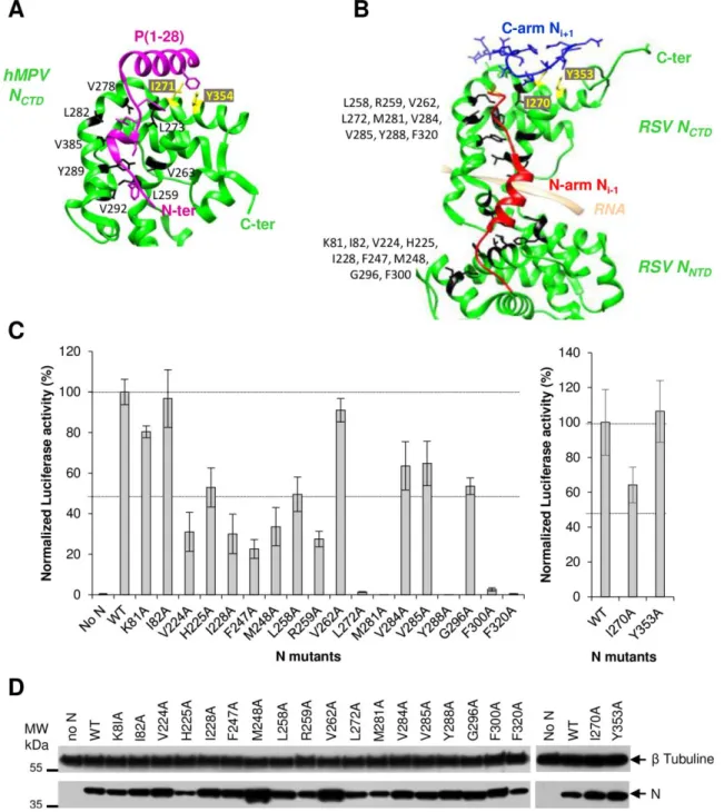
Investigation of the P40 binding site at the surface of the N protein by site-directed mutagenesis We then tried to identify the binding site of P40 on monomeric N. By analogy with the structure of N0 -P complexes of other Mononegavirales, it is expected that P plays a chaperone activity by binding to a surface of N that overlaps with the fixation site of the N-arm of the Ni+1 protomer within the nucleocapsid, thus impairing N oligomerization. The recent structural elucidation of the hMPV N0-P complex revealed that P binding also compete with the C-arm of the Ni-1 protomer involved in oligomerization (Fig. 2A) (16). Based on these data and on the structure of RSV N-RNA rings (21), we identified 20 residues at the surface of N protomer that could be involved in the interaction with P40 (Fig. 2B). Among these residues, 18 residues are implicated in the interaction with the N-arm of Ni+1 protomer, and the two residues I270 and Y353, are involved in the interaction with the C-arm of Ni-1 protomer. We first attempted to determine the impact of alanine substitution of these residues on the polymerase activity using the minigenome assay, as described previously (22) (23). Preventing the formation of a N0-P complex competent for genomic or antigenomic RNA encapsidation would result in a decrease in N-RNA template formation, and therefore of mRNA transcription and expression of the Luciferase (Luc) reporter. Whereas no main impact on N expression was observed (Fig. 2D), alanine substitution of the 5 residues L272, M281, Y288, F300, and F320 totally abrogated polymerase activity, and 11 other mutants displayed a reduction of luciferase activity of about or more than 50% compared to wild type N protein (Fig. 2C). Only 4 mutations (K81A, I82A, V262A, and Y353A) had no impact on the polymerase activity.
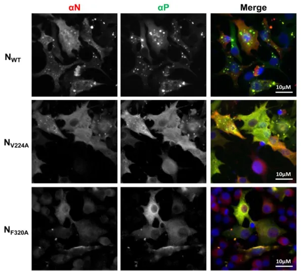
To further investigate the impact of these mutations, we then studied the cellular localization of N. As previously described, co-expression of N and P proteins leads to the formation of inclusion bodies (IBs) similar to the structures observed during RSV infection (24) (25) (Fig. 3) that were recently shown to be the sites of RSV replication and transcription
by guest on September 3, 2019
http://www.jbc.org/
4
(26). No impact on IBs morphology was detectedupon substitution of residues K81, I82, V262, V285, G296, and Y353 (not shown and Table 1). On the contrary, a total loss of IBs formation was observed when mutating residues L272, M281, Y288, F300, and F320 (Fig. 3 and Table 1). More surprisingly, the absence of IBs was also observed in the presence of N substitutions M248A and L258A that respectively displayed 30% and 50% of polymerase activity (Table 1). Finally, a main defect on IBs formation (decrease in number and size of IBs compared to the wild type) was observed upon mutation of residues V224, H225, I228, F247, R259, I270 and V284 (Fig. 3 and Table 1).
Taken together, these results show that, among the 20 targeted N residues, 16 are critical for N function within the polymerase complex. However, given the specific localization of targeted residues, the strong effect of mutations on polymerase activity and N localization could be attributed to either a major defect in: i/ intrinsic N conformation, ii/ N-N interaction, or iii/ N-P40 interaction.
Identification of N residues involved in P40 binding
We then assessed the potential impact of N mutations on its structure and its capacity to interact with RNA. As previously described, co-expression in bacteria of N with GST-PCT, which corresponds to a fusion protein between the GST and the fragment P[161-241] that interacts with the NNTD, allows purifying N-RNA rings using the GST tag (21) (23). Mutations were thus introduced in the pET-N vector, and the ability of N (wt or mutants) to co-purify with GST-PCT was evaluated by SDS-PAGE and Coomassie blue staining (Fig. 4A). Among the 18 N variants tested, only 8 (V224A, M248A, L258A, V262A, V285A, G296A, I270A and Y353A) were co-purified with GST-PCT with similar amounts compared to wild type N. The mutations of the 5 residues H225, I228, F247, M281 and V284 seemed to alter the interaction with PCT. Finally, the 5 mutants R259A, L272A, Y288A, F300A, and F320A were not pulled-down by GST-PCT, suggesting a strong impact of these mutations on the structural integrity of N. These results correlate with those obtained for the functional minigenome assay and IBs morphology (Table 1). We then focused on the mutants that could still interact with GST-PCT and tried to decipher if the targeted residues could be involved in the interaction with either P40 or the N-arm of N. To this end, 11 mutations were introduced in pET-NΔ30 vector. NΔ30 protein (wt or mutant) was co-expressed in bacteria together with either GST-N30 (30 N-terminal residues of N) or GST-P40, and the ability of monomeric N to be pulled-down was evaluated by SDS-PAGE and Coomassie blue staining. As shown in Figure 4B, the 5 mutations V224A, M248A, L258A, M281A, and V284A induced a loss of interaction between NΔ30 and GST-N30. On the
contrary, these mutations poorly affected the interaction with GST-P40, except for L258A and V284A substitutions that seemed to interact less efficiently with GST-P40. More interestingly, the mutation I270A specifically abrogated the interaction with GST-P40 without affecting the interaction with GST-N30 (Fig. 4B). Finally, the Y353A substitution, which had no impact on minigenome activity or on N oligomerization, induced a defect of interaction of NΔ30 with both GST-N30 and GST-P40. We thus cannot exclude a defect of NΔ30 folding for this mutant protein. Although our results suggest that most of the introduced mutations strongly affect N conformation, we clearly reveal the direct role of N residue I270 in the interaction with P40. The data also suggest a role of N residues L258 and V284 in the interaction. These results confirm the strong structural homology between RSV and hMPV N0-P complexes, with the P binding domain on N0 overlapping both the Ni-1 N-arm and Ni+1 C-arm binding sites involved in N oligomerization (Fig. 2A).
Modeling the structure of RSV N0-P complex in solution
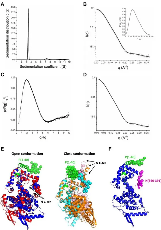
In order to gain information on the structure of the N0-P complex, small angle X-scattering (SAXS) and analytical ultracentrifugation (AUC) approaches were then combined to provide information about the overall size, shape and stoichiometry of this complex in solution (Fig. 5). After purification of the NΔ30-P40 complex, we first analyzed the complex by AUC (Fig. 5A). One main species with a sedimentation coefficient of 3.4 ± 0.15 S and a frictional ratio of 1.3 compatible with a complex form of one N0 and one peptide P40 was detected (Table 2). This single population suggests a really stable interaction between N0 and P peptide as previously measured (17). On the other hand, the molecular weights derived from Guinier analysis of the SAXS data reveal no sample aggregation or radiation damage. In agreement with AUC experiments (Table 2), we found that the NΔ30-P40 complex behaves as a monodisperse distribution of monomers in one to one complex in solution. The estimated molecular mass of the complex derived from the extrapolated intensity I(0) at the origin is consistent with the theoretical value of 45.3 kDa (Table 2 and supplementary Table S1). The maximum distance (Dmax) and the radius of gyration (Rg) of the NΔ30-P40 complex derived from the electron pair distance distribution function P(r) were respectively 9.41 nm and 2.74 nm (Table 2, Fig. 5B). The normalized Kratky plot is characteristics of a multidomain protein with flexible regions (Fig. 5C). To gain further understanding of the structural arrangement of the complex, we used Coral to generate 50 structures of the NΔ30-P40 complex, taking as starting configuration a model based on the X-ray structures of RSV N (PDB code 2WJ8) and
by guest on September 3, 2019
http://www.jbc.org/
5
hMPV N0-P complex (PDB code 5FVD), with arandom arrangement of the C-terminal end of P40. Briefly, during the modeling process, the P40 peptide and the globular domain (256-359) of N were fixed in position in order to maintain the contact between the NCTD surface and the P40 peptide based on the crystal structure of the hMPV N0-P complex. During the process, the last 13 amino acids in the C-terminal end of P40, the linker between the two globular domains of N as well as the C-arm of N were in random conformation, generating a large amount of interdomain and C-arm orientations. The chi2 value, deduced from the difference between the calculated SAXS curves of the models and the experimental data, as well as the calculated theoretical sedimentation values were used to select the best models that were representative of the SAXS curve. Out of the 50 models generated by Coral, only 18 were compatible with the AUC data.
Out of these 18, the best 5 models that were in agreement with both the experimental SAXS data and the AUC hydrodynamic parameters are presented here (Fig. 5E). Interestingly, these selected conformers display two main possible orientations of the C-arm of N depending on the relative position of the NCTD and NNTD domains. In one case (2 superimposed models in Fig. 5E left), the RNA groove access is blocked by the positioning of the N C-arm in the groove and the NCTD and NNTD domains are maintained in an open conformation. In the second case (3 superimposed models in Fig. 5F right), the NCTD and NNTD domains are maintained in a close conformation and N C-arm is flexible. However, the best model presented in Figures 5D and 5F suggests that the RNA groove access is blocked by the positioning of the N C-arm in the groove as already described for hMPV (16). In conclusion, our models support the strong structural similarity between the N0-P complexes of RSV and hMPV, and suggest a high flexibility of the N C-arm. Mutations in the N C-arm strongly affect the RNA polymerase activity
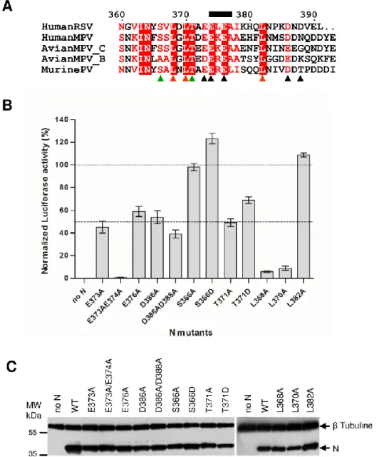
Based on our results and hMPV structure, we hypothesized that the C-arm of N is a flexible region that impairs interaction between the neosynthesized N0 nucleoprotein and RNA, by blocking the access of the RNA binding groove of N. Hence, in addition to the chaperone role of P, the C-arm of N would also contribute to the transition from the N0-P to the N-RNA bound complexes. It is noteworthy that the N C-termini of RSV and other pneumoviruses are characterized by the presence of conserved acidic and hydrophobic amino acid residues (Fig. 6A). Such residues could play a major role by interacting with the positively charged N RNA-groove. We thus investigated the role of these N residues on the activity of the polymerase complex by site-directed mutagenesis using the RSV minigenome assay. The C-terminal acidic (E373, E376, D386, D388,
E373+E374, D386+D388) and hydrophobic (L368, L370; and L382) residues of N were substituted by alanine, and the functionality of these N variants within the polymerase complex was assayed. Similarly, the potential phosphorylated residues S366 and T371 were substituted by either alanine or aspartic acid. As shown in Figure 6B, no major effect on the RNA polymerase activity was observed for S366A, S366D and L382A variants. However, the E373A, E376A, D386A, D386A/D388A, T371A and T371D substitutions resulted in a approximatively 50% reduction of luciferase activity compared to wild type N. Finally, the E373A/E374A, L368A, and L370A substitutions had the strongest effect, with a reduction of more than 90% of the polymerase activity. It is noteworthy that all N mutants were shown to be expressed in similar amounts in BSRT7 cells compared to wild type N protein (Fig. 6C).
Altogether, these results show that conserved acidic and hydrophobic residues of the C-arm of N play a major role in the function of N within the polymerase complex.
The N C-arm is directly involved in the inhibition of RNA binding
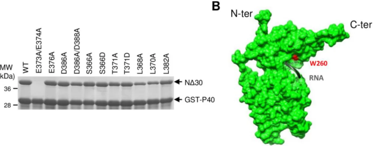
To determine whether the decrease in the polymerase activity observed with the N C-arm Ala mutants was correlated to a defect of N0-P complex conformation, the same mutations were introduced in the plasmid expressing NΔ30 in E.coli, to generate 11 N variants. GST-P40 was coexpressed with the wild type or mutant forms of NΔ30 in E.coli and purified on glutathione-Sepharose beads. The resulting purified GST-P40-NΔ30 complexes were analyzed by SDS-PAGE. Only the E373A/E374A mutant could not be purified, suggesting that this double mutation has a dramatic impact on the P40-NΔ30 complex formation (Fig. 7A). All the 10 others N mutant proteins were efficiently purified using GST-P40, showing that these mutations did not affect the P-N interaction. Next, the GST tag was cleaved by TEV protease and the OD260nm/OD280nm ratio of isolated NΔ30-P40 complexes was measured and compared to either N-RNA rings or NΔ30 alone (purified with a C-terminal His tag). As shown in Table 3, the OD260nm/OD280nm ratio of NΔ30 purified alone or in the presence of P40 were 0.87 and 0.63, respectively, confirming that P40 is required to efficiently block the interaction of N with RNA. Although all the NΔ30-P40 complexes displayed a OD260nm/OD280nm ratio ˂ 1 and close to 0.6 like wild type NΔ30, a slight increase of this ratio was detected for most of the mutants, especially for mutants L368A, and L370A (Table 3), suggesting the presence of some amounts of RNA associated to these complexes.
Given that N-RNA and N0-P forms of N present similar secondary structure as revealed by CD spectra (Fig. 1D), it is likely that binding of the N C-arm into the RNA-binding groove induces only
by guest on September 3, 2019
http://www.jbc.org/
6
minor conformational changes. To investigate theimpact of mutations in the N C-arm on N folding, we took advantage of the presence of a single Tryptophan residue (Trp260) in N, located close to the N RNA-binding groove (Fig. 7B), since λmax of emission of a Tryptophan reflects the polarity of its environment. In a hydrophilic environment, λmax shifts to 400 nm and the intensity, whereas in an apolar environment, λmax shifts to 300 nm. In order to validate that the fluorescence of Trp260 could be used to characterize the local structural change of N close to the RNA binding groove, we first measured the Trp λmax of N-RNA rings and of NΔ30 purified alone or in the presence of P40. As shown in Table 3, the λmax values of these proteins are 327 nm, 339 nm, and 330 nm respectively. When interacting with RNA, the Trp260 residue of N is in an apolar environment. The slight difference of λmax value and intensity detected between N-RNA and NΔ30-P40 suggests that Trp260 is maintained in a constrained environment within the N0-P complex. This hypothesis is supported by the important shift of λmax value measured for the NΔ30 alone that clearly revealed the polar environment of the Trp. The strong difference between the λmax values measured for NΔ30-P40 and NΔ30 alone could be attributed to i/ the binding of P40 on N, and/or ii/ the interaction of the N C-arm close to the RNA groove. We then investigated the impact of mutations in the N C-arm on the λmax of purified recombinant NΔ30-P40 complex. Although no major change in the λmax values was detected for most of the mutants, a slight change in the fluorescence spectra was detected when mutating residues S266 or T271, and a strong switch of the Trp λmax values, up to 337 nm, was detected for mutants L368A and L370A (Table 3). We consider that these changes were significant as they correspond to tendencies over many data points obtained for two independent protein purifications. These variations in Trp fluorescence reveal that Trp260 is more exposed to the solvent when mutating the residues L368 and L370.
Altogether, these data indicate that the residues L368 and L370 are critical for stabilization of the N0-P complex, and could be directly involved in the interaction of the C-arm close to the N-RNA binding groove, confirming our initial hypothesis.
Discussion
The viral RNA genome and anti-genome of Mononegavirales are coated by the viral N protein at all times. Efficient replication of genomic and antigenomic RNAs depends on the neosynthesis of the N protein that has to be maintained monomeric and RNA-free (N0) to specifically encapsidate these replication products as they are synthesized. It is noteworthy that all these N proteins present a strong propensity to oligomerize and to bind to RNA, and that these two mechanisms are concomitant. As a consequence, both mechanisms have to be impaired at the same time to maintain a pool of N competent
for RNA encapsidation. For this, viruses of the Mononegavirales order share a common strategy, which depends on the chaperone activity of the P N-terminal region that binds to N, forming a N0-P complex (5). It is noteworthy that the N proteins of Mononegavirales present a similar architecture and are composed of two globular domains (NNTD and NCTD) linked by a hinge region that forms the RNA groove, and N- and C-arms that are involved in N oligomerization. However, besides strong similarities between N0-P complexes, the mechanisms involved in the inhibition of RNA binding to N differ (10) (11) (13) (14) (12) (15).
Here, by deleting the N- and C-arms of RSV N, we observed that both the presence of the peptide P[1-40] and the deletion of the N-arm of N (NΔ30) are required to purify a stable monomeric N protein from E. coli. We also noticed that the C-arm of N is directly involved in the inhibition of the interaction of N with RNA (Fig. 1). Based on the crystal structures of RSV N-RNA and hMPV N0-P complexes, we then tried to characterize the surface of N involved in P40 binding. We identified some residues potentially involved in P40 binding, generated N mutants, and used both cellular and biochemical assays to validate the role of these residues. Although most of the mutations resulted in impaired N purification, we identified the residue I270 as directly and specifically involved in the interaction with P40. Our results also revealed a role of N residues L258 and V284 in the P40-N interaction.
Structural information on the RSV NΔ30-P40 complex in solution was then obtained by combining SAXS and AUC approaches. Models were generated by fixing in position the P40 peptide at the NCTD surface based on the crystal structure of the hMPV N0-P complex (16). These models show that monomeric N easily accommodates the structure of an N protomer from the N-RNA ring 3D structure, with a high flexibility of the N C-arm and of the linker region between NCTD and NNTD (Fig. 5E). Although some models suggest that NCTD and NNTD domains could be maintained in a close conformation with the N C-arm flexible, the best model suggests that the N C-arm could bind close to the RNA groove to impair its accessibility (Fig. 5F), as described for the N0-P complex of hMPV. In order to confirm these results, we generated N mutants of the N C-arm and showed that the acidic and hydrophobic residues of the C-arm of N are critical for its activity. More specifically, residues L368 and L370 of N were shown to be directly involved in the inhibition of RNA binding. These results correlate with the observation of Renner et al. (16) reporting semi-conserved LGLT-motif within the CTD-arms of the N proteins of Paramyxoviridae which is followed by a stretch of residues with helical propensity. It is noteworthy that the C-arms of N proteins of the Pneumoviridae family are composed of conserved acidic and hydrophobic residues. Such
by guest on September 3, 2019
http://www.jbc.org/
7
acidic and hydrophobic motifs are reminiscent ofeukaryotic acidic activation domains, in which acidic residues are used to promote long-range electrostatic interactions to attract their basic targets, and then undergo an induced structural transition to an alpha-helix to enable the contact with the target through hydrophobic residues (27).
In conclusion, our results point out the strong structural homology between RSV and hMPV N0-P complexes. These viruses present an original mechanism among Mononegavirales to impair RNA binding that involves i/ a specific interaction of the C-arm of N with the RNA groove, and ii/ the binding surface of P peptide that overlaps with both interaction sites of neighboring N protomers within N-RNA rings (i.e. of Ni+1 C-arm and Ni-1 N-arm). It is noteworthy that the crystal structure of the monomeric nucleoprotein (NP) of influenza virus that belongs to Orthomyxoviruses and which does not present homolog of the P protein, also revealed that the C-terminus of the NP monomer is bound to the side of the RNA binding surface (28). This binding allows to reduce the positive charge of the RNA groove to impair RNA binding. These observations thus suggest that the members of the negative strand viruses present conserved mechanisms to control the specificity of viral genome encapsidation. Although the 3D structure of the RSV N0-P still remains to be solved, these structural data paves the way to the rational design of specific protein-protein interaction inhibitors for these viruses that represent main respiratory pathogens because compounds that stabilize or destabilize the monomer may slow down viral infection. We and others already proposed that inhibitory peptides mimicking the P peptide could represent potent new antivirals not only against RSV but also for others Mononegavirales (29) (11) (17). Such approaches were developed to interfere with the fusion step of RSV, using stapled peptides (30) (31). Finally, these structural data revealed that the surface of interaction between the N C-arm and the RNA groove could constitute a second target to develop antivirals against the N0-P complex. In this case, stabilization of the interaction using small molecules could represent an attractive strategy (32). Experimental procedures
Plasmid constructions - All the viral sequences were derived from the hRSV strain Long, ATCC VR-26 (Genbank accession n° AY911262.1). N- and/or C-terminal N deletions were made by PCR using Pfu DNA polymerase (Stratagene). PCR products were cloned in the pET28a+ vector to produce truncated N proteins either with no tag or with a C-terminal 6xHis tag. The pGEX-N30 plasmid coding for the 30 N-terminal residues of N fused to GST was obtained by cloning annealed oligonucleotides at BamHI restriction site of the pGEX4T3 vector. The pGEX-PCT and pGEX-P40 vectors coding respectively for P[161-241] and P[1-40] in fusion with GST were
previously described (17). Point mutations were introduced in pET-N by site-directed mutagenesis, using the Quikchange site-directed mutagenesis kit (Stratagene). Sequence analysis was carried out to check the integrity of all the constructs.
Plasmids for eukaryotic expression of the hRSV N, P, M2-1, and L proteins designated pN, pP, pM2-1 and pL, have been described previously (33) (34). The pM/Luc subgenomic minigenome which encodes the firefly luciferase (Luc) reporter gene under the control of the M/SH gene start sequence was derived from the pM/SH subgenomic replicon (35) and has been described previously (22). Point mutations were introduced in pN by site directed mutagenesis as described above.
Antibodies - The following primary antibodies were used for immunofluorescence and/or immunoblotting: a mouse monoclonal anti-N protein (Serotec, Oxford, UK), rabbit anti-P and rabbit anti-N antisera (34) (23), and a mouse monoclonal anti-β-tubulin (Sigma, Saint-Louis, USA). Secondary antibodies directed against mouse and rabbit Ig G coupled to HRP were used for immunoblotting (P.A.R.I.S., Compiègne, France), and goat anti-rabbit and anti-mouse antibodies coupled to Alexa-448 and Alexa-594 were used for immunofluorescence.
Cell culture and transfections - BHK-21 cells (clone BSRT7/5) constitutively expressing the T7 RNA polymerase (36) were grown in Dulbeco Modified Essential Medium (Lonza, Cologne, Germany) supplemented with 10% fetal calf serum (FCS), 2 mM glutamine, and antibiotics. Cells were transfected using Lipofectamine 2000 (Invitrogen, Cergy-Pontoise, France) as described by the manufacturer.
Minigenome assay- Cells at 90% confluence in 48-well dishes were transfected with a plasmid mixture containing 125 ng of pM/Luc, 125 ng of pN, 125 ng of pP, 62.5 ng of pL, and 31 ng of pM2-1 as well as 31 ng of pRSV-β-Gal (Promega) to normalize transfection efficiencies (22). Transfections were done in triplicate, and each independent transfection was performed three times. Cells were harvested 24 h post-transfection, then lyzed in luciferase lysis buffer (30 mM Tris pH 7.9, 10 mM MgCl2, 1 mM DTT, 1% Triton X-100, and 15% glycerol). The luciferase activities were determined for each cell lysate with an Infinite 200 Pro (Tecan, Männedorf, Switzerland) and normalized based on β-galactosidase (β-Gal) expression.
Fluorescence microscopy - Immunofluorescence microscopy was performed with cells grown on coverslips and previously transfected with pN and pP. At 24 h post-transfection, cells were fixed with 4% paraformaldehyde (PFA) for 30 min. Fixed cells were made permeable and blocked for 15 min with
by guest on September 3, 2019
http://www.jbc.org/
8
PBS containing 0.1% Triton X-100 – 3% bovineserum albumin (BSA). Cells were then successively incubated for 1 hour at room temperature with primary and secondary antibody mixtures diluted in PBS containing 3% BSA, 0.05% Tween. Coverslips were mounted with Prolong gold antifade reagent containing DAPI (Invitrogen, CA, USA), and observed with a Nikon TE200 microscope equipped with a CoolSNAP ES2 (Photometrics, Tucson, USA) camera. Images were processed by using Meta-Vue (Molecular Devices, San Jose, USA) and ImageJ software.
Expression and purification of recombinant proteins - E. coli BL21 bacteria (DE3) (Novagen, Madison, WI) transformed with pET-N-derived plasmids alone, or together with pGEX-PCT, pGEX-P40, or pGEX-N30 plasmids were grown at 37°C for 8 h in 100 ml of Luria Bertani (LB) containing kanamycin (50 µg/ml) or ampicillin and kanamycin, respectively. The same volume of LB was then added and protein expression was induced by adding 80 µg/ml isopropyl-ß-D-thio-galactoside (IPTG) to the medium. Bacteria were incubated for 15 h at 28°C and then harvested by centrifugation. For the purification of the recombinant GST-fusion proteins, bacterial pellets were re-suspended in lysis buffer (50 mM Tris-HCl pH 7.8, 60 mM NaCl, 1 mM EDTA, 2 mM DTT, 0.2% Triton X-100, 1 mg/ml lysozyme) supplemented with complete protease inhibitor cocktail (Roche, Mannheim, Germany), incubated for 1 h on ice, sonicated, and centrifuged at 4°C for 30 min at 10,000g. Glutathione-Sepharose 4B beads (GE Healthcare, Uppsala, Sweden) were added to clarified supernatants and incubated at 4°C for 3 h. Beads were then washed two times in lysis buffer and three times in PBS 1X, and stored at 4°C in an equal volume of PBS. To isolate GST-free P40-NΔ30 complex, beads containing bound complex were incubated with TEV protease for 16 h at 20°C. For NΔ30-6xHis fusion protein purification, bacterial pellets were re-suspended in lysis buffer (20 mM Tris-HCl pH8, 500 mM NaCl, 0.1% TritonX-100, 10 mM imidazole, 1 mg/ml lysozyme) supplemented with complete protease inhibitor cocktail (Roche, Basel, Switzerland). After sonication and centrifugation, lysates were incubated 30 min with chelating Sepharose Fast Flow beads charged with Ni2+ (GE Healthcare). Finally, beads were successively washed in the washing buffer (20 mM Tris-HCl, pH 8, 500 mM NaCl) containing increasing concentration of imidazole (25, 50, and 100 mM), and proteins were eluted in the same buffer with 500 mM imidazole. Purified recombinant proteins were loaded onto a Sephacryl S-200 HR 16/30 column (GE Healthcare, Chicago, USA), eluted in 20 mM Tris-HCl pH 8.5, 150 mM NaCl and characterized. The presence of RNA was determined by measuring the OD260nm/OD280nm absorption ratio.
Circular Dichroism (CD) Spectroscopy - CD experiments were performed on a J-810 spectropolarimeter (Jasco, Tokyo, Japan) in a thermostated cell holder at 20°C. Purified recombinant N proteins were dialyzed against 5 mM phosphate, 100 mM NaF, pH 8.5. Far-UV spectra (180-260 nm) were recorded using a bandwidth of 1 nm and an integration time of 1s, in a 0.5 mm path-length quartz cell with proteins at concentrations of 20 µM. Each spectrum was the average of 6 scans, with a scan rate of 100 nm/min. The spectra were corrected by subtracting the signal from the buffer, smoothed using the FFT filter (Jasco Software, Tokyo, Japan), and were treated as previously described (37).
Fluorescence spectroscopy - Fluorescence measurements were performed with an FP-750 spectrofluorimeter (Jasco, Tokyo, Japan) at 20°C, using a 1-cm path length quartz cell, with proteins at a concentration of 10 µM. The excitation wavelength was fixed at 295 nm, and emission spectra were recorded from 300 to 360 nm at a scan rate of 125 nm.min-1. The maximum emission wavelength (λmax) represent the average of three values obtained from emission spectra. The measurements were performed on two independent purification of each recombinant protein.
Analytical ultracentrifugation (AUC) - Sedimentation velocity experiments were carried out at 20 °C in a ProteomeLab XL-I analytical ultracentrifuge (Beckman Coulter, Brea, CA, USA) equipped with double-UV and Rayleigh interference detection. The purified protein–peptide complexes (1.1 mg/ml) were centrifuged at 42000 rpm using an AN60-Ti rotor and 12 mm thick epon double sector centerpieces. Absorbance and interference profiles were recorded every 5 min. Buffer viscosity (η= 1.021 cP) and density ( = 1.0050° g.mL-1) at 20 °C were estimated with SEDNTERP 1.09. Partial specific volumes at 20 °C were estimated based on amino acid sequences using SEDNTERP 1.09 software. Data were analyzed with SEDFIT 15.3 using a continuous size distribution c(S) model. Theoretical sedimentations of the complex were generated using hydropro 10 (38).
Small-angle X-ray scattering (SAXS) experiments - Small-angle X-ray scattering (SAXS) data were collected on the SWING beamline at Synchrotron Soleil (France) using the online HPLC system. SAXS samples were prepared in buffer Tris 20 mM pH 8, NaCl 150 mM, glycerol 5%. Protein complexes of NΔ30–P40 complexe (50 µL) at 6 mg.ml-1 were injected into a size exclusion column (biosec 3-300 Agilent column) cooled at 25 °C and eluted directly into the SAXS flow-through capillary cell at a flow rate of 200 µL.min-1. The data were analyzed using FOXTROT and PRIMUS from ATSAS 2.7 (39), from which Guinier was generated.
by guest on September 3, 2019
http://www.jbc.org/
9
Scattering curves were selected for stable radius ofgyration (Rg) at the apex of the elution profile, and the selected curves were averaged and buffer signal was substracted. From these corrected scattering curves, the pair distribution functions was computed using GNOM (40) and the normalized Kratky plot was generated (41). Furthermore, 50 low-resolution models, obtained from independent reconstructions, were generated using coral (42) and a starting model of NΔ30-P40 complex based on the X-ray structure of a N protomer derived from the N-RNA rings
structure (PDB code 2WJ8) (21). The starting structure of N promoter was defined from N terminal end as a succession of 3 folded domains (30-249; 256-359; 374-382) connected through 2 linker regions between the first two domains and the last two domains. The P40 peptide was positioned on the NCTD (residues 256-359) based on X-ray structure of hMPV N0-P complex (16) with the C terminal end of P40 defined as a flexible region of 13 amino acids.
Acknowledgements: Molecular graphics images were produced using Chimera and PyMol softwares.
We thank the synchrotron facilities SOLEIL (St Aubin) for allocating regular beam time and their
dedicated staffs for technical help with the beamlines SWING. We warmly thank Origène Nyanguile
and Christina Sizun for skillful discussions and critical review of the manuscript. This work was carried
out with the financial support of the french Agence Nationale de la Recherche, specific program ANR
Blanc 2013 “Respisyncycell” (
ANR-13-IVS3-0007).
Conflict of interest: The authors declare that they have no conflicts of interest with the contents of this
article.
Author contributions: M.G., B.R. and J.F.E. designed experiments. C.E., J.F. and M.G performed
cellular and biochemical assays, C.A.R. performed gel filtration, P.R. performed SAXS measurements,
B.R and S.B. performed AUC experiments, P.R. and B.R. performed structural modeling. B.R, M.G.,
wrote the paper, and J.F.E. edited the manuscript. All authors commented on the manuscript.
References
1.
Collins PL, and Melero JA. (2011) Progress in understanding and controlling respiratory
syncytial virus: Still crazy after all these years. Virus Res. 162, 80-99
2.
Afonso, C. L., Amarasinghe, G. K., Banyai, K., Bao, Y., Basler, C. F., Bavari, S., Bejerman,
N., Blasdell, K. R., Briand, F. X., Briese, T., Bukreyev, A., Calisher, C. H., Chandran, K.,
Cheng, J., Clawson, A. N., Collins, P. L., Dietzgen, R. G., Dolnik, O., Domier, L. L., Durrwald,
R., Dye, J. M., Easton, A. J., Ebihara, H., Farkas, S. L., Freitas-Astua, J., Formenty, P., Fouchier,
R. A., Fu, Y., Ghedin, E., Goodin, M. M., Hewson, R., Horie, M., Hyndman, T. H., Jiang, D.,
Kitajima, E. W., Kobinger, G. P., Kondo, H., Kurath, G., Lamb, R. A., Lenardon, S., Leroy, E.
M., Li, C. X., Lin, X. D., Liu, L., Longdon, B., Marton, S., Maisner, A., Muhlberger, E.,
Netesov, S. V., Nowotny, N., Patterson, J. L., Payne, S. L., Paweska, J. T., Randall, R. E., Rima,
B. K., Rota, P., Rubbenstroth, D., Schwemmle, M., Shi, M., Smither, S. J., Stenglein, M. D.,
Stone, D. M., Takada, A., Terregino, C., Tesh, R. B., Tian, J. H., Tomonaga, K., Tordo, N.,
Towner, J. S., Vasilakis, N., Verbeek, M., Volchkov, V. E., Wahl-Jensen, V., Walsh, J. A.,
Walker, P. J., Wang, D., Wang, L. F., Wetzel, T., Whitfield, A. E., Xie, J. T., Yuen, K. Y.,
Zhang, Y. Z., and Kuhn, J. H. (2016) Taxonomy of the order Mononegavirales: update 2016.
Arch. Virol. 161, 2351-2360
3.
Bakker, S. E., Duquerroy, S., Galloux, M., Loney, C., Conner, E., Eleouet, J. F., Rey, F. A., and
Bhella, D. (2013) The respiratory syncytial virus nucleoprotein-RNA complex forms a
left-handed helical nucleocapsid. J. Gen. Virol. 94, 1734-1738
4.
Liljeroos, L., Krzyzaniak, M. A., Helenius, A., and Butcher, S. J. (2013) Architecture of
respiratory syncytial virus revealed by electron cryotomography. Proc. Natl. Acad. Sci. U S A
110, 11133-11138
5.
Jamin, M., and Yabukarski, F. (2017) Nonsegmented Negative-Sense RNA Viruses-Structural
Data Bring New Insights Into Nucleocapsid Assembly. Adv. Virus Res. 97, 143-185
by guest on September 3, 2019
http://www.jbc.org/
10
6.
Cowton, V. M., McGivern, D. R., and Fearns, R. (2006) Unravelling the complexities of
respiratory syncytial virus RNA synthesis. J. Gen. Virol. 87, 1805-1821
7.
Morin, B., Kranzusch, P. J., Rahmeh, A. A., and Whelan, S. P. (2013) The polymerase of
negative-stranded RNA viruses. Curr. Opin. Virol. 3, 103-110
8.
Ruigrok, R. W., Crepin, T., and Kolakofsky, D. (2011) Nucleoproteins and nucleocapsids of
negative-strand RNA viruses. Curr. Opin. Microbiol. 14, 504-510
9.
Karlin, D., and Belshaw, R. (2012) Detecting remote sequence homology in disordered proteins:
discovery of conserved motifs in the N-termini of Mononegavirales phosphoproteins. PloS one
7, e31719
10.
Leyrat, C., Yabukarski, F., Tarbouriech, N., Ribeiro, E. A., Jr., Jensen, M. R., Blackledge, M.,
Ruigrok, R. W., and Jamin, M. (2011) Structure of the vesicular stomatitis virus N(0)-P
complex. PLoS Pathog. 7, e1002248
11.
Yabukarski, F., Lawrence, P., Tarbouriech, N., Bourhis, J. M., Delaforge, E., Jensen, M. R.,
Ruigrok, R. W., Blackledge, M., Volchkov, V., and Jamin, M. (2014) Structure of Nipah virus
unassembled nucleoprotein in complex with its viral chaperone. Nat. Struct. Mol. Biol. 21,
754-759
12.
Guryanov, S. G., Liljeroos, L., Kasaragod, P., Kajander, T., and Butcher, S. J. (2015) Crystal
Structure of the Measles Virus Nucleoprotein Core in Complex with an N-Terminal Region of
Phosphoprotein. J. Virol. 90, 2849-2857
13.
Kirchdoerfer, R. N., Abelson, D. M., Li, S., Wood, M. R., and Saphire, E. O. (2015) Assembly
of the Ebola Virus Nucleoprotein from a Chaperoned VP35 Complex. Cell Rep. 12, 140-149
14.
Zhu, T., Song, H., Peng, R., Shi, Y., Qi, J., and Gao, G. F. (2017) Crystal Structure of the
Marburg Virus Nucleoprotein Core Domain Chaperoned by a VP35 Peptide Reveals a
Conserved Drug Target for Filovirus. J. Virol. 91 (18), e00996-17
15.
Aggarwal, M., Leser, G. P., Kors, C. A., and Lamb, R. A. (2018) Structure of the Paramyxovirus
Parainfluenza Virus 5 Nucleoprotein in Complex with an Amino-Terminal Peptide of the
Phosphoprotein. J. Virol. 92 (5), e01304-17
16.
Renner, M., Bertinelli, M., Leyrat, C., Paesen, G. C., Saraiva de Oliveira, L. F., Huiskonen, J.
T., and Grimes, J. M. (2016) Nucleocapsid assembly in pneumoviruses is regulated by
conformational switching of the N protein. eLife 5, e12627
17.
Galloux, M., Gabiane, G., Sourimant, J., Richard, C. A., England, P., Moudjou, M.,
Aumont-Nicaise, M., Fix, J., Rameix-Welti, M. A., and Eleouet, J. F. (2015) Identification and
Characterization of the Binding Site of the Respiratory Syncytial Virus Phosphoprotein to
RNA-Free Nucleoprotein. J. Virol. 89, 3484-3496
18.
Pereira, N., Cardone, C., Lassoued, S., Galloux, M., Fix, J., Assrir, N., Lescop, E., Bontems, F.,
Eleouet, J. F., and Sizun, C. (2017) New Insights into Structural Disorder in Human Respiratory
Syncytial Virus Phosphoprotein and Implications for Binding of Protein Partners. J. Biol. Chem.
292, 2120-2131
19.
El Omari, K., Scott, K., Dhaliwal, B., Ren, J., Abrescia, N. G. A., Budworth, J., Lockyer, M.,
Powell, K. L., Hawkins, A. R., and Stammers, D. K. (2008) Crystallization and preliminary
X-ray analysis of the human respiratory syncytial virus nucleocapsid protein. Acta Crystallogr.
Sect. F-Struct. Biol. Cryst. Commun. 64, 1019-1023
20.
Shapiro, A. B., Gao, N., O'Connell, N., Hu, J., Thresher, J., Gu, R. F., Overman, R., Hardern, I.
M., and Sproat, G. G. (2014) Quantitative investigation of the affinity of human respiratory
syncytial virus phosphoprotein C-terminus binding to nucleocapsid protein. Virol. J. 11, 191
21.
Tawar, R. G., Duquerroy, S., Vonrhein, C., Varela, P. F., Damier-Piolle, L., Castagne, N.,
MacLellan, K., Bedouelle, H., Bricogne, G., Bhella, D., Eleouet, J. F., and Rey, F. A. (2009)
Crystal structure of a nucleocapsid-like nucleoprotein-RNA complex of respiratory syncytial
virus. Science 326, 1279-1283
22.
Tran, T. L., Castagne, N., Dubosclard, V., Noinville, S., Koch, E., Moudjou, M., Henry, C.,
Bernard, J., Yeo, R. P., and Eleouet, J. F. (2009) The respiratory syncytial virus M2-1 protein
forms tetramers and interacts with RNA and P in a competitive manner. J. Virol. 83, 6363-6374
23.
Galloux, M., Tarus, B., Blazevic, I., Fix, J., Duquerroy, S., and Eleouet, J. F. (2012)
Characterization of a viral phosphoprotein binding site on the surface of the respiratory syncytial
nucleoprotein. J. Virol. 86, 8375-8387
by guest on September 3, 2019
http://www.jbc.org/
11
24.
Garcia, J., Garcia-Barreno, B., Vivo, A., and Melero, J. A. (1993) Cytoplasmic inclusions of
respiratory syncytial virus-infected cells: formation of inclusion bodies in transfected cells that
coexpress the nucleoprotein, the phosphoprotein, and the 22K protein. Virology 195, 243-247
25.
Garcia-Barreno, B., Delgado, T., and Melero, J. A. (1996) Identification of protein regions
involved in the interaction of human respiratory syncytial virus phosphoprotein and
nucleoprotein: significance for nucleocapsid assembly and formation of cytoplasmic inclusions.
J. Virol. 70, 801-808
26.
Rincheval, V., Lelek, M., Gault, E., Bouillier, C., Sitterlin, D., Blouquit-Laye, S., Galloux, M.,
Zimmer, C., Eleouet, J. F., and Rameix-Welti, M. A. (2017) Functional organization of
cytoplasmic inclusion bodies in cells infected by respiratory syncytial virus. Nat. Comm. 15 ; 8,
563
27.
Uesugi, M., Nyanguile, O., Lu, H., Levine, A. J., and Verdine, G. L. (1997) Induced alpha helix
in the VP16 activation domain upon binding to a human TAF. Science 277, 1310-1313
28.
Chenavas, S., Estrozi, L. F., Slama-Schwok, A., Delmas, B., Di Primo, C., Baudin, F., Li, X.,
Crepin, T., and Ruigrok, R. W. (2013) Monomeric nucleoprotein of influenza A virus. PLoS
Pathog. 9, e1003275
29.
Castel, G., Chteoui, M., Caignard, G., Prehaud, C., Mehouas, S., Real, E., Jallet, C., Jacob, Y.,
Ruigrok, R. W., and Tordo, N. (2009) Peptides that mimic the amino-terminal end of the rabies
virus phosphoprotein have antiviral activity. J. Virol. 83, 10808-10820
30.
Bird, G. H., Boyapalle, S., Wong, T., Opoku-Nsiah, K., Bedi, R., Crannell, W. C., Perry, A. F.,
Nguyen, H., Sampayo, V., Devareddy, A., Mohapatra, S., Mohapatra, S. S., and Walensky, L.
D. (2014) Mucosal delivery of a double-stapled RSV peptide prevents nasopulmonary infection.
J. Clin. Invest. 124, 2113-2124
31.
Gaillard, V., Galloux, M., Garcin, D., Eleouet, J. F., Le Goffic, R., Larcher, T., Rameix-Welti,
M. A., Boukadiri, A., Heritier, J., Segura, J. M., Baechler, E., Arrell, M., Mottet-Osman, G.,
and Nyanguile, O. (2017) A Short Double-Stapled Peptide Inhibits Respiratory Syncytial Virus
Entry and Spreading. Antimicrob. Agents Chemother. 61, e02241-16
32.
Zarzycka, B., Kuenemann, M. A., Miteva, M. A., Nicolaes, G. A. F., Vriend, G., and Sperandio,
O. (2016) Stabilization of protein-protein interaction complexes through small molecules. Drug
Discov. Today 21, 48-57
33.
Tran, T. L., Castagne, N., Bhella, D., Varela, P. F., Bernard, J., Chilmonczyk, S., Berkenkamp,
S., Benhamo, V., Grznarova, K., Grosclaude, J., Nespoulos, C., Rey, F. A., and Eleouet, J. F.
(2007) The nine C-terminal amino acids of the respiratory syncytial virus protein P are necessary
and sufficient for binding to ribonucleoprotein complexes in which six ribonucleotides are
contacted per N protein protomer. J. Gen. Virol. 88, 196-206
34.
Fix, J., Galloux, M., Blondot, M. L., and Eleouet, J. F. (2011) The insertion of fluorescent
proteins in a variable region of respiratory syncytial virus L polymerase results in fluorescent
and functional enzymes but with reduced activities. Open Virol. J. 5, 103-108
35.
Hardy, R. W., and Wertz, G. W. (1998) The product of the respiratory syncytial virus M2 gene
ORF1 enhances readthrough of intergenic junctions during viral transcription. J.Virol. 72,
520-526
36.
Buchholz, U. J., Finke, S., and Conzelmann, K. K. (1999) Generation of bovine respiratory
syncytial virus (BRSV) from cDNA: BRSV NS2 is not essential for virus replication in tissue
culture, and the human RSV leader region acts as a functional BRSV genome promoter. J. Virol.
73, 251-259
37.
Chenal, A., Nizard, P., Forge, V., Pugniere, M., Roy, M. O., Mani, J. C., Guillain, F., and Gillet,
D. (2002) Does fusion of domains from unrelated proteins affect their folding pathways and the
structural changes involved in their function? A case study with the diphtheria toxin T domain.
Protein Eng. 15, 383-391
38.
Ortega, A., Amoros, D., and Garcia de la Torre, J. (2011) Prediction of hydrodynamic and other
solution properties of rigid proteins from atomic- and residue-level models. Biophys. J. 101,
892-898
39.
Konarev, P. V., Volkov, V. V., Sokolova, A. V., Koch, M. H. J., and Svergun, D. I. (2003)
PRIMUS: a Windows PC-based system for small-angle scattering data analysis. J. Appl.
Crystallogr. 36, 1277-1282
by guest on September 3, 2019
http://www.jbc.org/
12
40.
Svergun, D. I. (1992) Determination of the Regularization Parameter in Indirect-Transform
Methods Using Perceptual Criteria. J. Appl. Crystallogr. 25, 495-503
41.
Durand, D., Vives, C., Cannella, D., Perez, J., Pebay-Peyroula, E., Vachette, P., and Fieschi, F.
(2010) NADPH oxidase activator p67(phox) behaves in solution as a multidomain protein with
semi-flexible linkers. J. Struct. Biol. 169, 45-53
42.
Petoukhov, M. V., Franke, D., Shkumatov, A. V., Tria, G., Kikhney, A. G., Gajda, M., Gorba,
C., Mertens, H. D., Konarev, P. V., and Svergun, D. I. (2012) New developments in the ATSAS
program package for small-angle scattering data analysis. J. Appl. Crystallogr. 45, 342-350
FOOTNOTES: This work was carried out with the financial support of the french Agence Nationale
de la Recherche, specific program ANR Blanc 2013 “Respisyncycell” (
ANR-13-IVS3-0007).
The abbreviations used are: hRSV, human respiratory syncytial virus; hMPV, human Metapneumovirus;
N, nucleoprotein; P, phosphoprotein; RdRp, RNA-dependent RNA polymerase; SAXS, small angle
X-ray scattering; AUC, analytical ultracentrifugation; VSV, vesicular stomatitis virus; PIV, parainfluenza;
CTD, C-terminal domain; NTD N-terminal domain.
by guest on September 3, 2019
http://www.jbc.org/
13
by guest on September 3, 2019
http://www.jbc.org/
14
by guest on September 3, 2019
http://www.jbc.org/
15
by guest on September 3, 2019
http://www.jbc.org/
16
Figure 1: Purification and characterization of truncated N
recombinant proteins. (A) Schematic
representation of the primary sequence of N and of the different deletions generated. The subdomains
of N are colored: N-arm (blue), N
NTD(yellow), N
CTD(red), and C-arm (green). Numbers indicate amino
acid positions. (B) SDS PAGE analysis of co-purification of GST-P40 and truncated N proteins; the
OD
260nm/OD
280nmratio indicating the presence or absence of RNA is indicated for each sample down the
lanes. (C) Elution profiles of N[13-391] (gray) and N[31-391] (black) from gel filtration, at 280 nm
(solid lines) and 260 nm (dash lines). (D) Far-UV CD spectra showing the absence of major
conformational changes between N-RNA rings (dash lines) and monomeric N[31-391]-P40 (solid lines).
by guest on September 3, 2019
http://www.jbc.org/
17
Figure 2: Search for the P40 binding site at the surface of N. (A) Ribbon representation of the hMPV
N
CTD(green) in interaction with the peptide of P (residues 1-28, purple). The residues of N involved in
the interaction with P are colored in black and yellow and the side chains are shown (PBD code 5FVD)
(B) Ribbon representation of a part of RSV N protomer (green) from N-RNA 3D crystal structure (PBD
code 2WJ8) showing binding of the N
i-1N-arm (in red) and of the N
i+1C-arm (in blue). Residues involved
in N
i-1N-arm or in N
i+1C-arm binding, and that could also interact with P40, are colored in black and
yellow, respectively, and their side chains are shown, (C) Effect of N mutations on RSV polymerase
activity. BSRT7/5 cells were transfected with plasmids encoding the WT P, M2-1 and L proteins, the
pMT/Luc minigenome, and WT or mutant N proteins, together with pCMV-βGal for transfection
standardization. Viral RNA synthesis was quantified by measuring the luciferase activity after cell lysis
24 h after transfection.
Each luciferase minigenome activity value was normalized based on
β-galactosidase expression and is the average of three independent experiments performed in triplicate.
Error bars represent standard deviations calculated based on three independent experiments made in
triplicate. (D) Western blot showing the expression of N protein variants in BSRT7/5 cells.
by guest on September 3, 2019
http://www.jbc.org/
18
Figure 3: Impact of N mutations on the formation of cytoplasmic inclusion bodies. N and P proteins
were coexpressed in BSRT7/5 cells; cells were then fixed 24 h post-transfection and labeled with
anti-P (green) and anti-N (red) antibodies, and the distribution of viral proteins was observed by fluorescence
microscopy. Nuclei were stained with Dapi. Scale bars, 10 µm.
by guest on September 3, 2019
http://www.jbc.org/
19
Figure 4: Impact of N mutations on N-P and N-N interactions. (A) Full length N constructs (wild
type or mutants) were co-expressed with GST-PCT in E.coli and complexes were pulled down using
GST tag. (B) N
Δ30constructs (wild type or mutants) were co-expressed with GST-P40 or GST-N30 in
E.coli and complexes were pulled down using GST tag. Co-purification of N (or N
Δ30) with GST
constructs was analyzed by SDS-PAGE and stained with Coomassie blue.
by guest on September 3, 2019
http://www.jbc.org/
20
Figure 5: Structural characterization of RSV N
Δ30–P40 in solution.
(A) Analytical ultracentrifugation sedimentation distribution analysis of RSV N
Δ30-P40, (B) Scattering
curve of RSV N
Δ30-P40 complex (triangle) with fitted curve (plain line) used to generate distance
distribution. (Inset) distance distribution functions obtained using the program GNOM, (C)
Dimensionless Kratky plot, (D) Fit of the best coral model (see F), (E) Presentation of the five best coral
models of RSV N
Δ30-P40 complex superimposed based on the two main possible orientations of the
C-arm of N and on the relative position of the N
CTDand N
NTDdomains. Left, the RNA groove access is
blocked by the positioning of the N C-arm in the groove and the N
CTDand N
NTDdomains are maintained
in an open conformation. Right, the N
CTDand N
NTDdomains are maintained in a close conformation and
N C-arm is flexible. The P40 peptide is represented in green and the C-terminus of N is indicated, (F)
Ribbon representation of the best structural coral model of RSV N
Δ30-P40 complex. The N protomer
(residues 31-382) is represented in blue, the C-arm of N (residues 383-391) in purple, and the P40
peptide in green. Linker regions are represented by spheres.
by guest on September 3, 2019
http://www.jbc.org/
21
Figure 6: Effect of mutations in the C-arm of N on the RSV polymerase activity. (A) Sequence
alignments of the C-arms of N proteins from viruses belonging to the Pneumoviridae family. Uniprot
accession
codes:
NCAP_HRSVA
(Human
RSV);
A1DZS3_9MONO
(Human
MPV),
A4KZ90_9MONO (Avian MPV_C); A7DV59_9MONO (Avian MPV_B), NCAP_MPV15 (Murine
PV). PV: Pneumonia Virus, MPV: MetaPneumoVirus. Invariant residues are highlighted in white font
on a red background. The short α-helix observed in the crystal structure of N (21) is indicated above the
sequence by a black rectangle. Triangles under the alignment mark mutated residues: acidic (black),
hydrophobic (red), and Ser/Thr (green). (B) RSV polymerase activity in the presence of N mutants.
BSRT7/5 cells were transfected with plasmids encoding the WT P, M2-1 and L proteins, the pMT/Luc
minigenome, and WT or mutant N proteins, together with pCMV-βGal for transfection standardization.
Viral RNA synthesis was quantified by measuring the luciferase activity after cell lysis 24 h after
transfection.
Each luciferase minigenome activity value was normalized based on β-galactosidase
expression and is the average of three independent experiments performed in triplicate. Error bars
represent standard deviations calculated based on three independent experiments made in triplicate. (C)
Western blot showing the expression of N protein variants in BSRT7/5 cells.
by guest on September 3, 2019
http://www.jbc.org/
22
Figure 7: Purification of C-arm mutants of N from E. coli. (A) N
Δ30constructs (wild type or mutants)
were co-expressed with GST-P40 in E.coli and complexes were pulled down using GST tag.
Co-purification of WT and mutants of N
Δ30with GST-P40 was analyzed by SDS-PAGE and Coomassie
blue staining. (B) Surface view of an RSV N monomer showing the position of RNA and of the W260
residue. N- and C-termini of N are indicated.
by guest on September 3, 2019
http://www.jbc.org/
Richard, Jenna Fix, Jean-François Eléouët and Marie Galloux
Camille Esneau, Bertrand Raynal, Pierre Roblin, Sébastien Brûlé, Charles-Adrien
solution
-P complex in
0
Biochemical characterization of the respiratory syncytial virus N
published online January 9, 2019 J. Biol. Chem.
10.1074/jbc.RA118.006453
Access the most updated version of this article at doi: Alerts:
When a correction for this article is posted
•
When this article is cited
•
to choose from all of JBC's e-mail alerts
Click here
by guest on September 3, 2019
http://www.jbc.org/