PEARLS
Exploitation of the Host Cell Membrane
Fusion Machinery by Leishmania Is Part of the
Infection Process
Christine Matte, Albert Descoteaux*
INRS-Institut Armand-Frappier and Centre for host-parasite interactions, Laval, Quebec, Canada
Successful vacuolar pathogens have developed sophisticated strategies to hijack the endomem-brane system of host cells, sabotage their signal transduction pathways, and evade antimicro-bial responses. The protozoan parasiteLeishmania, the causative agent of leishmaniases in
humans, is particularly adept at transforming the macrophage into a hospitable host cell. Here, we describe howLeishmania promastigotes subvert the macrophage membrane fusion
machinery to create an intracellular compartment favorable to the establishment of infection and to manipulate host immune responses.
What Is the Membrane Fusion Machinery?
Cellular functions such as phagocytosis and cytokine secretion rely heavily on a complex net-work of vesicle trafficking pathways that interconnect most membrane-bound intracellular compartments [1]. A critical step in the exchange of cargoes between vesicles in this network is the process of membrane fusion, which is mediated by SNARE (soluble
N-ethylmaleimide-sensitive factor attachment protein receptor) proteins. This superfamily of integral and periph-eral membrane proteins displays the distinctive SNARE motif, a stretch of heptad repeats that form a coiled-coil structure with a conserved arginine (R) or glutamine (Q) residue at the cen-tral “0” layer. Fusogenic SNARE complex formation requires the parallel association of an R-SNARE domain on the vesicle membrane with three cognate Q-SNARE domains on the target compartment. The subsequent “zippering” of this four-helix bundle from the mem-brane-distal amino termini towards the membrane-proximal carboxyl termini brings the two apposed membranes into close proximity and provides sufficient mechanical force to over-come the energy barrier for the formation of the fusion pore [1].
Whereas SNAREs make up the core of membrane fusion machinery, a large number of additional proteins are required for the spatiotemporal orchestration of the entire process [2]. For instance, the family of Rab GTPases are master regulators of virtually all events lead-ing to membrane fusion. Indeed, Rab effectors include motor proteins (for vesicle traffick-ing along the cytoskeleton) and membrane tethers (for the initial, loose attachment to the target compartment). A set of chaperones, such as the proteins of the Sec1/Munc18 (SM) family, Munc13 and Complexins, oversee the timing of cognate SNARE pairing by restrict-ing SNARE accessibility at first, then initiatrestrict-ing partial SNARE complex formation and keep-ing it in a “release-ready” state until the appropriate moment for membrane fusion. The final trigger for pore formation is provided by synaptotagmins (Syts), a family of calcium-sensing membrane proteins that control fusion via interactions with SNARE proteins and membrane lipids. Ultimately, SNARE complexes are disassembled by the ATPase NSF (
N-a11111
OPEN ACCESS
Citation: Matte C, Descoteaux A (2016) Exploitation of the Host Cell Membrane Fusion Machinery by Leishmania Is Part of the Infection Process. PLoS Pathog 12(12): e1005962. doi:10.1371/journal.ppat.1005962 Editor: Laura J Knoll, University of Wisconsin Medical School, UNITED STATES
Published: December 8, 2016
Copyright:© 2016 Matte, Descoteaux. This is an open access article distributed under the terms of theCreative Commons Attribution License, which permits unrestricted use, distribution, and reproduction in any medium, provided the original author and source are credited.
Funding: This work was supported by Canadian Institutes of Health Research grant MOP-125990 to AD. The Centre for Host-Parasite Interactions is supported by the Fonds de Recherche du Que´bec -Nature et Technologies (Regroupement Strate´gique). The funders had no role in study design, data collection and analysis, decision to publish, or preparation of the manuscript. Competing Interests: The authors have declared that no competing interests exist.
ethylmaleimide-sensitive factor) and the adaptor proteinα-SNAP (soluble NSF attachment protein).
What Is the Role of Vesicle Trafficking and Membrane Fusion
during the Phagocytic Process?
Binding and internalization of infectiveLeishmania stages to macrophages involves multiple
phagocytic receptors [3]. Phagocytosis of those large particles requires the expense of a con-siderable amount of plasma membrane for pseudopod extension around the prey. Various membrane-bound intracellular compartments lend a hand to this process by fusing with the cell surface and rapidly providing endomembrane required for particle engulfment [4]. Focal exocytosis of recycling endosomes, for instance, contributes not only to phagocytosis but also allows for rapid secretion of preformed inflammatory cytokines including TNF and IL-6 [5]. Fusion of recycling endosomes with the cell surface is mediated by the R-SNARE VAMP3 (vesicle-associated membrane protein 3) [1,4] and is regulated by Syt V [6]. Inter-estingly, Syt XI also localizes to recycling endosomes and is recruited to nascent phago-somes, but acts as a negative regulator of phagocytosis and cytokine secretion [7]. Late endosomes and lysosomes assist large particle phagocytosis as well, in a VAMP7- and Syt VII-dependent manner [1,4,8]. Contribution of the endoplasmic reticulum (ER) as a source of endomembrane varies according to the nature of the phagocytosed particle and requires the ER Q-SNARE Stx18 [4,9].
Upon the completion of particle internalization, the phagosome undergoes a series of “kiss-and-run” fusion and fission events with vesicles of the endocytic pathway, culminating in the creation of a highly microbicidal and immunologically competent compartment, the phagoly-sosome. Various components of the membrane fusion machinery participate in the genesis of this organelle, including the endosomal R-SNARE VAMP8 for the recruitment of the NADPH oxidase NOX2 [10], Syt V for the acquisition of the v-ATPase (vesicular proton-ATPase) [11], and the ER R-SNARE Sec22b for the recruitment of ER components required for antigen crosspresentation [12].
Do Leishmania Parasites Disrupt the Membrane Fusion Machinery
to Tamper with Macrophage Responses?
Pathogens use a variety of tactics to manipulate membrane fusion and vesicle trafficking to cause disease [13]. The intracellular bacteriaChlamydia and Legionella, for instance, produce
proteins with SNARE-like motifs that interact with host SNAREs and inhibit SNARE-medi-ated membrane fusion. The best-known example is the specific cleavage of SNAREs by clos-tridial neurotoxins, which are potent blockers of neurotransmission in peripheral cholinergic nervous system synapses [14].Leishmania promastigotes use two abundant surface
GPI-anchored virulence factors to interfere with vesicle trafficking and fusion: GP63 (glycoprotein 63), a zinc-dependent metalloprotease, and LPG (lipophosphoglycan), a polymer of repeating Galβ1,4Manα1-PO4units. Upon internalization of the parasites, GP63 and LPG are rapidly
redistributed throughout infected cells (Fig 1). Akin to the clostridial neurotoxins, GP63 cleaves components of the host cell membrane fusion machinery, including VAMP3, VAMP8, and Syt XI (Table 1) [10,15]. The consequences of these cleavage events are diverse. In macro-phages and dendritic cells, processing of exogenous antigens for crosspresentation on MHC I molecules is controlled by the NADPH oxidase NOX2: phagosome oxidation prevents exces-sive acidification and destruction of peptides destined for recognition by T cells [16]. Since VAMP8 is involved in the recruitment of NOX2 to phagosomes, GP63-mediated cleavage of VAMP8 results in increased phagosomal proteolytic activity, ensuing in defective
crosspresentation ofLeishmania antigens to T cells [10]. In parallel, during a noncanonical autophagic process referred to as LC3-associated phagocytosis (LAP), NOX2-mediated phago-somal oxidation promotes the recruitment of the autophagy-related protein LC3 to a subset of phagosomes. Several roles have been attributed to LAP, including increased phagosomal microbicidal activity and enhanced antigen presentation on MHC II molecules [17]. By cleav-ing VAMP8 and preventcleav-ing phagosomal recruitment of NOX2, GP63 allowsLeishmania Fig 1. Distribution of L. major virulence factors LPG and GP63 across an infected macrophage. Murine primary macrophages were
infected for 6 h with L. major promastigotes, fixed and stained for confocal immunofluorescence microscopy using DAPI (DNA, blue) and antibodies against LPG (red) and GP63 (green). Arrowheads point to parasite nuclei. Scale bar = 5μm.
major promastigotes to evade LAP [18], possibly contributing to the impairment of phago-some maturation and further inhibiting antigen presentation to T cells. Consistent with the role of Syt XI as a negative regulator of cytokine secretion, cleavage of this endosomal protein by GP63 fromL. major promastigotes increases the postinfection release of TNF and IL-6 [15]. These proinflammatory cytokines are responsible for the augmentation of neutrophil and inflammatory monocyte influx to the parasite inoculation site, which contributes to the spread and maintenance of infection.
Insertion of LPG into host cell lipid microdomains causes remodeling of the phagosome and delays its maturation into a highly microbicidal phagolysosome, as a result of reduced fusogenicity towards late endosomes and lysosomes [19]. The current model of LPG-mediated phagosome remodeling is that LPG disrupts membrane lipid microdomains and thereby inter-feres with the clustering of host molecules at these sites [19]. For instance, SNAREs and other members of the membrane fusion machinery are typically concentrated in cholesterol-rich membrane rafts [1]. Therefore, one consequence of LPG-mediated microdomain disorganiza-tion is the exclusion of Syt V from the phagosome (Table 1). This, in turn, abrogates v-ATPase recruitment and impedes phagosome acidification [11].
Aside from GP63-mediated cleavage and LPG-induced phagosomal exclusion of SNAREs and Syts,Leishmania parasites also target components of the membrane fusion machinery at
the transcriptional level. Infection of human macrophages withL. major or L. amazonensis
leads to the up-regulation of Syt II, VI, and VIII [20]. The underlying mechanism, including the parasite factors and/or host proteins involved, and the repercussions on PV (parasitophor-ous vacuole) formation and infection outcome have yet to be uncovered. Of note, the genome ofL. major encodes a repertoire of 27 putative SNARE proteins, which can be segregated into
R-SNAREs and Q-SNAREs based on the classification system used for other organisms. Most of these proteins display the expected structural characteristics of functional SNAREs, while a few show peculiarities. For instance, four proteins lack a predicted transmembrane domain or GPI-anchor, and only two out of the four possess potential lipidation sites for attachment to membranes [21]. Whether any of these 27 putative SNAREs play a role in the manipulation of host cell fusion events byLeishmania parasites is unknown.
Do Leishmania Parasites Promote Vesicle Fusion to Drive PV
Formation?
Old WorldLeishmania species (L. major, L. donovani, and L. tropica) reside in small,
tight-fit-ting PVs that undergo fission shortly after parasite replication, therefore rarely containing
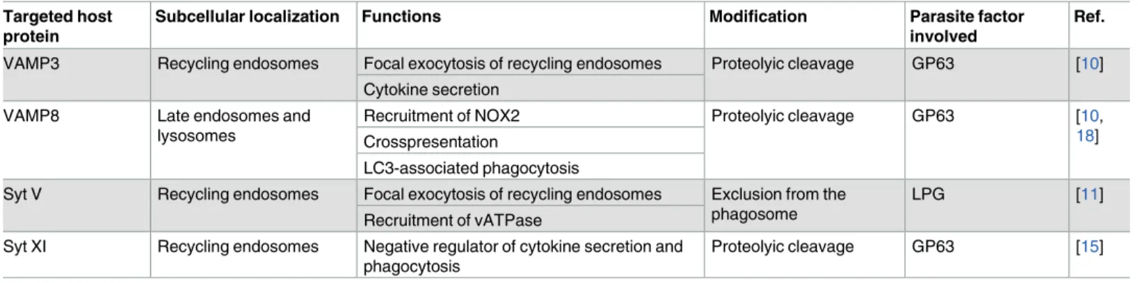
Table 1. Components of the host cell membrane fusion machinery targeted by Leishmania parasites. Subcellular localization and function in
macro-phages of each host protein are indicated.
Targeted host protein
Subcellular localization Functions Modification Parasite factor
involved
Ref.
VAMP3 Recycling endosomes Focal exocytosis of recycling endosomes Proteolyic cleavage GP63 [10] Cytokine secretion
VAMP8 Late endosomes and lysosomes
Recruitment of NOX2 Proteolyic cleavage GP63 [10,
18] Crosspresentation
LC3-associated phagocytosis
Syt V Recycling endosomes Focal exocytosis of recycling endosomes Exclusion from the phagosome
LPG [11]
Recruitment of vATPase
Syt XI Recycling endosomes Negative regulator of cytokine secretion and phagocytosis
Proteolyic cleavage GP63 [15]
more than a single amastigote. On the other hand, the establishment of a successful infection by New World species (L. mexicana, L. amazonensis, and L. pifanoi) requires the formation of
spacious, communal vacuoles that can harbour numerous parasites. Our understanding of the mechanisms allowing the development of individual versus communal PVs is very limited. Both types of PVs continuously interact with the host cell reservoir of acidic [22] and ER-derived vesicles [23], most likely to accommodate for the high membrane demand. Biogenesis of large communal PVs involves homotypic fusion between smaller PVs (Fig 2) [22], which may rely on the hijacking of specific components of the membrane fusion machinery. In sup-port of this model, targeting the ERGIC (ER-Golgi intermediate compartment) Q-SNARE Stx5 or the ER R-SNARE Sec22b and its cognate Q-SNARE partners Stx18 and D12 restricts the expansion ofL. amazonensis PVs and is detrimental to parasite replication [24]. Whether the molecular basis for this fundamental difference in the lifestyle of these two groups of Leish-mania is related to differential expression or activity of virulence factors such as GP63 that
directly target specific components of the host cell machinery remains to be investigated.
Conclusion
As our understanding of the function of membrane fusion mediators deepens, we are able to get a better insight into the challenges faced byLeishmania parasites upon entry into host cells
and, in parallel, the mechanisms of parasite virulence and pathogenesis. Conversely, Leish-mania represents a superb tool for the identification of novel roles for the membrane fusion
machinery in macrophages, by investigating the functional consequences of host protein cleavage or intracellular redistribution on cell and immune functions. Components of the membrane fusion machinery might emerge as targets for novel therapeutic interventions in infectious and inflammatory diseases.
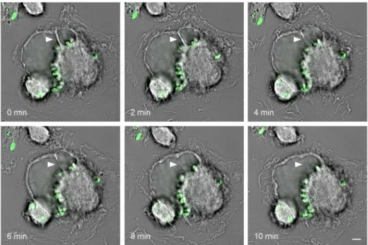
Fig 2. Two communal vacuoles undergoing homotypic fusion. Murine primary macrophages were
infected for three days with carboxyfluorescein succinimidyl ester (CFSE)-stained L. amazonensis parasites (green) and then monitored every two minutes by live-cell imaging. Arrowheads point to the septum between two parasitophorous vacuoles throughout the fusion process. Scale bar = 5μm.
References
1. Stow JL, Manderson AP, Murray RZ. SNAREing immunity: the role of SNAREs in the immune system. Nature reviews Immunology. 2006; 6(12):919–29. Epub 2006/11/25. doi:10.1038/nri1980PMID: 17124513
2. Malsam J, Kreye S, Sollner TH. Membrane fusion: SNAREs and regulation. Cellular and molecular life sciences: CMLS. 2008; 65(18):2814–32. Epub 2008/08/30. doi:10.1007/s00018-008-8352-3PMID: 18726177
3. Ueno N, Wilson ME. Receptor-mediated phagocytosis of Leishmania: implications for intracellular sur-vival. Trends in parasitology. 2012; 28(8):335–44. Epub 2012/06/26. doi:10.1016/j.pt.2012.05.002 PMID:22726697
4. Huynh KK, Kay JG, Stow JL, Grinstein S. Fusion, fission, and secretion during phagocytosis. Physiol-ogy (Bethesda). 2007; 22:366–72. Epub 2007/12/13.
5. Murray RZ, Stow JL. Cytokine Secretion in Macrophages: SNAREs, Rabs, and Membrane Trafficking. Frontiers in immunology. 2014; 5:538. Epub 2014/11/12. doi:10.3389/fimmu.2014.00538PMID: 25386181
6. Vinet AF, Fukuda M, Descoteaux A. The exocytosis regulator synaptotagmin V controls phagocytosis in macrophages. J Immunol. 2008; 181(8):5289–95. Epub 2008/10/04. PMID:18832684
7. Arango Duque G, Fukuda M, Descoteaux A. Synaptotagmin XI regulates phagocytosis and cytokine secretion in macrophages. J Immunol. 2013; 190(4):1737–45. Epub 2013/01/11. doi:10.4049/ jimmunol.1202500PMID:23303671
8. Czibener C, Sherer NM, Becker SM, Pypaert M, Hui E, Chapman ER, et al. Ca2+ and synaptotagmin VII-dependent delivery of lysosomal membrane to nascent phagosomes. The Journal of cell biology. 2006; 174(7):997–1007. Epub 2006/09/20. doi:10.1083/jcb.200605004PMID:16982801
9. Braun V, Niedergang F. Linking exocytosis and endocytosis during phagocytosis. Biology of the cell / under the auspices of the European Cell Biology Organization. 2006; 98(3):195–201. Epub 2006/02/17.
10. Matheoud D, Moradin N, Bellemare-Pelletier A, Shio MT, Hong WJ, Olivier M, et al. Leishmania evades host immunity by inhibiting antigen cross-presentation through direct cleavage of the SNARE VAMP8. Cell host & microbe. 2013; 14(1):15–25. Epub 2013/07/23.
11. Vinet AF, Fukuda M, Turco SJ, Descoteaux A. The Leishmania donovani lipophosphoglycan excludes the vesicular proton-ATPase from phagosomes by impairing the recruitment of synaptotagmin V. PLoS pathogens. 2009; 5(10):e1000628. Epub 2009/10/17. doi:10.1371/journal.ppat.1000628PMID: 19834555
12. Cebrian I, Visentin G, Blanchard N, Jouve M, Bobard A, Moita C, et al. Sec22b regulates phagosomal maturation and antigen crosspresentation by dendritic cells. Cell. 2011; 147(6):1355–68. Epub 2011/ 12/14. doi:10.1016/j.cell.2011.11.021PMID:22153078
13. Wesolowski J, Paumet F. SNARE motif: a common motif used by pathogens to manipulate membrane fusion. Virulence. 2010; 1(4):319–24. Epub 2010/12/24. doi:10.4161/viru.1.4.12195PMID:21178463
14. Montal M. Botulinum neurotoxin: a marvel of protein design. Annual review of biochemistry. 2010; 79:591–617. Epub 2010/03/18. doi:10.1146/annurev.biochem.051908.125345PMID:20233039
15. Arango Duque G, Fukuda M, Turco SJ, Stager S, Descoteaux A. Leishmania promastigotes induce cytokine secretion in macrophages through the degradation of synaptotagmin XI. J Immunol. 2014; 193 (5):2363–72. Epub 2014/07/27. doi:10.4049/jimmunol.1303043PMID:25063865
16. Savina A, Jancic C, Hugues S, Guermonprez P, Vargas P, Moura IC, et al. NOX2 controls phagosomal pH to regulate antigen processing during crosspresentation by dendritic cells. Cell. 2006; 126(1):205– 18. Epub 2006/07/15. doi:10.1016/j.cell.2006.05.035PMID:16839887
17. Romao S, Gasser N, Becker AC, Guhl B, Bajagic M, Vanoaica D, et al. Autophagy proteins stabilize pathogen-containing phagosomes for prolonged MHC II antigen processing. The Journal of cell biology. 2013; 203(5):757–66. Epub 2013/12/11. doi:10.1083/jcb.201308173PMID:24322427
18. Matte C, Casgrain PA, Seguin O, Moradin N, Hong WJ, Descoteaux A. Leishmania major promasti-gotes evade LC3-associated phagocytosis through the action of GP63. PLoS pathogens. 2016; 12(6): e1005690. Epub 2016/06/10. doi:10.1371/journal.ppat.1005690PMID:27280768
19. Moradin N, Descoteaux A. Leishmania promastigotes: building a safe niche within macrophages. Fron-tiers in cellular and infection microbiology. 2012; 2:121. Epub 2012/10/11. doi:10.3389/fcimb.2012. 00121PMID:23050244
20. Fernandes MC, Dillon LA, Belew AT, Bravo HC, Mosser DM, El-Sayed NM. Dual transcriptome profiling of Leishmania-infected human macrophages reveals distinct reprogramming signatures. mBio. 2016; 7 (3). Epub 2016/05/12.
21. Besteiro S, Coombs GH, Mottram JC. The SNARE protein family of Leishmania major. BMC genomics. 2006; 7:250. Epub 2006/10/10. doi:10.1186/1471-2164-7-250PMID:17026746
22. Real F, Mortara RA. The diverse and dynamic nature of Leishmania parasitophorous vacuoles studied by multidimensional imaging. PLoS neglected tropical diseases. 2012; 6(2):e1518. Epub 2012/02/22. doi:10.1371/journal.pntd.0001518PMID:22348167
23. Ndjamen B, Kang BH, Hatsuzawa K, Kima PE. Leishmania parasitophorous vacuoles interact continu-ously with the host cell’s endoplasmic reticulum; parasitophorous vacuoles are hybrid compartments. Cellular microbiology. 2010; 12(10):1480–94. Epub 2010/05/26. doi:10.1111/j.1462-5822.2010.01483. xPMID:20497181
24. Canton J, Ndjamen B, Hatsuzawa K, Kima PE. Disruption of the fusion of Leishmania parasitophorous vacuoles with ER vesicles results in the control of the infection. Cellular microbiology. 2012; 14(6):937– 48. Epub 2012/02/09. doi:10.1111/j.1462-5822.2012.01767.xPMID:22309219