Vaccinia virus strain Western Reserve protein B14
is an intracellular virulence factor
Ron A.-J. Chen, Nathalie Jacobs and Geoffrey L. Smith
CorrespondenceGeoffrey L. Smith glsmith@imperial.ac.uk
Department of Virology, Faculty of Medicine, Imperial College London, St Mary’s Campus, Norfolk Place, London W2 1PG, UK
Received 5 December 2005 Accepted 2 February 2006
A characterization of the B14R gene from Vaccinia virus (VACV) strain Western Reserve (WR) is presented. Computational analyses of the B14R gene indicated high conservation in
orthopoxviruses but no orthologues outside the Poxviridae. To characterize the B14 protein, the B14R gene was expressed in Escherichia coli and recombinant protein was purified and used to generate a rabbit polyclonal antiserum. This antiserum recognized a 15 kDa cytoplasmic protein in mammalian cells that were transfected with the B14R gene or infected with VACV WR, but not from cells infected with a VACV mutant (vDB14) from which the B14R gene was deleted. Compared to wild-type and revertant virus controls, vDB14 had normal growth kinetics in cell culture. The virulence of vDB14 was assessed in two in vivo models. Mice infected intranasally with vDB14 had similar weight loss compared to the controls, but in C57BL/6 mice infected intradermally vDB14 induced a smaller lesion size compared with controls. Moreover, intradermal infection with vDB14 caused an increased infiltration of cells into the infected lesion despite the smaller lesion size. Therefore, B14 is an intracellular protein that is non-essential for virus replication in cell culture but contributes to virus virulence in vivo and affects the host response to infection.
INTRODUCTION
Vaccinia virus (VACV) is a member of the genus Orthopox-virus of the Poxviridae, a family of large, dsDNA Orthopox-viruses replicating in the cytoplasm (Moss, 2001). The VACV strain Copenhagen genome (191 kb) was predicted to encode around 200 genes (Goebel et al., 1990) and the central region of the genome (~100 kb), like that of other chordopox-viruses, is highly conserved and contains 90 genes that are present in all sequenced chordopoxviruses (Upton et al., 2003; Gubser et al., 2004). Most of these genes encode proteins with essential functions for virus replication. In contrast, genes located in the terminal regions are more variable and encode proteins that are non-essential for virus replication but which affect virus virulence, host range and modulation of the host response to infection (Smith & McFadden, 2002). Immunomodulators from VACV and other poxviruses may act inside or outside the infected cell and may block the action of cytokines, chemokines and interferons, or intracellular signalling pathways leading to apoptosis or gene activation (Seet et al., 2003).
The B14R gene of VACV strain Western Reserve (WR) is located in the right terminal region that is rich in immuno-modulators. For instance, the upstream gene encodes an intracellular protein that inhibits caspase-1 activity and thereby induction of apoptosis (Dobbelstein & Shenk, 1996; Kettle et al., 1997) and the downstream gene encodes a soluble interleukin (IL)-1b-binding protein (Alcami & Smith, 1992; Spriggs et al., 1992) that inhibits development of fever during systemic infection (Alcami & Smith, 1996). VACV strain WR B14R gene is predicted to encode a 149 aa protein (Howard et al., 1991; Smith et al., 1991). The equivalent gene in VACV strain Copenhagen is called B15R due to disruption of the preceding open reading frame (ORF) into fragments called B13R and B14R (Goebel et al., 1990). Unusually for a gene located in a terminal region of the genome, VACV WR B14R gene is highly conserved among different orthopoxviruses, suggesting an important function (Gubser & Smith, 2002; Gubser et al., 2004). Although the B14R gene is conserved, hitherto there has been no characterization of the encoded protein or func-tional analysis with viruses lacking this gene. We selected the B14R gene for study as part of our ongoing interest in VACV immunomodulatory proteins because of its location and conservation.
In this study, we identified the B14 protein in infected cells using specific antiserum, and constructed a VACV mutant lacking the B14R gene (vDB14) and a revertant in which the
Published online ahead of print on as DOI 10.1099/vir.0.81736-0. Supplementary figures showing analysis of recombinant VACV genomes, growth kinetics of vDB14 and the affect of virus virulence in a murine intranasal model are available as supplementary material in JGV Online.
gene was reinserted into the deletion mutant (vB14-rev). The mutant virus was compared in vitro and in vivo to the wild-type (vB14) and revertant (vB14-rev) controls. Data presented show that although B14 is non-essential for virus replication in cell culture, it promotes VACV virulence in a murine intradermal (i.d.) model (Tscharke & Smith, 1999; Tscharke et al., 2002; Reading & Smith, 2003) and influences the infiltration of cells into the infected lesion. As such, it represents another intracellular virulence factor that is also an immunomodulator. Its mechanism of action is under investigation.
METHODS
Cell culture.Rabbit kidney RK13cells, African green monkey kidney
cells BS-C-1 and CV-1 cells were grown in Dulbecco’s modified Eagle’s medium (DMEM) supplemented with 10 % heat-inactivated fetal bovine serum (FBS) (Harlan Sera-Lab), 50 IU penicillin (Gibco-BRL) ml21, 50 mg streptomycin (Gibco-BRL) ml21 and 2 mM
L -glutamine (Gibco-BRL). HeLa cells were purchased from the European Collection of Cell Cultures.
Construction of plasmid vectors. Wild-type or modified B14R gene fragments were produced by PCR using VACV WR genomic DNA as template and were then cloned into pSJH7 (Hughes et al., 1991) via EcoRI and XbaI restriction sites. To produce plasmid pDB14, a DNA fragment containing the left and right flanking regions of the B14R gene but lacking the entire B14R ORF was pro-duced by overlapping PCR. The 59 fragment was generated with oligonucleotides 59-GGAATTCCTTCGGTTCAACTGGAGATTA-39 (L-FA), containing an EcoRI restriction site (underlined) and 59-AAATGTCAAGATGTACAACCTTACCAATTGCAATTGGAA-39, con-taining nucleotides from the 39 fragment (italics) at the 59 end. The 39 fragment was generated with oligonucleotides 59-GTTGTACAT-CTTGACATTT-39, complementary sequence to the 59 fragment and 59-GCTCTAGAGCATTGCTACCATTATCTATC-39 (R-FB), contain-ing an XbaI restriction site (underlined). Based on the overlappcontain-ing region, these two fragments were joined by PCR using the L-FA and R-FB oligonucleotides to become an FA–FB fragment. To generate pB14-rev, a DNA fragment containing the entire B14R gene and flanking regions was generated with oligonucleotides FA and FB. To construct a C-terminal haemagglutinin (HA)-tagged B14 VACV (vB14-HA), two fragments were generated and then joined by overlapping PCR. The first fragment was produced by PCR with oligonucleotides L-FA and 59-AACATCGTATGGGTACATATTC-ATACGCCGGAATATGA-39, encoding part of an HA peptide DNA sequence (italics). The second fragment was amplified by PCR with oligonucleotides R-FB and 59-ATGTACCCATACGATGTTCCAGA-TTACGCTTGATGAGTTGTACATCTTGA-39, containing the entire HA peptide DNA sequence (italics). These two fragments were joined with the L-FA and R-FB to become pSJH7-B14-HA.
To express the wild-type B14 protein and B14 tagged at the C terminus with HA in mammalian cells, DNA fragments were generated by PCR using pSJH7-B14-HA as template. For wild-type B14 expression (pCI-B14), a DNA fragment containing the B14R gene was produced by PCR with oligonucleotides 59-GGAATTCCATGACGGCCAACTTTAGTA-CC-39 (L-B14), containing an EcoRI restriction site (underlined) and 59-GCTCTAGAGCTCATCAATTCATACGCCGGAA-39, containing an XbaI restriction site (underlined). For expression of C-terminal HA tagged B14 (pCI-B14-HA), a DNA fragment encoding the HA peptide fused to the 39 end of B14R gene was generated by PCR with oligonucleotides L-B14 and 59-GCTCTAGAGCTCATCAAGCGTAA-TCTGGAAC-39, containing XbaI restriction site (underlined) and nucleotides for the HA peptide (italics). These two fragments were
cloned into the pCI vector (Promega) by EcoRI and XbaI restriction sites to become pCI-B14-HA. The fidelity of the PCR-derived regions from all plasmids was verified by DNA sequencing.
Construction of recombinant viruses.A VACV deletion mutant lacking the B14R gene coding sequences (vDB14), a revertant virus with these sequences reintroduced (vB14-rev) and a virus containing a B14R gene encoding a C-terminal HA-tag (vB14-HA) were con-structed. The modified and wild-type B14R gene were inserted into plasmid pSJH7 (Hughes et al., 1991), containing the E. coli guanine xanthine phosphoribosyltransferase gene as a selectable marker (Boyle & Coupar, 1988). The plasmids were then transfected into either VACV WR-infected CV-1 cells (to make vDB14) or vDB14-infected cells (to make vB14-rev and vB14-HA), and recombinant viruses were isolated by transient dominant selection (Falkner & Moss, 1990).
Immunoblotting.Transfected or infected cells were lysed and pre-pared for immunoblotting as described previously (Parkinson & Smith, 1994).
Immunofluorescence. Cells used for fluorescent and confocal microscopy were grown on sterilized glass coverslips (borosilicate glass; BDH) in six-well plates. The cells were washed three times in ice-cold PBS, fixed in 4 % paraformaldehyde in PBS for 1 h at room temperature and incubated with 0?1 % Triton X-100 (Sigma) for 10 min at room temperature. The samples were blocked with 10 % FBS in PBS for 1 h at room temperature (or 4uC overnight), incu-bated with primary antibody (Ab) at room temperature for 1 h and washed three times in PBS. Samples were then incubated with fluorescein or tetramethylrhodamine isothiocyanate-conjugated secondary Ab at room temperature for 1 h, washed three times with PBS and mounted in Mowiol-4’,6-diamidino-2-phenylindole medium. Samples were examined with a Zeiss 512 laser scanning confocal microscope and images were reconstructed and processed using Confocal Assistant and Adobe Photoshop software.
Virus growth curves. Monolayers of BS-C-1 or CV-1 cells were infected with either 10 or 0?01 p.f.u. per cell for measurement of one-step or multi-step growth kinetics, respectively. The culture supernatant was removed at the indicated times and centrifuged at 800 g at 4uC for 10 min to collect detached cells. Attached cells were scraped into DMEM/2?5 % FBS, added to the cells collected from the supernatant, frozen and thawed three times and sonicated to obtain the cell-associated virus. Infectious virions were determined by plaque assay on duplicate BS-C-1 cell monolayers.
Virulence assays. For murine intranasal (i.n.) models, female BALB/c mice (5–6 weeks old) were anaesthetized and infected intra-nasally with the indicated viral dosage in 20 ml PBS (Williamson et al., 1990). For the murine i.d. model, female C57BL/6 mice (6–8 weeks old) were anaesthetized and injected intradermally in the left ear pinnae with either 104p.f.u. per 10 ml PBS per ear as described previously (Tscharke & Smith, 1999).
Analysis of cell populations in infected ears by flow cytometry.
At the indicated times, intradermally infected mice were sacrificed and ears were removed. The infiltrate was collected and analysed as described previously (Reading & Smith, 2003; Jacobs et al., 2006). Briefly, cells in the i.d. compartment migrated into RPMI 1640 medium (Gibco-BRL) containing 50 IU penicillin (Gibco-BRL) ml21, 50 mg streptomycin (Gibco-BRL) ml21, 10 % FBS and 2?5 mM HEPES, pH 7?4 in a plastic plate. Adherent and non-adherent cells were pooled, washed once with RPMI/10 % FBS, once with Tris/ NH4Cl buffer (0?14 M NH4Cl in 17 mM Tris, pH 7?2) and twice
with FC buffer (0?1 % BSA, 0?1 % NaN3in PBS). Dermal cells were
(PE) anti-CD3; Becton Dickinson] on T lymphocytes, Ly6-G (PE anti-Ly6-G; Becton Dickinson) on neutrophils and F4/80 (tri-color-anti-F4/80; Caltag Laboratories) on macrophages.
Statistical analysis. Student’s t-test (two tailed, unpaired) was used to examine the significance of raw data.
RESULTS
Computational analysis of the B14 protein The VACV WR B14R gene (GenBank accession no. AAO89475) was predicted to encode a 17?3 kDa protein (Howard et al., 1991; Smith et al., 1991) without a trans-membrane domain or a secretory signal peptide (see www. poxvirus.org). Computational comparisons detected no orthologues outside poxviruses but found very similar proteins predicted to be encoded by many orthopoxviruses including other VACV strains, Variola virus, Camelpox virus, Monkeypox virus, Ectromelia virus and Cowpox virus with 94–98 % amino acid identity. The phylogenetic relationships of these proteins are shown in a rooted tree (Fig. 1, group I) produced from the aligned amino acid sequences. Another group of more distantly related ortho-poxvirus proteins, typified by protein B22 from VACV Copenhagen, was identified and had between 41 and 42 % amino acid identity and between 57 and 61 % amino acid similarity (Fig. 1, group II). In some other chordopoxvirus
genera, e.g. Yatapoxvirus, Capripoxvirus, Leporipoxvirus and Suipoxvirus, there are proteins related to B14 that have between 30 and 41 % amino acid identity and between 49 and 63 % amino acid similarity (Fig. 1 group III). However, these proteins clustered more closely with group II proteins from orthopoxviruses than with B14 and the group I proteins. For additional analysis of this family of protein see www.poxvirus.org. The B14 family is part of Poxvirus A4/B15 family (PF06225) and, interestingly, proteins related to B14 are not found in the genera Avipoxvirus and Mollusci-poxvirus (see www.sanger.ac.uk/Software/Pfam).
The generation of B14R mutant VACV-WR To study the function of B14 protein in virus-infected cells several recombinant viruses were constructed based on VACV strain WR (Methods). These included a plaque-purified wild-type (vB14), deletion mutant (vDB14), revertant (vB14-rev) and HA-tagged B14 (vB14-HA). The genomes of these viruses were analysed by PCR and restriction enzyme digestion using DNA extracted from purified intracellular mature virus (IMV) (see Supple-mentary Fig. S1 and S2 available in JGV Online). PCR using primers for the B14R gene locus confirmed the presence of the B14R gene in vB14, vB14-rev and vB14-HA and its absence in vDB14 (Supplementary Fig. S1 available in JGV Online). Digestion of genomic DNA with SphI showed similar fragments for all viruses, except that a fragment of approximately 3 kb was reduced in size by 500 bp in vDB14 compared with controls (Supplementary Fig. S2 available in JGV Online), consistent with the loss of the B14R coding sequences. Similar analysis with enzymes HindIII and SalI found no differences outside the B14R locus (data not shown).
Identification of B14 expression using polyclonal antiserum
To identify the B14 protein in mammalian cells, the B14R gene was expressed in E. coli with an N-terminal His-tag and the recombinant protein was purified by Ni2+-affinity column (data not shown) and used to raise a rabbit anti-B14 polyclonal Ab. Immunoblot analysis using this Ab identified a 15 kDa protein in extracts of cells transfected with a plasmid containing the B14R ORF driven by the human cytomegalovirus immediate-early promoter (Fig. 2a) and in cells infected by VACV WR (Fig. 2b). The specificity of the Ab was confirmed by its recognition of the 15 kDa protein (i) only in cells transfected with the B14R ORF but not the empty vector, and (ii) in cells infected with wild-type and revertant viruses but not vDB14 (Fig. 2b, lower panel). The protein was not detected in highly purified VACV IMV virions or in the tissue culture supernatant (data not shown). The B14 protein was also expressed in mammalian cells with an HA-tag fused to the C terminus. This protein was recognized by an anti-HA monoclonal antibody (mAb) and by the anti-B14 Ab, whereas the wild-type protein was recognized only by the latter (Fig. 2a). To determine the time of expression of B14 protein during VACV infection,
Fig. 1. A rooted phylogenetic tree of VACV WR B14 and related poxvirus proteins. The sequences of the individual pro-teins were aligned using programCLUSTAL W and a rooted tree was derived from this alignment using PHYLIP (phylogeny infer-ence package, version 3.2) via Biology WorkBench 3.2 website (see http://workbench.sdsc.edu/). The bootstrap values from 1000 replica samplings are indicated. See www.poxvirus.org for details of B14 protein related proteins.
cells were infected in the presence or absence of cytosine arabinoside (AraC), an inhibitor of viral DNA replication and late protein expression, and extracts of cells were pre-pared at different times post-infection (p.i.) and were then analysed by immunoblotting (Fig. 2c). B14 was detected from 2 h p.i., and at all times examined, and was present at higher levels at 8 h p.i. It was still detected in cells in the presence of AraC, indicating the protein was made during the early phase of infection. In contrast, AraC blocked the expression of D8, a protein known to be expressed late in the VACV life cycle (Niles & Seto, 1988).
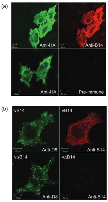
The location of the B14 protein was examined in HeLa cells transfected with the plasmid encoding the HA-tagged B14 protein. Immunofluorescence analysis with the anti-HA mAb and anti-B14 Ab detected a diffuse cytoplasmic stain-ing with no obvious co-localization to specific organelles or the cell surface and with relatively little staining in the nucleus (Fig. 3a, upper panels). Only a weak background signal was detected by pre-immune rabbit serum in the HA-tag-positive transfected cells (Fig. 3a, lower right panel). This indicated the specificity of anti-B14 Ab. To compare the location of B14 in transfected cells with those infected by VACV, at 8 h p.i. vB14-infected cells were stained with the rabbit Ab and this detected a similar intracellular distribu-tion (Fig. 3b, upper right panel). In vDB14-infected cells, the B14 Ab showed very little staining (Fig. 3b, lower right panel). The infection with both viruses was confirmed using a mAb against the VACV D8 protein that detects punctate cytoplasmic structures (Fig. 3b, left panels) that are virions as reported previously (van Eijl et al., 2002; Carter et al., 2003).
B14 is non-essential in cell culture
The role of B14 protein in the life cycle of VACV-WR was examined by characterizing the phenotype of vDB14 in cell culture. The plaque size formed by vDB14 was smaller than that formed by vB14 and vB14-rev in CV-1 cells and slightly smaller in BS-C-1 but not in RK13cells (Fig. 4). The
reduc-tion in plaque size of CV-1 cells and BS-C-1 was 40 and 20 %, respectively (n=20 plaques; P<0?05, Student’s t-test). To examine the effect of B14 on the viral life cycle in more detail in vitro, one-step (Fig. 5a) and multi-step (Fig. 5b) growth curves were studied in BS-C-1 and CV-1 cells (Supplementary Fig. S3 available in JGV Online). In both cell types vDB14 showed similar growth kinetics to control viruses after high (10 p.f.u. per cell) or low multiplicity (0?01 p.f.u. per cell) of infection. Therefore, the basis for the cell type-specific plaque size reduction in BS-C-1 and CV-1 cells was not a reduced virus yield.
B14 affects virus virulence and the recruitment of leukocytes
The virulence of vDB14 was compared to control viruses in two murine models. In an i.n. model, no significant differ-ence in weight loss or signs of illness (data not shown) was observed in animals infected with vDB14 at 104 or
Fig. 2. Characterization of B14 by immunoblotting. (a) Wild-type and C-terminal HA-tagged B14 were expressed transiently in HeLa cells. At 24 h post-transfection, extracts of cells were prepared using RIPA buffer (20 mM Tris/HCl, pH 8?0, 150 mM NaCl, 1 % Nonidet P-40, 1 % SDS) and analysed by immuno-blotting with either rabbit anti-B14 Ab (left) or an anti-HA mAb (right). (b) HeLa cells were infected by the indicated VACVs at 10 p.f.u. per cell for 16 h, or mock infected or transfected with pCI-B14 for 24 h. The cells were lysed in RIPA buffer and extracts were analysed by immunoblotting with anti-D8 mAb (top panel) or anti-B14 Ab (lower panel). (c) HeLa cells were infected with VACV WR at 10 p.f.u. per cell in the presence (+) or absence (”) of 40 mg AraC ml”1. At the indicated times
p.i., the cells were washed once with PBS and lysed by addi-tion of 200 ml protein loading buffer. Each cell lysate (20 ml) was resolved by SDS-PAGE and analysed by immunoblotting with anti-D8 mAb (top panel) or anti-B14 Ab (lower panel).
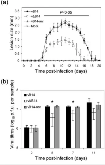
36103p.f.u. compared to control viruses (see Supplementary Fig. S4 available in JGV Online). However, in an i.d. infec-tion model, vDB14 induced a significantly (P<0?05) smaller lesion size from days 6 to 17 p.i. (Fig. 6a). Measurement of infectious virus in infected lesions indicated that although the titres of infectious virus increased equally up to 2 days p.i., thereafter (days 5 and 7) titres were reduced in
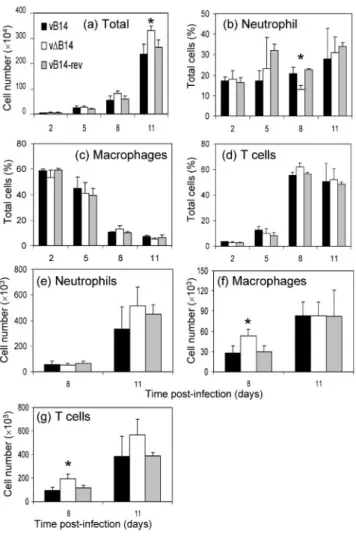
vDB14-infected ear lobes compared with controls (Fig. 6b). These observations indicate that B14 enhances virus virul-ence, at least partly by restricting clearance of virus. To start to address the mechanism by which B14 affects the outcome of infection in vivo, the cells in the infected ears were extracted, quantified by trypan blue exclusion and identified by flow cytometry (Methods). This revealed a statistically (P<0?05) higher number of cells in vDB14-infected ears at day 11 p.i. compared with tissue vDB14-infected by control viruses (Fig. 7a). Interestingly, in vDB14-infected cells at day 8 p.i., neutrophils represented a reduced per-centage of the total cell population compared with controls (Fig. 7b), while the percentage of macrophages (Fig. 7c) and lymphocytes (Fig. 7d) were slightly higher than the controls. However, when the absolute number of each cell type was analysed, rather than the percentage of total cells, the number of macrophages (Fig. 7f) and T cells (Fig. 7g) were significantly higher in ears infected by vDB14 than in ears infected by controls on day 8 p.i. By this criterion the number of neutrophils was not significantly different between the groups (Fig. 7e). In addition, CD4, CD8, T-cell receptor cd cells were also examined but showed no significant difference (data not shown). Therefore, the decrease in percentage of neutrophils on day 8 was likely to be attributable to the corresponding increase of macro-phages and lymphocytes. A similar result was observed on day 5 p.i. when cells were extracted from ears by digestion with collagenase (Jacobs et al., 2006) (data not shown).
Fig. 3. Subcellular localization of B14. (a) B14 with a C-terminal HA tag was expressed transiently by transfection in HeLa cells for 24 h. Cells were fixed and permeabilized as described in Methods and stained with anti-HA mAb (left panels), anti-B14 Ab (top right panel) or rabbit pre-immune serum Abs (bottom right panel). (b) HeLa cells were infected with vB14 (top panels) or vDB14 (lower panels) at 10 p.f.u. per cell for 8 h. Cells were fixed and permeabilized as in (a) and stained with either anti-D8 mAb (left panels) or anti-B14 Ab (right panels).
Fig. 4. Plaque formation by vDB14. Monolayers of CV-1, RK13
or BS-C-1 cells were infected by vB14, vDB14 or vB14-rev and overlaid with DMEM/2?5 % FBS/1 % carboxymethyl cellu-lose. After 48 (CV-1 and BS-C-1 cells) or 72 h (RK13 cells),
the monolayers were stained with 0?1 % crystal violet in 15 % ethanol and photographed.
DISCUSSION
This paper provides a characterization of the B14 protein of VACV strain WR that was uncharacterized hitherto. Data presented show that B14 is an intracellular, 15 kDa protein that is expressed early during infection and is non-essential for the viral life cycle. Loss of the B14 protein reduced virus virulence in a murine i.d. but not i.n. model. The attenua-tion was manifested by a decrease in lesion size, reduced virus titres and enhanced recruitment of macrophages and T cells. Collectively, this indicates that B14 is an intracellular immunomodulator.
In both infected and transfected cells B14 was predomi-nantly cytoplasmic, indicating that other VACV proteins do not influence the location of this protein. Under the con-ditions examined, there was no apparent co-localization with specific organelles, although a weak nuclear staining was also apparent. Active nuclear import is dependent on a nuclear localization signal or carrier protein(s) such as importin that shuttle between the cytoplasm and nucleus. Alternatively, small proteins (less than 20–40 kDa) could pass through nuclear pores by passive diffusion (Macara, 2001; Xu & Massague, 2004). Therefore, the presence of
some B14 in the nucleus might be via an unknown active transport system or simply by passive diffusion into the nucleus due to the small molecular mass of B14.
B14 is a non-essential protein based on in vitro character-ization of vDB14. Although the mutant displayed reduced plaque size compared with wild-type and revertant control viruses in BS-C-1 and CV-1 cells, the plaque formed by each virus was indistinguishable in size in RK13cells. However,
vDB14 failed to show a significant decrease in growth
Fig. 5. Growth kinetics of vDB14. BS-C-1 cells were infected at either 10 (a) or 0?01 (b) p.f.u. per cell and aliquots of infected cells were collected at the indicated times p.i. Cells were frozen and thawed three times, sonicated and the virus infectivity was titrated in duplicate on BS-C-1 cell monolayers. Data are presented as the mean log10p.f.u.±SD.
Fig. 6. Virulence assay in murine i.d. model. (a) Female C57BL/6 mice (n=6) were infected i.d. with 104p.f.u. of the indicated viruses and the size of the lesion was measured daily with a micrometer. Horizontal bar indicates the days on which the lesion size caused by vDB14 was statistically different (P<0?05) from both vB14 and vB14-rev. Data are means of lesion size±SD. (b) Infectious virus in infected lesions. Mice were infected in both ears as in (a) and at the indicated time p.i. the animals were sacrificed, ears were removed and infec-tious virus was determined by plaque assay on duplicate mono-layers of BS-C-1 cells. Asterisks indicate days on which the titre of virus present in ears infected with vDB14 was signifi-cantly different from both vB14 and vB14-rev-infected tissue. Data are means of viral titres±SD.
kinetics, even in BS-C-1 and CV-1 cells. The major factor determining plaque size is the ability to form virus-tipped actin tails beneath cell-associated enveloped virus particles at the cell surface that drive surface virions into surrounding cells, for review see Smith et al. (2002). However, an analysis of actin-tail formation in cells infected by vDB14 showed no difference from controls (data not shown). Other VACV proteins affect the plaque size or morphology without inhibiting actin-tail formation. For instance, a VACV strain lacking kelch-like protein C2 produced plaques with a
distinct morphology but the same size (Pires de Miranda et al., 2003) and either the deletion (Parkinson et al., 1995) or overexpression (Sanderson et al., 1996) of the A38 protein reduced plaque size. Overall, B14 is not required for the virus replication and the basis for the restricted plaque size is not due to a cell-line-specific defect in viral growth or inability to form any actin tails.
In vivo analysis showed that B14 contributed to virus virul-ence in a murine i.d. (local infection) but not i.n. (systemic infection) model. Previously, other VACV immunomodu-lators were shown to exhibit a phenotype in either one model or the other and this may be explained by the different inflammatory responses following infection with VACV via the i.n. and i.d. routes (Tscharke et al., 2002; Reading & Smith, 2003). Analysis of infectious virus titres in infected dermal tissue showed that by 2 days p.i. the virus titres had increased for all viruses to a similar extent; therefore, there was no replicative defect associated with loss of B14 in vivo. However, subsequently (days 5 and 7 p.i.) the titres of virus in ears infected with vDB14 was reduced compared with controls. This finding indicated that B14 impaired the ability of the host response to control virus growth, and so in its absence smaller lesions were produced. To examine the effect of B14 in vivo further, cells that were present in the infected lesions were extracted and analysed by staining with specific mAb and FACS analysis. Although there was a trend showing an increased infiltration of cells into the vDB14-infected ears on days 5 and 8 p.i. this was only significantly different from controls at day 11 p.i. The absolute number of neutrophils present in infected lesions was not altered at any time point, but the number of macrophages and T cells rose significantly at day 8 p.i. in vDB14-infected lesions compared with controls. This increase in leukocytes indicates an enhanced host inflam-matory response in the absence of B14, and may contribute to the size restriction and early resolution of the resulting lesion.
The recruitment of leukocytes to the skin is dependent on the expression of chemokines and cytokines (Gillitzer & Goebeler, 2001; Schon et al., 2003). Among these mediators, chemokines CCL2 and CCL3 (formerly monocyte chemoat-tractant 1 and macrophage inflammatory protein-1a, respectively) play important roles in recruiting T cells and macrophages (Kunstfeld et al., 1998; von Stebut et al., 2003). Therefore, the production of such chemokines early after infection will affect the cellular infiltrate. Infection by vDB14 caused a greater infiltrate in infected tissues on days 5–11 p.i. compared with infection by control viruses (although this was only significantly different on day 11). Notably these infiltrates contained increased numbers of T cells and macrophages (day 8 p.i.). Taken together, these findings suggest that B14 may modulate the inflammatory responses, possibly by affecting the expression of pro-inflammatory chemokines such as CCL2 and CCL3. However, the mechanism by which B14 exerts such immunomodulation remains to be investigated.
Fig. 7. Characterization of infiltrating leukocytes in the C57BL/ 6 mice infected intradermally with 104p.f.u. of the indicated viruses. Cells were extracted (Methods) from both ears of an infected mouse and pooled. (a) Total numbers of viable cells were determined by trypan blue exclusion. Data are means of cell counts±SD. In these cells, neutrophils (b), macrophages (c) and T cells (d) were identified by corresponding surface markers as described in Methods. The absolute number of neu-trophils (e), macrophages (f) and T cells (g) at days 8 and 11 p.i. were determined as described in Methods. The mean±SD
of data from three infected mice were analysed. Asterisks indicate the day on which data for vDB14 are significantly different from the vB14 and vB14-rev groups (P<0?05, Student’s t-test).
In summary, B14 is an intracellular virulence factor that is highly conserved in orthopoxviruses and modulates the host response to infection. These findings contribute to our understanding of poxvirus pathogenesis and suggest a means by which attenuated orthopoxvirus vaccines may be produced.
ACKNOWLEDGEMENTS
This work was supported by grants from the Wellcome Trust, the UK Medical Research Council and Imperial College London. R. A.-J. C. is the recipient of an Overseas Research Award scholarship and G. L. S. is a Wellcome Trust Principal Research Fellow.
REFERENCES
Alcami, A. & Smith, G. L. (1992).A soluble receptor for interleukin-1b encoded by vaccinia virus: a novel mechanism of virus modula-tion of the host response to infecmodula-tion. Cell 71, 153–167.
Alcami, A. & Smith, G. L. (1996).A mechanism for the inhibition of fever by a virus. Proc Natl Acad Sci U S A 93, 11029–11034.
Boyle, D. B. & Coupar, B. E. (1988).A dominant selectable marker for the construction of recombinant poxviruses. Gene 65, 123–128.
Carter, G. C., Rodger, G., Murphy, B. J., Law, M., Krauss, O., Hollinshead, M. & Smith, G. L. (2003). Vaccinia virus cores are transported on microtubules. J Gen Virol 84, 2443–2458.
Dobbelstein, M. & Shenk, T. (1996).Protection against apoptosis by the vaccinia virus SPI-2 (B13R) gene product. J Virol 70, 6479–6485.
Falkner, F. G. & Moss, B. (1990). Transient dominant selection of recombinant vaccinia viruses. J Virol 64, 3108–3111.
Gillitzer, R. & Goebeler, M. (2001).Chemokines in cutaneous wound healing. J Leukoc Biol 69, 513–521.
Goebel, S. J., Johnson, G. P., Perkus, M. E., Davis, S. W., Winslow, J. P. & Paoletti, E. (1990).The complete DNA sequence of vaccinia virus. Virology 179, 247–266, 517–263.
Gubser, C. & Smith, G. L. (2002).The sequence of camelpox virus shows it is most closely related to variola virus, the cause of smallpox. J Gen Virol 83, 855–872.
Gubser, C., Hue, S., Kellam, P. & Smith, G. L. (2004). Poxvirus genomes: a phylogenetic analysis. J Gen Virol 85, 105–117.
Howard, S. T., Chan, Y. S. & Smith, G. L. (1991). Vaccinia virus homologues of the Shope fibroma virus inverted terminal repeat proteins and a discontinuous ORF related to the tumor necrosis factor receptor family. Virology 180, 633–647.
Hughes, S. J., Johnston, L. H., de Carlos, A. & Smith, G. L. (1991).
Vaccinia virus encodes an active thymidylate kinase that complements a cdc8 mutant of Saccharomyces cerevisiae. J Biol Chem 266, 20103–20109.
Jacobs, N., Chen, R. A.-J., Gubser, C., Najarro, P. & Smith, G. L. (2006). Intradermal immune response after infection with vaccinia virus. J Gen Virol 87, 1157–1161.
Kettle, S., Alcami, A., Khanna, A., Ehret, R., Jassoy, C. & Smith, G. L. (1997).Vaccinia virus serpin B13R (SPI-2) inhibits interleukin-1b-converting enzyme and protects virus-infected cells from TNF- and Fas-mediated apoptosis, but does not prevent IL-1b-induced fever. J Gen Virol 78, 677–685.
Kunstfeld, R., Lechleitner, S., Wolff, K. & Petzelbauer, P. (1998).
MCP-1 and MIP-1a are most efficient in recruiting T cells into the skin in vivo. J Invest Dermatol 111, 1040–1044.
Macara, I. G. (2001).Transport into and out of the nucleus. Microbiol Mol Biol Rev 65, 570–594.
Moss, B. (2001). Poxviridae: the viruses and their replication. In Virology, 4th edn, pp. 2849–2883. Edited by B. N. Fields, D. M. Knipe, P. M. Howley, R. M. Chanock, J. Melnick, T. P. Monath, B. Roizman & S. E. Straus. Philadelphia: Lippincott-Raven Publishers.
Niles, E. G. & Seto, J. (1988). Vaccinia virus gene D8 encodes a virion transmembrane protein. J Virol 62, 3772–3778.
Parkinson, J. E. & Smith, G. L. (1994).Vaccinia virus gene A36R encodes a Mr 43–50 K protein on the surface of extracellular
enveloped virus. Virology 204, 376–390.
Parkinson, J. E., Sanderson, C. M. & Smith, G. L. (1995). The vaccinia virus A38L gene product is a 33-kDa integral membrane glycoprotein. Virology 214, 177–188.
Pires de Miranda, M., Reading, P. C., Tscharke, D. C., Murphy, B. J. & Smith, G. L. (2003).The vaccinia virus kelch-like protein C2L affects calcium-independent adhesion to the extracellular matrix and inflam-mation in a murine intradermal model. J Gen Virol 84, 2459–2471.
Reading, P. C. & Smith, G. L. (2003).A kinetic analysis of immune mediators in the lungs of mice infected with vaccinia virus and comparison with intradermal infection. J Gen Virol 84, 1973–1983.
Sanderson, C. M., Parkinson, J. E., Hollinshead, M. & Smith, G. L. (1996).Overexpression of the vaccinia virus A38L integral membrane protein promotes Ca2+influx into infected cells. J Virol 70, 905–914.
Schon, M. P., Zollner, T. M. & Boehncke, W. H. (2003). The molecular basis of lymphocyte recruitment to the skin: clues for pathogenesis and selective therapies of inflammatory disorders. J Invest Dermatol 121, 951–962.
Seet, B. T., Johnston, J. B., Brunetti, C. R. & 7 other authors (2003).
Poxviruses and immune evasion. Annu Rev Immunol 21, 377–423.
Smith, G. L. & McFadden, G. (2002).Smallpox: anything to declare? Nat Rev Immunol 2, 521–527.
Smith, G. L., Chan, Y. S. & Howard, S. T. (1991). Nucleotide sequence of 42 kbp of vaccinia virus strain WR from near the right inverted terminal repeat. J Gen Virol 72, 1349–1376.
Smith, G. L., Vanderplasschen, A. & Law, M. (2002).The formation and function of extracellular enveloped vaccinia virus. J Gen Virol 83, 2915–2931.
Spriggs, M. K., Hruby, D. E., Maliszewski, C. R., Pickup, D. J., Sims, J. E., Buller, R. M. & VanSlyke, J. (1992).Vaccinia and cowpox viruses encode a novel secreted interleukin-1-binding protein. Cell 71, 145–152.
Tscharke, D. C. & Smith, G. L. (1999).A model for vaccinia virus pathogenesis and immunity based on intradermal injection of mouse ear pinnae. J Gen Virol 80, 2751–2755.
Tscharke, D. C., Reading, P. C. & Smith, G. L. (2002). Dermal infection with vaccinia virus reveals roles for virus proteins not seen using other inoculation routes. J Gen Virol 83, 1977–1986.
Upton, C., Slack, S., Hunter, A. L., Ehlers, A. & Roper, R. L. (2003).
Poxvirus orthologous clusters: toward defining the minimum essential poxvirus genome. J Virol 77, 7590–7600.
van Eijl, H., Hollinshead, M., Rodger, G., Zhang, W. H. & Smith, G. L. (2002). The vaccinia virus F12L protein is associated with intra-cellular enveloped virus particles and is required for their egress to the cell surface. J Gen Virol 83, 195–207.
von Stebut, E., Metz, M., Milon, G., Knop, J. & Maurer, M. (2003).
Early macrophage influx to sites of cutaneous granuloma formation is dependent on MIP-1a/b released from neutrophils recruited by mast cell-derived TNFa. Blood 101, 210–215.
Williamson, J. D., Reith, R. W., Jeffrey, L. J., Arrand, J. R. & Mackett, M. (1990).Biological characterization of recombinant vaccinia viruses in mice infected by the respiratory route. J Gen Virol 71, 2761–2767.
Xu, L. & Massague, J. (2004).Nucleocytoplasmic shuttling of signal transducers. Nat Rev Mol Cell Biol 5, 209–219.