The Effectiveness of an Upper Extremity Neuromuscular Training Program on the
Shoulder Function of Military Members with a Rotator Cuff Tendinopathy:
A Pilot Randomized Controlled Trial
Mémoire
Amanda Ager
Maîtrise en sciences cliniques et biomédicales
Maître ès sciences (M. Sc.)
Québec, Canada
The Effectiveness of an Upper Extremity Neuromuscular Training Program on the
Shoulder Function of Military Members with a Rotator Cuff Tendinopathy:
A Pilot Randomized Controlled Trial
Mémoire
Amanda Ager
Sous la direction de:
Luc J. Hébert, directeur de recherche
Jean-Sébastien Roy, codirecteur de recherche
iii
RÉSUMÉ
INTRODUCTION: La tendinopathie de la coiffe des rotateurs (TCR) entraine au quotidien des douleurs et faiblesses musculaires et une diminution du contrôle moteur à l'épaule. OBJECTIFS: Les objectifs de cette étude étaient i) d'effectuer une revue de littérature pour identifier les méthodes de quantification de la proprioception de l'épaule utilisées en laboratoire et en clinique et d’en présenter les qualités métrologiques, ii) d'évaluer l'efficacité d’un programme d’entrainement neuro-musculaire en comparant son efficacité à réduire la douleur à l’épaule et en améliorer la fonction à celle obtenue par des soins usuels de physiothérapie. MÉTHODES: i) Une revue de 5 bases de données a été conduite d’octobre 2015 à juillet 2016 pour documenter les propriétés métrologiques de protocoles d’évaluation de la proprioception à l'épaule. Les études incluses ont été évaluées à l'aide de l’outil de contrôle QualSyst et de l'échelle COSMIN à 4 points. ii) Trente-trois soldats en service actif au sein des Forces armées canadiennes ont été assignés au hasard à 1) programme standardisé supervisé d’entrainement neuromusculaire et contrôle moteur (Exp) ou à 2) soins usuels de physiothérapie (Ctl). Les variables principales étaient les symptômes, la capacité fonctionnelle et les limitations physiques évalués avec le questionnaire Disabilities of the Arm, Shoulder and Hand (DASH) et la variable secondaire était l'indice Western Ontario Rotator Cuff (WORC). Toutes les variables ont été mesurées au départ (T0) et à 6 (T6) et 12 (T12) semaines après l'intervention. La comparaison des effets des interventions a été évaluée à l'aide d’une analyse per protocole (APP), analyse intention-traitement (AIT) et avec une analyse de variance à mesures répétées à 2 voies. RÉSULTATS: i) Vingt et une études (n = 407 participants, 553 épaules) ont été retenues. Les études analysées confirment d'excellents scores méthodologiques avec l’outil QualSyst (88,1 ± 9,9%) et de bons scores avec le COSMIN pour la fidélité (71,1%) et un score de qualité modérée à faible (50%) pour la validité de critère. Les coefficients de corrélation intraclasse (CCI) pondérés pour la fidélité intraévaluateur étaient les plus élevés pour le sens du positionnement articulaire passif et la kinesthésie soit 0,92 ± 0,07 (n = 214) et 0,92 ± 0,04 (n = 74), respectivement. Le mouvement et l'outil les plus fidèles sont la rotation interne à 90 ° d'abduction (CCI = 0,88 ± 0,01 (n = 53)) et le dynamomètre (CCI = 0,92 ± 0,88 (n = 225)). Aucune étude n’a rapporté d’indices de sensibilité au changement. ii) Aucune interaction significative (p ≥ 0,101) de groupe × temps (p ≥ 0,101) n'a été démontrée. Par contre, nous avons observé un effet de temps significatif (p <0,001) pour le questionnaire DASH et l'indice WORC.
iv
CONCLUSION: Ces données préliminaires suggèrent que les deux approches proposées conduisent à des améliorations comparables. L'utilisation d'une intervention de groupe axée sur l'exercice a le potentiel d'être aussi efficace qu'une approche un à un plus exigeante en terme de temps de traitement. Ces résultats permettront de fournir aux cliniciens des lignes directrices pour la mesure de la proprioception à l'épaule et l’utilisation d’une approche novatrice de traitement en groupe pour la TCR.
Mots clés: Épaule, tendinopathie, contrôle moteur, proprioception, programme d'exercices, soins en physiothérapie
v
ABSTRACT
INTRODUCTION: The shoulder is the most mobile joint of the body which means that it heavily relies of an important level of neuromuscular control at all times. A rotator cuff (RC) complex provides stability to the shoulder and often times falls victim to injury, which can produce functional limitations during activities of daily living and work tasks. Individuals affected by an RC tendinopathy often have neuromuscular and proprioceptive deficits. OBJECTIVES: The objectives of this study are to (i) conduct a systematic review to identify methods of quantifying shoulder proprioception in a laboratory and clinical setting and to present the associated psychometric properties. (ii) To evaluate the effectiveness of a novel neuromuscular training program for the upper extremities versus one-on-one physiotherapy care (manual therapy, range of motion exercises, strengthening) for the reduction of shoulder pain and improvement in function with soldiers affected by an RC tendinopathy. METHODS: (i) A review of five databases was conducted from conception to July 2016 to identify studies that reported at least one psychometric property of a shoulder proprioception protocol. The included studies were evaluated using the QualSyst checklist and the 4-point COSMIN scale. (ii) Thirty-three military personnel with the Canadian Armed Forces were randomly assigned to one of the following interventions: 1) Upper Extremity Neuromuscular Training Program; (2) usual physiotherapy care. The main outcomes included symptoms and functional capacity assessed using the Disability of the Arm, Shoulder, and Hand (DASH) questionnaire. A secondary outcome included the Western Ontario Rotator Cuff (WORC) Index. Outcome measures were evaluated at baseline (T0) and 6 (T6) and 12 (T12) weeks post-intervention. The effects of the interventions were evaluated using repeated 2-way variance measures (ANOVAs) for a per-protocol analysis and intention-to-treat. RESULTS: i) Twenty-one studies were included, resulting in 407 participants and 553 evaluated shoulders (n). The weighed intraclass correlation coefficients (ICC) for intra-rater reliability were highest for passive joint position sense and kinesthesia, ICC = 0.92 ± 0.07 (n = 214) and ICC = 0.92 ± 0.04 (n = 74), respectively. The most reliable direction of movement and equipment used were internal rotation at 90° abduction, ICC = 0.88 ± 0.01 (n = 53), and the dynamometer, ICC = 0.92 ± 0.88 (N = 225). ii) No significant group (p ≥ 0.1) or group × time interactions (p ≥ 0.1) were found; though a statistically significant time effect (p < 0.001) was established for the DASH questionnaire and WORC Index. Our preliminary data suggests a marginally better improvement with the control group with all outcomes over 12 weeks. CONCLUSION: The evaluation of shoulder
vi
proprioception is most reliable when using a passive protocol with an isokinetic dynamometer for internal rotation at 90° shoulder abduction. The preliminary results of our pilot RCT suggest that both groups statistically improved with a time effect, but that the usual care group further demonstrated clinically significant gains. The results of this study will provide clinicians with potential guidelines for measuring shoulder proprioception in a clinical setting, as well as an innovative approach to group therapy that is potentially less costly and equally as effective as conventional one-on-one physiotherapy.
Key words (4-6): Shoulder, tendinopathy, motor control, proprioception, exercise program, physiotherapy care
vii
TABLE OF CONTENTS
RÉSUMÉ ... iii
ABSTRACT ... v
LIST OF ABBREVIATIONS AND COMMON TERMS ... xvi
ACKNOWLEDGEMENTS ... xix
FOREWORD ... xx
CHAPTER 1 ... 1
INTRODUCTION ... 1
1.0 The justification of our research ... 1
1.1 The shoulder joint ... 2
1.1.1 The prevalence, incidence, and etiology of shoulder pain ... 3
1.2 Anatomy and biomechanics ... 4
1.2.1 Static structures and mechanoreceptors ... 5
1.2.2 Shoulder musculature... 5
1.2.3 Biomechanics of shoulder movement ... 6
1.3 Motor control and proprioception ... 9
1.4 Rotator cuff tendinopathy ... 11
1.4.1 Classification of tendon injuries and contributing factors ... 12
1.4.2 Intrinsic factors ... 12
1.4.3 Extrinsic factors ... 13
1.4.4 Cortical influence and central sensitization ... 14
1.5 Military members and shoulder injuries ... 15
1.6 Physical rehabilitation for a rotator cuff tendinopathy ... 16
1.6.1 Acupuncture and electro modalities... 17
1.6.2 Stretching and range of motion exercises ... 18
1.6.3 Manual therapy ... 18
1.6.4 Exercise prescription ... 19
1.6.5 Effectiveness of a structured program approach ... 20
1.6.6 Effectiveness of a group treatment approach ... 21
1.6.7 Motor control and proprioceptive exercises ... 22
1.6.8 The development of the Upper Extremity Neuromuscular Training Program ... 24
1.7 Objectives ... 25
1.7.1 The purpose of this thesis ... 25
viii
1.8 Pilot randomized control trial ... 26
1.8.1 Scientific question ... 26
1.8.2 Statement of hypothesis ... 26
1.8.3 Specific objectives ... 26
1.9 The systematic review ... 27
1.9.1 Research questions ... 27
1.9.2 Specific objective of the systematic review ... 27
CHAPTER 2 ... 28
METHODOLOGY: PILOT RCT ... 28
2.1 Research protocol ... 28
2.2 Study design ... 28
2.3 Participants ... 30
2.4 Instrumentation and outcome measures ... 31
2.4.1 Questionnaires... 33
2.4.2 Secondary outcome measures ... 33
2.5 Randomization and blinding ... 34
2.6 Interventions ... 35
2.6.1 The development of the Upper Extremity Neuromuscular Training Program ... 35
2.6.2 UpEx-NTP parameters ... 39
2.6.3 The development of the Usual Physiotherapy Care (UPC) guidelines ... 40
2.6.4 The usual physiotherapy care (UPC) protocol and parameters ... 41
2.7 Statistical analysis... 41
2.8 Feasibility, potential risks, and ethics ... 42
2.8.1. Feasibility ... 42
2.8.2 Potential patient risks ... 42
2.8.3 Ethics... 42
2.9 Funding ... 42
CHAPTER 3 ... 43
SHOULDER PROPRIOCEPTION: HOW IS IT MEASURED AND IS IT RELIABLE? A SYSTEMATIC REVIEW ... 43
3.1 Problem Statement ... 44
3.1.1 What is already known on this topic ... 44
3.1.2 What our study adds... 44
ix
ABSTRACT ... 47
3.3 Introduction ... 48
3.4 Methodology ... 49
3.4.1 Literature search and study identification ... 49
3.4.2 Study Selection ... 49
3.4.3 Data extraction and shoulder proprioception measurements ... 50
3.4.4 Quality Assessment ... 50
3.4.4.1 Standard Quality Assessment Criteria for Evaluating Primary Research Papers ... 51
3.4.4.2 COSMIN 4-point scale ... 51
3.4.5 Data analysis ... 51
3.5 Results ... 52
3.5.1 Description of the studies... 52
3.5.2 Quality of the included studies ... 52
3.5.3 Specific findings ... 53
3.5.3.1 Population ... 53
3.5.3.2 Type of proprioception evaluated (Figure 6)... 53
3.5.3.3 Direction of movement (Figure 7) ... 54
3.5.3.4 Equipment (Figure 8) ... 54
3.5.3.5 Validity, reliability, and responsiveness ... 55
3.6 Discussion ... 55
3.6.1 Ecological validity and the clinical application of proprioception ... 56
3.6.2 Lack of standardization ... 57
3.6.3 Strength and limitations of the review ... 58
3.7 Conclusion ... 58
3.7.1 Take Home Message for Clinicians ... 59
3.7.2 Role of the Funding Source ... 59
3.7.3 Conflict of Interest Statement ... 59
3.7.4 Author Contributions ... 59
CHAPTER 4 ... 66
THE EFFECTIVENESS OF AN UPPER EXTREMITY NEUROMUSCULAR TRAINING PROGRAM ON THE SHOULDER FUNCTION OF MILITARY MEMBERS WITH A ROTATOR CUFF TENDINOPATHY: A PILOT RANDOMIZED CONTROLLED TRIAL 66 4.1 Résumé / Abstract ... 67
x 4.1.1 Résumé ... 67 4.1.2 Abstract ... 69 4.2 Background ... 71 4.3 Methodology ... 73 4.3.1 Participants ... 73 4.3.2 Study Design ... 73
4.3.3 Randomisation and blinding ... 74
4.3.4 Interventions ... 74
4.3.5 The group-supervised Upper Extremity Neuromuscular Training Program ... 74
4.3.6 One-on-one Usual Physiotherapy Care (UPC) ... 75
4.4 Outcomes ... 76
4.4.1 Symptoms and disability ... 76
4.4.2 Muscle impairment ... 76
4.4.3 Physical limitations ... 77
4.4.4 Perceived level of change ... 77
4.4.5 Data analysis ... 78
4.5 Results ... 78
4.5.1 Level of symptoms and disability ... 81
4.5.2 Muscle strength impairments and physical limitations ... 85
4.5.3 Perceived Level of Change, adherence to treatment schedule, and blinding ... 86
4.6 Discussion ... 87
4.6.1 Multimodal versus exercise-based treatments ... 88
4.6.2 A supervised approach for common MSK conditions ... 88
4.6.3 Group exercise and access to physiotherapy services... 89
4.6.4 Strengths and limitations of this study ... 89
4.6.5 Take home message for clinicians ... 90
4.6.6 How to increase adherence to a group exercise program ... 90
4.7 Conclusion ... 91
4.7.1 Acknowledgements ... 91
4.7.2 Funding ... 91
4.7.3 Availability of data and materials ... 92
4.7.4 Ethical Approval ... 92
4.7.6 Author contributions ... 92
xi
DISCUSSION ... 93
5.0 The pilot RCT ... 93
5.1 Potential central adaptations with both interventions ... 94
5.2 Improvement to our UpEx-NTP ... 95
5.3 Our study amongst the scientific literature ... 96
5.4 The impact of our pilot RCT ... 97
5.5 The shoulder proprioception systematic review ... 98
5.6 The future is promising... 99
5.6.1 Lessons learned ... 99
5.6.2 Recommendations for clinicians ... 100
5.7 Future research ... 100 CHAPTER 6 ... 102 CONCLUSIONS ... 102 REFERENCES ... 103 SUPPLEMENTARY ANNEX I ... 121 SUPPLEMENTARY ANNEX II ... 124
SUPPLEMENTARY ANNEX III ... 127
SUPPLEMENTARY ANNEX IV ... 128
SUPPLEMENTARY APPENDICES ... 129
APPENDIX A: Recruitment poster for the military participants, Quebec ... 130
APPENDIX B: Recruitment poster for the military clinicians, Valcartier Garrision, Quebec 131 APPENDIX C: Information package for participants ... 132
APPENDIX D: Consent form for participants ... 139
APPENDIX E: Subjective telephone interview evaluation form... 140
APPENDIX H: DASH questionnaire (French Canadian) ... 149
APPENDIX I: WORC Index (French Canadian) ... 152
APPENDIX J: Edinburg Handedness Inventory (French) ... 162
APPENDIX K: Questionnaire of perception of change and satisfaction (French) ... 163
APPENDIX L: Usual Physiotherapy Care Intervention form (French) ... 164
APPENDIX M: Upper Extremity Neuromuscular Training Program (Visual Guide) ... 166
APPENDIX N: Upper Extremity Neuromuscular Training Program (Patient Tracking Sheet) ... 174 APPENDIX O: Scientific approval from the Scientific Committee of the CIRRIS / IRDPQ 181
xii
APPENDIX P: Ethical Approval (CIRRIS - IRDPQ) ... 182
APPENDIX Q: Letter from Surgeon General of the Canadian Armed Forces ... 184
APPENDIX R: Awarded Grant: REPAR-OPPQ Program 4.2 for a clinical study ... 185
APPENDIX S: Awarded Student Bursary from CIRRIS and Laval University ... 187
LIST OF TABLES
TABLE 1………15 TABLE 2………29 TABLE 3………30 TABLE 4………32 TABLE 5………60 TABLE 6………81 TABLE 7………83 TABLE 8………85TABLE CAPTIONS
TABLE 1: Minimal Physical Fitness Standards (MPFS) for universality of service for an active Canadian Armed Forces member. Retrieved from: http://www.forces.gc.ca/en/about-policies-standards-medical-occupations/op-def-performance-standards-minimum-tasks.page.
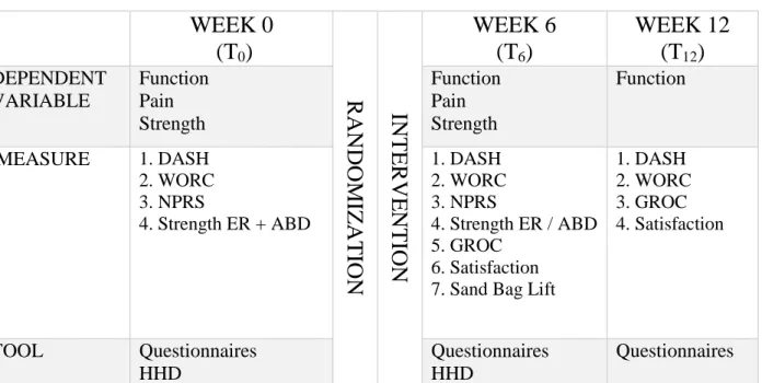
TABLE 2: Evaluation and intervention timeline with associated outcome measures for RCT. TABLE 3: Statistical properties of clinical diagnostic tests for RC tendinopathy.
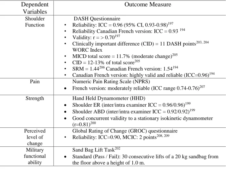
TABLE 4: The psychometric properties of the special tests and outcome measures used for the pilot RCT.
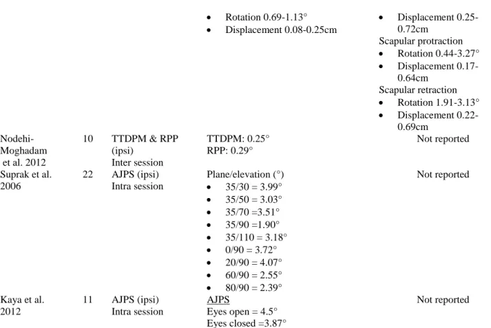
TABLE 5: Comparison of psychometric properties of different shoulder proprioception protocols for our systematic review.
TABLE 6: Means and standard deviations of baseline characteristics of the participants, according to intention-to-treat analysis (n = 33). Also presented are the results from the statistical analysis, demonstrating no statistical significant differences between the Ctl and Exp groups for their baseline demographics.
TABLE 7: Mean scores and standard deviations of DASH-CF and WORC-CF Questionnaires in relation to baseline values for the Ctl and Exp Groups (PPA n = 21, ITT n = 33) Data presented as mean change (± standard deviation). Δ Denotes a change from the baseline score (indicated at T0 in bold) DASH: Disabilities of the Arm, Shoulder and Hand questionnaire (lower score indicates higher disabilities, therefore a negative change from baseline indicates an improvement); WORC:
xiii
Western Ontario Rotator Cuff index (higher score indicates higher functional capacity, therefore a positive change from baseline indicates an improvement).
TABLE 8: Mean scores and standard deviations of maximal voluntary isometric contractions (MVIC), expressed as muscle strength in Newton meters (Nm) of injured and healthy shoulder for the Ctl and Exp Groups at T0 and T6 (per-protocol analysis, n = 21 and intention-to-treat, n = 33). MVIC values reported in Newton-meters (Nm).
ABD: abduction, ER: external rotation. No statistically significant results have been found for a time or group × time interaction, nor for a group × time × shoulder interaction for either ABD or ER isometric strength.
xiv
LIST OF FIGURES
FIGURE 1 ... 8 FIGURE 2 ... 14 FIGURE 3 ... 31 FIGURE 4 ... 62 FIGURE 5 ... 63 FIGURE 6 ... 64 FIGURE 7 ... 64 FIGURE 8 ... 65 FIGURE 9 ... 80 FIGURE 10 ... 82 FIGURE 11 ... 84 FIGURE 12 ... 87FIGURE CAPTIONS
FIGURE 1: Scapulothoracic normalized kinematics of the shoulder complex. FIGURE 2: Extrinsic and intrinsic mechanisms of a rotator cuff tendinopathy. FIGURE 3: Effect size calculation for our pilot RCT.
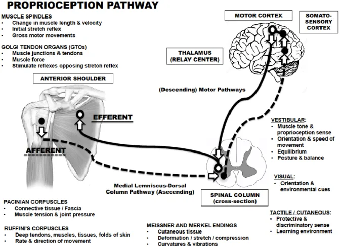
FIGURE 4: Graphical depiction of the shoulder proprioception pathway. Proprioception includes bilateral symmetry with crossed representation. This suggests that proprioception information from the right side of the body is process in the somatosensory cortex in the left hemisphere. FIGURE 5: An organogram describing the literature selection process according to the PRISMA (Preferred Reporting Items for Systematic Reviews and Meta-Analyses) statement.
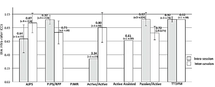
FIGURE 6: Weighted averages (WA) ICCs for intra-rater reliability of proprioception measures of the glenohumeral joint, except for PJMR for which a WA ICC could not be calculated. Columns without standard deviation (SD) bars represent data from a single study (s = number of studies included, n=number of shoulders evaluated).
FIGURE 7: Weighted averages (WA) ICCs for intra-rater reliability for various movements of the glenohumeral joint, except for Abduction (ABD) for which a WA ICC could not be calculated. Columns without standard deviation (SD) bars represent data from a single study (s = number of studies included, n=number of shoulders evaluated).
FIGURE 8: Weighted averages (WA) ICCs for intra-rater reliability for various proprioceptive equipment used for quantifying shoulder proprioception of the glenohumeral joint. Columns without standard deviation (SD) bars represent data from a single study (s = number of studies included, n=number of shoulders evaluated).
xv
FIGURE 9: Recruitment algorithm for an intention-to-treat (ITT) and per-protocol analysis (PPA). FIGURE 10: Mean scores of DASH-CF and WORC-CF over time (T0, T6, and T12), per-protocol analysis (PPA: n = 21) and intention-to-treat (ITT: n = 33). A higher WORC Index score represents a functional improvement, whereas a lower DASH score represents improvement.
FIGURE 11: Pain levels at rest for both groups at T0 and T6, represented as (X̅ ± SD), per protocol analysis (PPA: n = 21) and intention-to-treat (ITT: n = 33) of the injured shoulder. Using the NPRS scale where 0 represents "no pain at all" and 10 represents "worst pain imaginable".
FIGURE 12: Results of secondary outcome measure: perceived level of change (GROC), represented as response rate (%) for both groups, as a per-protocol analysis (n = 21).
xvi
LIST OF ABBREVIATIONS AND COMMON TERMS
Abbreviation Term
CAF Canadian Armed Forces
MPFS Minimum Physical Fitness Standards
AAOMPT American Academy of Orthopedic Manual Physical Therapists
JPS Joint position sense
MC Motor control
RC tendinopathy Rotator cuff tendinopathy
MSK Musculoskeletal
UPC Usual Physiotherapy Care
UpEx-NTP Upper Extremity Neuromuscular Training Program
Ctl Control group
Exp Experimental group
RCT Randomized controlled trial
SGHRP Canadian Armed Forces Surgeon General Health Research
Program
IR Internal rotation
ER External rotation
ABD Abduction
SIS Subacromial impingement syndrome
MDC Minimal detectable change
MICD Minimally important clinical difference
MVIC Maximum voluntary isometric contraction
CID Clinically important difference
SEM Standard error of measurement
DASH Disabilities of the Arm, Shoulder and Hand questionnaire
WORC Index Western Ontario Rotator Cuff Index
GROC Global rating of change scale
xvii
Pain: Pain is a complex pattern of sensory system activations that are intimately linked to the activity of other cortical systems including, but not limited to, the emotional, cognitive and modulatory processes.1 It
is important to note that not every trauma to tissues will result in the manifestation of pain.
Proprioception: Proprioception can be understood as our sixth sense,2 through the gathering of internal
sensory information through our peripheral and central nervous system. Proprioception has been defined as the awareness of, and ability to, sense the position of our limbs and trunk in space (position sense), as well as kinesthesia, the awareness of motion of the human body (motion sense).3, 4 Proprioception is essential
for well-adapted sensorimotor control. It fulfills the roles of feedback and feed forward sensorimotor control and consequently, the regulation of muscle stiffness, movement acuity, joint stability, coordination, and balance.5
Joint Position Sense (JPS): Joint position sense is a sub-modality of our conscious awareness of proprioception and refers to our ability to detect the positioning of our limbs and trunk within our surrounding environment.6
Neuromuscular control: Neuromuscular control is defined as a system of collaborative networks of the cerebral cortex, the spinal column, neurons and muscle fibers involved in the control of movement and posture.7 Neuromuscular control further encompasses the efferent motor responses to sensory information,
such as proprioception and kinesthesia. Neuromuscular control involves both a feed forward, planning of movements and preparatory muscle activity, and feedback mechanisms, which involve the regulation of muscle activity through reflexive pathways and top-down cortical commands.8
Neuromuscular training program: Neuromuscular training can be defined as "... training enhancing unconscious motor responses by stimulating both afferent signals and central mechanisms responsible for dynamic joint control".9 In the case of the upper or lower extremities, it may include motor control,
proprioceptive, and functional training.
Motor control: Motor control can be understood as the physiological mechanism behind how the peripheral and central nervous system produces purposeful, coordinated movements so that our limbs and trunk can interact with the rest of our body as well as our surrounding environment.10
Tendon: Tendons are mechanically loaded tissues that generally connect muscles to bone and are responsible for the tensile force transmission of muscle cells.11
Tendinitis: The inflammation of a tendon as a result of micro-tears when the musculo-tendinous unit is mechanically acutely overloaded with a tensile force.12
Tendinosis: Refers to the degeneration of the collagen within the tendon due to chronic overuse without an adequate healing period. This is generally the case with repetitive strain injuries.12
Rotator cuff (RC) tendinopathy: The progressive degeneration of a / several rotator cuff tendons13 of the
shoulder complex.
Shoulder impingement syndrome: A shoulder impingement syndrome refers to the dysfunctional biomechanics of the shoulder complex, which results in the physical pinching or encroachment of soft tissues (such as the tendons or bursae) under the acromion during shoulder movements.14 The most common
clinical signs of an impingement dysfunction include localized pain to the shoulder during elevation or overhead reaching, as well as positive clinical tests such as the Full Can, Empty Can, a painful arch, the lift off sign, and painful and weakened external rotation and abduction of the shoulder.15
xviii
To my devoted husband, Justin Christian Thibault
xix
ACKNOWLEDGEMENTS
Creating new knowledge requires the harmonious synchronization of many individuals and organizations. This project is of no exception and many thanks are due. Firstly, to my directors Luc J. and Jean-Sébastien, who guided me throughout two years of over ambitiousness. To the Center for Interdisciplinary Research in Rehabilitation and Social Integration (CIRRIS), Laval University, and the Quebec Rehabilitation Research Network (REPAR) for their financial contributions, both through student bursaries as well as a research grant which allowed for the material realization of our rehabilitation project. To all the physiotherapists from the Valcartier Garrison (Quebec City, QC) who have believed in me from the very beginning, particularly France Gamache, Sophie Bernard, Pierre-Marc Vézina, Myriam Cyr, and Valérie Charbonneau, who all acted as the pillars to this project. A sincere thank you to Marie-Élise Prémont who is one of the hardest working people I know. Her role as an evaluator during the winter of 2017 allowed this project to continue to move forward during a difficult friction point. Lastly, to my cherished husband, Justin, who has supported me literally throughout our adventures around the world, and who will always be my infinite source of inspiration.
xx
FOREWORD
The presentation of this thesis is the result of collective work performed by the Motor Control Laboratory at the Center for Interdisciplinary Research in Rehabilitation and Social Integration (CIRRIS) / Institut de réadaptation en déficience physique de Québec (IRDPQ) as well as in collaboration with the Canadian Armed Forces Surgeon General Health Research Program and the Valcartier Garrison of the Canadian Armed Forces.
The aggregate of the scientific efforts has been compiled to form the basis for my Master's in Clinical and Biomedical Sciences (concentration in rehabilitation) through Laval University and under the supervision of my Director Dr. Luc J. Hébert and my Co-Director Dr. Jean-Sébastien Roy. The following overture is presented as a Master level thesis with the insertion of two articles, the first being a systematic review and the second being the results from our pilot randomized controlled trial, in the presentation of six chapters. The first chapter encompasses the introduction to the subject of shoulder pain, specifically caused by a rotator cuff (RC) tendinopathy, and the underlying biomechanical and motor control deficits that are associated with this disorder. The first chapter is further developed by exploring the scientific literature on the management of an RC tendinopathy as well as dissects the two possible approaches to shoulder pain management, specifically usual physiotherapy care (UPC) and an exercise-based group approach. The first chapter also explores the concepts of motor control and proprioception as it pertains to the rehabilitation efforts of the most mobile joint in the body, the shoulder. The first chapter concludes by introducing the overall aims of this thesis and presenting the objectives of our systematic review on shoulder proprioception and our pilot randomized control trial (RCT). The second chapter outline the methodology behind our pilot RCT. The third chapter offers the summation of our publication in the Journal of Hand Therapy, entitled Shoulder Proprioception, how is it Measured and Is It Reliable: A Systematic Review. The fourth chapter includes our recent manuscript submission: The Effectiveness of an Upper Extremity Neuromuscular Training Program on the Shoulder Function of Military Members with a Rotator Cuff Tendinopathy: A Pilot Randomized Controlled Trial to the Journal of Military Medicine. Chapters five and six finalizes our findings by presenting the ensemble of our discussion and conclusions while offering guidance to clinicians
xxi
for evidence-based rehabilitation for the management of a shoulder rotator cuff tendinopathy and the measurement of shoulder proprioception within a clinical setting.
I am the principle author of both articles as well as the sole author of said thesis. I fully participated in the theoretical inception, the development of the methodology, the collection of the data, the analysis, as well as the realization of the submitted manuscripts. The authors of the systematic review include my directors, as well as three collaborators, Marianne Roos, Amélie Fournier Belley, and Dr. Ann Cools from Ghent University in Belgium. The authors of the pilot RCT include my directors as well as a former colleague, France Gamache, PT, from the Valcartier Garrison who collaborated on the development and implementation of the Upper Extremity Neuromuscular Training Program. All authors made significant contributions to the development and achievement of the studies.
All information and studies presented are part of an overarching goal of bringing motor control and proprioception to the forefront of shoulder rehabilitation. This research was made possible by a collaborative student bursary between the CIRRIS and Laval University, as well as a research grant from the OPPQ-REPAR 4.2 program for clinical research.
1
CHAPTER 1
INTRODUCTION1.0 The justification of our research
Our interactions with our surrounding environment greatly depend on our physical health. Reaching, pulling, and lifting, for example, are all activities that heavily rely on the health of our upper limbs, and of our shoulders in particular. Shoulder pain is one of the most common musculoskeletal (MSK) symptoms, with up to one-quarter of the Western population reporting a problem at any one time and up to two-thirds of all adults reporting pain over a lifetime.16 Shoulder pain is the third most common reason to consult a physiotherapist,17 but yet the management of shoulder pain and injuries are considered to be one of the most challenging areas of MSK medicine today.18
In Canada, statistics collected between 2009-2010 through the Canadian Community Health Survey (CCHS), found that among serious MSK injuries, involving a ligament, muscle sprain or strain, dislocations or fractures, 13.2% occurred at the shoulder, elbow, or the arm.19 In most cases, muscle, tendon, or nerve injuries happen as a result of overuse or repetitive movements over an extended period of time. The shoulder complex is of no exception, acting as the leading site for repetitive strain trauma, accounting for 22.6% of all bodily strain injuries.19
The rotator cuff (RC) complex is one of the most common sites for shoulder injuries and is the leading cause for shoulder pain and physical impairments among an adult population.20 This is exceptionally relevant to manual labourers engaged in repetitive movements of the upper extremities,21, 22 which includes an active military population. It is well documented that such injuries of the shoulder can translate into significant time off work and a significant cost to the employer, both in terms of human resources and loss of productivity.23 The military follows this trend, as shoulder injuries among soldiers are the fourth leading site for MSK injuries, leading to a medical discharge from active service.24 Although studies have identified shoulder pain as being an important burden for a military population,24-26 few studies have attempted to provide treatment guidelines for this specialized group.
2
There is currently an extend need for effective and efficient treatment approaches for shoulder disorders among serving military members. Although there are studies addressing neck and shoulder pain,27 shoulder instability,28 or post-operative repair among a military cohort,29 to our knowledge, there are no treatment guidelines for the management of a shoulder rotator cuff tendinopathy or impingement syndrome for soldiers.
This project has the purpose of exploring the effectiveness of a supervised group-based exercise program in comparison to usual physiotherapy care for the management of a RC tendinopathy among active military service personnel. If the results suggest a comparable functional improvement between both treatment approaches, this could potentially spark a new discussion regarding the allocation of rehabilitation resources in terms of materials, time, and treating physiotherapists. This project has the potential to open a discussion regarding the efficiency and resource-effectiveness of a group approach for common MSK rehabilitation efforts across Canada.
1.1 The shoulder joint
The shoulder joint, anatomically understood as the glenohumeral (GH) articulation, is a very functionally important joint of the body. Being the most proximal joint of the upper extremity, the GH joint is involved in all upper quadrant movements and determines the success of our ability to execute movements involving our upper limbs to effectively interact with our environment. The GH joint does not act in isolation, but rather requires a complex choreography of surrounding joints, both active and passive structures, as well as the guidance from the nervous system to execute a purposeful motor task. For this reason, it is functionally more accurate to refer to the GH joint not only as the shoulder but as a shoulder complex, in order to be inclusive of the neighboring joints and structures that contribute to the coordinated movements of the shoulder and upper extremity.
The shoulder joint is known to be the most mobile articulation of the body,30-32 with 360° of azimuth, it has 3 degrees of freedom, and consequently 6 movements within 3 anatomical planes. The shoulder complex is an important site for muscle attachment, with over 15 muscles33 that act in-sync to allow us
3
to gainfully perform activities of daily living. The shoulder is heavily involved in common tasks such as reaching, pulling, pushing, and lifting.34, 35 Often times, it is the gross motor movements of the shoulder that allow us to use our proximal joints for fine motor tasks such as preparing a meal, hygiene activities, sports and leisure, and even the menial task of typing on a computer.35 Because of it's vast mobility and heavy implication in daily tasks,34 the shoulder is a popular site for dysfunction and injury.
1.1.1 The prevalence, incidence, and etiology of shoulder pain
A shoulder injury can be functionally devastating to an individual, significantly impacting the most basic activities of daily living,36 and can potentially place unnecessary financial stress on our health care system.37 The actual etiology of shoulder pain is not fully known, but it is well known that shoulder pain is quite common and results in an annual incidence of shoulder disorders, ranging from 7 - 26% in a Western general population.16
According to the National Health Service and Society in the United Kingdom, approximately 1% of their population consults a medical practitioner with a new presentation of shoulder pain each year, which equates to an estimated cost of £310 million (an estimated $510 million Canadian) in health care related spending.38 In the Netherlands, up to 50% of the cost associated with musculoskeletal (MSK) pain has been attributed to sick leave from paid employment.39 Similarly in Quebec, a report of the Commission
de la santé de la sécurité du travail (CSST), estimates that for the period of 2005-2007, the total annual
expenses associated with shoulder disorders, including the human cost and those associated with lost of productivity from work, are estimated to be $393,204,738.23, 40 Similarly, shoulder pain has been noted among Canadian Armed Forces military members, representing 14% of all reported MSK injury cases as well as being third in prevalence, tied with spinal injuries, and following closely behind ankle and knee injuries.41, 42 We can therefore definitively concede that shoulder pain is a costly problem for both the civilian and military population.
Shoulder pain is currently among the most common reasons to visit a general practitioner or a physiotherapist today.43 It is third in prevalence to back and neck pain44 and nearly two-thirds of adults suffer from shoulder pain at some point during their lives.20 A few commonly diagnosed shoulder dysfunctions include bicipital tendonitis, adhesive capsulitis (frozen shoulder), GH and AC arthritis,
4
instabilities and labral tears,37 as well as an impingement syndrome (SIS) or a RC disorder.15, 45 RC disorders, specifically a RC tendinopathy, is among the leading cause for medical consultation for shoulder pain.20 The incidence itself of RC tendinopathies varies between 0.3% to 5.5%, with an estimated annual prevalence of 0.5% to 7.4%.46 To best appreciate the potentially extensive limitations a shoulder injury can have on a person's quality of life, it is imperative to understand the intricacies of the underlying anatomy and biomechanics of the shoulder complex.
1.2 Anatomy and biomechanics
The shoulder complex involves 3 physiological joints, notably the glenohumeral (GH) joint, the acromioclavicular (AC) joint, the sternoclavicular (SC) joint, as well as a "functional joint" known as the scapulothoracic (ST) joint. The SC joint is the only bony attachment site of the upper extremity to the axial skeleton. The ST joint involves the gliding movement of the scapula along the rib cage during upper extremity movements and does not include a physical bone-to-bone attachment. The GH joint is of particular interest when understanding the mechanism of shoulder injuries because it is osteologically predisposed to instability.47, 48 The GH joint is comprised of a ball and socket synovial joint, where the head of the humerus (convex surface) articulates with the glenoid fossa (concave surface) of the scapula. Because of the relatively large surface area of the humeral head in relation to the fossa, the joint itself has limited bony congruency, and consequentially heavily depends on surrounds soft tissues for structural support. Moreover, it is estimated that only 25% of the humeral head articulates with the glenoid fossa at any one time during movement.49 The surrounding passive structures (the labrum, joint capsule, and ligaments) as well as the active structures (the muscles and associated tendons) act cooperatively in a healthy shoulder to maintain dynamic stability throughout movement.
An area most often involved in the cases of shoulder pain is the subacromial space, which includes the theoretical space between the coracoacromial arch and the head of the humerus.13, 50 More specifically, the subacromial canal lies underneath the acromion, the coracoid process, the AC joint and the coracoacromial ligament.51, 52 The space itself includes a bursa which provides lubrication for the RC tendons, the insertion for the long head of the biceps tendon, and the RC tendons themselves.13, 50-52
5
1.2.1 Static structures and mechanoreceptors
The static structures of the shoulder complex, which includes the labrum (a fibrocartilaginous ring), the capsule, cartilage, ligaments, and fascia collectively act as the physical restraints to the osseous matter and provides a deepening effect to the shallow glenoid fossa.53 Further to their passive stabilization role, they also provide additional protection via the various mechanoreceptors embedded within their fibers. Mechanoreceptors can be understood as the neural sensors that provide afferent input to the central nervous system for motor processing and descending motor commands for the execution of movements.54-56 Mechanoreceptors are characterized by their specialized nerve endings that are sensitive to the mechanical deformations of tissues,57-59 and therefore contribute to the modulation of motor responses of the adjacent muscles. Mechanotendinous receptors (muscle spindles and golgi tendon organs), capsuloligamentous receptors (ruffini and pacinian corpuscles) as well as cutaneous receptors (meissner, merkel and free nerve endings) are responsible for our sense of touch, vibration, proprioceptive positioning, as well as provide the feedback regarding muscle length, tension, orientation, further to the speed and strength of the contractions of the muscle fibers.47, 60 It is therefore, resoundingly clear that the passive structures of the shoulder provide a neurological protection mechanism through feed forward and feedback input, that directly mediates reflex musculature stabilization about the glenohumeral joint.55
1.2.2 Shoulder musculature
Further to the intricate network of passive ligatures that conjoin adjacent bones, the importance of the surrounding musculature cannot be overstated. Active muscle contractions are essential for maintaining the stability of the shoulder complex.47 The musculature of the shoulder region can be subdivided into the global movers of the shoulder and the fine-tuning stabilizers of the individual articulations. The larger muscles such as the trapezius, the levator scapula, the pectorali, the deltoids, the serratus anterior, the latissimus dorsi, the rhomboids, the teres major, the biceps, the coracobrachialis, and triceps muscles are responsible for various synergistic activities during shoulder movements. Conjointly as agonist and antagonist couplings, they allow for the gross motor movements of the upper quadrant. More specifically to the GH joint, the fine-tuning stabilizers are just as important to the shoulder complex as the global movers for coordinated and smooth shoulder movements.
6
The stabilizing muscles of the GH articulation, the supraspinatus, subscapularis, infraspinatus, and teres minor, are often summarized as the rotator cuff (RC) complex, and attach to the humeral head within the glenoid fossa. Collectively, they act as the dynamic stabilizers of the GH joint by maintaining a centralized positioning of the humeral head within the glenoid fossa,61, 62 in both static and dynamic conditions. It has been suggested that the tendons of the rotator cuff muscles blend with the ligaments and the glenoid labrum at their respected sites of attachments, so that the muscle contractions can provide additional stability by tightening the static structures during movement.63 The synchronized contractions of the RC muscles must maintain the centralized positioning of the humeral head during movements in order to avoid the physical encroachment of tissues, predominantly anteriorly or superiorly to the GH joint, which has been linked to injury and pain amongst the shoulder region. As previously noted, due to the anatomical passage of the common RC tendon within the subacromial space, the RC tendons are particularly vulnerable to compression, abnormal friction, and ultimately an impingement (pinching) during active tasks.13, 50 Proper alignment of the glenohumeral head is important for the healthy engagement of the shoulder joint in activities of daily living.
1.2.3 Biomechanics of shoulder movement
To further grasp the contributing factors of shoulder pain and associated dysfunctions, it is essential for researchers and clinicians alike to understand the biomechanics of the shoulder complex. In the interest of a specific injury of the shoulder, notably the rotator cuff (RC) tendinopathy, the biomechanics of the GH and ST joints will be discussed within this section.
The natural arthrokinematics of the GH joint of the shoulder complex during an open-chain movement supports various directional glides of the humeral head within the glenoid fossa.64, 65 Del Maso and colleagues have estimated that a maximum of 7.5 mm of upward translation of the humeral head may occur during range of motion movements,65 which is not an insignificant amount of migration for a large bony structure to experience within a compact space during a dynamic task. The success of a coordinated movement of the humeral head with normalized arthrokinematics, avoiding an impingement situation, requires the harmonious co-contraction of the RC tendons. Abnormal glenohumeral translations have been linked to pathological shoulders and it has been suggested to be a contributing factor for shoulder pain and discomfort, and may also lead to the damage of encompassing structures.65, 66
7
As illustrated by the force-vectors of their respected moment arms, the RC tendons collectively have been accredited with the compression of the humeral head within the glenoid fossa during movements.67 The individualized tendons of the RC complex are directly affiliated with limiting the translation of the humeral head in specific directions. The supraspinatous muscle contributes to preventing excessive superior translation, the infraspinatus and teres minor limit excessive superior and posterior translation, and the subscapularis controls excessive anterior and superior translation of the humeral head, respectively.68 An imbalance in the neural activation of any one of the RC muscles could easily cause a misalignment of the humeral head thus giving rise to an impingement of the subacromial structures during movement. Both the superior and anterior translation of the humeral head during movements are the leading biomechanical causes for an impingement syndrome,69 and a contributing factor to the development of a rotator cuff tendinopathy.65, 66, 69
The movement of the scapula along the thoracic cage also directly influences the biomechanics of the shoulder complex as a whole, and can moreover predispose the development of an impingement syndrome. The healthy movement of the scapula along the thorax during arm elevation includes protraction, posterior tilting and lateral rotation, depending on the plane of movement (Figure 1).70-73
8
Figure 1 Caption: Scapulothoracic normalized kinematics of the shoulder complex. Retrieved from Zhao et al. 2015.74
Although posterior tilting is generally understood as primarily an acromioclavicular joint motion, the tilting that occurs at the scapula during arm elevation is crucial in order to minimize the encroachment of soft tissues passing under the acromial arch.73 The normal contribution of the ST joint is generally expressed as the ratio of ST movement with regards to that occurring simultaneously at the GH articulation. The scapulohumeral rhythm is quantified by dividing the total amount of shoulder elevation (humeralthoracic) by the scapular upward rotation (scapulothoracic).70 Within the scientific literature, the scapulohumeral rhythm is generally accepted to be 2:1, which represents 2° of humeral elevation for every degree of scapular upward rotation.71, 75, 76
9
The stability of the ST joint relies on the coordinated activity of the 18 muscles that directly attach to the scapula.77 The scapular muscles must dynamically control the positioning of the glenoid so that the humeral head remains centered and permits arm movement to occur. When a weakness or neuromuscular dysfunction of the scapular musculature is present, normal scapular arthrokinematics become altered,76 and ultimately predisposes an individual to an injury of the GH joint.75-77 The pathological kinematics of the ST joint include, but are not limited to 1) increased medial rotation, 2) decreased superior rotation and 3) decreased posterior tilting74, 78, 79 These movement alterations are believed to increase the proximity of the rotator cuff tendons to the coracoacromial arch or glenoid rim,73, 80 however, there are still points of contention as to how the movement pattern deviations directly contribute to the reduction of the subacromial space.73 For the sake of clarification, the current literature differentiates between an internal impingement and an external impingement. An impingement that involves a decreased space towards the coracoacromial arch is said to be an external impingement, whereas an internal impingement involves the glenoid rim,73 and can be associated with a GH instability.81 Regardless of the classification, the dysfunctional shoulder mechanisms can further the progression of rotator cuff disease82 and must therefore be understood as a neuromuscular impairment.
The neuromuscular control of the scapula relies on the balanced team-work between the global movers and the fine-tuning stabilizing muscles of the shoulder complex. Again, because of the floating nature of the scapula along the thorax, it too, must rely on the kinship between the cortical direction provided by the nervous system and the resulting action of the MSK system. We can therefore affirm, that the shoulder complex is among the most kinematically complex regions of the human body,80 and requires a high level of neuromuscular stability throughout movement. The neuromuscular control of the shoulder also requires a well-developed sense of motor control and proprioception.
1.3 Motor control and proprioception
For the purpose of this thesis, motor control can be defined as the ability of our peripheral nervous system (specifically, our mechanoreceptors, sensory receptors, and neural relay pathways) and our central nervous system (spinal and cortical processing) to produce purposeful, coordinated joint movements to facilitate internal interactions (of our limbs and body) and external interactions (our environment) for every day life.10 It is a process that varies in complexity from a reflexive spinal loop, to higher processing neural networks that involve cortical control. In the case of the shoulder complex, it involves using all
10
senses to produce normalized and non-pathological movement patterns. The complex nature of the shoulder joint implies that numerous muscles must act together to provide both stability and motion. 75-77, 83 Moreover, the normalized mechanism of the shoulder complex involves the input from the nervous system, both peripherally and centrally, to successfully interact with our environments and sustain from injury.
Dynamic stability of the shoulder joint requires highly attuned motor control and an intact sense of proprioception. Proprioception is a concept that is associated with motor control, but should not be misunderstood as representing the same physiological concept. Proprioception is accredited with being our sixth sense,2 and can best be appreciated as our ability to detect the position of our trunk and limbs in space in the absence of visual feedback.3 Proprioceptive input is collected by the mechanoreceptors located within our passive, dynamic, as well as cutaneous tissues, and is sent via the posterior column-medial lemniscus pathway (PCML) for higher processing within the postcentral gyrus and cerebral cortex.84 Descending commands from the motor cortex directs the neuromuscular synchronicity about an articulation for purposeful and (motor) controlled movements. As noted by Clark and colleauges,5 both proprioception and motor control are absolutely essential for a well-adapted sensorimotor control, particularly with regards to highly mobile joints such as the shoulder. Proprioceptive feedback facilitates shoulder motor control by regulating muscle stiffness, movement acuity, joint stability, coordination, and balance.5 It further contributes to motor control by providing sensory feedback for inter-limb coordination,85, 86 correcting and updating movement strategies,87 and for the formation of muscle synergies.88,89 Proprioception is the sensory input that helps the nervous system implement efficient and effective motor strategies for healthy movement.
Motor control and proprioceptive deficits have been associated with MSK injuries90-95 and have also been linked to the recurrence and persistence of physical impairments such as shoulder pain, decreased range of motion and strength.91, 95, 96 As outlined by Contemori & Biscarini,90 deficiencies in afferent proprioceptive information may results in the poor accuracy of descending motor commands and impairment of the shoulder neuromuscular function, leading to reduced shoulder functional stability, and ultimately an increased risk of injury. Furthermore, proprioception and motor control have been recognized as being disturbed among MSK disorders due to pain, effusion, trauma, and fatigue,5 all of which frequently occur within the scope of a shoulder injury. More precisely, it has been well
11
documented that individuals affected by an RC tendinopathy or SIS often exhibit motor control97, 98 and proprioceptive deficits.95, 99, 100
1.4 Rotator cuff tendinopathy
Among shoulder disorders, the RC tendons are the leading source of shoulder pain.99, 101-103 Due to their role in providing dynamic joint stability, they are often highly susceptible to injury.13, 104 Like any tendon, the RC tendons can become pathological due to several mechanisms, but most commonly, it is the result of a shoulder mechanical impingement. A RC tendinopathy is commonly referred to as a subacromial impingement syndrome (SIS),104 however, it is important to note that despite the use of the term "impingement" in a diagnostic capacity, a RC impingement is a clinical sign, not a diagnosis.61, 105 To best understand the biomechanics behind the concept of a SIS, it is important to outline both the intrinsic and extrinsic factors that contribute to the possible irritation or degeneration of the RC tendons. Although the exact pathophysiological etiology of the RC tendinopathy is not entirely clear,61 there is a growing consensus that an impingement occurs when the RC tendons, collectively passing within the subacromial space, are subjected to repetitive stresses such as pinching, most often caused during repetitive overhead activities.106 The RC tendons become "pinched" within this space during movements among individuals with decreased shoulder girdle motor control, consequently causing irritation, swelling and damage to the tendons.61, 106 The exact pathophysiological reasoning behind the changes to the tendons or the subarcromial space is not currently known.61 For the purpose of this thesis, a RC tendinopathy will refer to the clinical presentation of a collection of cluster signs and symptoms, determined by clinical diagnostic tests, which suggest an underlying degeneration of the tendons or a compression of subacromial structures (the RC tendons, the bursa, and / or the long head of the biceps tendon). An RC tendinopathy can be provoked by either a trauma or an impingement mechanism. It is important to note that not every person with a clinical diagnosis of tendinopathy will experience shoulder pain.104 Because there is currently a poor understanding of the source of pain in an RC tendinopathy, shoulder pain alone cannot be the only clinical indicator of a pathology.
12
1.4.1 Classification of tendon injuries and contributing factors
The terms tendinitis, tendinosis, and tendinopathy are often used synonymously by researchers and clinicians.45 In recent scientific trends, greater emphasis has been placed on improving the precision of tendon injury taxonomy. It has become increasingly important for both researchers and health care providers to systematically define the source of the injury so that the underlying mechanism can be correctly identified, and subsequently successfully treated. A tendinopathy is an overarching term which indicates damage, and at times pain, in and around the tendons.107 The term encompasses both a tendinitis and a tendinosis. For precision sake, a tendinitis traditionally refers to the acute inflammation of the tendon,12 whereas the tendinosis refers to the separation and degeneration of collagen bundles of the tendons due to repetitive and often long-term stresses.12, 108 Controversially, basic scientific research suggests that factually, little to no inflammation, is present among these tendon conditions.107
1.4.2 Intrinsic factors
The intrinsic factors of a RC tendinopathy are known to be associated with the degeneration of the tendinous tissues.109 As outlined by Seitz and colleagues,109 the intrinsic factors include pathophysiological elements such as tendon vascularity, morphology and composition, as well as the natural biology or genetics of a person. Khan and colleagues110 have also suggested that intrinsic factors should include the resultant effects of an acute or traumatic event, such as inflammation, which can potentially provoke pathophysiological changes to the involved tissues.
Inflammation and degeneration of a tendon can also occur from excessive loading. Excessive loading occurs when external forces exerted on the soft tissue exceeds its maximal tolerance, thus causing micro-tearing over time. Tendons are load bearing structures and their main role is to transmit forces from muscle to bone. Loading is essential for maintaining tendon homeostasis, however excessive loading can lead easily led to degeneration and tearing.111 This resultant mechanism of overloading can encourage the RC tendons to become pathological with overuse and repetitive activities.112
13
1.4.3 Extrinsic factors
The extrinsic factors of a RC tendinopathy are defined as those that cause a compression of the RC tendons.109 The compression is linked to the narrowing of the subacromial space,106 which could be due to an excessive angulation of the acromion,106 a type II or III acromion morphology,110 inadequate stabilization of the scapula,113 abnormal shoulder kinematics,80, 114-116 specific muscular weaknesses (rotator cuff, serratus anterior), or muscular tightness / shortening (pectoralis minor which pulls the scapula into a protracted position),61 globally resulting in a RC and/or scapular muscles performance deficit.80, 117, 118 This inadequate scapulothoracic muscle control is believed to contribute to a reduction in amplitude in posterior tilting and lateral rotation of the scapula,80 which causes the acromion to remain in a lower anterolateral position resulting in a dynamic narrowing of the subacromial space.97, 119It is also noted that the elevation of the humeral head may provoke an imbalance between the humeral head elevators (deltoid muscle) and the stabilizers (notably the rotator cuff muscles). This noted imbalance may encourage a superior migration of the humeral head,106 consequently further narrowing the space for the passage of the RC tendons and resulting in further damage to the tissues.97, 119 Along the same resultant biomechanics of the superior or anterior migration of the humeral head, a shoulder impingement can sometimes be associated with a shoulder instability,120 where individuals exhibit hypermobility and significant capsular laxity,62, 121 furthering the mobility of the humeral head and encroaching on the subacromial space. Collectively, these deficits contribute to the impingement of subacromial structures and often lead to the symptoms associated with a RC tendinopathy. More often than not, the underlying mechanisms of a RC tendinopathy can best be understood as a combination of both intrinsic and extrinsic factors61, 109 (Figure 2).
14
Figure 2 Caption: Extrinsic and intrinsic mechanisms of rotator cuff tendinopathy. Lines indicate non-directional evidence of these relationships, as described by Seitz et al. (2011).109
1.4.4 Cortical influence and central sensitization
Further to the peripheral mechanisms of a tendinopathy, there is also growing support for a possible central cortical component. It is becoming increasingly recognized that a shoulder tendinopathy can be associated with pain radiating down the arm, cutaneous hypersensitivity,122 as well as bilateral upper extremity symptoms,123, 124 which could suggest changes to the central nervous system, or central sensitization. To support this theory, a study by Ngomo and colleagues noted a decreased in the corticospinal excitability of the infraspinatus muscle of the shoulder with an RC tendinopathy compared to the uninjured shoulder,125 suggesting central adaptations to the nervous system associated with the injury. This is important to understand for rehabilitation purposes, because the management of a RC tendinopathy should therefore include both the management of the local problematic biomechanics of the shoulder, but should also address the cortical reorganization of the shoulder region. The following FIGURE 2
15
section will outline current rehabilitation practices for the management of an RC tendinopathy, with an introduction to our population of study, active military members.
1.5 Military members and shoulder injuries
The military population was chosen for our study due to a soldier's high susceptibility to MSK injuries41, 42, 126 and because rehabilitation occurs in a very unique context. Soldiers are expected to maintain a high level of physical fitness and must recover quickly and effectively from their injuries in order to maintain operational readiness. Canadian soldiers who cannot meet the minimal physical standard, known as the Test Force (See Table 1), are put on medical restrictions, which could potentially lead to a medical discharge from active service. Physical health is an integral part of a Canadian Armed Forces (FAC) member's career.
TABLE 1 Functional
Task
Minimum requirements for a pass
1. Sandbag Lift
30 consecutive lifts of a 20 kg sandbag from the floor above a height of 1.0 m. The member alternates between left and right sandbags separated by 1.25 m.
To be completed in 3 minutes and 30 seconds.
2. Intermittent Loaded Shuttle
10 consecutive shuttles (1 shuttle = 20 m there, 20 m back), alternating between loaded shuttles with a 20 kg sandbag and unloaded shuttles, totalling 400 m.
To be completed in 5 minutes and 21 seconds.
Sandbag Drag Carry one 20 kg sandbag and pull a minimum of four on the floor over 20 m without stopping. Number of sandbags being dragged depends on the type of floor.
No minimum time limit.
20 Meter Rushes
Starting from the prone position, complete two shuttle sprints (1 shuttle = 20 m there, 20 m back) dropping to the prone position every 10 m for a total of 80 m.
To be completed in 51 seconds or less.
Retrieved from: http://www.forces.gc.ca/en/about-policies-stan dards-medical-occupations/op-def-performance-standards-minimum-tasks.page.
Table 1 Caption: Test Force: Minimal Physical Fitness Standards (MPFS) for universality of service for an active Canadian Armed Forces member.
Physiotherapy within a military context must be efficient, effective, and allow the member to return to optimal physical capability for mission readiness.127 For this reason, the approach and interventions of our project have been specifically designed for a military population. Our interventions have been framed
16
within the realities of a military context, which equates to more difficult and functional exercises, parameters that encourage endurance, as well as a time-frame that optimizes a rapid return to operational readiness for the member.
Because of the level of physical fitness required to perform basic soldier duties, military members are often characterized as highly trained athletes. Although there are comparable features between the two populations, the reality remains that military members need to be functionally fit and agile in a variety of environments. As such, military members often fall victim to MSK injuries, whether they be acute from a traumatic event or chronic repetitive-strain injuries. Interestingly enough, the majority of the MSK injuries are not caused by military exercises or combat missions, but rather are non-combat related injuries brought forth by sporting activities or physical training.24, 25 As astutely reported by Hébert (2016),24 MSK injuries are a not only a hindrance to the health and wellbeing of the CAF, they also represent a significant cost for military healthcare expenditures. To offer further perspective, within a United States context, MSK injuries remain the number one reason for military personnel to seek medical care. Nonfatal injuries (which include MSK injuries) result in almost 25 million days of limited duty (sick leave or modified work restrictions) annually.128, 129 In Canada, both the 2010 and 2014 Surgeon General's Medical Reports, indicate that MSK injuries are responsible for between 43% and 66% of medical releases from the CAF for members who were considered disable, unfit to perform their duties, or otherwise unemployable by the military.130, 131
The shoulder is among the leading sites for MSK injuries for active military personnel.41, 132 Despite being identified as an important source of pain and injury, the exact prevalence and profile of shoulder injuries among a military population is currently unknown.28 Moreover, to our knowledge, the etiology and prevalence of a shoulder RC tendinopathy, specific to a military population, remains to be clearly identified. What is known, is the devastating effect that a shoulder injury can bring to a soldier, in terms of the longevity of their career, quality of life, and ultimately their livelihood. Because of the nomadic nature of a soldier's work environment, establishing an efficient and effective physiotherapy treatment plan for shoulder injuries remains a challenge to this day. The following section will outline the current rehabilitation efforts for the management of a RC tendinopathy.
17
Currently there is no resounding consensus as to how shoulder pain should be treated in a rehabilitation setting.133 The current non-operative trends include a combination of modalities,134, 135 stretching,136 manual therapy,137-139 acupuncture techniques,140 and exercise prescription for strengthening and motor control of the surrounding musculature.141-144 The literature currently favors a combination of treatment approaches,135, 141, 145 depending on the presented etiology and symptomology of the patient. There is no clear-cut rehabilitation pathway for addressing shoulder pain,35 which also includes the management of a RC tendinopathy. Traditionally, a RC tendinopathy has been addressed by a combination of physiotherapy, the prescription of non-steroidal anti-inflammatory drugs (NSAIDs), and as a last resort, surgical intervention.146 Physiotherapy is often the first line of defense for a RC tendinopathy,147 but a clear set of clinical guidelines for treatment over time has yet to be well established. The following sections will outline the scientific evidence for the various physical therapy approaches currently being practiced by clinicians for the management of a RC tendinopathy.
1.6.1 Acupuncture and electro modalities
Acupuncture and electro modalities such as ultrasound, transcutaneous electrical nerve stimulation (TENS), pulsed electromagnetic field therapy (PEMF), microcurrent electrical stimulation (MENS), acetic acid iontophoresis and microwave diathermy, as well as shockwave therapy, are common physiotherapy treatments used to induce a localized analgesic effect for shoulders affected by a RC tendinopathy. Recent systematic reviews148, 149 reported no significant differences between acupuncture and a placebo for short-term shoulder improvement, and found very limited evidence concerning the effectiveness of acupuncture for improving pain or shoulder function over time.
Along the same vain, the effects of electrophysical agents among patients with a RC tendinopathy were explored in a systematic review by Page and colleagues (2016),135 which included 47 trials and 2388 participants. Due to the low quality evidence and poor statistical power, it is unclear whether therapeutic modalities provide additional benefits to the management of a RC tendinopathy. There may be evidence to support the use of pulsed ultrasound for short-term benefits in individuals with calcific rotator cuff tendinitis, but further high quality placebo-controlled trials are needed to support these results.135 Further support for the discontinuation of electro modalities for a rotator cuff tendinopathy comes from Desmeules and colleagues, who found no additional benefit when using ultrasound for the management of pain or functional gains among this population.150 Moreover, their systematic review from 2016