598 Assessment of Left Ventricular Function (Mancini et a!)
Quantitative
Assessment
of Global
and
Regional
Left Ventricular
Function
With
Low-contrast
Dose Digital
Subtraction
Ventricu log raphy*
G.
B. John
Mancini,
M.D.;John
McB.
Hodgson,
M.D.;
Victor
Legrand,
M.D.;
Eric
R.
Bates,
M.D.;
Fred
M.
Aueron,
M.D.;
Michael
T LeFree,
B. S.; Joseph
S.
Smith,
M.D.;
Glenn
J.
Beauman,
M. S.;
and
Robert
A.
Vogel,
M.D.
Few
studies
have
compared
the
use
of low-contrast
dose
digital
subtraction
ventriculography
with
conventional
yen-triculographyfor quantitative
assessment
ofboth
global and regionalleft ventricular
function.
Accordingly,
34
patientsunderwent
conventional
ventriculography
using
36 ml of
ionic
contrast
material
and
digital
ventriculography
(mask-mode) using10 ml of contrast
diluted
in 10 ml of saline
and
injected
over
two seconds. Data from twopatients
were
excluded
because
of ectopy
during
cmneventriculography
and
from
one
because
of ectopy
during
both
studies.
End-diastolic
and
end-systolic
volumes
were
calculated
from
Jjigital
intravenous
(IV)
ventriculography
has
recently
been
shown
to provide
accurate
deter-minations
of global
and
regional
left
ventricular
func-tion
compared
with
conventional
contrast
ventriculog-raphy.
‘-Although
this
technique
provides
high-quality
ventricular
images
in
a
minimally
invasive
fashion
and
improved
resolution
compared
with
other
noninvasive imaging techniques, the use of relatively
large
doses
ofcontrast
material
limits
its usefulness
in
certain
patient
subgroups
intolerant
to
the
hemo-dynamic
effects
of
contrast
or
in
whom
repeated
ventriculograms
are
needed.
The
hemodynamic
per-turbations
induced
by contrast
material
have
recently
been
shown
to be
similar
whether
contrast
is
adminis-For
editorial
comment
see
page
560
tered
IV
or
directly
into
the
left
As
a
consequence,
several
groups
have
proposed
the
use
of
direct,
low-contrast
dose
left
ventriculography
to
over-come
these
drawbacks
while
forgoing
the
advantage
of
less
invasive
IV techniques.
Relatively
few studies
have
compared
this
new
method
with
conventional
contrast
ventriculography
for
the
assessment
of volume
and
*From the Division of Cardiology, University of Michigan Medical School, Veterans Administration Medical Center, Ann Arbor. Supported in part by the Research Service of the Veterans Administration.
Manuscript received August 6; revision accepted September 21. Reprint requests: Dr Mancini, VA Medical Center, Ann Arbor48lO5
both studies by an area-length method and used to calculate
ejection
fractions.
Regional
wall
motion
was quantitated
by
the centerline method. Results of linear regression analysisdemonstrated high correlations for all parameters
(end-diastolic
volume,
r0.85;
end-systolic
volume,
r0.93;
ejection
fraction,
r
0.92; quantitative regional wallmo-tion,
r
= 0.90).Thus,
low-contrast
dose
digital
subtraction
ventriculography provides anaccurate
assessment
of both
gI obal
and
regional
ventricular
function
and
minimizes
the
requireddose
and
inherent
risks
of contrast
media.
ejection
fraction
measurements.6#{176}’
What
is
more,
comparative
assessment
of
regional
function
on
a
quantitative
basis
has
rarely
been
The
purpose of
this
study
was
to
compare
standard
left
ventriculography
with
low-contrast
dose
digital
yen-triculography
for
the
quantitative
measurement
of
both
global
and
regional
left
ventricular
function.
Patient Population
METHODS
All patients referred for cardiac catheterization were eligible for
this study except when renal insufficiency was present. Thirty-four patients, 43 to 67 years ofage, were studied. On the basis of coronary arteriography, five were normal and 29 had significant (>50 percent)
coronary stenoses ofat least one major coronary artery. The majority
of patients were taking cardioactive drugs up to the time of cardiac catheterization, but no new medications were administered between
the
acquisition of the ventriculograms, which were obtained within 15 minutes of each other during a single cardiac catheterization.Clinical Protocol
All patients underwent coronary arteriography by the Sones or Judkins technique with multiple angulated views. A 7-French pigtail catheter was advanced into the left ventricle, and a low-contrast dose digital ventriculogram was acquired using 10 ml of sodium meglumine diatrizoate (Renografin 76) diluted and agitated in 10 ml ofnormal saline solution just prior to power injection at 10 ml/second
fbr two seconds. This technique caused no significant changes in left ventricular systolic or end-diastolic pressures. Ten to fifteen minutes later, a standard cineventriculogram was acquired using 36 ml of contrast injected over three seconds. All images were obtained in the 30#{176}right anterior oblique projection.
Imaging System
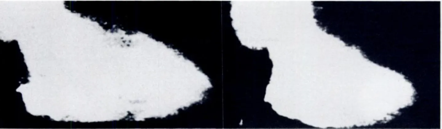
FIGURE 1. End-diastolic (left) and end-systolic (right) frames of a low-contrast dose, direct digital
ventriculogram. Severe inferior hypokinesis is evident.
.L..4L,
--- 20 100____I 40:E:
0 p-,I#{149}1 +4 +2 I. #{149}“ 0 ‘I , ., .,..,..,uiI #{149}_
2 H Y P 0\
),.,. \. I_V’
#{149}\ 4 Cl ‘..,i-
\ijj cThe angiographic facility utilizes a Philips Optimus M200 unit that provides standard 35-mm cineradiography, as well as ECG-gated, multiple-frame per cardiac cycle, 5-msec/frame exposures onto a 1,000:1 signal-to-noise ratio Plumbicon television camera. Radiation exposure is 25 mcR/frame. For digital ventnculography, the video signals are digitized on-line through a 10-bit A to D converter onto a digital disk with a 7 megabyte/second transfer rate, using a commer-cial minicomputer (ADAC, DPS-4100C) for final image processing. The ventricular images are digitized at 30 frames per second into a 256 x256 x 8-bit matrix and displayed after digital-to-analogue con-version.
Data Analysis
The entire digital ventriculographic acquisition was analyzed to obtain precontrast mask frames and the frames constituting the single best opacified beat, excluding ectopic or immediately postec-topic beats. In the early phase ofthe study, the mask was formed by integrating up to 16 frames precontrast injection, whereas subse-quently mask-mode subtraction was performed by matching both precontrast and postcontrast images to the same phase ofthe cardiac cycle (ECG-synchronized mask-mode subtraction). Because patient motion was kept to a minimum, neither method of mask selection produced significant differences in ventricular quantitation. The processed beat was contrast enhanced to optimize border definition, and the outlines ofthe largest and smallest ventricular images were drawn and stored (Fig 1). A magnification correction factor was obtained by imaging a 1-cm, cross-hatched grid at the same distance as the distance measured from the image intensifier to the midchest level ofthe subject. End-diastolic and end-systolic volumes and the ejection fraction were then determined by an area-length
Regional wall motion was quantitated by the centerline method developed and validated at the University of Washington (Seat-tle).6 Briefly, this method measures motion at 100 equidistant chords constructed perpendicular to a centerline drawn midway between the end-diastolic and end-systolic contours. To adjust for heart size, the chord lengths are divided by the perimeter ofthe end-diastolic contour and expressed in terms of a dimensionless percent shortening fraction. The shortening fraction for each chord is then normalized by subtracting the mean chordal shortening of a normal group and dividing by the SD of chord motion of the normal population. Thus, wall motion is expressed in terms ofSDs per chord (SD/C). This standardization allows comparisons between different segments of the same heart and between different hearts. Quan-titative wall motion was determined for anterior and inferior regions. Abnormalities of motion in the anterior wall were ascribed to significant stenoses of the left anterior descending or nondominant circumflex systems. Inferior abnormalities were considered to be due to disease in the right coronary or dominant circumflex systems
(Fig 2).
The same methods of volume and wall motion analysis were applied to the cineventriculograms after selection, digitization, and outlining ofthe largest and smallest endocardial images.
To determine the reproducibility of the measurements, parame-ters were recalculated from the low dose digital ventriculograms from ten patients by one observer Ofl two occasions at least six weeks apart and by a second independent observer. Correlations were determined by linear regression analysis.
REsulTs
Data
from
two
patients
who
had
ectopic
heats
during
cineventriculography
and
one
patient
who
had
ectopy
during
both
conventional
and
digital
ventriculography
were
excluded.
Volume
and
Ejection
Fraction
Measurements
Figure
3
demonstrates
the
relationship
between
end-diastolic
volumes
determined
from
the
cine-ventriculograms
compared
to
the
digital
ventric-ulograms.
The
correlation
coefficient
was
0.85
(y=
0.80x+27
ml,
SEE20.1
ml,
p<.OOl).
End-systolic
volumes
(Fig
4) showed
a higher
correlation
H
i F’ F:.J
ii P f1 I’FIGURE 2. Video display of the quantitative centerline method
applied to ventricular outlines (above) from a patient with anterior dyskinesis. Regional wall motion is expressed in SDs per chord and shown in the bottom graph, which has the chord number on the abscissa and the SDs on the ordinate.
‘(*0.80 x + 27.0 0.85 SEE. 20.1 ML p .001 n* 31
Y:0.93x
+6.0
r
=0.92 S.E.E.: 6.7% p cOOln= 31
><
-J
0
z
0
I-.-cc
U-z
0
0
w
w
.END-DIASTOLIC VOLUME (CINE)(ML) 20 40 60 80
FIGURE 3. Relationship between end-diastolic volumes determined
by conventional ventriculography (CINE) and low dose, digital ventriculography (DIGITAL). The regression line is solid and the line of identity is dashed. S. E. E. = standard error of the estimate.
(r=0.93,
y=0.80x+7.9
ml,
SEE=11.2
ml,
p<.OOl).
The
ejection
fraction
measurements
(Fig
5) also
dis-played
a
high
correlation
(r
=0. 92,
y
=0. 93x
+
6.0
percent,
SEE
=6.7percent,
p<.OOl).
Regional Wall Motion
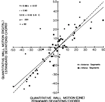
Figure 6 demonstrates a high degree ofcorrelation for quantitative wall motion assessed from the digital and cineangiographic measure-ments (r= .090. y=0.88x+ .02 SD/C, SEE =0.92 SD/C, p<.OOl). No differences were noted in the ability to define either anterior or inferior wall motion.
Reproducibility
The intraohserver variability was: end-diastolic volume, ± 11. 7 ml
(
± 7.4 percent); end-systolic volume, ± 8.7 ml(
± 12.9 percent);Y0.80x + 7.9
100
r: 0.93 &E.E. 11.2 ML Pc .001 n a 31 Y’088x +002 r ‘0.90 SEE *0.92 S.D. C. pn 001 n.62-I
a
w 50 40 30 20 U 10 U. U , / U #{149} / / / / / / ,7’ U U,y
((U z& Qz S #{149} O: .J> -Jw #{149} <0 2< 1-0 0 U 20 3.0 40 50100
END-SYSTOLIC
VOLUME
(CINE)(ML)
FIcuRE 4. Results ofend-systolic volume correlations. Format and
abbreviations as in Figure 3.
600 Assessment of Left Ventricular Function (Mancini et a!)
-J -J 0
w
-J 0 > 0 -J C,) 0 0z
w
EJECTION
FRACTION
(CINE)(%)
FIGURE 5. Results of ejection fraction correlations. Format and
abbreviations as in Figure 3.
ejection fraction, ± 6.4 units (± 11.1 percent); regional wall motion,
±0.57 SD/C. The interobserver variability was: end-diastolic
vol-ume, ± I2.3 ml (±7 percent); end-systolic volume, ± 4.8 ml
(
± 8.0 percent); ejection fraction, ± 3.4 units(
± 6.0 percent); regional wallmotion, ± 0.66 SD/C.
DIsCUSsION
The
results
of this
investigation
confirm
and
extend
the
observations
of previous
investigators
in
determin-ing
the
relative
accuracy
with
which
low-contrast
dose
digital
ventriculography
can
be
used
to
quantitate
#{149}sAnter,or Segmnts U Inferior Segments
QUANTITATIVE WALL MOTION (CINE)
(STANDARD DEVIATIONS/CHORD)
FIGURE 6. Results of quantitative wall motion correlations. Format
and abbreviations as in Figure 3. No differences in quantitation of anterior vs inferior regional motion was noted. Results shown represent pooling of results for anterior and inferior segments from each patient.
global
ventricular
function.6’11’3
The
correlation
pa-rameters
reported
herein
are
intermediate
to
those
previously
published.
For
studies
reporting
greater
correlations,
the
differences
compared
with
this
study
that
may
account
for
the
slightly
disparate
results
include
exclusion
of
patients
with
abnormal
ventri-des”
and
the
combining
ofboth
end-diastolic
and
end-systolic
volumes
to determine
only
one
volume
regres-sion.6
In
a study
reporting
inferior
results
for
end-diastolic
volumes,
the
relevant
difference
might
be the
use
of
small,
undiluted
doses
of contrast
in
a study
population
similar
to the
current
one
which
contained
a substantial
proportion
of patients
with
large
ventri-des
and
low
ejection
fractions.’
Although
not
directly
examined
in
this
study,
empirically
it
appears
that
small,
undiluted
doses
of contrast
do
not
consistently
provide
diagnostic
images
in
patients
with
large,
poorly
contracting
ventricles
or
when
tachycardia
is
present.
The
dilution
factor
used
in
this
study
was
based
on this
observation,
and
the
additional
observa-tion
that
more
excessively
diluted
contrast
doses’3
sometimes
also
yield
inadequate
opacification.
It
should
be emphasized
that
although
a rigid
protocol
is
required
for investigative
studies,
the
optimal
amount,
dilution,
and
rate
of contrast
injection
will
be
predi-cated
by
individual
patient
characteristics
and
should
be
adjusted
accordingly
as is practiced
with
standard
ventriculography.
The
quantitation
ofventricular
function
is facilitated
by
on-line
acquisition
and
digitization
of ventricular
images,
eliminating
the
sometimes
cumbersome
proc-ess
of separately
digitizing
cine
or
video
images.
In
digital
form,
the
images
can
be
easily
and
routinely
analyzed
by
objective
methods.
Despite
this,
little
attention
has
been
paid
to
the
quantitation
of digital
ventricular
images
for
determination
of regional
wall
motion.
In large
measure,
this
is due
to the
absence
of
a universally
accepted
method
of wall
motion
analy-sis,’7.’9 but
the
need
for an objective
means
of
quantita-tion
for
correlative
studies
is
underscored
by
the
degree
of observer
variability
when
subjective
tech-niques
are
used.42#{176}Only
one
previous
study’3
used
a
quantitative
method
to analyze
and
compare
regional
function
from
low
dose
digital
ventriculograms
and
conventional
ventriculograms
.A moderate
correlation
(
r
=0. 81) was
shown,
which
the
authors
ascribed
to a
suboptimal
image
frame
rate
of only
10/second,
not
to
any
potential
deficiency
of
the
digital
ventriculo-graphic
images.
Thus,
the
most
significant
contribu-tion
of the
current
study
is that
itextends
the
limited
conclusions
of
that
one
prior
study
regarding
the
accuracy
with
which
regional
wall
motion
can
be
assessed
from
low-contrast
dose
digital
ventriculo-grams
and
confirms
that
with
a
more
appropriate
framing
rate
(30/second),
the
quantitation
of regional
wall
motion
with
low
dose
digital
ventriculography
correlates
highly
(r
=0. 90)
with
similar
quantitation
of
standard
cineventriculograms.
While
forgoing
the
advantage
ofIV
digital
ventricu-lography,
which
is
minimally
invasive,
low-contrast
dose
direct
ventriculography
avoids
the
hemodynamic
perturbations
seen
with
both
IV digital
ventriculogra-phy
and
conventional
ventriculography.
10,12.13This
technique
may
not
be
as
appropriate
for
screening
patients
or
for
outpatient
procedures,
but
itis
of
definite
clinical
advantage
as
an
adjunct
to
cardiac
catheterization
facilities,
particularly
when
studying
patients
who
do
not
tolerate
the
effects
of the
usual
doses
of contrast,
including
those
with
impaired
renal
function,
aortic
stenosis,
unstable
angina,
or
con-gestive
failure.
In
addition,
Tobis
and
co-workers’22’
have
demonstrated
the
usefulness
of
this
technique
when
multiple
ventriculograms
are
needed
to assess
interventions
such
as
atrial
pacing
or
when
biplane
imaging
is desired
but
unavailable.
This
group
also
demonstrated
fewer
premature
ventricular
contrac-tions
with
the
low dose
protocol
than
with
conventional
ventriculography
(0 vs 20 percent
of patients,
respec-tively).
In
the
current
study,
this
advantage
was
also
evident
but
less
dramatic,
in
that
only
three
cm-eangiograms
and
one
digital
ventriculogram
of
34
could
not
be
analyzed
because
of excessive
ectopy.
Even
so,
when
a digital
acquisition
run
demonstrates
ectopic
beats,
subtraction
and
contrast
enhancement
frequently
still
allow
diagnostic
imaging
of
faintly
opacified
sinus
beats
relnote
from
the
ectopic
beat,
an
option
not
possible
with
conventional
cineangiogra-phy.
Low-contrast
dose
digital
subtraction
ventriculogra-phy
can
be
used
accurately
to determine
both
global
and
regional
left
ventricular
function
while
minimizing
the
required
dose
and
inherent
risks
ofcontrast
media.
ACKNOWLEDGMENTS: The authors are grateful to investigators at the University of Washington, Seattle (Florence H. Sheehan, Edward L. Bolsoit, and Harold T Dodge), for providing assistance in implementing the centerline method ofwall motion analysis used in this study. The secretarial assistance of Ms. Diane Bauer is also greatly appreciated.REFERENCES
1 Mancini GBJ, Norris SL, Peterson KL, Gregoratos G, Widmann T, Ashburn WL, et al. Quantitative assessment ofseginental wall motion abnormalities at rest and after atrial pacing using digital intravenous ventriculography.
J
Am Coll Cardiol 1983; 2:70-762 Norris SL, Slutsky BA, Mancini GBJ, Gregoratos C, Ashburn WL, Peterson KL, et al. Comparison of digital intravenous vetitriculography with direct left ventriculography for quantita-tion of left ventricular volumes and ejection fractions. Am
J
Cardiol 1983; 51:1399-14033 Vas R, Diamond GA, Forrester JS, Whiting JS, Swan HJC. Computer enhancement of direct and venous-injected left ventricular contrast angiography. A,n Heart
J
1981; 102:719-28 4 Vas R, Diamond GA, Forrester J5, Whiting J5, Pfaff MJ,Levisinan JA, et al. Computer-enhanced digital angiography: correlation of clinical assessment of left ventricular ejection
602 Assessment of Left Ventricular Function (Mancini eta!) fraction and regional wall tnotion. Am Heart
J
1982; 104:732-95 TobisJ, Nalcioglu 0, Johnston WD, Seibert A, Iseri LT, Roeck W, et al. Left ventricular imaging with digital subtraction angiogra-phy using intravenous contrast injection and fluoroscopic expo-sure levels. Am Heart
J
1982; 104:20-276 Kronenberg MW, Price RR, Smith CW, Robertson RM, Perry M, Pickens DR. et al. Evaluation ofleft ventricular performance using digital subtraction angiography. Am
J
Cardiol 1983; 51: 837-427 Goldberg HL, Borer JS, Moses JW. Fisher
J,
Cohen B, SkellyNT Digital subtraction intravenous left ventricular intravenous
left ventricular angiography: comparison with conventional in-traventricular angiography.
J
Am CoIl Cardiol 1983; 1:858-628 Nisseit SE, Booth D, Waters
J,
Fassas T, DeMaria AN. Evalua-tion of left ventricular contractile pattern by intravenous digital subtraction ventriculography: comparison of cineangiography and assessment of interobserver variability. AmJ
Cardiol 1983;52: 1293-8
9 Eiigels PHC, Ludwig JW, Verhoeven LAJ. Left ventricular evaluation by digital video subtraction angiography. Radiology
1982; 144:471-4
10 Mancini GBJ, Ostrander DR, Slutsky RA, Shabetai R, Higgins CB. Comparative effects of ionic contrast agents injected in-travenously or directly into the left ventricle: implications for digital angiography. AJR 1983; 140:425-30
11 Sasayama S, Nonogi H, Kawai C, Eiho 5, Kuwahara M. Auto-mated method for left ventricular volume measurement by
cineventriculography with minimal doses of contrast medium.
Am
J
Cardiol 1981; 48:746-5312 Tobis JM, Nalcioglu 0, Johnston WD, Seibert A, Roeck W, Iseri L1 et al Correlation of 10-milliliter digital subtraction
ventriculograms compared with standard cineangiograms. Am Heart
J
1983; 105:946-5213 Nichols AB, Martin EC, Fles TP, Stugensky KM. Balancio LA, Casarella WJ, et al. Validation of the angiographic accuracy of digital left ventriculography. Am
J
Cardiol 1983; 51:224-30 14 Sandler H, Dodge HT The use ofsingle plane angiocardiogramsfor the calculation ofleft ventricular volume in man. Am Heart
J
1968; 75:325-3415 Bolson EL, Kilman S. Sheehan FH, Dodge HT Left ventricular segmental wall motion: a new method using local direction information. IEEE Comput Cardiol 1980; 245-8
16 Sheehan FH, Dodge HT, Mathey DG, Bolson EL, Mitten S. Application of the centerline method: analysis of change in regional left ventricular wall motion in serial studies. IEEE Comput Cardiol 1982; 97-100
17 Gelberg JH, Brundage BH, Glantz S, Parmley WW Quan-titative left ventricular wall motion analysis: a comparison of area, chord and radial methods. Circulation 1979; 59:991-1000
18 Karsch KR, Lamm U, Blanke H, Rentrop KR Comparison of nineteen quantitative methods for assessment of localized left ventricularwall motion abnormalities. Clin Cardiol 1980; 3:123-8 19 Clayton PD, Jeppson GM, Kla#{252}sner SC. Should a fixed external
reference system be used to analyze left ventricular wall motion.
Circulation 1982; 65:1518-21
20 Chaitman BR, DeMoto H, BristowJD, FoschJ, Rahimtoola SH. Objective and subjective analysis ofleft ventricular angiograms. Circulation 1975; 52:420-5
21 TobisJ, Nalcioglu 0, Johnston WD, Seibert A, Iseri LT, Roeck W, et al. Digital angiography in assessment of ventricular function and wall motion during pacing in patients with coronary artery disease. Am