Cooperative interaction
withVolume17Number 13 1989 Nucleic Acids Research
Cooperativeinteraction of chicken lysozymeenhancersub-domainspartially overlappingwitha
steroidreceptorbinding site
JoachimAltschmied+, Marc Muller, Aria Baniahmad, Christof Steiner and Rainer Renkawitz*
Genzentrum, Max-Planck-Institut fiir Biochemie, D-8033 Martinsried, FRG
ReceivedApril 18, 1989;Revised and AcceptedJune5, 1989
ABSTRACT
Expression of the lysozyme gene is a marker for the differentiation of macrophages, lysozyme transcription being gradually increased during maturation. We have analyzed the fine structure and function of two macrophage-specific enhancer elements of the chicken lysozymegene (E-2.7 kb andE-0.2kb). Bothincrease their activities upon LPS induction, both contain multiple binding sites forsimilar or identical nuclear factors and both can be divided into two functional modules. For the E-0.2 kb enhancer we found a synergistic activity of the modules to be dependent on their distance. Binding sites for nuclear proteins within enhancer E-0.2kb overlapsubstantially with the previously identifiedprogesterone/glucocorticoid receptor binding site, which isrequired for steroid induction oflysozyme transcription in the oviduct.
INTRODUCTION
One of themain contemporary challenges in molecular genetics istheelucidation ofthe mechanisms underlying tissuespecific gene expression. Several tissue specific promoter andenhancerelements have been identified (1). Manyof the modelsystemsanalyzedare limitedinthat onlymaturecells ortissuesareavailablefor testing,whereasgeneregulation mayinvolve amulti step processduring differentiation fromprecursor to maturecells. Thehaematopoetic cell populationisgeneratedbyoneof thefewdifferentationpathways whichcanbestudiedstepbystepinvitroaswellasin vivo. One of thecelltypesgenerated in this system is the macrophage cell. Expression ofthe lysozyme gene is a specific differentiation marker, being gradually turnedonduring maturation ofmacrophages (2). In addition to this constitutive macrophage expression, the chicken lysozyme gene is inducible by steroid hormones in the tubular gland cells of the oviduct (3).
Threemacrophage-specific enhancer elements of the chickenlysozymegenehave been identified, one each at -6.1 kb (4), -2.7 kb (5) and -0.2 kb (5) upstream of the transcription startsite. The lattertwoelementsarereferredtohereusingtheabbreviated forms E-2.7 kb and E-0.2 kb according to their positions. Furthermore, two silencer elementswhicharespecifically inactiveinmaturemacrophages(S-2.4kband S-0.25kb), andonewhich is active in all cell types tested(S-1.0kb) havebeen described(5,6). Steroid inducedexpressionin theoviduct ismediatedbyatleasttwodifferenthormoneresponsive elements, one at -200 bp (7) and one at -1.9 kb (8).
Herewe report the fine structure of the twomacrophage specific enhancerelements E-2.7 kb and E-0.2kb,their functionalactivityand theircapacitytobind nuclearproteins. Thebinding patterninthe multifunctionalenhancer E-0.2kb,whichalso contains functional steroid receptor binding sites (7), suggests acompetition between steroid receptors and Nucleic Acids Research
Nucleic Acids Research . ...*... ... ... .'Aw g.: ** ml4i :.; -lIUC tll?] ..S.S.l _. a I i I 4. lS.N g ...I:...0 *
Xt...
Iuss*auJ1...
n * * =|1~~~~
I
s?
R,1 51
U
......18...
ipL2~~~~~~~~~~~~~~~~~~~~~
___. W..Y.. '' ISW Di P44Nucleic Acids Research enhancer factors allowing steroid induction in the oviduct and constitutive expression in macrophages.
MATERIALS AND METHODS Plasmid Constructions
The identityof the recombinant plasmids was confirmed by restriction mapping and dideoxy-sequencing using the Sequenase-kit from USB.
The plasmids pICfrag21 and pUClys-280/+15 were created by inserting the HindIHI/BglIH-lysozyme genefragment from -2.71 kb to -2.54 kb and the HindIHI/XhoI-fragmentcarrying thelysozyme-promoter (-280 to + 15) fromplysCATA -280 (5) into the corresponding sites in the polylinker ofpIC20R (9).
Twoseries of different plasmids containing subregions of lys E-0.2 kb were used, all ofwhich arederivativesofthe plamidspBLCAT2 (10) orplys-208/-67a tkCAT (5). Oneseriescontainsaspacerof varyinglengthbetween lys -67/- 161 and lys - 162/-208 in front of tkCAT. This series was constructed by introducing a 20 bp synthetic oligonucleotide containing recognition sitesforSnaBI, Stul,NruIandSallinto the natural RsaI-site inposition -161 inplys-208/-67atkCAT, andperforming subsequent deletions using these endonucleases. Plasmids with spacer-lengthsof 5, 8, 10, 13, 15, 17 and 20 bp were constructed.
The second series contains various combinationsofthe lysozyme fragments from -208 to -162 and -161 to -67 in front ofthe tkCAT-fusion gene. These plasmids were constructedfromasubcloneoflys -67/-208in pUC 18, of lys -95/-208in pUC 18, of plysCATA208 and pBLCAT2 (10). Selected lysozyme sequences were inserted in antisense direction in front of the tk-promoter to test them directly for enhancer-characteristics.
pE-0.2-1 was generated by ligating the lysozyme BamHI/AccI fragment from pUC lys -208/-95 into the BamHI site of pBLCAT2.
pE-0.2 -2 contains the lysozyme sequences from -208 to -162 as an RsaI/BamHI-fragment cloned into the BamHI and filled-in Sall sites of pBLCAT2.
Bycloningablunt-endedHindIII/RsaI-fragmentfrom pUClys-208/-67 harboring the region from -161 to -67 into thefilled in BamHI- orHindIII-site ofpBLCAT2 orthe BamHI-site of pE-0.2 -2,weobtained the plasmidspE-0.2-3, -4, -5, -6and -2x3. For a double insertion of the distal domain ofE-0.2 kb this sequence was cut out of pE-0.2 -2 with BamHI andHindu, filled in and re-inserted into the blunt-ended HindIll site ofthe same plasmid (pE-0.2-2x2).
ForpSVtkCAT and E-2.7 constructions see Steiner et al., 1987 (5). Nuclear Extract Preparation
The nuclearextractswerepreparedasdescribed(11-13).Subconfluent cellswereharvested fromabout 60 culture dishes(15cmindiameter)andwashed inphosphatebufferedsaline. Allfollowing manipulationswereperformedat0°Cusing precooled solutions,tubes and
Figure1.DNaseIprotectionassayonthe E-2.7 kb(A)and theE-0.2 kb enhancer(B).The upper(upper)and lower strands(lower)were3' end-labelled anddigestedwithDNaseIin the presence ofHD1I nuclear extract (+)orwithout nuclearextract(-). The G+Asequencing productsareshownasmolecularweightmarkers. The shadeddiagramontherightdepictsthefootprintingpatternachieved,boxesindicateprotectedsequences,
arrowsshowpositionsof strongDNaseIhypersensitivitycausedbynuclearextractsand horizontal linespoin
jX I'| U'* r)~~j '. I2 * * - I~___.111 S|.|... **" aI _. .,__ t 14 it , ,b ''
ag
. ! . z ,;**P4.#: _a _t2 4....: } It: m|
..|
.. ... Ir
LL,,,--K
*J*, ...@@@ -@
tHU~~ 3.,2 ... If~~~~ 3: :;I S' I .1 31.1 U. It I I .: I I :, I .I.:.I
I....H I ....F:4
Nucleic Acids Research centrifuges. Thepelleted cells were suspended in five packed cell volumes (PCV) of buffer
A' (10 mMHepes/KOH (pH 7.6), 10 mMKCI,0.15 mMspermine, 0.5 mMspermidine,
1mM EDTA, 0.5 mM DTT) and allowed to stand on ice for 10min.The cells werepelleted bycentrifugation and suspended in 2 PCV. The cells were broken by homogenization in a glass-glass douncehomogenizer. After addition of one-tenth volume of buffer B'(1OmM
Hepes-KOH (pH 7.6), 1M KCl, 0.15 mM spermin, 0.5 mM spermidin, 1mM EDTA,
0.5 mM DTT) thenuclei werepelleted by centrifugation (2400 rpm; 10 min). This pellet was used toprepare the nuclear extract as described (12) with the modification that the volume of nuclear lysis buffer was 1 PCV and that theprecipitated nuclear proteins were dissolved in 1/10 PCV ofthe dialysis buffer. The opaquenuclear extractwith aprotein concentration of 5-20 mg/ml (Biorad protein assay) was frozen in smallaliquots in liquid nitrogen and stored at -85°C.
DNaseI-Footprinting and Band Shift Assays
To obtain probes labelled on one strand the plasmids pICfrag2l and pUClys-280/+15 were cut with EcoRI andlabelledasdescribed (14). The footprint reactions were performed according to Cereghini et al., 1987 (15) with the described modifications (14). The following blunt ended, double strandedoligonucleotideswere used: lysozyme sequences -208/- 162, -193/-180, -146/-123, -125/-93, -96/-66; SV40 core:
GTTAGGGTGT-GGAAAGTCCCCAG;SPI SV: CGACTGATCAGTTCCGCCCATTCTCCGCCCCAG
(with AccI/BamHI sticky ends); Spl dhfr: CTTGGTGGGGGCGGGGCCTAAGCTG
(16); AP4: GATCACCAGCTGTGGAAT (17).
Band shiftassays wereperformedin atotal volume of 10ylcontaining: 250 ng poly(dIdC)
(Pharmacia), 25 ng denatured calf thymus DNA, 5% glycerol; 25 mM Hepes/KOH pH
= 7.6, 25 mM KCI, 5 mM MgCl2, 0.4
jig
nuclear extract and32P-labelled
doublestrandedoligonucleotide (30,000 to 40,000 cpm). The nuclear extract was preincubated for 15 min on ice before addition of thelabelled probe. After further 20 min incubation onicethe samples were loadeddirectlyon a5% acrylamidegel(acrylamide:bisacrylamide ratio of 79: 1 in 192 mM glycin / 25 mM Tris buffer).
The gel was run for 1.5 hrs at 15 to 20 mA at +4°C, fixed, dried andautoradiographed. Cell Culture, Transfectionand CAT-Assays
HD11 [=HBC1=LSCC-HD(MC/MA1) (18)] and DU249 (19) cells were maintainedin DME-medium (Biochrom) supplemented with 8% fetal calfserum, 2% chicken serum, 100 units/ml penicillin and 100
jig/ml
streptomycin. All transfectionexperiments were done in triplicate and confirmed in a second series of tests. Transfections were carried out asdescribed (5)with thehelp ofan automated 'precipitator' (20). LPS treatmentof HD11 cells was doneby applying 0.1 tg/ml LPS tothe dishes at 22hrs after transfer. Allcells wereharvested 2dafterDNAtransferand assayed for CATactivityorfor mRNA amounts (5).Figure 2. Competitions of thefootprintsonthe enhancer E-2.7 kbandE-0.2 kb. Footprintreactions without nuclear extract (-) and with HDII nuclear extract either without competitor (/) or with double stranded oligonucleotides (1), (2), (3)or(4) inamountsof 0.5pmole(leftlanes)or2.5pmole(rightlanes).Vertical lines indicate the positions of protectedregions, whereas the dots pointoutcompeted footprints. Thefollowing 3' end-labelled DNA fragmentswereused: upper strand of E-2.7 kb(A), upper strand of E-0.2kb(B)and, for better resolution, lower strandof E-0.2 kb (C). Panel D depictsthe different oligonucleotidesused.
AM .
Figure 3. Gel shiftexperimentswithlysozyme (lys-96/-66)andSpI (SpIdhfr)oligonucleotides.HD11 nuclear extract wasassayed forspecificDNAbinding activity. Competitionexperimentsweredoneusingtwodifferent
concentrations(0.1 pmoleandlpmolein the firstand secondlane,respectively)ofdouble strandedoligonucleotides
lys -96/-66, Spl SV and Spl dhfr. The AP4oligonucleotidewas usedat aconcentration of 1 pmole. The arrowpoints tothe majorretarded band.
A 4 A r*t~yr 1* .r 'P S t 4L It
Figure 4. ComparisonofHD11 andDU249nuclear extracts infootprintreactions. The lower strand of E-2.7
kb(A)and the lower strand ofE-0.2kb(B) were3' end-labelled and incubated withHD11 (HD) orDU249
extract(DU)and DNase Idigested. Forcomparisonfootprint reactionswithoutnuclear extract(-) andG +A
sequencing products(GA)areshown.Strongandweak DNase Ihypersensitivesites arepointedoutby horizontal
lines.
Nucleic Acids Research
F:::rl
xK.
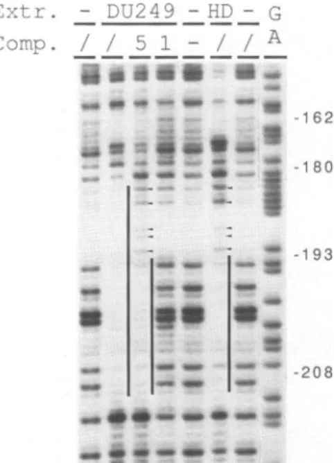
- Dli249 G-1-DD Com. // 51 _//A s162 -F
4 ee- 1i -180 - ~....-,u ._ .~ @1 m:s : *~~~~~~wFigure5. Competitionofthe liver cells specific footprintonthe E-0.2 kb enhancer. Footprint reactions with DU249(DU),withHD11(HD)orwithout(-)nuclearextractareshown.Competitionswith unlabelled double strandedoligonucleotide 1or5(-208/-162or-193/-180lysozymesequences;seeFigure 2D)werecarried
outwith 3-10pmoleascompetitorDNA[(1)and(5);(I)is withoutcompetitorDNA]. Verticallines indicate thefootprintregionsand thearrow headspoint tohypersensitivesites.
RESULTS
Several Factors Bindto Both Enhancers E-2.7 kb and E-O.2 kb
We have
previously
identified twomacrophage specific
enhancer elements upstream of the chickenlysozyme
gene(5).Sequences
ofabout 150bp
each arerequired
foroptimal
function of both enhancersequences. Since the sizeoftheseelements
suggested
thepossible
presence of several
binding
sites for nuclearproteins,
we carried outfootprinting
experiments
using
total nuclearextracts. Nuclear extracts werepurified
from a chickenpromacrophage
cell line (HD 11) in which both enhancer elements are functional (5).Footprinting experiments
onbothenhancersequences demonstrated theprotection
from DNase Idigestion
ofmultiple regions (figure
1).Sequence analysis
ofboth enhancer elements has revealed a common sequence motifpresent several times in E-2.7 kb and in E-0.2 kb (5). This motifis similar tothe SV40 enhancer core sequence (21). Sincemostofthese sequenceelementsare
protected
from DNase Idigestion
inthefootprinting
experiments
(seefigure
6), we tested forrelationship
between the boundproteins.
We used fourdifferentoligonucleotides
tocompetetheprotein
binding
infootprint
reactions(figure
2).Oligonucleotide
[1]
containslysozyme
sequences from -208bp
to -162bp
including
twocore motifhomologies; oligonucleotides
[2] and [3], whichcorrespond
tothe
regions
-146bp
to -123bp
and -125bp
to -93bp, respectively,
containone core motifeach. Theoligonucleotide
[4] contains theoriginal
SV40 core sequence [SV40 sequences233to255;BBBsystem(22)].Thefootprint competitions
show that theproteins
F3 F4 F5 -2550 F4 F5 F6 . . _E-E--470 Identity Reference + + TGACTATGACTACTGGC TGACTA TGACTA F2 + + AGGAGAATGAGGAACTAGC GAAaGAGGAACT F3 + + GACCACCATGGAGTCACCCA GAGTCA F4 + + ACTGGTATTTGGAAATAATAA TTTGGAAAT TgTGGAAAT ttt F5 + + TCTGAATTGCAAAGC aTGCAAAt AAaaCCACA tTT E-0.2 Fl + + GATATTGCAACAGACTA AaaaCcACA TTt F2 - + TATAAAATTCCTCTG AAATTCCTCTG F3 + - TGTGGCTTAGCCAATGTGGT TTAcaCAATGTGGT Taaa F4 + + AGAAATTTGGCAAGTTTAG TTTGGCAAG TTTGGaAAt TgTGGaAG ttt F5 + + GAAGTGTTGGGAAATTTCTG TTtGGAAAT TgtGGAAAg ttt F6 + + CAAGAGGGCGTTTTTG GAGGGCGT SV40 PU-box 25 GCN4 - AP1 24 lys. cons. 5 AP3 - C/EBP 28 octa AP3 - C/EBP 29,30 28 AP3 - C/EBP 28 NRD I C/EBP AP3 - C/EBP NF1 lys. cons. AP3 - C/EBP 32 31 26,28 15 5 26,28 lys. cons. 5 AP3 - C/EBP 26,28 Spl 33 E-2. 7 -2680 E-0. 2 -210 Extract M L Fl F2 I
F..1
-7
.I Fl F2 F3 tGR R) t ~PR ) Lysozyme Seguence Similarity E-2.7 Fl GCN4 - AP1 GCN4 - AP1 24 24Nucleic Acids Research (figure 2). Although differences in binding efficiences are seen-each oligonucleotide showing the best competition for the footprint on its corresponding region-it is obvious that all the 'core'-containing sequences show qualitatively identical competitor function incomparison to footprints which are not changed, such as the footprint upstream of -217 bp (figure 2C), which lies within the silencer element S-0.25 kb (6) and the E-2.7 kb footprints outsidethe 'core' sequence-containing region (E-2.7 kb sequences -2600 bp to -2700bp; figure 2A).
One of thefootprint regions (sequences -90 to -74 ) in the E-0.2 kb enhancer could only be detected quite weakly, and since thesequence resembles aSpl binding site (see figure 6), we performed band shiftexperiments with lysozyme DNA -96/-66 (figure 3). One major shifted band can be seen in addition to several minor bands. All can be competed specifically by the lysozyme sequence (-96/-66) and by two different Spl binding sites [one from the SV 40 promoter (Spl SV) and one from the dihydrofolate reductase gene (SpI
dhfr)],
except for one minor band. An AP4 binding site cannot compete. This complex pattern of several shifted bands is typical for Spl as can be seen in the reciprocal experiment (figure 3): the labelled Spl dhfr DNA shows the same pattern of shifted bands which is competable by itself, the Spl SV and the lys -96/-66 oligonucleotides. The SpI and lys -96/-66 oligonucleotides differ only in their binding affinities, that of the lysozyme sequence being weakest.Tissue Specific Differences inBinding Patterns of Nuclear Proteins
Inorder to identify those regions of the lysozyme enhancers involved in binding tissue specific proteins, we compared nuclear extracts from a chicken liver cell line (DU249, inwhich thelysozyme gene is inactive) with theHD1 1 promacrophage extract. Some extract specific differences can be detected forbothenhancer elements in their promoter distal parts(figure4). The E-2.7 element manifests itsspecificity in
HD1
1 specifichypersensitive sites within the large footprint area between nucleotides -2635 and -2675 (figure 4A: domainA). Celltypespecificfootprint patternscanbeseen onthe E-0.2 elementbetween nucleotides -155 and -192 (figure 4B: domain A). One HD11 specific footprint (-1551- 175)andoneextendedfootprint caused by the DU249 extract (-180/- 192) are evident (figure 4B).Theextended footprint seen in the region -193bpto -180bp with the DU249extract wasalsoseenwithanextractfromanotherlysozyme negative cell line,HD3erythroblasts (Baniahmad, Steiner andRenkawitz,unpublishedresults). Thisfootprintmay bethe result of a single additional binding protein absent in HD11, or may reflect the binding ofa
completely differentsetofbinding proteinsin eachextract. Todistinguishbetweenthese twopossibilities, we carriedout footprinting reactions with the liver cell extract in the presence ofanexcess ofvarious competitor oligonucleotides (figure 5). Asacontrol we
usedthe oligonucleotide -208/-162(oligonucleotide 1, seefigure 2) whichcovers the livercell-specificfootprint plusbothflanking footprints. Uponcompetitionthiscomplete Figure6.Regionsof the E-2.7 kb and the E-0.2 kb enhancer boundbynuclearproteins. Bindingsequences
asdeterminedbyfootprintingand bandshiftexperimentsareboxed.DNAregionscoveredbytheprogesterone (PR)orglucocorticoid(GR)receptorsareindicated(1,22).Sequenceswith identicalshadingcancompete each other. The table lists all thelysozymesequences(underlined) protectedinfootprintexperiments (numberedE-2.7F1
toF5andE-0.2F1toF6)with their similarities(capitalletters)toknowntranscriptionfactorbindingsites(see text). AP3-C/EBPbindingsequencesareeither taken fromSV40(26)orfrompolyoma(28). (+)and(-)indicate the presenceorabsenceofaparticularfootprintwithextractsfrompromacrophages (M)orfrom liver cells(L).
Enhancer activity promacro- liver phages cells F1F2 F3 F4 F5 E-2.7 100% 14% 2.7-1 107% 13% 2.7-2 71% 13% 2.7-3 7% 6% domain: A B
Figure 7. Functionalactivityof E-2.7 kb enhancer derivatives. The E-2.7 element andsubregionsthereof were cloned in front of tk CAT andtransfected intoHDI1(promacrophages)orDU249(liver)cells. CATexpression isnormalizedtotheexpressionoftk CAT andenhanceractivityof E-2.7 in HD1I isset to100%. Footprint
regions Fl toF5are shown. The function of domainsAand B isdiscussed in the text.
region becomes DNaseI sensitive. However, competition with the shortoligonucleotide
-193/-180 (oligonucleotide 5, see figure 2), which coversonly the liver cell specific footprint,generatesacluster ofDNaseI-sensitive sitesinthis centralregionwithout affecting theflanking footprints. Thispatternofprotectedregions is identicaltothatachievedwith themacrophageextract inthe absence ofcompetitor(figure 5). Therefore, weconclude that thisextract-specificdifference in the footprint patterns iscausedbyanadditionalfactor bindingontheDNAbetween -193bpand -180bpupstreamofthetranscriptionalstart site, this factor being absent in promacrophage nuclei.
Figure 6 summarizes thefootprintingresultsdescribedabove.Foreach ofthelysozyme sequencesprotectedinthe footprintingexperiments (E-2.7F1 toF5 andE-0.2 Fl toF6) similaritiestoothernuclearfactorbinding sitesareindicated.E-2.7F1 containstworepeats oftheGCN4proteinbinding site which is theyeasthomologuetotranscription factorAPI (23). This particular binding site has only alow affinity for GCN4 (24), but since itis repeated, bothmayfunctionin asynergisticmanner.E-2.7F2is quitesimilartotheSV40 PU-box, which can bebound by a lymphoid specific protein (25). E-2.7F3 contains a strongbinding sequenceforGCN4-AP1 (24). Sequences inE-2.7F4, E-0.2F4and F5 are similartoeach other(lys.cons.) (5) and to the 'core' enhancersequence (21). This motif
canbebound bythe AP3protein(26,27)or byC/EBP(28). Inaddition E-0.2F4issimilar
to a 'halfbinding site' ofNFl (15). E-2.7F5 shows similarities to AP3-C/EBP and to the'octa'-sequence(29,30). AP3-C/EBPsimilaritiy is also seen with the footprintsequences E-0.2F1and F3 with F3containingaCAAT-box, whichisbound byC/EBPaswell (31). All of thefootprint sequences withAP3-C/EBPsimilarity arecompeteableby theSV40 'core'-sequence (see above) which is recognized by AP3 (27). The footprint E-0.2F2 contains asequence identical to the negative NRDI region of the human
13-interferon
gene (32)andE-0.2F6containsabinding sequence identicaltotheSplbinding site of thehuman U2small nuclear RNA gene (33). Inaddition to the regions defined by the footprintingNucleic Acids Research experiments with whole nuclear extracts the glucocorticoid and progesterone receptor
binding sites are shown on the E-0.2 enhancers (7,34).
Sincetissuespecific footprintingpatternsare seenonthe distal halvesof eachenhancers,
we carried out functional tests both on the completeenhancers and on isolatedenhancer
elements.
TissueSpecificEnhancer ActvityisAchievedbyaCombinatorialEffectofEnhancerModules
Havingfoundmultiple bindingsites for similarnuclear factorsonboth enhancers,wewanted
toknowwhetherornotallofthesesitesarerequiredfor function,andifinteractionbetween sitescouldbedetected. Wecloned several enhancer subfragments in variousorientations andcombinationsupstreamofthethymidinekinase (tk) promoter,which inturnwas fused
tothechloramphenicol acetyl transferase(CAT)gene(10). The resultingcontructs (figures
7and 8) weretransfected intochicken liver cells (DU249) and chickenpromacrophages (HD1 1), and cell extracts were analyzed for CAT-activity. To normalize for different transfection efficiencies into thesetwo cell lines, weused a plasmid carrying the SV40
enhancer upstream ofthe tkCAT fusion gene as a standard. Both lysozyme enhancers
showedabouthalfof theSV40 enhanceractivity in HD1 1 cells(datanotshown), whereas
they remained inactive in DU249 cells (figures 7 and 8).
The E-2.7enhancer either intact, orwitha5' deletion (2.7-1), which doesnotaffect
thefootprint region, shows full enhancer activityin promacrophage cellsandlowactivity
inliver cells (figure 7). Thetwo enhancerparts (2.7-2and 2.7-3) show very different
activities: the distal domain (A) isclearly active by itself, but the proximal domain (B) isnot.Onlythecombinationofboth(2.7-1) leadstofull activityin HD11 promacrophages.
Similarly, dissectionoftheE-0.2enhancerdemonstratesthe function oftwodomains(figure 8). 3' deletions up todomain A (construct 0.2-2) showa progressive lossofenhancer
activity indicating the requirement of domain B for enhancer function. Neitherdomain
AnorBisactiveinisolation. Duplicationleadsto strongactivity of domainA(0.2-2x2), whereasdomain B(0.2-2x3) remains inactive. Interestingly, duplication of 'A' shows
wildtype enhancer activity in HD11 promacrophages, but does not show the tissue
specificity:clearenhanceractivity is seen evenin livercells (figure 8A).Therecombined enhancer with both halves arranged in a head to head orientation (construct 0.2-4)
maintained wildtype enhancer activity and tissue specificity. Upon separation of thetwo
halvesby 23 bp withthe distal domain in sense orientation(construct 0.2-5) orby 46
bp withthe distal domain in anti-sense orientation (construct0.2-6) enhanceractivity
was considerably reduced.
In order toconfirm that the observed CAT-activities reflect theamount ofcorrectly
initiated RNA, RNase mapping experiments werecarried out on RNA generated after transfectingselectedconstructs in HD11 cells. Theseexperiments confirmedthe results
of the CAT-assays (figure 8B).
To examine in more detail the observed disruption of enhancer activity following
separation ofthe enhancerhalves, we inserted linkers of different length (5, 8, 10, 13,
15, 17 and 20 bp) between positions -161 bp and -162 bp. CAT-activities recorded
aftertransfectionintoHD11cellsareshowninfigure 9. Insertionof5bp reducedenhancer
activity considerably, withafurtherreduction causedbyan8bpspacer.However,aspacer
of10bpleadstoanincreaseinenhanceractivity,presumably allowingthe proteinsbound
to thetwoenhancer halves tobepositioned in a threedimensional arrangement similar
tothat inthewildtypeenhancer. Thisresultindicates therequirementforacertain distance
A
Orx
Of - r-»~~~~~~~~~~~~~~~~~~~~~~~~~~~~ ( .} .-S ..t .. .7B
r. *S 4. inuumwsFigure8. Functionalactivityof E-0.2 kb enhancerderivatives. E-0.2recombinant sequenceswerecloned in front of tk CAT and transfected intoHD1 1orDU249 cells (seelegend forfigure7). A. CATactivity.Footprint
regions FltoF6 andbindingsites for theprogesterone(PR) andglucocorticoid (GR)receptorsareindicated. B. RNasemapping of selectedconstructsafter transfection intoHDl 1 cells. Lane (P) is the untreatedprobe,
lane(M)end-labelledmarker DNAand lane(tRNA) theprobehybridizedwith tRNA anddigestedwithRNase
AandT1.(C) isalabelled control DNAfragmentcarriedthroughthe RNasemappingproceduretojudge complete
recoveryof nucleic acids. (+1) indicates thepositionofcorrectly initiatedRNAand(P) thesizeof theprobe. The diagramdepicts thearrangementofexpressionplasmid, ofthetranscriptand of theRNA probe.
2..., I i .--l -... ".: ,. -) 60 1. I., %NW
Nucleic Acids Research
resulted inlevels which, although suggestiveof aperiodicity inactivities, lie within the deviation ofthe experiment.
Weconclude that activity of the 150 bpE-0.2 kbenhancer is a result of asynergistic, orientation independent interactionbetween domains A and B, and that tissue specificity is achieved by thecombinatorial effect of both domains.
Both Enhancerare LPSInducible
Sincelysozyme expression is gradually increasedduring macrophage differentiation (2) andsince bothenhancer elements showahigheractivity in primary maturemacrophages ascompared toHD1 1promacrophages(5) weanalyzed the enhancer activity in LPS treated HD11 cells. LPSis knowntoactivateand toinducedevelopmentalchanges inmacrophages [for review see (35)]. To test, whether the activity of the lysozyme enhancer elements is sensitive to LPS treatment of the cells, we carried out DNA transferexperiments in the presenceofLPS. TransientexpressionofpE-0.2 tk CAT andpE-2.7 tk CAT in HD 1I cellswasclearly inducedby LPS, whereastheptkCAT controlplasmidwas notinduced (figure 10).
DISCUSSION
The complexregulationofthe chickenlysozymegeneby steroidhormones intheoviduct and by the maturation ofmacrophages is achieved by the combined action of several enhancer, silencer andsteroid responsiveelements [forreview see(36)]. Whether all of theseelementsactindependently onthepromoter orwhethertheyinteract with eachother remains to be shown. Here we describe the detailed analysis of both lysozyme gene-enhancers E-2.7 kb and E-0.2 kb, which consist of several proteinbinding sites coinciding withfunctional modules. Some ofthebinding proteinsaretissue-specific, othersarepresent inliver cellsaswellasinpromacrophages. Such aconcomitantbinding of cell-type specific and ubiquitous factors is also found in other enhancer elements, for example in that of themousecl-antitrypsingene(37). Thelysozyme enhancers E-2.7 kb and E-0.2 kb together contain six 'core' motifhomologies which arecompletely protectedby nuclearproteins fromdigestion with DNaseI(figure 6). Competition experiments demonstrate theimportance of the 'core' homology in thebinding,ascross-competitionisapropertyof allprotected regionswhichshare only thesesequences. Thisindicates that proteinswith atleast similar DNA-binding domains recognize differentparts ofboth enhancers E-2.7 kb and E-0.2 kb. Althoughwefindmultiple similarbinding sites, the enhancer domains show different activities, andspecificities and therefore thebindingproteinsmaybedifferent. Itispossible that thebindingproteins described herearerelatedtothepreviouslycharacterised factors AP3 orC/EBP(26,28), although this hastobeprovenbyanalyzingthe respectiveclose contactsitesontherecognitionsequences. Asimilarpuzzlingsituation of celltypespecific andunspecific effects causedbythesamebindingsequencehas been found for theoctamer
sequence[forreviewsee(38)]. BoththeexpressionofB-cellspecificandof'housekeeping genes' can be driven by octamer factors.
Characterization ofthe protein-binding sites haspromptedus to dissect E-0.2 kb into
two domains. Although neither ofthese domains hasstrong enhancer
activity
alone asasinglecopy,duplicationofthe distaldomain(-208/
-162)
resultsinanactivity
similartothat of thecompleteenhancer,but without its tissue
specificity. Only
the combinatorial effectofboth domainsleadstowildtypeactivityandspecificity.
Thiswasalso shownfor the twodomains of the E-2.7 kb enhancer(figure
7). Thesynergistic
effectof enhancer domainswasfurther substantiatedbythe detectionof enhanceractivity
evenwithcompletely
dP spacer 0 100 En (4\ a,) 04 X 80 0) 60 ,~40 tkCAT 0 0 5 10 15 20 Spacer length (bp)
Figure 9. CAT activitiesofspacingmutants of theE-0.2kb enhancer. Different spacer lengthswereinserted
betweennucleotides -162and -161 of theE-0.2 kbenhancer and tested for their effectonenhanceractivity
upstreamoftkCAT. CATactivities achieved aftertransfecting HD1I cells intriplicate experimentsareshown
with standard deviations, activity oftkCAT withoutenhancer sequences is indicated by the arrow.
rearranged modules. Investigation ofthe requirementforaparticulardistance ofthetwo
domains indicatedadependenceofenhanceractivityonthe relativepositionof themodules. Similareffects of thestereospecificalignmentontheefficiency oftranscriptionregulating elements has been shown for the SV 40 enhancer (39) or for the interaction of the glucocorticoid receptorand the CACCC-protein (14). In each case optimal activity was
only observed after introduction ofmultiplesofacompletehelicalturnof the DNAbetween the proteinbinding sites. In the case oflys E-0.2 kb, we observe a strong decrease of activity upon introducing additionalnucleotides between itstwo domainswith apeak of increasedactivityupon insertion of 10bp. Asynergistic interaction of the boundproteins inthis constellationwouldbe indicative ofabendingofDNAbetweenthetwodomains,
aflexibleprotein domain and/or the involvement ofa 'bridgingprotein'that mediates the interaction of the DNA-bound factors and could confer theflexibilitytothe wholesystem.
Participation ofnon-DNA-binding proteins intranscription regulation has been shown in
some cases (40-44).
Domain structures suchas those described here are also found in other transcription regulatory elementsand seem to be ageneral phenomenon (37,45,46). A modular structure may be adapted during evolution to a variety of regulatory processes by using various
combinations ofa limited number of differentbinding sites.
Cleartissue-specific differences inthe bindingpatternof liver cell andpromacrophage
extracts were observed between -2675 bp and -2635 bp and -192 bp and -155 bp
(figure 6). Theregions of both enhancers (domain A) harboring these binding sites show
Nucleic Acids Research ,^- X = No inducer E = LPS 'i, 6001 0 04 400->1 4. 2001 C-) 0 tk E-0.2 E-2.7
Figure10. LPS induces enhanceractivity.Bothenhancer constructspE-0.2 tk CAT (E-0.2) and pE-2.7 tk CAT (E-2.7) were transfected into LPS treated or untreatedHD11promacrophages. CAT activities are compared to theexpression at ptk CAT (tk).
and 8). Whether the increase of enhancer activity during differentiation or during LPS treatment involvesone orseveral ofthesebinding proteins remainstobe shown. Inthe caseofE-0.2 kb itseemsthat liver cells containanextraDNAbinding proteininaddition
to those found in the promacrophage line. A possible negative effect ofthis protein is also suggested by the remarkable similarity of the binding sequence with the negative regulatory domain I (NRDI) ofthe human fl-interaction gene (32).
The E-0.2 kb enhancerofthe chicken lysozymegene canalso bind steroid-hormone-receptors in vitro, andis involvedinthe hormonal control oflysozyme synthesis inthe oviduct[(7)andfigure6]. Since thereceptorbinding sitesareoverlappingwiththe nuclear factorbinding sites described inthis paper, a degree of interference betweenregulatory proteinsmaybepostulated,possiblyinvolving ablock ofexpression in the oviductbeing
overcomebyasteroid stimulus. Inothercells,althoughtheappropriatereceptorsarepresent, nuclear factorsmightpreventbinding of steroid receptors. Such atissuespecific steroid regulation hasrecentlybeendemonstratedforthe chickenovalbumin gene(47). The E-0.2 kbenhancerevidently acts as amultifunctional element: it contributestothe constitutive gene expression inmacrophages and the hormone-induction inthe oviduct, the regions mediating these effects being structurally, and probably functionally, highly integrated.
ACKNOWLEDGEMENTS
We thank DoritWeigand andDr. Ronald Mertz for synthesizing theoligonucleotides,
Dr. Michael Cross forcritically reading the manuscriptand Karin Schulz and Dagmar Wolf for excellent technical assistance.
This work was supportedby the Bundesministerium furForschungundTechnologie, by the Deutsche Forschungsgemeinschaft (Re 433/6-2) andby a Kekule stipend given to
*To whomcorrespondence should be addressed
'Presentaddress: Salk Institute, La Jolla, CA 92037, USA
REFERENCES
1. Maniatis, T., Goodbourn, S. and Fischer, J. A. (1987) Science, 236, 1237-1245.
2. Cross, M., Mangelsdorf, I., Wedel, A. and Renkawitz, R. (1988). Proc. Nati. Acad. Sci. USA 85,
6232-6236.
3. Palmiter, R.D. (1972) Biol. Chem., 247, 6450-6461.
4. Theisen, M., Stief, A. and Sippel, A.E. (1986) EMBOJ., 5, 719-724.
5. Steiner,Ch., Muller, M., Baniahmad, A. and Renkawitz, R. (1987) Nucleic Acids Res., 15, 4163-4177.
6. Baniahmad, A., Muller, M., Steiner, Ch. and Renkawitz, R. (1987) EMBO J., 6, 2297-2303.
7. Renkawitz, R., Schiutz, G., von der Ahe, D. and Beato, M. (1984) Cell 37, 503-510.
8. Hecht, A., Berkenstam, A., Stromstedt, P.-E., Gustafsson, J.-A. and Sippel, A.E. (1988) EMBOJ., 7,
2063 -2073.
9. Marsh, J. L., Ertle, M. and Weykes, E.J. (1984) Gene 32,481-485.
10. Luckow, B. and Schutz, G. (1987) Nucleic Acids Res., 15, 5490.
11. Dignam, J.D., Lebowitz, R.M. andRoeder, R.G. (1983) Nucleic Acids Res., 11, 1475-1489. 12. Gorski, K., Carneiro, M. and Schibler, U. (1986) Cell 47, 767-776.
13. Parker, C.S. and Topol, J. (1984) Cell 36, 357-369.
14. Schule, R., Muller, M., Otsuka-Murakami, H. and Renkawitz, R.(1988) Nature 332, 87-90. 15. Cereghini, S.,Raymondjean,M., Carranca, A.G.,Herbomel, P. and Yaniv, M. (1987)Cell,50, 627-238.
16. Dynan, W.S., Sazer, S., Tjian, R. and Schimke R.T. (1986) Nature, 319, 246-248.
17. Mermod, N., Williams, T.J. and Tjian, R. (1988) Nature, 332, 557-561.
18. Beug, H., von Kirchbach, A., Doderlein, G., Conscience, J.F. and Graf, T. (1979) Cell, 18, 375-390. 19. Langlois, A.J., Ishizaki, R., Beaudreau, G.S., Kummer, J.F., Beard J.W. and Bolognesi, D.P. (1976)
Cancer Res., 36, 3894-3904.
20. Steiner, Ch. and Kaltschmidt, C. (1989) Trends Genet., in press.
21. Weiher, H., Konig, M. and Gruss, P. (1983) Science 219, 626-631.
22. Tooze, J. (ed.). (1982) Cold Spring Harbor Laboratory, New York.
23. RauscherIII, F.J., Sambucetti, L.C., Curran, T., Distel, R.J. and Spiegelmann, B.M. (1988) Cell, 52,
471-480.
24. Arndt, K. and Fink, G.R. (1986) Proc. Natl. Acad. Sci. USA, 83, 8516-8520.
25. Pettersson, M. and Schaffner, W. (1987), Genes & Development, 1, 962-972.
26. Chiu, R., Imagawa, M., Imbra, R.J., Bockoven, J.R. and Karin, M. (1987) Nature, 329, 648-651.
27. Mercurio, F. and Karin, M. (1989) EMBO J., 8, 1455-1460.
28. Johnson, P.F., Landschulz, W.H., Graves, B.J. and McKnight, S.L. (1987) Genes & Development, 1,
133-146.
29. Falkner, F.G. and Zachau, H.G. (1984) Nature, 310, 71-74.
30. Parslow, T.G., Blair, D.L., Murphy, W.J. and Granner, D.K. (1984) Proc. Natl. Acad. Sci. USA, 81,
2650-2654.
31. Landschutz, W.H., Johnson, F., Adashi, E.Y., Graves, B.J. and McKnight, S.L. (1988) Genes & Development,
2, 768-800.
32. Goodbourn, S. andManiatis, T. (1988) Proc. NatI. Acad. Sci. USA, 85, 1447-1451.
33. Janson, L., Bark, C. and Pettersson, U. (1987) NucleicAcids Res., 13, 4997-5017.
34. v.d.Ahe, D., Renoir, J.M., Buchou, T., Baulieu, E.M. and Beato, M. (1986). Proc.Natl.Acad. Sci. USA,
83, 2817-2821.
35. Hamilton, T.A. and Adams, D.O. (1987) Immunol. Today, 8, 151-158.
36. Sippel, A.E. andRenkawitz, R. (1989) In: Renkawitz, R. (ed.), Tissue Specific Gene Expression. VCH
Verlagsgesellschaft, Weinheim, in press.
37. Grayson,D.R., Costa, R.H.,Xanthopoulos,K.G.and Darnell,J.E.(1988) Mol. Cell. Biol., 8, 1055-1066.
38. Schreiber,E.,Muller, M.M., Schaffner,W. andMatthias, P. (1989) In: Renkawitz, R. (ed.), TissueSpecific
GeneExpression. VCH Verlagsgesellschaft, Weinheim, in press.
39. Takahashi, K., Vigneron, M., Matthes, H.,Wildeman, A., Zenke, M. and Chambon, P. (1986) Nature,
319, 121-126.
40. Tsai, S.Y., Sagami, I., Wang, H., Tsai, M. andO'Malley, B.W. (1987) Cell, 50, 701-709. 41. Zheng, X., Moncollin, V., Egly, J.M. andChambon, P. (1987) Cell, 50, 361-368.
Nucleic Acids Research
42. Curran, T. and Franza, B.R. (1988) Cell, 55, 395-397. 43. Lillie, J.W. and Green, M.R. (1989) Nature, 338, 39-44.
44. Triezenberg, S.J., LaMarco, K.L. and McKnight, S.L. (1988) Genes &Development,2, 730-742.
45. Firak, T.A. and Subramanian, K.N. (1986)Mol. Cell. Biol., 6, 3667-3676.
46. Kruse, F., Komro, C.T., Michnoff, C.H. and MacDonald, R.J. (1988)Mol. CellBiol., 8, 893-902.
47. Tora,L.,Gronemeyer, H.,Turcotte,B.,Gaub, M.-P. and Chambon, P. (1988) Nature, 333, 185-188.
This article, submittedondisc, has beenautomatically