Received: 01 September 2014 / Accepted: 11 March 2015 / Published (online): 01 June 2015
Biomechanical Analysis of Abdominal Injury in Tennis Serves. A Case Report
François Tubez
1,2,3, Bénédicte Forthomme
1,3,4, Jean-Louis Croisier
1,3,4, Caroline Cordonnier
1,3,
Olivier Brüls
1,5, Vincent Denoël
1,6, Gilles Berwart
1,3, Maurice Joris
7, Stéphanie Grosdent
3,4and
Cédric Schwartz
11 Laboratory of Human Motion Analysis (LAMH), University of Liège; 2 Physiotherapy Department, Haute École
Rob-ert Schuman (HERS), Libramont; 3 Department of Sport and Rehabilitation Sciences, University of Liège; 4 University Hospital Center of Liège, Liège; 5 Department of Aerospace and Mechanical Engineering (LTAS), University of Liège;
6 Structural Engineering, Department ArGEnCo, University of Liège; 7 Medical Centre Trixhay Sport and Art, Liège,
Belgium
Abstract
The serve is an important stroke in any high level tennis game. A well-mastered serve is a substantial advantage for players. However, because of its repeatability and its intensity, this stroke is potentially deleterious for upper limbs, lower limbs and trunk. The trunk is a vital link in the production and transfer of energy from the lower limbs to the upper limbs; therefore, kin-ematic disorder could be a potential source of risk for trunk injury in tennis. This research studies the case of a professional tennis player who has suffered from a medical tear on the left rectus abdominis muscle after tennis serve. The goal of the study is to understand whether the injury could be explained by an inappropriate technique. For this purpose, we analyzed in three dimensions the kinematic and kinetic aspects of the serve. We also performed isokinetic tests of the player’s knees. We then compared the player to five other professional players as refer-ence. We observed a possible deficit of energy transfer because of an important anterior pelvis tilt. Some compensation made by the player during the serve could be a possible higher abdominal contraction and a larger shoulder external rotation. These partic-ularities could induce an abdominal overwork that could explain the first injury and may provoke further injuries.
Key words: Kinematics, tennis, overarm throwing,
perfor-mance, pathology, abdomen.
Introduction
The serve is an important stroke in high level tennis. A well-mastered serve is a substantial advantage for players (Girard et al., 2005; Johnson et al., 2006). However, the serve is extremely complex and requires a wide range of technical and physical skills (Elliott, 2006; Girard et al., 2005; Kovacs and Ellenbecker, 2011). This stroke is learned and improved upon throughout the entire player career development process, from beginner to profession-al level (Whiteside et profession-al., 2013). Because of its repeatabil-ity and its intensrepeatabil-ity, this stroke is potentially deleterious (Kibler and Safran, 2005; Martin et al., 2013a; Renstrom and Johnson, 1985). It could lead to various muscular and articular pathologies of the upper and lower limbs (Campbell et al., 2014; Kibler and Safran, 2000; Perkins and Davis, 2006; van der Hoeven and Kibler, 2006) but also of the trunk (Maquirriain et al., 2007). The trunk is at the center of energy flow (Martin et al., 2014) observed
during the proximo-distal sequence (Kovacs and Ellenbecker, 2011; Kibler and Van Der Meer, 2001; Liu et al., 2010). Previous studies show that abdominal mus-cle disorder could be a source of potential risk for local injury in tennis (Natsis et al., 2012; Sanchis-Moysi et al., 2010), however, it is not yet demonstrated that a specific serve kinematic could cause abdominal disorder during this energy transfer (Bahamonde, 2000; Girard et al., 2005; 2007b).
The two-dimensional method has been used for a long time to analyze tennis serving (Bahamonde, 2000; Sprigings et al., 1994). However, 3D methods enable more objective quantification of this stroke. Indeed, 3D methods precisely measure the kinematic of the body segments (Elliott et al., 2003; Tanabe and Ito, 2007). Authors collect high accuracy and high frequency 3D data in all three planes of space. In addition to 2D or 3D, re-searchers utilize force plates, radar and isokinetic dyna-mometer to evaluate performance (Antunez et al., 2012; Croisier et al., 2008; Elliott et al., 1986; Forthomme et al., 2013; Girard et al., 2007b; Julienne et al., 2012; Silva et al., 2006). The combination of all these techniques in a kinematic and kinetic analysis could be an original way to better understand the tennis serve mechanism and so optimize performance and prevent injury (Abrams et al., 2011; Elliott and Reid, 2008; Kovacs and Ellenbecker, 2011; Knudson, 2007).
Biomechanics play an important role in compre-hension, prevention and management of injuries caused by sport practice (Abrams et al., 2011; Chan et al., 2008). The literature describes generalities of the tennis serve movement (Kovacs and Ellenbecker, 2011) but the throw-ing gesture, and particularly the service action itself is unique and specific for each individual player. It is there-fore interesting to provide an individualized analysis of the player kinematic. In this case report, we performed a kinematic analysis of a high level tennis player with a previous history of abdominal injury.The injury original-ly appeared during a tennis service movement. We dis-cuss retrospectively his kinematic during his serve. We expect that a combination of medical examination and kinematic analysis can help us to better understand the injury mechanisms. In order to have a reference, this study compares the previously injured player with a non-injured reference group composed of five international
Professional Tennis Association (ATP) ranked players. The aim of our study is to provide a hypothesis of the injury mechanism based on a biomechanical evaluation.
Case report
The injured athlete was a 22 year-old international tennis player (height: 1.80 m and weight: 69.8 kg). He is right-handed and was ranked in the top 50 of the ATP in 2014.
History: The player suffered from a medical tear
on the left rectus abdominis muscle. According to the player, the pain “appeared in the beginning of the trunk flexion when the trunk was in extension and starting the flexion”. At that moment of the stroke, abdominis mus-cles would have been at the end of eccentric contraction and at the beginning of concentric contraction.
A 12 mm tear located on third bottom of left rectus abdominis was objectified by clinic and para-clinic exam-inations. MRI (Magnetic Resonance Imaging) showed a hypertrophy of rectus abdominal muscle and was con-firmed by ultrasound diagnosis. This hypertrophy had already been demonstrated for other professional players (Sanchis-Moysi et al., 2010) as a specific localized site of injuries caused by the tennis serve (Maquirriain et al., 2007, Natsis et al., 2012, Chow et al., 2009, Balius et al., 2012).
Treatment and back assessment: Following the
di-agnostic, the player performed 18 sessions of physiother-apy treatments. Thereafter, an experienced physiothera-pist performed an isometric evaluation of the player trunk muscles (flexors, extensors, lateral-flexors and rotators) using specific trunk dynamometers (the David 110, 120, 130 and 150) and in accordance with the manufacturer’s instructions regarding placement (David Back™, David Health Solutions Ltd, Helsinki, Finland) (Grosdent et al., 2014). Results showed a weakness of the right lateral-flexors (2.67 N.m.Kg-1) in comparison with the left lat-eral-flexor muscles (3.32 N.m.Kg-1). In addition, we ob-served that the agonist/antagonist ratio (flexors/extensors) for this player is 0.77 which is higher compared to the
classical value seen in professional tennis players (0.57), highlighting dominance of flexors muscles of the player (Grosdent et al., 2014).
After treatment, and with the aim of better under-standing the abdominal injury, the player carried out a 3D kinematic evaluation of his serve as well as functional evaluations: passive joint mobility and isokinetic force. Afterward, we compared the results of the player with the reference population who had performed the same as-sessments in standardized conditions.
Follow up: A few weeks after these evaluations,
the player presented a new injury, a tear on the distal insertion of the right psoas muscle. This injury caused a temporary cessation of competition.
Methods
The study protocol reported is approved by the Medical Ethics Committee of the University of Liège. The estab-lished protocol provides reproducible results when ana-lyzing the tennis serve.
Reference population: We compared the results of
the injured player with those of five professional players among the top 600 ATP rankings. All the players are right-handed, 22 years old (± 3), 75 kg (± 4) and 1.81m (± 0.02). At the time of testing, all players were considered as being fit for competitive practice. Except for our case study subject, no other player reported abdominal tear history. No players reported significant joint injury, histo-ry of pain or surgehisto-ry on the dominant arm or their legs. They performed all the evaluations (a 3D evaluation, a passive joint mobility and an isokinetic force assessment) within a one to three week period.
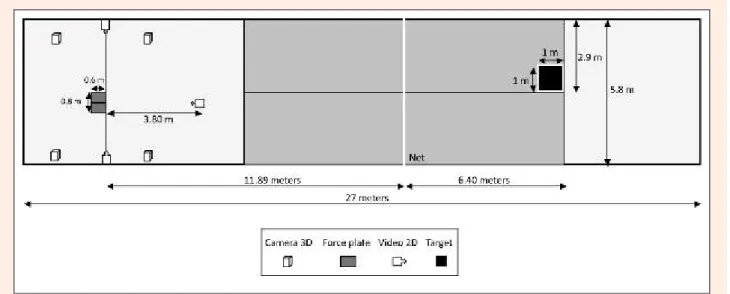
3D kinematic and kinetic evaluation: In the
labora-tory, we reproduced one half of a tennis court (Figure 1). The width of our court was smaller (5.8 m) than the nor-mal size (8.23 m) in order to fit into the laboratory. Play-ers served from two force plates located behind the base-line. We placed the net at a regulatory distance and height (International Tennis Federation, Roehampton, England) from the baseline and ground.
A B
Figure 2. Representation of body (A) and racket (B) marker (circle) and additional anatomical points (square) placement.
Before the tests, the players performed a general cardio-vascular warm-up with lower limb, (skipping rope, running and/or ergometric bicycle) and upper limb (rub-ber band) exercises. Afterward, they undertook a general short stretching routine for legs and arms. Finally, players engaged in a specific warm-up procedure for tennis serves, first without markers and then with markers placed on the skin. This specific warm-up allowed players to get familiar with the laboratory context (field, target and markers on the skin). Each player decided the number of serves necessary for warming-up and for familiariza-tion with a maximum of 30 serves allowed in order to avoid fatigue.
After the general and specific warm-up, the test began and the players served 25 times each, with 30 se-conds between each serve. The instructions were to serve in the target (“T” area) with the highest ball speed possi-ble and minimal ball rotation (flat serve). Afterward, the three best serves were kept for analysis (Reid et al., 2015, Whiteside et al., 2014) in order to consider the derivation of accurate and representative movement kinematics (Mullineaux et al., 2001). The selection criteria were precision (serve performed successfully in the 1 m2 area or “T” zone of the deuce square (Gillet et al., 2009)) and highest forward velocity of the racket at impact (Reid et al., 2014, Whiteside et al., 2014).
We used a three-dimensional optoelectronic sys-tem (Codamotion™, Charnwood Dynamics, Rothley, UK) to measure the movements. We tracked the 3D posi-tions of the player’s racket, dominant arm and forearm, trunk, pelvis and legs with 28 markers and four Codamo-tion CX1 units. The acquisiCodamo-tion rate was equal to 200 Hz. We placed three markers on the trunk, three mark-ers on the pelvis, four markmark-ers on both legs, four markmark-ers on the dominant arm, four makers on the dominant fore-arm and three on the dominant hand in accordance with the recommendations of the International Biomechanical Society (ISB) (Wu et al., 2002; 2005) (Figure 2A). We also placed three markers on the racket: one on each side and one on the top (Martin et al., 2014, Martin et al., 2012) (Figure 2B). We identified additional anatomical points by reference to the placed markers: T8, left and right posterior-superior iliac spine, dominant side lateral epicondyle, dominant side medial epicondyle and center of dominant side glenohumeral joint (Figure 2A).
The marker placement allowed us to measure the ankle, knee, pelvis and shoulder joints and segments’ amplitude (°); the linear velocity (m∙s-1) of markers and anatomic points for pelvis, shoulder, elbow, wrist and racket; also the ankle, knee, pelvis and shoulder angular velocity (°∙s-1) in frontal, transverse and/or sagittal plane(s). We additionally analyzed the kinematic chain (Kibler et al., 2013) with the observation of the sequence of motion (Liu et al., 2010). To achieve that goal, we measured the maximal forward linear velocity of domi-nant side markers.
The most important position in the tennis serve is the moment of impact (ball-racket contact). During a serve, the impact position timing corresponds to the max-imal forward linear velocity of the racket (Tanabe and Ito, 2007, Gordon and Dapena, 2006). We measured the rack-et velocity with the centroid of the three rackrack-et markers to better align the racket speed with ball impact location.
Figure 3. Representation of pelvis and trunk motions in frontal (right and left lateral tilt), sagittal (anterior-posterior tilt) and transverse (right and left rotations) planes.
In our 3D kinematic evaluation, we measured the maximal external rotation of the shoulder. For pelvis and trunk motion analysis, we measured the maximal rotation in frontal (right and left lateral tilt), sagittal (anterior-posterior tilt) and transverse (right and left rotations) planes in reference to the ground (Figure 3).
We also measured the maximal ground reaction force and the impulsion with two force plates (Kisler™ type 9281 EA, Kisle AG, Switzerland). Each force plate measured 60 cm by 40 cm so the players were able to push on both feet for either foot-up or foot-back tech-nique. The results represent the normalized peak ground reaction force (N∙Kg-1) and normalized impulsion (Ns∙Kg -1) (Linthorne, 2001).
Passive joint mobility and muscle flexibility: With
a Cochin goniometer (MSD™ Europe BVBA, Londerzeel – Belgium) used in accordance with suggested guidelines (Swann and Harrelson, 2012), we measured passive mo-bility (°) of the main joints including ankles (flexion-extension) and shoulders (glenohumeral rotations) (Moreno-Perez et al., 2015, Forthomme et al., 2013). We also evaluated hamstring length using a straight leg raise flexibility test (Neto et al., 2014). These procedures were performed before the 3D warm-up and carried out by the same examiner.
Isokinetic force: We used a CybexNorm™
isoki-netic dynamometer (Henley Healthcare, Sugarland, Tex-as) to measure voluntary maximal strength developed by quadriceps and hamstrings. We assessed absolute peak torque (PT; N.m) and body mass relative to peak torque (per kg; Nm∙Kg-1).
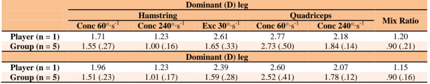
We performed lower limb measurements on quad-riceps (Q) and hamstrings (H) using protocol modalities based on previous studies (Croisier et al., 2002). Selected isokinetic speeds are 60°∙s-1 and 240°∙s-1 in concentric mode and 30°∙s-1 in eccentric mode. We also measured agonist-antagonist ratios (Hamstrings/Quadriceps) and determined a mixed ratio (combination of antagonist PT in the eccentric 30°.s-1 mode and agonist PT in the con-centric 240°.s-1 mode) to represent more specifically mus-cle contractions in a knee extension.
Figure 4. Velocity of the racket at impact for player and group. Each plot represents one of the 3 best serves of the
player. Each symbol represents a different player. We express means
of player and group as meter per second (m∙s-1). Values for the player (n
= 1; black) and the group (n = 5; grey).
Results
We analyzed kinematics, muscular and joint information of the injured player (‘the player’) compared to the con-trol group (‘other players’, ‘the group’). We select and describe remarkableresults in this section.
3D analysis during tennis serve
Velocity of the racket at impact: Racket velocity of the
player (38.9 ± 0.4 m∙s-1) is higher than for four out of five of the other players (37.4 ± 2.3 m∙s-1) (Figure 4).
A
B
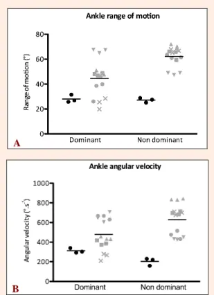
Figure 5. Range of motion (A) and maximal angular velocity
(B) of ankle plantar flexion for player and group. Each plot
represents one of the 3 best serves of a player. Each symbol represents a different player. We express means of player and group as degree (°) or degree per second (°∙s-1). Values for the player (n = 1; black) and the
group (n = 5; grey).
Range of motion and maximal angular velocity of
ankles and knees joints: Bilaterally, the ankle plantar
flexion ROM and maximal angular velocity during the serves is lower for the player compared to the group (Fig-ure 5A; 5B). This difference occurs mainly on the non-dominant side.
We observe similar knee extension maximal angu-lar velocity (°∙s-1) for the player and the group (dominant: 532.2 ± 18.5 °∙s-1 vs 519.2 ± 46.1 °∙s-1; non-dominant: 431.1 ± 8.6 °∙s-1 vs 429.3 ± 61.8 °∙s-1). However, the non-dominant knee extension ROM (front knee) is lower for the player than the group (48.4 ± 0.3° vs 63.7 ± 11.0°) (Figure 6A). Moreover, when the player leaves the force plate, he has bilaterally a more important knees flexion (dominant: 14.5 ± 2.6°; non-dominant: 27.6 ± 4.8°) in comparison to the group (dominant: 8.8 ± 3.0°; non-dominant: 18.3 ± 4.8°) (Figure 6B).
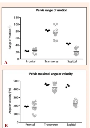
Pelvis range of motion and maximal angular
ve-locity: From maximal position to impact position, anterior
pelvic tilt ROM is higher for the player than for the group (44.2 ± 1.9° vs 22.0 ± 9.0°) (Figure 7A). Concerning frontal and transverse planes, we observe no particular difference (Frontal: 23.2 ± 1.1° vs 24.3 ± 6.4°; Trans-verse: 82.6 ± 2.2° vs 75.2 ± 16.9°).
A
B
Figure 6. Extension knee range of motion (A) and knee
an-gular flexion when leaving force plate (B) for player and
group. Each plot represents one of the 3 best serves of a player. Each
symbol represents a different player. We express means of player and group as degree (°).Values for the player (n = 1; black) and the group (n = 5; grey).
The pelvis maximal angular velocity of the player is particularly higher compared to the group in sagittal plane (439.3 ± 16.0 °∙s-1 vs 222.5 ± 28.9 °∙s-1) (Figure 7B). We observe no material difference in the frontal and transversal planes (Frontal: 191.2 ± 3.5° vs 184.4 ± 62.3°; Transverse: 456.9 ± 11.9° vs 423.0 ± 49.6°).
Maximal forward linear velocity of anatomic
points: Regarding the kinetic chain, we observe that the
maximal forward linear velocity is similar for the player and the group on the pelvis (1.1 ± 0.1 m∙s-1 vs 0.9 ± 0.2 m∙s-1), elbow (8.4 ± 0.3 m∙s-1 vs 8.0 ± 1.1 m∙s-1) and wrist (12.0 ± 0.1 m∙s-1 vs 11.9 ± 0.9 m∙s-1). However, on the dominant shoulder, maximal forward linear velocity is higher for the player (5.3 ± 0.4 m∙s-1 vs 4.4 ± 0.5 m∙s-1) (Figure 8).
Active shoulder external rotation: During the
serve, we observe a larger maximal external rotation for the player compared to the group (132 ± 1° vs 121 ± 9°) (Figure 9). However, we do not observe a higher shoulder internal rotation maximal angular velocity (Player: 1632 ± 149 °∙s-1; Group: 1851 ± 381°∙s-1).
Passive mobility (Goniometry)
We do not observe particularities for passive dominant shoulder external rotation by the player. Concerning low-er limbs, we obslow-erve a bilatlow-eral ankle rigidity in plantar and dorsal flexion for the player compared to the group (Table 1). Also, we do not observe greater hamstring flexibility for the player from the straight leg raise flexi-bility test (Table 2).
A
B
Figure 7. (A) Pelvis range of motion (ROM) from the maxi-mal lateral flexion/rotation/tilt position until the impact
position in the 3 planes (frontal, transverse, sagittal). (B)
Maximal angular velocity of pelvis girdle in the 3 planes
(frontal, transverse, sagittal). Each plot represents one of the 3 best
serves of a player. Each symbol represents a different player. We ex-press means of player and group as degree (°) and degree per second (°∙s-1). Values for the player (n = 1; black) and the group (n = 5; grey).
Figure 8. Maximal forward linear velocity (MFLV) of
domi-nant side joints in the kinetic chain. We express values in meters
per second (m∙s-1) for markers and anatomic points placed on pelvis,
shoulder, elbow and wrist. Values for the player (n = 1; black) and the group (n = 5; grey).
Ground reaction force and impulsion: We observe lower
vertical leg drive impulsion for the player than for the group (0.6 ± 0.2 Ns∙Kg-1 vs 1.1 ± 0.1 Ns∙Kg-1) (Figure 11A). Compared to the group, the player has a lower maximal ground reaction force (N.Kg-1) in the forward direction (1.5 ± 0.4 N∙Kg-1 vs 2.7 ± 0.8 N∙Kg-1) (Figure 10) and similar maximal ground reaction force (N.Kg-1) in the vertical direction (20.2 ± 1.0 N∙Kg-1 vs 21.2 ± 2.7 N∙Kg-1) (Figure 11B).
Table 1. Passive amplitude in degrees (°) of plantar and dorsal ankle’s flexion and shoulder’s external rotation. Passive measure of straight leg raise flexibility test. D = dominant (right); ND = non-dominant (left). Data are means (±SD).
Ankle Shoulder
D ND D ND
Plantar Dorsal Plantar Dorsal RE RE
Player (n =1) 32 ° 5 ° 32 ° 4 ° 92 ° 80 °
Group (n=5) 64 (7) ° 12 (2) ° 64 (11) ° 10 (3) ° 96 (7) ° 92 (9) °
Figure 9. Maximal external rotation for shoulder. Each plot represents one of the 3 best serves of a player. Each symbol represents a different player. We express means of player and group as degree per second (°∙s-1). Values for the player (n = 1; black) and the group (n = 5;
grey).
Figure 10. Vertical leg drive impulsion. Each plot represents
one of the 3 best serves of a player. Each symbol represents a
different player. We express means of player and group as relative impulsion (Ns∙Kg-1). Values for the player (n = 1; black) and the group
(n = 5; grey).
Table 2. Passive measure of hamstrings flexibility with straight leg raise flexibility test. D = dominant (right); ND = non-dominant (left). Data are means (±SD).
Hip Hamstring
D ND
Player (n =1) 80 ° 85 °
Group (n=5) 86 (6) ° 87 (7) °
Isokinetic assessment: Peak torque relative to body
mass of the player is generally higher for the player than for the group in all conditions (hamstrings and quadri-ceps; fast and slow speed; concentric and eccentric mode). We observe better performances for hamstrings in eccentric mode and quadriceps in fast concentric mode for the player than for the group. Mixed ratios are also higher
for the player compared to the group (Table 3).
A
B
Figure 11. Maximal ground reaction forces in the forward
(A) and vertical (B) directions. Each plot represents one of the 3
best serves of a player. Each symbol represents a different player. We express means of player and group as relative force (N∙Kg-1). Values for
the player (n = 1; black) and the group (n = 5; grey).
Discussion
This case study relates to a top level tennis player with a particular medical history. The player presented a muscle tear on the non-dominant rectus abdominis. The goal of our analysis was to be able to discuss retrospectively, through the analysis of the stroke action of the player, the potential injury mechanism. We performed several kine-matic and kinetic analyses. We performed tests to assess muscle strength, passive articular amplitudes, muscular measures and 3D kinematic of the tennis serve.
In the kinetic chain, the pelvis is the link between legs and trunk, and abdominis the link between the pelvis girdle and the shoulder girdle. Pelvis and trunk are the vital links in the sequence of actions during service (Kibler and Van Der Meer, 2001; Martin et al., 2014) and abdominis muscles are essentials part of the pelvis and trunk link (Maquirriain et al., 2007). During the cocking phase, players move the racket to the back with an abduc-tion and an external shoulder rotaabduc-tion. Then, lumbar spine
Table 3. Peak torque (PT, Nm∙kg-1) and body mass relative to peak torque (per kg) of the player and the group observed for
quadriceps and hamstring by isokinetic test. Data are means (±SD).
Conc = concentric, Ecc = eccentric. Mix ratio “hamstring (Ecc30)/quadriceps (conc240)”. D = dominant; ND = non-dominant. gets in hyperextension. This movement allows an
in-creased racket distance that generates speed and power to the racket and the ball at impact (Maquirriain et al., 2007). The eccentric contraction of non-dominant rectus abdominis followed by concentric contraction during the cocking phase of the serve motion is related as a specific tear injury mechanism (Maquirriain et al., 2007) as en-countered in our case.
We observed that our case study player had one of the best serving performances in comparison to the group. Indeed, we noted our player’s better racket speed com-pared to the mean of the group (38.9 ± 0.4 m∙s-1 vs 37.4 ± 2.3 m∙s-1). There is a correlation between the racket veloc-ity at impact and ball speed (Gordon and Dapena, 2006; Tanabe and Ito, 2007), which is a contributor to overall service performance (Girard et al., 2007b; Fleisig et al., 2003; Tanabe and Ito, 2007).
There is a proximo-distal sequence to perform the serve in tennis (Elliott, 1986; 2003, Ellenbecker, 2004, Marshall and Elliott, 2000; Martin et al., 2013b; Pugh et al., 2003). Legs are the start of the energy production from the lower limbs to the upper limbs (Elliott and Colette, 1993; Elliott et al., 2003; Girard et al., 2005). According to Elliott and Colette (1993), "It is significant to understand that power (force) is not developed by the trunk and arm. The primary source of power is generated from the ground in the form of ground reaction forces" (Elliott and Colette, 1993). So, leg extension is a key parameter in the search for efficiency in the tennis serve (Elliott and Colette, 1993; Girard et al., 2007a; 2007b) and rapid leg extension is a contributor of serve speed (Campbell et al., 2014; (Girard et al., 2007a; Reid et al., 2008).
Energy from the legs is transmitted along the ki-netic chain (Martin et al., 2014). In the case of our player we observe a lower leg drive impulsion result. This ob-servation is not due to a deficit of strength because the isokinetic results show better muscle qualities. Also, it is not due to a deficit of knee extension velocity because we observe similar maximal angular velocity of knees (serve kinematic). However, during leg drive, we note our play-er’s ankle maximal angular velocity and ROM in plantar flexion is below the mean of the group and there is in-complete knee extension at the instant of leaving the force plate. This lack of energy generation must be recovered by other movements in the kinematic chain in order to obtain one of the best performances of serve velocity. Lintner and al. (2008) describe this kind of compensation in terms of a “catch-up” concept (Lintner et al., 2008).
The compensations required to produce better racket velocity may appear along all the kinematic chain.
The pelvis maximal forward linear velocity is not higher for the player than the group but the dominant shoulder maximal forward linear velocity reaches a higher peak for the player, indicating higher energy generation between pelvis and shoulders for the player. Distally, from elbow to racket, we observe no particularities. In-deed, the player’s dominant shoulder for linear and angu-lar velocity, the forearm pronation, the wrist flexion and the ulnar deviation are similar to the rest of the group. These observations highlight absence of distally compen-sation and possible compencompen-sation between pelvis and shoulder.
Because of the incomplete leg drive, we hypothe-size that the abdominis work more to transfer and add energy from the pelvic girdle to the scapular girdle. In the energy flow (Martin et al., 2014) of our player, this lack of energy potentially appears at the pelvis level.
In fact, we observe that the pelvis moves with a larger anterior tilt ROM and maximal angular velocity. The important observed anterior pelvis tilt induces addi-tional hamstring tension that may explain the incomplete leg drive and consequently a lower energy production from the lower limbs. This increased anterior tilt of the pelvis induces a specific lumbar lordosis in combination with development of abdominal pre-stretch during the eccentric phase of the abdominal contraction (pre-stretched abdominal muscles). The eccentric phase, quick-ly followed by the concentric phase of the abdominal muscles, can cause a very important muscle request dur-ing the startdur-ing phase of trunk flexion (Maquirriain et al., 2007) and lead to a tear.
The maximal external rotation dominant in the player’s values as observed in passive measure (goniome-ter) are similar to the group but are larger during the ac-tive motion (3D) assessment suggesting the addition of constraints on the shoulder in dynamic situations. This could also add higher lumbar lordosis in addition to the abdominal eccentric tension.
We hypothesize that the particular pelvis kinematic induces a lack of leg drive and consequently of energy flow, which leads to various compensations including abdominal overwork and larger shoulder external rotation. Overwork on a link of the kinetic chain increases the risk of injury. In our opinion, the abdominal muscle overwork may explain the injury mechanism. The player compen-sates for the lack of energy transfer by important ab-dominal pre-stretch. This specific movement can cause
Dominant (D) leg
Hamstring Quadriceps
Mix Ratio
Conc 60°∙s-1 Conc 240°∙s-1 Exc 30°∙s-1 Conc 60°∙s-1 Conc 240°∙s-1
Player (n = 1) 1.71 1.23 2.61 2.77 2.18 1.20
Group (n = 5) 1.55 (.27) 1.00 (.16) 1.65 (.33) 2.73 (.50) 1.84 (.14) .90 (.21)
Dominant (D) leg
Player (n = 1) 1.96 1.23 2.39 2.60 2.07 1.15
important contraction of the abdominis at the start of the cocking stage.
Because of the retrospective approach of our study, we cannot affirm if this specific kinematic is the cause rather than the consequence of the abdominal injury. Forced external rotation in combination with a pelvis anterior tilt and an abdominal eccentric tension could also be a risk of following injury in the shoulder and the ab-dominis. However, we observed a new injury in a pelvis muscle (psoas) a few weeks after the tests. This develop-ment was not entirely surprising because the psoas muscle is actively involved in pelvic anterior tilt movement. A significant contraction of the psoas-iliac muscle can ex-plain the important pelvis anterior tilt observed and an important lumbar lordosis. Repeated contractions of this muscle may also explain the origin of this new lesion. This prospective follow-up injury is supported by our previous observations.
Our study contributes to the need for awareness by medical staff of the importance of a pre-season check-up. We would suggest to the medical staff (trainer, physic trainer, doctor, and physiotherapist) a corrective program based on these particular observations. In the case of this player, it would be judicious to propose specific leg drive exercises using complete knee extension jumps, associat-ed to a controllassociat-ed pelvis kinematic with abdominal core strengthening. This could potentially limit the strength of the abdominis and psoas muscles contraction and the shoulder external rotation compensation. Monitoring the muscle activity of the abdominis muscles with surface electromyography could help to evaluate the effectiveness of the rehabilitation program.
Limitations of the study
Our study highlights the importance of measuring tools to improve and objectivize players’ kinematics. These tech-nologies could ameliorate player performance and prevent lesion risk. We also demonstrate through our work the benefit of a multidisciplinary analysis of a gesture using several techniques simultaneously. In the future, it would be also interesting to combine our evaluations with elec-tromyography (EMG). These measures are focused on muscle activity and may provide further insights into injury mechanisms.
1. This retrospective study cannot establish with certain-ty if our observations are a cause or a consequence of the injury. If our observations are the cause of the jury, there is a risk of recurrence and it would be in-teresting to continue to provide prevention work in the pelvis region. It would also be interesting to perform a prospective study to evaluate the effect of a specific rehabilitation program.
2. Our reference group is small. It limits us in the com-parison analysis of data. However, our participants are amongst the highest international level players, which improves the relevance of data. We also checked the reproducibility of the whole protocol on ten partici-pants. These unpublished results showed that the kin-ematic, kinetic and clinical measurements are repro-ducible.
3. Three dimensional technology with an active markers system is a source of pitfalls in the context of a com-plex and fast gesture analysis such as that as encoun-tered in throwing sports (Abrams et al., 2011, Gordon and Dapena, 2006).
Conclusion
The case study player’s racket velocity at impact was superior to the mean of group. To overcome a deficit of energy transfer due to an uncompleted leg drive and a specific pelvis kinematic, it is likely that the player com-pensated involuntarily thanks to other parameters in-volved in the production of racket velocity (Kovacs and Ellenbecker, 2011). We observe an important external rotation during the serve. The incomplete transmission of the energy of the legs to the pelvis may also have been compensated by a larger abdominis contraction. These particularities could be a retrospective explanation of medical history concerning the abdominal muscles and also highlight the risk of future pathologies.
Similarities between the observations of the expe-rienced eye and the 3D analysis are numerous. However, the 3D kinematic evaluation is an indispensable tool for an objective evaluation of the kinematic in the tennis serve. Coaches are familiar with the performance analysis of the serve but less so with its preventive counterpart.
In this case report, we demonstrate that three di-mension analysisan effective solution to better understand and highlight some injury mechanisms. Also, we con-clude that the application of several evaluation techniques together helps to provide a more complete overall and individualized comprehension of the athlete.
Aknowledgement
The authors wish to thank the Wallonia-Brussels Federation for their assistance in this study.
References
Abrams, G.D., Sheets, A.L., Andriacchi, T.P. and Safran, M.R. (2011) Review of tennis serve motion analysis and the biomechanics of three serve types with implications for injury. Sports
Biomechanics 10, 378-390.
Antunez, R.M., Hernandez, F.J., Garcia, J.P., Vaillo, R.R. and Arroyo, J.S. (2012) Relationship between motor variability, accuracy, and ball speed in the tennis serve. Journal of Human Kinetics 33, 45-53.
Bahamonde, R.E. (2000) Changes in angular momentum during the tennis serve. Journal of Sports Sciences 18, 579-592.
Balius, R., Pedret, C., Galilea, P., Idoate, F. and Ruiz-Cotorro, A. (2012) Ultrasound assessment of asymmetric hypertrophy of the rectus abdominis muscle and prevalence of associated injury in professional tennis players. Skeletal Radiology 41, 1575-1581. Campbell, A., O'Sullivan, P., Straker, L., Elliott, B. and Reid, M. (2014)
Back pain in tennis players: a link with lumbar serve kinematics and range of motion. Medicine and Science in Sports and
Exercices 46, 351-357.
Chan, K.M., Fong, D.T., Hong, Y., Yung, P.S. and Lui, P.P. (2008) Orthopaedic sport biomechanics - a new paradigm. Clinical
Biomechanics (Bristol, Avon) 23(Suppl 1), S21-30.
Chow, J.W., Park, S.A. and Tillman, M.D. (2009) Lower trunk kinematics and muscle activity during different types of tennis serves. Sports Medicine, Arthroscopy, Rehabilitation, Therapy
& Technology: SMARTT 1, 24.
Croisier, J.L., Forthomme, B., Namurois, M. H., Vanderthommen, M. and Crielaard, J.M. (2002) Hamstring muscle strain recurrence
and strength performance disorders. The American Journal of
Sports Medicine 30, 199-203.
Croisier, J.L., Ganteaume, S., Binet, J., Genty, M. and Ferret, J.M. (2008) Strength imbalances and prevention of hamstring injury in professional soccer players: a prospective study. The
American Journal of Sports Medicine 36, 1469-1475.
Ellenbecker, T.S. (2004) Chapter 17 - Analysis of Sport Technique: Tennis and Overhead Throwing Model. In: Ellenbeacker, T.S. (ed.) Clinical Examination of the Shoulder. Saint Louis: W.B. Saunders.
Elliott, B. (2003) Developing racquet velocity. Biomechanics of
advanced tennis. London: ITF Ltd.
Elliott, B. (2006) Biomechanics and tennis. British Journal of Sports
Medicine 40, 392-396.
Elliott, B. and Colette, D. (1993) Biomechanics. ITF Coaching and
Sport Science Review, 1, 11.
Elliott, B., Fleisig, G., Nicholls, R. and Escamilia, R. (2003) Technique effects on upper limb loading in the tennis serve. Journal of
Science and Medicine in Sport 6, 76-87.
Elliott, B., Marsh, T. and Blanksby, B. (1986) A three-dimentional cinematographical analysis of the tennis serve. International
Journal of Sport Biomechanics 2, 260-270.
Elliott, B. and Reid, M. (2008) The Use of Technology in Tennis Biomechanics. ITF Coaching and Sport Science Review 15, 2-4.
Fleisig, G., Nicholls, R., Elliott, B. and Escamilla, R. (2003) Kinematics used by world class tennis players to produce high-velocity serves. Sports Biomechanics 2, 51-64.
Forthomme, B., Wieczorek, V., Frisch, A., Crielaard, J.M. and Croisier, J.L. (2013) Shoulder pain among high-level volleyball players and preseason features. Medicine and Science in Sports and
Exercice 45, 1852-1860.
Gillet, E., Leroy, D., Thouvarecq, R. and Stein, J.F. (2009) A notational analysis of elite tennis serve and serve-return strategies on slow surface. Journal of Strength and Conditioning Research 23, 532-539.
Girard, O., Eicher, F., Fourchet, F., Micallef, J.P. and Millet, G.P. (2007a) Effects of the playing surface on plantar pressures and potential injuries in tennis. British Journal of Sports Medicine 41, 733-738.
Girard, O., Micallef, J.P. and Millet, G.P. (2005) Lower-limb activity during the power serve in tennis: effects of performance level.
Medicine and Science in Sports and Exercices 37, 1021-1029.
Girard, O., Micallef, J.P. and Millet, G.P. (2007b). Influence of restricted knee motion during the flat first serve in tennis.
Journal of Strength and Conditioning Research 21, 950-957.
Gordon, B.J. and Dapena, J. (2006) Contributions of joint rotations to racquet speed in the tennis serve. Journal of Sports Sciences 24, 31-49.
Grosdent, S., Demoulin, C., Souchet, M., Tomasella, M., Crielaard, J.M. and Vanderthommen, M. (2014) Trunk muscle profile in elite tennis players with and without low back pain. The Journal of
Sports Medicine and Physical Fitness, (In press).
Johnson, C.D., McHugh, M.P., Wood, T. and Kibler, B. (2006) Performance demands of professional male tennis players.
British Journal of Sports Medicine 40, 696-699.
Julienne, R., Gauthier, A. and Davenne, D. (2012) Fatigue-resistance of the internal rotator muscles in the tennis player's shoulder: isokinetic and electromyographic analysis. Physical Therapy in
Sport 13, 22-26.
Kibler, W.B. and Safran, M. (2005) Tennis injuries. Medicine and Sport
Science 48, 120-137.
Kibler, W.B. and Safran, M. R. (2000) Musculoskeletal injuries in the young tennis player. Clinical Sports Medicine 19, 781-92. Kibler, W.B. and Van Der Meer, D. (2001) Mastering the kinetic chain.
In: World-Class Tennis Technique. Eds: Roetert, P. and Groppel, J. Champaign ed.
Kibler, W.B., Wilkes, T. and Sciascia, A. (2013) Mechanics and pathomechanics in the overhead athlete. Clinical Sports
Medicine 32, 637-651.
Knudson, D. (2007) Qualitative biomechanical principles for application in coaching. Sports Biomechanics 6, 109-118.
Kovacs, M. and Ellenbecker, T. (2011) An 8-stage model for evaluating the tennis serve: implications for performance enhancement and injury prevention. Sports Health 3, 504-513.
Linthorne, N.P. (2001) Analysis of standing vertical jumps using a force platform. American Journal of Physics 69, 1198-1204.
Lintner, D., Noonan, T.J. and Kibler, W.B. (2008) Injury patterns and biomechanics of the athlete's shoulder. Clinics in Sports
Medicine 27, 527-551.
Liu, H., Leigh, S. and Yu, B. (2010) Sequences of upper and lower extremity motions in javelin throwing. Journal of Sports
Sciences 28, 1459-1467.
Maquirriain, J., Ghisi, J.P. and Kokalj, A.M. (2007) Rectus abdominis muscle strains in tennis players. British Journal of Sports
Medicine 41, 842-848.
Marshall, R.N. and Elliott, B.C. (2000) Long-axis rotation: the missing link in proximal-to-distal segmental sequencing. Journal of
Sports Sciences 18, 247-254.
Martin, C., Bideau, B., Bideau, N., Nicolas, G., Delamarche, P. and Kulpa, R. (2014) Energy flow analysis during the tennis serve: comparison between injured and noninjured tennis players. The
American Journal of Sports Medicine 42, 2751-2760.
Martin, C., Bideau, B., Nicolas, G., Delamarche, P. and Kulpa, R. (2012) How does the tennis serve technique influence the serve-and-volley? Journal of Sports Sciences 30, 1149-1156. Martin, C., Bideau, B., Ropars, M., Delamarche, P. and Kulpa, R.
(2013a). Upper limb joint kinetic analysis during tennis serve: Assessment of competitive level on efficiency and injury risks.
Scandinavian Journal of Medicine and Science in Sports (In
press).
Martin, C., Kulpa, R., Delamarche, P. and Bideau, B. (2013b) Professional tennis players' serve: correlation between segmental angular momentums and ball velocity. Sports
Biomechanics 12, 2-14.
Moreno-Perez, V., Moreside, J., Barbado, D. and Vera-Garcia, F.J. (2015) Comparison of shoulder rotation range of motion in professional tennis players with and without history of shoulder pain. Manual Therapy 20(2), 313-318.
Mullineaux, D. R., Bartlett, R.M. and Bennett, S. (2001) Research design and statistics in biomechanics and motor control.
Journal of Sports Sciences 19, 739-760.
Natsis, K., Lyrtzis, C., Papathanasiou, E. and Anastasopoulos, N. (2012) Rectus abdominis overuse injury in a tennis athlete treated with traumeel. The American Journal of Case Reports 13, 3-6. Neto, T., Jacobsohn, L., Carita, A.I. and Oliveira, R. (2014) Reliability
of the active knee extension test and the straight leg raise test in subjects with flexibility deficits. Journal of Sport Rehabilitation, (In press).
Perkins, R.H. and Davis, D. (2006) Musculoskeletal injuries in tennis.
Physical Medicine and Rehabilitation Clinics of North America
17, 609-631.
Pugh, S.F., Kovaleski, J.E., Heitman, R.J. and Gilley, W.F. (2003) Upper and lower body strength in relation to ball speed during a serve by male collegiate tennis players. Perceptual and Motor
Skills 97, 867-872.
Reid, M., Elliott, B. and Alderson, J. (2008) Lower-limb coordination and shoulder joint mechanics in the tennis serve. Medicine and
Science ind Sports and Exercice 40, 308-315.
Reid, M., Giblin, G. and Whiteside, D. (2015) A kinematic comparison of the overhand throw and tennis serve in tennis players: How similar are they really? Journal of Sports Sciences 33,713-723. Renstrom, P. and Johnson, R.J. (1985) Overuse injuries in sports. A
review. Sports Medicine 2, 316-333.
Sanchis-Moysi, J., Idoate, F., Dorado, C., Alayon, S. & Calbet, J.A. (2010) Large asymmetric hypertrophy of rectus abdominis muscle in professional tennis players. PLoS One 5,1-8. Silva, R.T., Gracitelli, G.C., Saccol, M.F., Laurino, C.F., Silva, A.C. and
Braga-Silva, J.L. (2006) Shoulder strength profile in elite junior tennis players: horizontal adduction and abduction isokinetic evaluation. British Journal of Sports Medicine 40, 513-517. Springings, E., Marshall, R., Elliott, B. and Jennings, L. (1994) A
three-dimensional kinematic method for determining the effectiveness of arm segment rotations in producing racquet-head speed. Journal of Biomechanics 27, 245-254.
Swann, E. and Harrelson, G.L. (2012) Measurement in Rehabilitation. In: Physical Rehabilitation of the Injured Athlete. Ed: Wilk, J.R.A.L.H.E. Fourth Edition. Philadelphia: W.B. Saunders. Tanabe, S. and Ito, A. (2007) A three-dimensional analysis of the
contributions of upper limb joint movements to horizontal racket head velocity at ball impact during tennis serving. Sports
Biomechanics 6, 418-433.
Vand Der Hoeven, H. and Kibler, W.B. (2006) Shoulder injuries in tennis players. British Journal of Sports Medicine 40, 435-440.
Whiteside, D., Elliott, B., Lay, B. and Reid, M. (2013) The effect of age on discrete kinematics of the elite female tennis serve. Journal
of Applied Biomechanics 29, 573-582.
Whiteside, D., Elliott, B., Lay, B. and Reid, M. 2014. The effect of racquet swing weight on serve kinematics in elite adolescent female tennis players. Journal of Science and Medicine in Sport 17, 124-128.
Wu, G., Siegler, S., Allard, P., Kirtley, C., Leardini, A., Rosenbaum, D., Whittle, M., D'Lima, D.D., Cristofolini, L., Witte, H., Schmid, O., Stokes, I., Standardization and Terminology Comittee of the International Society of Biomechanics (2002) ISB recommendation on definitions of joint coordinate system of various joints for the reporting of human joint motion--part I: ankle, hip, and spine. International Society of Biomechanics.
Journal of Biomechanics 35, 543-548.
Wu, G., Van Der Helm, F.C T., Veeger, H.E.J., Makhsous, M., Van Roy, P., Anglin, C., Nagels, J., Karduna, A.R., McQuade, K., Wang, X., Werner, F.W. and Buccholz, B. (2005) ISB recommendation on definitions of joint coordinate systems of various joints for the reporting of human joint motion—Part II: shoulder, elbow, wrist and hand. Journal of Biomechanics 38, 981-992.
Key points
• In the proximal-distal sequence, energy is transmit-ted from lower limbs to upper limps via trunk. • The 3D analysis tool is an indispensable test for an
objective evaluation of the kinematic in the tennis serve.
• Multiple evaluations techniques are useful for fuller comprehension of the kinematics and contribute to the awareness of the player’s staff concerning pa-thologies and performance.
AUTHOR BIOGRAPHY
François TUBEZ Employment
PhD Student, Laboratory of Human Motion Analysis (LAMH). Department of Sport and Rehabilitation Sciences. University of Liège.
Degree
MSc
Research interests
Study of tennis serve gesture.
E-mail: [email protected]
Bénédicte FORTHOMME Employment
Professor, Department of Sport and Reha-bilitation Sciences. Laboratory of Human Motion Analysis (LAMH). University of Liège.
Degree
PhD
Research interests
Shoulder rehabilitation and biomechanics.
E-mail: [email protected]
Jean-Louis CROISIER Employment
Professor, Department of Sport and Reha-bilitation Sciences. Laboratory of Human Motion Analysis (LAMH). University of Liège.
Degree
PhD
Research interests
Isokinetic and eccentric contractions.
E-mail: [email protected]
Caroline CORDONNIER
Employment
PhD Student, Laboratory of Human Motion Analysis (LAMH). Department of Sport and Rehabilitation Sciences. University of Liège.
Degree
MSc
Research interests
Study of knee movement in jumps
E-mail: [email protected]
Olivier BRÜLS Employment
Professor, University of Liège.
Degree
PhD
Research interests
Development of numerical methods based on biomechanical models in order to evalu-ate and predict muscular loads.
E-mail: [email protected]
Vincent DENOËL Employment
Professor, University of Liège.
Degree
PhD
Research interests
Study about the effects of human excitation of flexible structures.
E-mail: [email protected]
Gilles BERWART Employment
Researcher, Laboratory of Human Motion Analysis (LAMH). Department of Sport and Rehabilitation Sciences. University of Liège.
Degree
MSc
Research interests
2D biomechanical evaluations in sports.
E-mail: [email protected]
Maurice JORIS Employment
Doctor, Centre Médical du Trixhay. Sport & Art.
Degree
MD
Research interests
Study in tennis pathomechanic.
Stéphanie GROSDENT Employment
PhD Student, University Hospital Centre of Liege, Belgium Degree MSc Research interests Trunk evaluation. E-mail: [email protected] Cédric SCHWARTZ Employment
Research engineer, Laboratory of Human Motion Analysis (LAMH). Department of Sport and Rehabilitation Sciences. Univer-sity of Liège.
Degree
PhD
Research interests
3D biomechanical evaluations in sports.
E-mail: [email protected]
François Tubez
Laboratory of Human Motion Analysis (LAMH), Department of Sport and Rehabilitation Sciences, University of Liège, 4, Allée des Sports 4000 Liège, Belgium