1495
Original
Article
Three-dimensional
Electron
Microscopy
of Ribosomal
Chromatin
in Two
Higher
Plants:
A Cytochemical,
Immunocytochemical,
and
In Situ
Hybridization
Approach’
PATRICK M.
MOTTE,2
ROLAND LOPPES, MONIQUE MENAGER,and
ROGER
DELTOUR
Laboratoire de Morphologie V#{233}gitale (PMM,RD) et de G#{233}n#{233}tiqueMol#{233}culaire (RL), D#{233}partement de Botanique, Universit#{233} de Lilge,
B4000 Liege, Belgium, andLaboratoire d’Histologie, Embryologic et Cytoginitique, Facult#{233}de M#{233}decine,51100 Reims, France (MM).
Received for publication January 15, 1991 and in revised form May 20, 1991; accepted May 31, 1991 (1A2 211).
We report the 3-D arrangement ofDNA within the
nudeo-lan subcomponents from two evolutionary distant higher plants, Zea mays and Sinapis alba . These species are partic-ularly convenient to study the spatial organization of plant intranudeolar DNA, since their nudeoli have been previ-ously reconstructed in 3-D from serial ultra-thin sections.
We used the osmium ammine-B complex (a specific DNA
stain) on thick sections ofLowiayl-embedded root fragments.
Immunocytochemical techniques using anti-DNA antibodies
and rDNA/rDNA in situ hybridization were also applied
on ultra-thin sections. We showed on tilted images that the
OA-B stains DNA throughout the whole thickness of the
section. In addition, very low quantities ofcytoplasmic DNA
were stained by this complex, which is now the best DNA
Introduction
The chromatin organization of the interphase nucleus has been
investigated for many years. From recent data, we can argue that interphase chromosomes occur in well-defined nuclear domains (8,21,29,30,36,37). Concerning the nucleolar chromatin, it is well known that each metaphasic nucleolus organizer region (NOR), which contains the genes coding for the precursors of nibosomal RNA (rDNA), is a well-defined part of an NOR-bearing chromo-some. The number of NOR-bearing chromosomes varies accord-ing to the species. At the end of telophase each NOR, or part of it, is responsible for the formation of one nucleolus (9,11,17,31). During interphase the nucleoli may fuse together. However, the exact localization ofthe nucleolar chromatin, i.e., ofthe rDNA mol-ecule, within the active nucleolus is still unknown and is the subject of controversy (see Discussion). Its three-dimensional distribution is still totally unknown. By electron microscopy, five subcomponents can be distinguished in a transcriptionally active nudeolus: the fibnillar
1Supported by grants from the FRFC (2.405.87 and 2.4521.89) and from “Action de Recherche Concert#{233}e” (88/93.129).
2Correspondence and present address: Dr. Patrick M. Motte,
Max-Planck-Institut f#{252}rZUchtungsforschung, 10 Carl-von-Linn#{233} Weg, D-5000 K#{246}ln30, Germany.
stain used in electron microscopy. Within the nudeoli the
DNA waslocalized in the fibrillar centers, where large dumps
of dense chromatin were also visible. In the two plant
spe-cies intranudeolar chromatin forms a complex network with
strands partially linked tochromosomal nudeolar-organizing
regions identified by in situ hybridization. This study
describes for the first time the spatial arrangement of the intranudeolar chromatin in nucleoli of higher plants using
high-resolution techniques.
(J
Histochem Cyrochem39:1495-1506, 1991)
KEY WORDS: Nucleolus; rDNA; Nucleolus-associated chromatin;
Root cells; Anti-DNA antibody; Osmium arnmine; Zea mays; Sinapis &ba.
centers (FCs), the dense fibnillar component (DFC) which surrounds
the FCs, the granular component (GC), the highly contrasted nucleolus-associated chromatin (NAC) observed at the periphery of the nucleolus, and small, less electron-opaque spaces, referred to as nucleolar vacuoles.
The unambiguous localization of rDNA in these subcompo-nents could be essential for the understanding of the functional organization of the nucleolus. In the present study we have used DNA-specific osmium ammine staining on thick sections, immu-nocytochemical technique, and in situ hybridization to localize rDNA and to study its spatial distribution in an active plant nucleo-lus at the electron microscopic level. Compared with animal mate-rial, the plant cell nucleolus exhibits specific advantages: (a) The NAC, restricted to heterochromatic knobs located at the nucleolar periphery, are parts of the interphasic NORS (32,36,37; and this report). On the contrary, in animal nucleoli, a peripheral ring of
NAC of an unknown nature is always observed. The relationships between this peripheral chromatin and the intranucleolar rDNA
are not well defined (43); (b) the DFC is large and constitutes the main structure of the plant active nucleolus; and (c) the active nucleoli ofthe two plant species here studied have been previously three-dimensionally reconstructed from serial ultra-thin sections (10,32).
I
1496 MOT1’E, LOPPES, MENAGER, DELTOUR
The localization and spatial arrangement ofnibosomal
chroma-tin is discussed and compared with the recent data on rDNA dis-tribution in both plant and animal nucleoli.
Materials
and
Methods
Germination
Procedure
Batches of 30 seeds ofZea mays and Sinapis alba were germinated in petri
dishes on cotton wool and filter saturated with distilled water. They were incubated in darkness in an Ehret-controlled temperature chamber at 16’C
and 9’C, respectively. Germination began when the seeds were placed in
contact with water. The maximum percentage of germinated seeds was
al-ways over 90%, and the rates ofgermination were very similar for both
spe-cies. One hundred twenty hours after germination embryonic roots were
about 3 cm in length.
Tissue
Preparation
The distal millimeter of roots was excised from quiescent embryos and,
120 hr after sowing, rapidly immersed in a solution of4% formaldehyde
(freshly prepared from paraformaldehyde) in 0.1 M Sorensen buffer (pH 7.0) and maintained for 2 hr at room temperature. The root caps were
cx-cised and discarded. The root fragments were washed three times in 0.1
M Sorensen buffer at 4’C. The samples were then embedded in Lowicryl K4M at low temperature according to Carlemalm et al. (6) with longer in-filtration periods. Semi-thin and ultra-thin sections were cut with a
dia-mond knife in the cortex 1 mm from the root tips with a Sorval Porter Blum MT2 ultramicrotome. After the reactions described below, the
5cc-tions were observed with a Siemens Elmiskop 101 or a Zeiss electron
micro-scope EM1O9 at 80 kV.
Osmium
Ammine-B
(OA-B)
Staining
OA-B was synthesized according to Olins et al. (33). Semi-thin sections about 0.5 tm thick realized in maize and Sinapis alba root fragments were
mounted on 300-mesh gold grids without formvar support. Before the OA-B
staining the sections were treated with 1 mg/mI pre-digested pronase
(Boehringer; Mannheim, ERG) for 1 hr at 37’C in Tnis-HCI 10 mM (pH 7.2). For the staining, the grids were first floated, sections facing the solu-non, on the surface ofa 5 N HCI solution for 25 mm at room temperature.
The grids were then thoroughly washed with distilled water and floated
on some drops ofwater until osmium ammine-B solution was ready. A 0.2% aqueous solution ofOA-B was bubbled with 502 for 20 mm. The staining of DNA was performed for 60 mm at 37’C in a well-covered container, sections floating on the solution. The grids were thoroughly jet-washed with distilled water. We performed successively two ultra-thin and one
semi-thin serial sections. The two ultra-thin sections were stained with uranyl and lead (Ur-Pb), while the thick section was stained with OA-B.
Some thick sections were observed with a Jeol 200CX at 200 kV. 5cc-tions were tilted with an eucentric goniometer stage from - 18 to +18’
by 6’ steps. Stereo pairs oftilted images were mounted to be observed with a pocket stereoviewer according to Ploton et al. (35).
Electron Microscopic Immunocytochemistry
Yellow ultra-thin sections of embedded Zea mays fragments were mounted on formvar-coated gold grids. For labeling the grids were processed at room temperature as described below. The sections were incubated on 20 tl of PBS, pH 7.5, containing 0.5% BSA, 0.1% Tween 20 (PBST), and normal goat serum diluted 1:20, for 30 mm. Then the grids were floated on 10
hI ofPBS containing 1gM mouse monoclonal DNA antibodies (30 Rg/ml; Boehninger) and 1:50 normal goat serum, for 3 hr. The grids were washed
with PBST twice for 10 mm, then were incubated on 20 tl of PBST (pH
8.2) containing 1:75 goat anti-mouse 1gM coupled to 5-nm gold particles
.._1_.
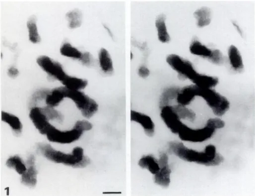
Figure 1. Stereo pair of Zea mays meta-phase chromosomes stained with OA-B complex, observed at 200 kV. One-rim thick section. Long chromosome segments lo-cated at different level in the section are stained all along their length without inter-ruptions. This demonstrates that the OA-B complex penetrates through the entire Lowicryl section. Note that when a chromo-some is longitudinally sectioned the portion within the section is less thick and thus is less stained. Original magnification x 6800. Bar = 1 rim.
;1i.._
V
!
.
C
.4r:
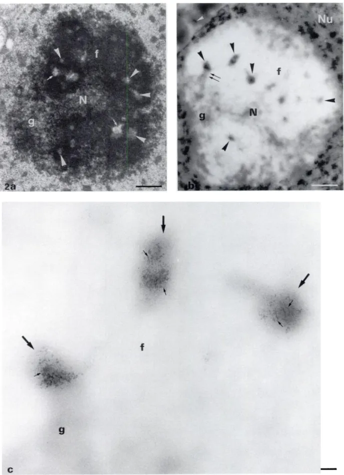
Figure 2. (a) Electron micrograph of a Lowicryl K4M-embedded nucleolus (N) of Zea mays, a monocotyledon. The subcomponents are visible and well discrimi-nated: the DFC (f) is the main component surrounding a few FCs (arrowheads) often associated with small vacuoles or interstices (small arrows). The whole fibrillar component (DFC and FCs) which forms the nucleolonema is enclosed within the granular component (g). Uranyl and lead staining. (b) Semi-thin section of a nuclear portion ofZea mayastained with osmium ammine-B complex. The DNA-containing structures are well contrasted in the nucleoplasm (Nu). At low magnifica-tion, the nucleolar subcomponents are still easily visible owing totheir remnant electron opacity after the pronase digestion. The DFC (f) appears like homogeneous bleached large zones and the GC (g) like interspersed clear gray and bleached zones. The FCs (arrowheads) appear like small contrasted areas embedded in the DFC and contain DNA stained by the OA-B complex. In some cases the granular component is directly in contact with a fibrillar center (double arrows). The cytoplasmic DNA is also visible (white arrowhead). (C) High magnification of the three upper left FC5 (arrows) of b. Clumps of chromatin fibers (small arrows) are well visible inside small areas probably corresponding to FCs. DNA fibers are not observed within the surrounding DFC (f). g, granular component. Original magnifications: a,b x 6000; c x 20,000. Bars: a,b = 1 tm; c = 0.1 tm.
1498
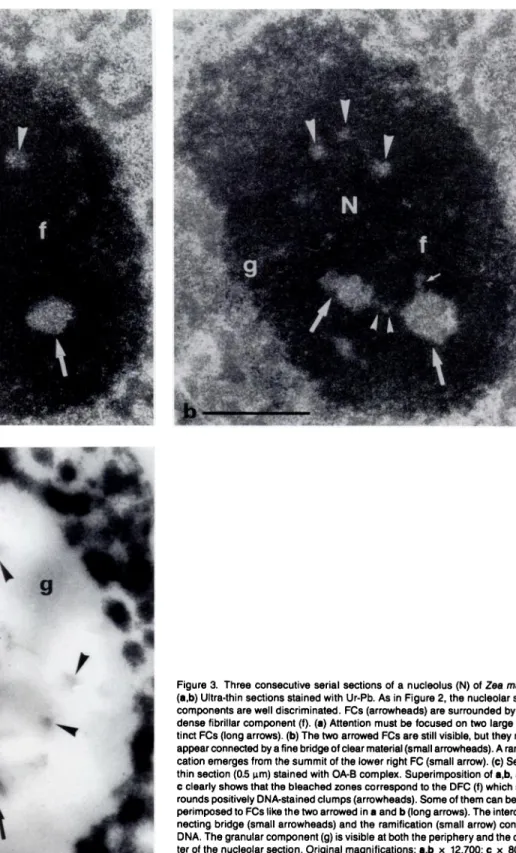
Figure a Three consecutive serial sections of a nucleolus (N) of Zea mays. (a,b) Ultra-thin sections stained with Ur-Pb. As in Figure 2, the nucleolar sub-components are well discriminated. FCs (arrowheads) are surrounded by the dense fibrillar component (f). (a) Attention must be focused on two large dis-tinct FCs (long arrows). (b) The two arrowed FCs are still visible, but they now appear connected by afine bridge ofclear material(small arrowheads). A
ramifi-cation emerges from the summit of the lower right FC (small arrow). (C)
Semi-thin section (0.5 tm) stained with OA-B complex. Superimposition of a,b, and
Cclearly shows that the bleached zones correspond to the DFC (f) which sur-rounds positively DNA-stained clumps (arrowheads). Some of them can be su-perimposed to FCs like the two arrowed in a and b (long arrows). The intercon-necting bridge (small arrowheads) and the ramification (small arrow) contain DNA. The granular component (g) is visible at both the periphery and the cen-ter of the nucleolar section. Original magnifications: a,b x 12,700; C x 8000. Bars = 1 tm.
Uanssen Life Sciences; Beerse, Belgium) for 60 mm. The grids were rinsed
three times for 10 mm in PBST and three times in distilled water. The
5cc-nons were stained with uranyl acetate. Controls were performed by incubating sections with either distilled water, PBS, or normal mouse serum in place of the anti-DNA antibody. Ultra-thin sections labeled with the anti-DNA
antibody were also stained by the OA-B as described above, without pronase
digestion.
In
Situ
Hybridization
Procedure
Preparation ofthe Biotinylated Probe. Plasmid pMrl(48) contains parts of the 255 and 185 rRNA coding regions and one complete spacer region of a maize rRNA transcription unit. The 0.75 KB Sma I fragment of the 255 rRNA coding sequence was subcloned into pUC9. The presence in the insert of Bgl II and Bam HI sites was verified. One microgram of the
plas--./
4
N
. 4 .:F
mid (pULG-1) was labeled with biotin-11-dUTP by nick translation using
the B1uGENE kit from Bethesda Research Laboratories (Gaithersburg, MD). The probe was purified by gel filtration on Sephadex G-50, precipitated
by ethanol, then dissolved in 50 tl sterile water and stored at -20C. Spots
of 6 pg (in 3 lil) of the probe were easily detected on nitrocellulose filters using streptavidin-alkaline phosphatase conjugate, then nitroblue tetrazo-hum and 5-bromo-4-chloro-3-indolylphosphate according to the recom-mendations of the supplier.
In Situ Hybridization (rDNA-rDNA). Yellow sections ofZea mays
sam-pies, collected on formvar-coated gold grids, were first submitted to enzy-matic digestions performed at 37’C in a moist chamber. The sections were
digested for 1 hr by 1 mg/mi pre-digested pronase in 10 mM Tris-HCI(pH
7.2), then treated for 1 hr by pre-boiled RNAse (1 mg/mI in 10 mM Tris-HCI, pH 7.2). The grids were then subjected to three 10-mm rinses in dis-tilled water and air-dried. Cellular DNA was denatured for 10 mm at 80’C
in 75% (v/v) deionized formamide in 2 x SSC (1 x SSC = 0.15 NaCI, 0.015 M sodium citrate, pH 7.0). The sectionswere then immediately dipped
into ice-cold 0.1 x SSC, dehydrated in an ethanol series of 50%, 75%,
and absolute ethanol, and air-dried for a few minutes. The probe diluted
in the hybridization buffer (50% deionized formamide/10% dextran sul-fate/i x Denhardt’s solution/i mM EDTA/600 mM NaCI/500 tg.ml’ herring sperm DNA/25 mM Tnis-HC1, pH 7.2) was denatured for 4 mm
at i00’C. Hybridization buffer contained biotinylated probe at 0.8-1.0
tg/ml. The grids were floated overnight at 40C in a moist chamber on 5 p3 ofthe denatured hybridization solution. Grids were then floated three times for i0 mm on drops of PBS, incubated successively in mouse
anti-biotin antibody (Dako; Santa Barbara, CA) and goat anti-mouse IgG
con-jugated to gold particles 3-5 nm in diameter (Janssen), and finally stained
with uranyl acetate before examination. Controls were performed by in-cubating sections on either hybridization buffer or hybridization buffer
con-taming the biotinylated pUC9 plasmid.
Results
One hundred twenty hours after the onset ofgermination, the fully
functional nucleolus was capable ofboth synthesizing and processing pre-nRNAs (50). It was composed ofwell-segregated granular and fibrillar components.
Osmium
Ammine-B
Staining
The osmium ammine complex is known to be a specific stain for
DNA in electron microscopy (12,13). To increase the contrast of the chromatin fibers and to study three-dimensionally the intranucleolar
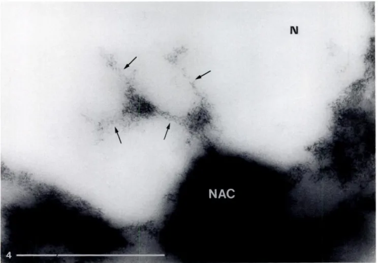
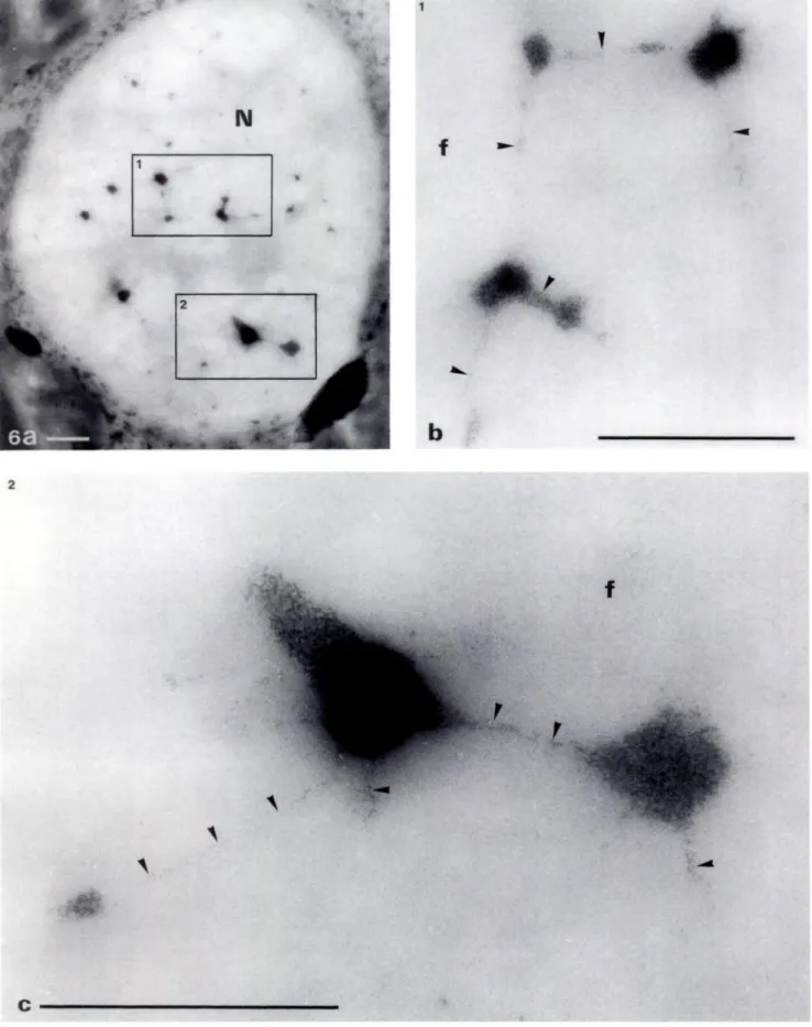
Figure 4. Semi-thin section of a maize nucleolus (N) stained with OA-B complex. One peripheral knob of the nucleolus-associated chromatin (NAC) is visible,
from which a continuous and multibranched DNA-containing structure intrudes into the nucleolus. The ramifications are shown by arrows. Original magnification x 22,000. Bar = 1 run.
LC
1500 MOTI’E, LOPPES, MENAGER, DELTOUR
DNA, the OA-B reaction was applied on semi-thin sections of
Lowicryl K4M-embedded root fragments. In this polar embedding resin, the OA-B complex stained the DNA-containing structures throughout all the semi-thin section thickness, as seen very well on the tilts of the metaphasic chromosomes (Figure 1). It must be noted that in Lowicryl K4M sections the OA reaction is specific
for the nucleic acids (12). However, the pre-treatment with
hydro-chlonic acid removes all the ribonucleic acids from the section. Differ-ent times of HCI hydrolysis (from 10-35 mm) were realized with-out modification ofthe specificity and quality ofthe staining (data not shown). When a DNAse digestion was performed, the
con-trast of DNA-containing structures disappeared (unpublished
results). We performed a pronase digestion before OA-B staining to strongly decrease the electron opac ity of the nucleolar section under the EM, which was mainly due to proteins. In our material
and working conditions very good staining ofchromatin fibers was obtained in Zea mays (a monocotyledon) as well as in Sinapis a/ba
(a dicotyledon) (Figures 2-6). After the OA staining on thick
5cc-tions the DNA was heavily stained in both the cytoplasmic organdIes
(mitochondnia and plastids) and the nucleus (Figures 2 and 5). In the nucleoplasm, irregular clumps ofchromatin, sometimes in con-tact with the nuclear envelope, were densely stained. From them, fine non-nucleosomal DNA filaments about 3 nm thick emerged and irradiated within interchromatin zones (data not shown). A large roundish space corresponding to the nucleolar body showed bleached areas surrounding some entities of higher electron opacity
containing contrasted DNA filaments. The bleached areas cor-respond to the dense fibnillar component, which is known to
con-tan a large amount ofproteins (25,28). The small areas surrounded
by the DFC correspond to the FCs. The granular component
ap-peared like interspersed clear gray and bleached zones. To unam-biguously identify the nucleolar subcomponents, we performed su-perimposition of successive ultra-thin sections stained with Un-Pb
and a thick section stained with the OA-B complex. Comparison of Figures 3a, 3b, and 3c clearly showed that the entities within
the DFC were FCs containing chromatin fibers. Moreover, the DNA localization was sometimes restricted to particular regions of the FCs. The DFC and the GC did not contain any detectable DNA. At higher magnification on ultra-thin sections stained with OA-B,
10-13-nm thick granules considered as nucleosomes were seen in the FCs (Motte et al. , manuscript in preparation). On some
5cc-lions we could see very well the continuity and the ramifications of the intranucleolar chromatin which is located in a complex net-work (Figures 4-6). The internal NAC appeared to be ramified as soon as it intruded within the nucleolus (Figure 4). The
ramifica-tions were often visible at the level of large masses of dense
chro-matin probably corresponding to the core of the heterogeneous FCs (Figure 6). In some rare sections, loops of internal NAC were observed (data not shown).
Immunocytochemical
Localization
of DNA
We localized the DNA by means ofa mouse monoclonal antibody directed against single- and double-stranded DNA. Immunolabel-ing was performed on ultra-thin Lowicryl sections. After the
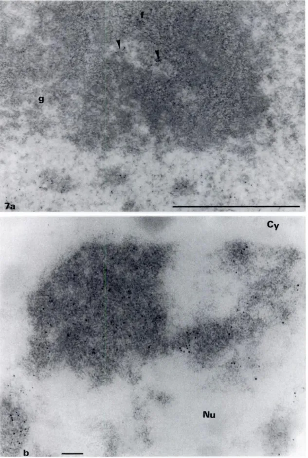
im-munoreactions gold particles were essentially observed over the dense chromatin in the nucleus (Figure 7) and over the clumps of dense NAC located at the nucleolar periphery (not shown). Large nucleoplasmic areas were devoid of labeling. Over the nucleolus, gold particles were present over electron-clear areas corresponding to FCs. On some sections the labeling was observed at the border ofthe FCs (Figure 7a). To compare the respective efficiency of the
OA-B complex and the anti-DNA antibody, we stained ultra-thin
Lowicryl sections by both methods. We observed that the gold par-tides were strictly located over structures stained with the osmium
ammine-B complex, but the density ofgold particles over the dense chromatin was weak (Figure 7b). This is due to the reaction of the anti-DNA antibody with only the DNA molecule at the surface level of the section, whereas the OA-B stains DNA included in the entire section thickness. Thus, the use of the anti-DNA antibody is insufficient to reveal the continuity of the intranucleolar chro-matin.
In
Situ
Hybridization
We performed in situ hybridization on ultra-thin Lowicryl sections ofquiescent maize embryos because the quiescent state offers cen-tan advantages: the NAC appears like two knobs adjacent to the periphery of the nucleolus, is very compact, and permits high
sig-nal/background labeling. Indeed, after the in situ hybridization we saw very well the gold particles concentrated in the region
con-responding to the NAC (Figure 8). Therefore, these knobs con-respond to the NORS which, as previously shown, stretch out and run inside the nucleolus during early germination.
Discussion
Localization ofDNA in the Interphase Nucleolus
Previous ultrastructunal and cytochemical studies have suggested the presence of DNA in the fibnillar component of plant and ani-mal nucleoli (1,17,28). More recently, by means ofthe OA reaction
Figure 5. (a) Electron micrograph of a nucleolus (N) of Sinapis alba, a dicotyledon. The dense fibrillar component (f) appears as large electron-opaque areas surrounding many small FCs (arrowheads). The granular component (g), which is more compact than in maize, envelopes the entire fibrillar component. (b) Semi-thin section of a nucleolus (N) ofSinapis a!be stained with OA-B complex. As in maize, one can clearly recognize the strongly bleached DFC (f) surrounding small FCs (arrowheads) which react positively with OA- The granular component (g) appears like a homogeneous clear gray zone. Note that, first, the GC is fre-quently observed in close contact with FCs (double arrows), and second, nucleolar vacuoles (long thin arrows) are present in both the DFC and GC. They appear as areas slightly more electron opaque than the GC. This is due to the intrinsic opacity ofthe acrylic resin, which is more abundant in this nucleolar subcomponent. In the DFC the vacuoles are always associated with the DNA internal to FCs. k is probably a karyosome. (C) High magnification of the three bottom right FCs (arrows) of b showing the DNA clumps located in the FCs. The DFC seems to be devoid of DNA. The contact between the GC (g) and the DNA internal to FCs is particularly well visible. Small threads of DNA (small arrowheads) originate from the large clumps. They are probably ramifications or interconnections between chromatin clumps, as more clearly observed in Figure 6. Original magnifications: a x 4700; b x 6000; c x 20,000. Bars: a,b 1 rim; C - 0.1 tm.
--
----.--- -.! f .2: b f t 44 1C
Figure 6. Semi-thin section of nucleolus (N) of Sinapis albe stained with OA-B complex. (a) The FCs form a complex network, the interconnections of which are small threads of DNA. (bc) High magnifications of the two nucleolar portions 1 and 2. The progressive stretching out of the compact DNA (arrows) is clearly apparent (arrowheads). Some ramifications are so thin that they are probably constituted of one or a few rDNA molecules. Original magnifications: a x 4000; b,c x 20,000. Bars - 1 tm.
-1
Figure 7. (a) Immunocytochemical localization of DNA on Lowicryl K4M sections of Zea maya using a monoclonal anti-DNA antibody. Uranyl staining. (a) View ofan FC surrounded by athick layer of DFC (f). Labeling is present in the FC (arrowheads). The DFC and the granular component(g) are devoid ofgold particles. The fibrillar texture ofthe DFC is well visible on this micrograph. (b) Portion of a nucleus ofZea maya labeled with anti-DNA antibody and stained with the OA-B complex. Note that the HCI hydrolysis does not remove the gold particles. If there is a good superimposition of the detection methods, the density of the gold particles is not sufficient to detect all the DNA and to delineate precisely the DNA-containing regions. In contrast, the latter can be strictly mapped by the OA-B
-: (_. . ..
.
‘: . ‘ ,. .. ,. ‘a . . .-.. N-_______________
1504 ., . . . . , -,.. . . . ‘. . .:-, :‘‘* ,..::.; ‘#{149}; -.. % 4.. . , . .... .. -.,..‘ . . - . .. ..,-. -- ,:
.., . “.. ._4_ ., . . ... . -:, .::.:‘.. - . . . . - .. . #{149}rMOTFE, LOPPES, MENAGER, DELTOUR
Figure 8. In situ hybridization on protease-ANAse-digested Lowicryl section of a quiescent maize nucleolus (N). The gold particles are restricted to the peripheral knob of the nucleolus-associated chromatin (arrows) in close association with fibers of DNA. The two knobs observed at the periphery of the maize nucleolus correspond to the two nucleolus-organizer regions present in this species. Original magnification x 20,000. Bar = 1 im.
on animal tissues, Derenzini et al. (13) have established that DNA is present within the FCs and probably in the DFC, and is con-stituted ofextended non-nucleosomal 3-nm thick filaments.
How-ever, after the OA reaction the identification of the FCs and tight DFC is not reliable in animal nucleoli. On the basis of qualitative (9,17) and quantitative (15) high-resolution autonadiognaphic
studies, it has generally been admitted that the DFC corresponds to the transcniptionally active pant of the interphase NOR.
How-ever, autoradiography can be criticized because a displacement of
labeled primary rRNA transcripts could rapidly occur and/or the labeling could also result from 55 nRNA synthesized elsewhere in the nucleus and rapidly transferred into the DFC (44). In this con-nection, some recent biochemical studies have disclosed that the
intengene spacer is transcribed in various animal cells (Xenopus,
Drosophis, mammalian species)(3,7,14,20,22,45). The spacer
tran-scripts are highly unstable in vivo and a rapid processing and degna-dation of them is likely to occur (20,45). Therefore, one can not state with certainty that DFC contains DNA. In addition, some recent immunocytochemical studies have led to challenge of this view. Scheer et al. (41,42) have detected the anti-RNA polymerase
I antibodies exclusively in the FCs of various cell lines. A similar nucleolan localization was also found for anti-topoisomerase I and anti-DNA antibodies (38,40,46). Benavente et al. (4,5), using a
different approach, support the idea that DFC is a genuine entity
not constituted by the superimposition of transcriptional units of rDNA and primary transcripts. The rDNA localization with in situ
7. Cassidy BG, Yang-Yen H-F, Rothblum LI: Additional RNA
polymer-hybridization techniques in active animal nucleoli provides con-troversial results (47,51). Th.ken together, these results cast doubts
on the localization of the primary sites of transcription in the DFC. In the present study, we detect DNA mainly in the FCs of inter-phase plant nucleoli. The penetration ofthe OA complex through-out the section thickness shows any very weak quantity of DNA and thus allows study of the 3-D organization of DNA on thick
sections. On semi-thin sections digested by pronase and stained by OA, the nucleolar subcomponents are well discriminated and recognizable by their electron opacity. The nucleolar subcompo-nents appear like a negative micrognaph of an ultra-thin section stained with uranyl and lead. This was confirmed by the supenim-position ofsuccessive ultra-thin sections and OA-stained thick sec-lion. It is beyond doubt that DNA chromatin fibers are located in FCs. A potential problem in the interpretation of our results is the possibility that the processes of the OA staining would spe-cifically destroy the DNA fibers contained in the DFC. Since the OA complex stains very small DNA amounts in the FCs, in the nucleoplasm, and in the cytoplasmic organdIes, it follows that if
DNA is present in the DFC it should be structurally different and more flimsy than the other DNA fibers. Possibly this could occur following the pronase digestion. However, because we know of no data reporting such observations, this process appears to us highly improbable. Therefore, our results support the studies suggesting
that rDNA is localized only in FCs in higher plant (36,37) as well
as in animal cells (2,38,40-42,46).
Spatial Organization of the
Intranucleolar
Chromatin
Early cytological studies on the nucleolus have already described, in light microscopy, an intranucleolan filamentous structure
gener-ally called the “nucleolonema” (see 11 for references). These
filamen-tous structures are detected in animal cells after drug treatment (16,18,26,39) or after removal ofarginine from the culture medium (19), and in plant cells after treatment of isolated nuclei with a detergent (23,24). The nucleolonema may also appear as a natural structure in the epidermal cells of Calpodes in relation to the con-tent ofhemolymph ecdysteroid (27). In situ hybridization has shown that in human glioma cells, nibosomal genes form intranucleolar strands 0.2 j.tm thick (29).
By means of 3-D neconstructions of nucleoli from serial
ultra-thin sections in the two species studied here, we have previously shown that the nucleolonema is ramified and appears as an arbores-cent structure mainly composed of fibnillar material, the axis of which appeared in transverse or oblique sections as homogeneous and heterogeneous FCs (10,32). However, we had no information on the localization of DNA and nibosomal genes in this filamen-tous structure. In the present study, we have shown a defined 3-D organization of the intranucleolan chromatin which forms a very complex filamentous structure within an active higher plant cell nucleolus. This structure is continuous and ramified. In 3-D recon-structions it was sometimes difficult to follow the continuity of the FCs. Indeed, we see very clearly on thick sections stained with the OA complex that the axis of the nucleolonema (containing DNA) can be so thin at some places (Figures 6 and 7) that it cannot be visible on ultra-thin sections stained with Un-Pb. This led to the
conclusion that the FCs are discrete entities. The body of the above data confirm here that each nucleolonema is fixed by its base to an interphase NOR-bearing chromosome from which it is a lateral
extension.
The nucleolonemata probably contain the nibosomal genes since,
during early germination, each of them comes from the stretching out and the intrusion within the nucleolus of the two peripheral
knobs in which in situ hybridization has disclosed the presence of all the rDNA. These peripheral knobs correspond to the NORS. The two NORS adjacent to the nucleolar periphery observed in quiescent maize cells are still partially visible in the transcription-ally active nucleolus. They probably correspond to transcription-ally inactive rDNA of the heterochnomatic segment of the
inter-phase NOR not involved in the formation of the nucleolus
(32,34,36,37). To our knowledge, this is the first time that in dcc-tron microscopy a very ordered spatial distribution of intranucleo-Ian chromatin is described in situ in a particular nucleolar subcom-ponent, the FCs. As previously shown (9), the FCs of maize and
Sinapis nucleoli are small entities (between 0.22-0.58 tm in
di-ameter), although the transcription units ofthese two species
mea-sure respectively 1.7 and 1.9 .tm. Moreover, the transcniptionally
active ribosomal genes in one nucleolus could number several hun-dred (34). This means that the rDNA should be highly compacted in the FCs. In animal cells, Tn#{246}stenet al. (49) have observed that these genes are compacted by a factor of 4.
Since we have observed arborescent nucleolonemata which some-times present loops in two evolutionary distant plant embryos, we can propose that the nucleolonematal organization is general for higher plants.
Acknowledgments
We thank Dr G. Feix (Institut f#{252}rBiologic III der Univers:tdt, Freiburg,
Germany) for the pMrl plasmidand Ms M. Dejxe forpatiently typing
the various successive versions ofthis manuscript. We also thank Ms F Motte-Toilet and Mr Gerstmans (Dept. of Chemistry, University ofLilge, Bel
gium) for help in the preparation ofthe osmium ammineB complex.
Literature
Cited
1. Anteunis M, Pouchelet M, Gansmullcr A, Robineaux R: The spatial
organization ofnucleolar DNA in phytohemagglutinin-stimulated
lym-phocytes of the guinea pig. Cell Tissue Res 235:65, 1984
2. Arroua ML, Hartung M, Devictor M. Bergc-Lefranc)L, Stahl A:
Locali-sation of nibosomal genes by in situ hybridization in the fibrillar centre
of the nucleolus in the human spermatocyte. Biol Cell 44:337, 1982 3. Bateman E, Paule MR: Promoter occlusion during ribosomal RNA
tran-scription. Cell 54:985, 1988
4. Benavente R, Reimer G, Rose KM. HUgle-Dorr B, Scheer U: Nuclco-lar changes after microinjection of antibodies to RNA polymerase I into the nucleus of mammalian cells. Chromosoma 97:115, 1988
5. Benavente R, Rose KM, Reimer G, Hugle-Dorr B, Scheer U: Inhibi-ion of nucleolar reformation after microinjection of antibodies to RNA polymerase I into mitotic cells. ) Cell Biol 105:1483, 1987
6. Canlemalm E, Ganavito RM, Villiger W: Resin development for dcc-tron microscopy and an analysis of embedding at low temperature.) Microsc 126:123, 1982
1506 MOTI’E, LOPPES, MENAGER, DEL1DUR
32. Motte P, Deltour R, Mosen H, Bronchart R: Three-dimensional dcc-ase I initiation site within the nontranscribed spacer region of the rat rRNA gene. Mol Cell Biol 7:2388, 1987
8. Comings DE: Arrangement ofchromatin in the nucleus. Hum Genet 53:131, 1980
9. Deltour R, Mosen H: Proposals for the macromolecular organization of the higher plant nucleolonema. Biol Cell 60:75, 1987
10. Deltour R, Mosen H, Bronchart R: Three-dimensional electron
micros-copy ofthc internal nucleolus-associated chromatin and ofthc nuclco-lar vacuoles during early germination of Sinapis alba. )Cell Sci 82:53, 1986
1 1. Deltour R, Motte P: The nucleolonema of plant and animal cells: a comparison. Biol Cell 68:5, 1990
12. Derenzini M, Farabegoli F: Selective staining ofnuclcic acids by
osmium-ammine complex in thin sections from lowicryl-embedded samples.
J
Histochem Cytochem 38:1495, 199013. Dcrenzini M, Hernandez-Vcrdun D, Farabegoli F, Pession A, Novello
F: Structure of ribosomal genes of mammalian cells in situ.
Chromo-soma 95:63, 1987
14. Dc Winter RFJ, Moss T The ribosomal spacer in Xcnopus iacvis is
tran-scnibed as part ofthe primary ribosomal RNA. Nudeic Acids Res 14:6041, 1986
15. Dupuy-Coin AM, P#{233}busquc MJ, SeIte R, Bouteille M: Localization of transcription in nucleoli of rat sympathetic neurons. A quantitative
ultrastructural autoradiography study.
J
Submicrosc Cytol 18:21, 198616. Ghosh 5, Lettr#{233}R, Ghosh I: On the decomposition on the nucleolus with special reference to its filamentous structure. Z Zellforsch 101:254, 1969
17. Goessens G: Nucleolar structure. mt Rev Cytol 87:107, 1984 18. Granick D: Nucleolar necklaces in chick embryo fibroblast cells. II.
Microscope observations ofthe effrct ofadenosine analogues on nucleolar necklace formation. ) Cell Biol 65:418, 1975
19. Granick 5, Granick D: Nucleolar necklaces in chick embryo myoblasts
formed by lack of arginine.
J
Cell Biol 51:636, 197120. Harnington CA, Chikaraishi DM: Transcription ofspaccrsequenccs flank-ing the rat 455 nibosomal DNA gene. Mol Ccii Biol 7:314, 1987 21. Hilliker A), Trusis-Coulter SN: Analysis ofthe functional significance
of linkage group conservation in Drosophila. Genetics 117:233, 1987 22. Labhart P. Reeder RH: Characterization of three sites of RNA 3’ end formation in the Xenopus nibosomal gene spacer. Cell 45:431, 1986 23. La Cour LF: The internal structure ofnucleoli. In Darlington CD, Lewis
KP, eds. Chromosomes today. London, Oliver and Boyd, 1966, 150 24. La Cour LF, Wells B: The loops and ultnastructure of the nucleolus of
Ipheon uniflorum. Z Zellforsch 82:25, 1967
25. Lafontainc)G, Lord A: An ultrastructural and radioautographic
inves-ligation ofthe nucleolonemal component ofplant interphase nucleoli.
J
Cell Sri 12:369, 197326. Lettr#{233}R, Siebs W, Pawelctz N: Morphological observations on the nucleolus of cells in tissue culture with special regard to its composi-tion. NatI Cancer Inst Monogr 23:107, 1966
27. Locke M, Leung H: Nucleolar necklace formation in response to
hemo-lymph ecdysteroid peaks. Tissue Cell 17:589, 1985
28. Luck BT, LafontaineJG: An ultracytochemical study ofnucleolar orga-nization in menistematic plant cells (Allium porrum).) Cell Sci 43:37,
1980
29. Manuelidis L: Indications ofcentromere movement during interphase
and differentiation. Ann NY Acad Sci 450:205, 1985
30. Manuelidis L: Individual interphase chromosome domains revealed by
in situ hybridization. Hum Genet 71:288, 1985
31. Miller OUr: The nucleolus, chromosomes, and visualization of genetic
activity.
J
Cell Biol 91:155, 1981ion microscopy of the nucleolus and nucleolus-associated chromatin
(NAC) during early germination ofZea mays L. Biol Cell 62:65, 1988
33. Olins AL, Moyer BA, Kim SH, Allison DP: Synthesis of a more stable
osmium ammine electron-dense DNA stain. ) Histochem Cytochem
37:395, 1989
34. Phillips RI, McMullen MD, Enomoto 5, Rubenstein I: Ribosomal DNA in maize. In Gustafson )P, Apels R, eds. Chromosome structure and function. New York, Plenum Publishing, 1988, 201
35. Ploton D, Beorchia A, M#{233}nagerM,)eannesson P, Adnet)J: The three-dimensional ultrastructure of interphasic and metaphasic nucleolar ar-gyrophilic components studied with high-voltage electron microscopy in thick sections. Biol Cell 59:113, 1987
36. Rawlins DJ, Shaw P): Localization of ribosomal and telomenic DNA sequences in intact plant nuclei by in-situ hybridization and three-dimensional optical microscopy. ) Microsc 157:83, 1990
37. Rawlins D), Shaw P): Three-dimensional organization ofnbosomal DNA
in interphase nuclei ofPisum sativum by in situ hybridization and op.
tical tomography. Chromosoma 99:143, 1990
38. Scheer U, Benavente R: Functional and dynamic aspects of the mam-malian nucleolus. BioEssays 12:14, 1990
39. Scheer U, Huglc B, Hazan R, Rose KM: Drug-induced dispersal of
tnan-scnibed rRNA genes and transcriptional products: immunolocalization and silver staining ofdiffrrent nucleolan components in rat cells treated
with 5,6-dichloro-13-D-nibofuranosylbenzimidazole.J Cell Biol 99:672,
1984
40. Scheer U, Messner K, Hazan R, Raska I, Hansmann P, Falk H, Spiess
E, Franke W: High sensitivity immunolocalization ofdouble- and single-stranded DNA by a monoclonal antibody. Eur) Cell Biol43:358, 1987
41. Scheer U, Raska I:Immunocytochemical localization OfRNA polymerase I in the fibnillar centers of nucleoli. In Stahl A, Luciani JM, Vagner-Capodano AM, eds. Chromosomes today. London, Allen &Unwin, 1987, 284
42. Scheer U, Rose KM: Localization ofRNA polymerase I in interphasic cells and mitotic chromosomes by light and electron microscopic im-munocytochemistry. Proc Nati Acad Sci USA 81:1431, 1984
43. Schwanzachen HG, Wachnier F: Nudeolus organizer regions and nudeoli:
cytological findings. In Stahl A, Luciani )M, Vagner-Capodano AM,
eds. Chromosomes today. London, Allen & Unwin, 1987, 253 44. Stcitz JA, Berg C, Hendrick )P, La Branche-Chabot H, Metspalu A,
Rinke J, Yario T: A 55 rRNA/L5 complex is a precursor to nibosome assembly in mammalian cells. ) Cell Biol 106:545, 1988
45. 1?autz D, Dover GA: Transcription of the tandem array of nibosomal DNA in Drosophila melanogasten does not terminate at any fixed point.
EMBO J 5:1267, 1986
46. Thiry M, Scheer U, Goessens G: Localization of DNA within Ehnlich
tumour cell nucleoli by immunoelectron microscopy. Biol Cell 63:27,
1988
47. Thiry M, Thiry-Blaise L: In situ hybridization at the electron micro-scope level: an improved method for precise localization of nibosomal DNA and RNA. Eur
J
Cell Biol 50:235, 198948. Toloczycki C, Feix G: Occurrence of 9 homologous repeat units in the
external spacer region ofa nudear maize rRNA gene unit. Nudeic Acids Res 14:4969, 1986
49. Tn#{246}sterH, Spring H, Meissner B, Shultz P. Oudet P, Trcndelenburg MF: Structural organization of an active, chromosomal nucleolar
or-ganizer region (NOR) identified by light microscopy, and subsequent STEM electron microscopy. Chromosoma 91:151, 1985
50. Van de Walle C, Bernier G, Deltoun R, Bronchart R: Sequence of
reac-tivation of nibonucleic acid synthesis during early germination of the
maize embryo. Plant Physiol 157:632, 1976
51. Wachtler F, Mosgollcr W, Schwarzacher HG: Electron microscopic in
situ hybridization and autoradiography: localization and transcription