HAL Id: inserm-01433144
https://www.hal.inserm.fr/inserm-01433144
Submitted on 12 Jan 2017HAL is a multi-disciplinary open access
archive for the deposit and dissemination of sci-entific research documents, whether they are pub-lished or not. The documents may come from teaching and research institutions in France or abroad, or from public or private research centers.
L’archive ouverte pluridisciplinaire HAL, est destinée au dépôt et à la diffusion de documents scientifiques de niveau recherche, publiés ou non, émanant des établissements d’enseignement et de recherche français ou étrangers, des laboratoires publics ou privés.
PPARbeta in macrophages and atherosclerosis
Giulia Chinetti-Gbaguidi, Bart Staels
To cite this version:
Giulia Chinetti-Gbaguidi, Bart Staels. PPARbeta in macrophages and atherosclerosis. Biochimie, Elsevier, 2017, �10.1016/j.biochi.2016.12.008�. �inserm-01433144�
PPAR in macrophages and atherosclerosis
G. Chinetti-Gbaguidi1 and B. Staels2
1 Université Côte d’Azur, CHU, CNRS, Inserm, IRCAN, France
2 Univ. Lille, Inserm, CHU Lille, Institut Pasteur de Lille, U1011, EGID, F-59000 Lille,
France
Corresponding author: Bart Staels
Inserm UR 1011, Institut Pasteur de Lille 1, rue du Professeur Calmette
BP 245, 59019 Lille, France Tel: +33-3-20-87-78-25 Fax: +33-3-20-87-73-60
ABSTRACT
Macrophages are central cells in the genesis and development of atherosclerosis, one of the major causes of cardiovascular diseases. Macrophages take up lipids (mainly cholesterol and triglycerides) from lipoproteins thus transforming into foam cells. Moreover, through the efflux pathway, macrophages are the main actors of the elimination of excessive tissue cholesterol toward extra-cellular acceptors. Macrophages participate in the control of inflammation by displaying different functional phenotypes, from the M1 pro-inflammatory to the M2 anti-inflammatory state.
The nuclear receptor Peroxisome Proliferator-Activated Receptor (PPAR) (also called PPARor PPAR) is expressed in macrophages where it plays a different role in the control of lipid metabolism, inflammation and phagocytosis of apoptotic cells.
This review will summarize our current understanding of how PPARregulates macrophage biology and its impact on atherosclerosis. Differences between studies and species-specific macrophage gene regulation will be discussed.
Keywords:
Highlights
- PPAR is expressed in macrophages
- PPAR controls the expression of genes involved in macrophage lipid handling - PPAR controls the macrophage inflammatory response
1. INTRODUCTION
A crucial step in atherosclerosis development is the migration of monocytes into the sub-endothelial space of large arteries where they differentiate into macrophages. Due to the expression at their surface of receptors such as the scavenger receptors A (SR-A) and CD36, macrophages accumulate lipids thus forming foam cells, leading to lipid deposition within the atherosclerotic lesions [1].
The net lipid accumulation in macrophages is the result of a balance between lipid uptake and elimination of the excess of cholesterol through the efflux, a pathway mediated by specific surface membrane receptors, such as the adenosine triphosphate (ATP)-binding cassette transporters (ABC)A1, ABCG1, ABCG4. The mobilization of cholesterol from intracellular pools to the plasma membrane determines its availability for further storage in lipid droplets or efflux to extracellular acceptors, such as high density lipoproteins (HDL) and their major apolipoproteins (apo), apoAI and apoE [2].
Beside their role in cholesterol and lipid handling and metabolism, macrophages are also involved in the control of the inflammatory status of the lesions. Indeed, while atherosclerosis has been considered for a long time a chronic inflammatory disease, recent data indicate that this can also be the consequence of an imbalanced anti-inflammatory response within the lesion [3]. In fact, macrophages do not constitute a homogeneous population. Very schematically, at least two populations of macrophages have been described from an inflammatory point of view, the prevalence of which is dictated by the macrophage micro-environment. While Th1 cytokines (TNF, IFN, IL1..) and lipopolysaccharides drive the macrophages toward the acquisition of a M1 pro-inflammatory phenotype leading to the production of pro-inflammatory cytokines and mediators, the Th2 cytokines (IL-4 and IL-13) lead to the alternative M2 macrophage differentiation [4]. These M2 macrophages exert anti-inflammatory, reparative and angiogenic activities. These different macrophage subtypes have been identified in atherosclerotic lesions by measuring the expression of markers specific for each phenotype [3].
2. Actions of PPARon macrophages in vitro
PPAR is present in monocytes and its expression further increases during their in vitro differentiation into macrophages [5, 6]. Moreover, relative PPAR expression has been determined in the different macrophages sub-types. Thus, PPAR expression level is higher in human M1 and unpolarized macrophages when compared to human M2 macrophages
differentiated in the presence of IL-4 [7, 8]. However, incubation with IL-13, another M2 polarizing cytokine, increases PPAR expression in mouse macrophages [9]. Since both IL-4 and IL-13 signal through the STAT6 pathway [10] but lead to different effects in human and murine cells, these results suggest that the regulation of PPAR expression in M2 macrophages is probably species-specific. As we will discuss below, also from a functional point of view, PPAR activities often differ between mouse and human macrophages.
2.1. Effects on lipid accumulation and metabolism
The effects of PPARon foam cell formation have been studied by different groups using both human and mouse macrophages. The first observations indicated that PPARactivation by 3-propyl-4-(3-(3-trifluoromethyl-7-propyl-6-benz-[35]-isoxazoloxy)-propylthio)phenylacetic acid (compound F) increases lipid accumulation in human
macrophages treated with either serum or oxidized LDL (OxLDL) [5]. Compound F increases the expression of genes involved in lipid uptake and storage such as SRA, CD36 and adipophilin. Generation of PPAR over-expressing THP-1 cells confirmed the effects of PPAR on macrophage lipid accumulation [5]. However, when mouse macrophages are incubated in vitro with OxLDL, treatment with the PPARligand GW0742 did not modify OxLDL uptake, while OxLDL degradation slightly increased [11]. While the expression of CD36 was highly induced, SRA expression was not affected by the PPARligand. Treatment with the GW0742 ligand in vivo did not reduce total cholesterol accumulation in foam cell macrophages isolated directly from the peritoneal cavity of LDLR-KO mice fed a high cholesterol diet, even though the levels of free cholesterol as well as triglycerides decreased [11]. Thus, the effects of PPARon cholesterol accumulation appear to be inconsistent, probably dependent on the model (in vitro or in vivo foam cell generation, ligand employed) as well as on the species (human vs mouse) Moreover, to date, no data are available concerning the actions of PPAR in the control of the intracellular cholesterol trafficking pathway, such as the transporters of the Niemann Pick type C (NPC) family.
PPAR activation by GW01516 in OxLDL-induced THP-1 foam cells increases the activity of sphingomyelin synthase (SMS)2, the last enzyme in the biosynthetic pathway of sphingomyelin and diacyl-glycerol, thus increasing the cell content of sphingomyelin [12]. SMS activity is positively correlated with the levels of sphingomyelin and diacyl-glycerol in lipid rafts thus contributing to cell lipid metabolism [12]. Since SMS expression and activity
have been associated with atherosclerosis, these observations suggest a potential negative effect of PPAR activation in macrophages on atherogenesis.
The effects of PPAR activation have been also studied in foam cell formation induced by VLDL, which leads to a strong accumulation of triglycerides (TG). Pre-treatment of human THP1 macrophages with GW01516 modestly, but significantly, reduced VLDL-derived TG accumulation [13]. Without changing lipoprotein lipase (LPL) mRNA expression, PPAR inhibits LPL activity by increasing the expression of angiopoietin-like protein 4 (Angptl4), a natural LPL inhibitor [13]. Interestingly, while PPAR ligands did not affect the expression of the VLDL receptor gene, its expression is strongly induced in PPAR KO macrophages, suggesting that this gene is either directly or indirectly suppressed by PPAR. PPAR does not regulate the expression of genes involved in VLDL/fatty acid uptake, such as apoB48 receptor or acyl-CoA synthetase. Furthermore, expression of both the chylomicron remnant receptor-1 (LRP1) and LDL-receptor (LDLR) was unaffected in PPAR KO macrophages [14]. Interestingly, macrophage PPAR is activated by VLDL-derived fatty acids released by LPL [14]. Indeed, transcriptional activation of PPAR by VLDL leads to the induction of adipophilin, which may potentially allow macrophages to store excess triglycerides, an effect completely lost in PPAR KO macrophages [14]. Moreover, activation of PPARby VLDL induces the expression of genes involved in peroxisomal and mitochondrial -oxidation as well as in carnitine biosynthesis [15]. PPAR regulates the expression of the transport secretion protein 2 (TTS2 or desnutrin) which acts as a lipase to increase fatty acid catabolism, thus modulating the availability of fatty acids from VLDL to activate PPAR. Interestingly, deletion of PPARderepresses expression of its target genes, a phenotype similar to that of ligand activation [15]. This suggests that unliganded PPARblocks fatty acid utilization by an active repressive mechanism that is reversed upon ligand activation. Finally, PPARligand upregulated CPT-1 gene expression and increased fatty acid -oxidation.
Collectively these data indicate that PPARreduced VLDL-induced TG accumulation by reducing VLDL TG hydrolysis and increasing fatty acid -oxidation and identify a transcriptional pathway mediated by PPAR activation through which dietary TG incorporated into VLDL can directly regulate gene expression in cells of the vessel wall. The
functional link between VLDL and PPAR appears as clear, making PPARa sensor for VLDL in macrophages (Figure 1)
2.2. Effects on cholesterol efflux
Treatment of human macrophages (primary and THP-1 cells) with the GW01516 compound increased the expression of the cholesterol transporter ABCA1 leading to the induction of apoAI-mediated cholesterol efflux [16, 17]. Similar results on ABCA1 and ABCG1 induction were obtained in THP-1 cells activated with GW0742 [18]. Controversially, treatment of THP-1 cells with compound F inhibited the expression of some cholesterol efflux genes, such as apoE and cholesterol-27-hydroxylase [5]. This conflicting result can be attributed to the modest selectivity of compound F on PPAR, compared to the higher selective GW01516 and GW0742 compounds. Moreover, it has been recently reported that both in human and murine macrophages, PPAR activation increases the expression of the phospholipid transfer protein (PLTP) [18], which can also contribute to the regulation of cholesterol efflux, since PLTP interacts and stabilizes ABCA1 thus enhancing cholesterol efflux [19]. However, regulation of ABCA1 and ABCG1 expression and ensuing apoAI-cholesterol efflux was not observed in mouse macrophages treated with GW0742 [11, 18] or GW01516 [20], thus highlighting a species-specific regulation of this functional process.
2.3. Effects on the inflammatory response and macrophage polarization
The effects of PPAR in the control of the macrophage inflammatory response have been largely studied. Investigations of the effects of synthetic PPARligands show an inhibitory effect on the expression of pro-inflammatory cytokines (iNOS, COX2, TNF) in murine macrophages by direct inhibition of NFB transactivation [21] as well as chemoattractant molecules such as MCP-1 and MCP-3 [20, 22]. However, bone marrow-derived macrophages from PPAR-deficient mice displayed reduced expression of pro-inflammatory genes such as MCP-1, IL-1 and MMP-9 [20]. Moreover, over-expression of PPARin RAW mouse macrophages increased the expression of MCP-1, MCP-3, MIP-1, IL-1 and MMP-9 compared to control cells, suggesting that PPARmay also exert pro-inflammatory properties [20, 22]. The global underlying molecular mechanism explaining these apparently contradictory observations is the fact that PPAR forms a complex with the transcriptional repressor protein B cell lymphoma 6 (Bcl6), thus preventing Bcl6 to repress cytokine genes [20]. Unliganded PPARthus has pro-inflammatory effects. However, when PPARis bound
to its ligand, Bcl6 is released from the complex, exerting anti-inflammatory activities. Indeed, ligand-activated PPARis anti-inflammatory [20] (Figure 2) More recently, global transcriptome analysis has been performed on human macrophages treated with the L165.041 ligand [6] confirming these observations. The results of this large scale study indicate that PPAR activation has predominantly anti-inflammatory effects (by reducing the expression of cytokines as well as components of the inflammasome complex). However, the study also indicated that PPAR activation can modulate the expression of anti-inflammatory and immune stimulatory genes [6]. This suggests that PPAR can exert context-dependent effects rather then purely inhibitory functions in immune regulation. Additionally, PPAR repressed the inflammatory response by targeting other signalling pathways. Indeed, activation of human macrophages by VLDL increased the expression of AP-1 dependent genes, such as ICAM-1, IL-1 and MIP1, via both Erk1/2, and AKT/FoxO1 dependent signalling mechanisms [14]. Treatment with synthetic PPARligands inhibited VLDL-induced pro-inflammatory cytokine expression by inhibiting VLDL-stimulated ERK1/2 activation and reversing VLDL-mediated inhibition of AKT/FoxO1 phosphorylation, by a mechanism independent of the stimulation of -oxidation or cellular TG reduction [14].
PPAR has been also investigated for its potential activity as regulator of macrophage polarization. Two important studies performed in mice show that PPAR bone marrow-specific deletion renders adipose tissue and liver macrophages unable of transition toward the M2 phenotype [9, 23]. This provokes inflammation and metabolic perturbations in adipocytes as well as hepatic dysfunction and systemic insulin resistance, respectively [9, 23]. However, when primary human monocytes are differentiated under M2 polarizing conditions, the presence of GW01516 during the differentiation period was without any effect on the expression of M2 markers [8]. Together, these results indicate that PPAR does not promote the M2 phenotype in human macrophages, contrary to what is observed in mouse macrophages. Moreover, in human atherosclerotic lesions no correlation is observed between the expression of PPARdid not correlate with M2 markers (CD206, CCL18, IL-10) [8].
2.4. Effects on efferocytosis
Later stages of atherosclerosis are characterized not only by the predominance of a pro- inflammatory status, but also by an inefficient clearance of apoptotic and dying cells (efferocytosis) by macrophages contributing to the aggravation of disease progression [24]. PPAR has been reported as a sensor of dying cells. Indeed, macrophage engulfment of
apoptotic cells increases and activates PPAR [25]. In turn, activation of PPARby GW0742 enhances the expression of opsonins, such as the complement component-1 (C1qa and C1qb), thrombospondin-1 and Mfge8, involved in bridging apoptotic cells to the macrophage surface receptor, thus increasing apoptotic cell clearance, both in human and mouse macrophages [25]. While opsonin regulation by PPAR also occurs in vivo in mice, whether this occurs in vivo in humans has to be clarified, especially since treatment of human macrophages with the PPAR ligand L165.041 decreased the uptake of fluorescent FITC-dextran, indicating a reduced phagocytotic activity [6]. Further studies are thus required to precisely understand the role of PPARin phagocytosis and efferocytosis.
3. Effects of PPARon atherosclerosis in vivo 3.1. Action of synthetic ligands
The effects of PPAR activation on atherosclerosis development have been extensively studied using different synthetic ligands.
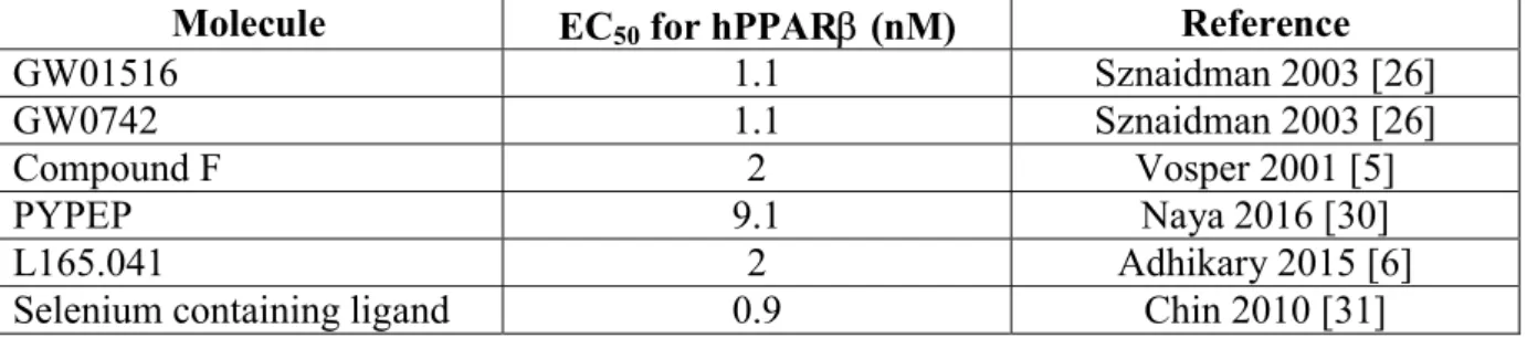
Activation of PPARby the potent synthetic ligand GW01516 (EC50: 1.1 nM) [26]
significantly reduced atherosclerotic lesion formation in apoE KO mice fed an atherogenic diet [22]. This effect was accompanied by an increase in circulating HDL levels and by a suppression of chemoattractant signalling in the aorta by down-regulation of chemokine expression levels. GW01516 administration in LDL-R KO mice on a high fat/high cholesterol diet markedly reduced aortic sinus lesions as well as the number of lesional macrophages [27]. Interestingly, GW01516 treatment also reduced the accumulation of cholesterol and TG within the lesions, in concert with the induction of CPT-1 and Angptl4, suggesting that GW01516 may stimulate aortic fatty acid -oxidation and inhibit aortic LPL activity, which could contribute to the reduced atherogenesis. Indeed, the expression of M1 markers (iNOS, TNF, CCL3, IL-6) was repressed, while the expression of the M2 marker arginase 1 (Arg1) is increased upon GW01516 treatment. However, while these results comfort the anti-inflammatory effects of PPAR ligands, only one marker of the M2 state was measured, rendering the conclusion that PPAR activation can orientate the macrophage toward an M2 phenotype in mouse atherosclerotic lesions in vivo preliminary.
Contradictory results have been obtained when another selective PPAR agonist GW0742 (EC50 : 1.1 nM, table 1) [26]was used. GW0742 reduced atherosclerosis by 30 % in female
LDLR-KO mice fed an atherogenic diet (0.25% cholesterol) [28] as well as in angiotensin II treated LDLR-KO mice [29]. However, the same ligand had no effect on male LDLR-KO
mice fed a 1.25% cholesterol atherogenic diet [11], whereas the expression of inflammatory genes (MCP-1, VCAM-1, ICAM-1, TNF) was reduced in the atherosclerotic lesions [11]. This suggests that the local anti-inflammatory activities of activated PPARare not sufficient to result in a net anti-atherogenic effect probably due to the extreme hypercholesterolemia in this experimental animal model.Overall, the differences between these studies could be due to the characteristics of the protocol designs (sex of animals, diet and drug dose). Moreover, in human apoB and CETP double transgenic mice fed an atherogenic diet (a model of human hypercholesterolemic dyslipidemia), oral administration of a novel PPAR agonist (PYPEP) reduced the progression of atherosclerotic lesions [30]. This was associated with increased serum cholesterol and ApoAI levels, concomitantly with a reduction in the non HDL-cholesterol and MCP-1 concentrations.
An isosteric selenoether-containing highly selective PPARligand (compound 1f, EC50 = 0.9
nM, table 1) attenuated atherosclerosis, increased HDL-C and suppressed the expression of ICAM-1 and IL-6 in aortic lesions [31].
Globally, in different atherosclerosis mouse models PPARactivation with different synthetic ligands did not result in significant changes in lesional or cultured macrophage mRNA expression levels of cholesterol metabolism genes (such as ABCA1, CD36, ABCG1), suggesting that the observed effects on atherosclerotic lesion size reduction are mainly related to a decrease of macrophage content and inflammatory status. This conclusion is further supported by the fact that GW01516 inhibits TNF-induced leukocyte rolling, adhesion, and emigration in the cremasteric microcirculation of C57BL6/SV129 mice demonstrated using intravital microscopy. Moreover, GW01516 reduced the expression of adhesion molecules (ICAM-1, VCAM-1, E-selectin) in the cremasteric post-capillary venules [32]. It remains to be understood why ligands with comparable selectivity on PPAR (such as GW01516 and GW0742, table 1) can induce opposite results in terms of atherosclerosis protection in mice. Explications reside probably in the different used methodologies: ligand concentration and administration ways, mouse models, duration of treatment.
3.2. Manipulation of PPAR expression levels
The role of PPAR in atherosclerosis has also been analysed by transplantation experiments of myeloid cells deficient for this transcription factor. Transplantation of LDLR-KO mice fed an atherogenic diet with PPAR-/-bone marrow resulted in > 50% reduction of aortic valve
lesion surface compared to wild type bone marrow transplanted mice [20]. These effects occur without modification of the plasma cholesterol and lipoprotein profiles, but are associated with a reduced expression of MCP-1 in aortic valve lesions.
More recently, a novel approach has been employed to decrease PPAR expression in myeloid cells. Transplantation of bone marrow cells infected with lentivirus expressing selective microRNA (miRNA) targeting PPAR into recipient LDLR-KO mice, resulted in a 35% reduction of aortic root atherosclerotic lesions, accompanied by a reduction of the number of macrophages present in the plaque [33]. Expression of MCP-1 and MMP-9 was also decreased in the atherosclerotic plaques of mice receiving the miRNA PPARbone marrow cells, whereas the level of plasma lipids did not change.
Comparison of the results obtained by PPARactivation with synthetic ligands with the findings obtained upon manipulating PPARexpression in myeloid cells leads to an apparent paradox. Indeed, both PPARactivation and deletion converge to a similar reduction of inflammation. As stated above (section 2.3), this can be explained by the ability of PPARto act as an inflammation switcher by binding or releasing the transcriptional repressor Bcl6, depending of the absence or the presence of a synthetic ligand, respectively [20] (Figure 2). In the absence of ligands PPARsequesters Bcl6 leading to inflammation, while in the presence of ligands PPARreleases Bcl6 which can exert anti-inflammatory properties. Whether additional mechanisms besides the PPAR/Bcl6 axis may contribute to explain this apparent paradox remains to be demonstrated.
CONCLUSION
Macrophages mainly act at two levels on the development of atherosclerosis: lipid uptake/metabolism and inflammation. While the effects of PPARon cholesterol accumulation and efflux are not completely coherent among the different studies, the functional link between PPARand VLDL metabolism / signalling appears more consistent. A global analysis of in vitro and in vivo studies using synthetic ligands identifies the activated PPARas an anti-inflammatory mediator with beneficial consequences for the development/evolution of atherosclerosis. However, studies using cells deleted for PPARindicate that in a genetically inactivated state this receptor displays pro-inflammatory properties. Altogether these data suggest that PPARcontrols an inflammatory switch in which both ligand activation and genetic receptor depletion lead toanti-inflammatory effects.
Whether PPARaffects alternative macrophage polarization in human cells and plaques, as well as in mouse atherosclerotic lesions, is still not completely clear.
It has to be kept in mind that species differences exist and have to be considered when concluding on the effect of PPARin macrophages and the consequences on disorders such as atherosclerosis. Continuing exploration of PPARsignalling and functions in macrophages, in particular in human cells, is necessary to identify novel mechanisms of action of PPAR which may lead to the development of potential approaches for human atherosclerosis.
ACKNOWLEDGMENTS
Grants from the Agence Nationale de la Recherche (AlMHA project), the Fondation de France, the Fondation pour la Recherche Médicale (DPC2011122981), the “European Genomic Institute for Diabetes” (EGID, ANR-10-LABX-46) and European Commission are acknowledged. B. Staels is a member of the Institut Universitaire de France.
FIGURE LEGENDS
Figure 1. PPARacts as VLDL sensor and controls VLDL metabolism in macrophages A. PPAR is activated by fatty acids (FA) released from VLDL by LPL. In turn, VLDL-activated PPARinduces the expression of genes involved in FA peroxisomal and mitochondrial -oxidation as well as in carnitine biosynthesis.
B. PPARreduces VLDL-induced TG accumulation by reducing VLDL TG hydrolysis (via the induction of Angptl4, a natural inhibitor of LPL) and by increasing fatty acid -oxidation, via the induction of CPT-1 expression.
Figure 2. Unliganded and liganded PPAR have different effects on inflammation
A. PPAR forms a complex with the transcriptional repressor Bcl6, thus preventing its repressive activity on pro-inflammatory genes. Unliganded PPARthus has pro-inflammatory activities.
B. When PPARisbound to its ligand, Bcl6 is released from the complex and subsequently exerts anti-inflammatory activities. Ligand activated PPARis thus anti-inflammatory, which contributes to the protective effects on atherosclerosis development observed in mice treated with PPARligands.
Declaration of interest Nothing to declare
REFERENCES
[1] Libby P, Aikawa M, Schonbeck U. Cholesterol and atherosclerosis. Biochim Biophys Acta 2000; 1529:299-309.
[2] Phillips MC. Molecular mechanisms of cellular cholesterol efflux. J Biol Chem 2014; 289:24020-24029.
[3] Chinetti-Gbaguidi G, Colin S, Staels B. Macrophage subsets in atherosclerosis. Nat Rev Cardiol 2015; 12:10-17.
[4] Gordon S, Taylor PR. Monocyte and macrophage heterogeneity. Nat Rev Immunol 2005; 5:953-964.
[5] Vosper H, Patel L, Graham TL et al. The Peroxisome Proliferator-activated Receptor promotes lipid accumulation in human macrophages. J Biol Chem 2001; 276:44258-44265. [6] Adhikary T, Wortmann A, Schumann T et al. The transcriptional PPARbeta/delta network in human macrophages defines a unique agonist-induced activation state. Nucleic Acids Res 2015; 43:5033-5051.
[7] Thulin P, Wei T, Werngren O et al. MicroRNA-9 regulates the expression of peroxisome proliferator-activated receptor delta in human monocytes during the inflammatory response. Int J Mol Med 2013; 31:1003-1010.
[8] Bouhlel MA, Brozek J, Derudas B et al. Unlike PPARgamma, PPARalpha or PPARbeta/delta activation does not promote human monocyte differentiation toward alternative macrophages. Biochem Biophys Res Commun 2009; 386:459-462.
[9] Kang K, Reilly SM, Karabacak V et al. Adipocyte-derived Th2 cytokines and myeloid PPARdelta regulate macrophage polarization and insulin sensitivity. Cell Metab 2008; 7:485-495.
[10] Rolling C, Treton D, Pellegrini S et al. IL4 and IL13 receptors share the gamma c chain and activate STAT6, STAT3 and STAT5 proteins in normal human B cells. FEBS Lett 1996; 393:53-56.
[11] Li AC, Binder CJ, Gutierrez A et al. Differential inhibition of macrophage foam-cell formation and atherosclerosis in mice by PPARalpha, beta/delta, and gamma. J Clin Invest 2004; 114:1564-1576.
[12] Mou D, Yang H, Qu C et al. Pharmacological activation of Peroxisome Proliferator-Activated Receptor {Delta} increases sphingomyelin synthase activity in THP-1 macrophage-derived foam cell. Inflammation 2016; 39:1538-1546.
[13] Bojic LA, Sawyez CG, Telford DE et al. Activation of peroxisome proliferator-activated receptor delta inhibits human macrophage foam cell formation and the inflammatory response induced by very low-density lipoprotein. Arterioscler Thromb Vasc Biol 2012; 32:2919-2928. [14] Chawla A, Lee CH, Barak Y et al. PPARdelta is a very low-density lipoprotein sensor in macrophages. Proc Natl Acad Sci USA 2003; 100:1268-1273.
[15] Lee CH, Kang K, Mehl IR et al. Peroxisome proliferator-activated receptor delta promotes very low-density lipoprotein-derived fatty acid catabolism in the macrophage. Proc Natl Acad Sci USA 2006; 103:2434-2439.
[16] Oliver WRJ, Shenk JL, Snaith MR et al. A selective peroxisome proliferator-activated receptor delta agonist promotes reverse cholesterol transport. Proc Natl Acad Sci USA 2001:5306-5311.
[17] Ogata M, Tsujita M, Hossain MA et al. On the mechanism for PPAR agonists to enhance ABCA1 gene expression. Atherosclerosis 2009; 205:413-419.
[18] Chehaibi K, Cedo L, Metso J et al. PPAR-beta/delta activation promotes phospholipid transfer protein expression. Biochem Pharmacol 2015; 94:101-108.
[19] Oram JF, Wolfbauer G, Vaughan AM et al. Phospholipid transfer protein interacts with and stabilizes ATP-binding cassette transporter A1 and enhances cholesterol efflux from cells. J Biol Chem 2003; 278:52379-52385.
[20] Lee CH, Chawla A, Urbiztondo N et al. Transcriptional repression of atherogenic inflammation: modulation by PPARdelta. Science 2003; 302:453-457.
[21] Welch JS, Ricote M, Akiyama TE et al. PPAR{gamma} and PPAR{delta} negatively regulate specific subsets of lipopolysaccharide and IFN-{gamma} target genes in macrophages. Proceedings of the National Academy of Sciences USA 2003; 100:6712-6717. [22] Barish GD, Atkins AR, Downes M et al. PPARdelta regulates multiple proinflammatory pathways to suppress atherosclerosis. Proc Natl Acad Sci U S A 2008; 105:4271-4276.
[23] Odegaard JI, Ricardo-Gonzalez RR, Red Eagle A et al. Alternative M2 activation of Kupffer cells by PPARdelta ameliorates obesity-induced insulin resistance. Cell Metab 2008; 7:496-507.
[24] Tabas I. Macrophage death and defective inflammation resolution in atherosclerosis. Nat Rev Immunol 2010; 10:36-46.
[25] Mukundan L, Odegaard JI, Morel CR et al. PPAR-delta senses and orchestrates clearance of apoptotic cells to promote tolerance. Nat Med 2009; 15:1266-1272.
[26] Sznaidman ML, Haffner CD, Maloney PR et al. Novel selective small molecule agonists for peroxisome proliferator-activated receptor delta (PPARdelta)--synthesis and biological activity. Bioorg Med Chem Lett 2003; 13:1517-1521.
[27] Bojic LA, Burke AC, Chhoker SS et al. Peroxisome proliferator-activated receptor delta agonist GW1516 attenuates diet-induced aortic inflammation, insulin resistance, and atherosclerosis in low-density lipoprotein receptor knockout mice. Arterioscler Thromb Vasc Biol 2014; 34:52-60.
[28] Graham TL, Mookherjee C, Suckling KE et al. The PPARdelta agonist GW0742X reduces atherosclerosis in LDLR(-/-) mice. Atherosclerosis 2005; 181:29-37.
[29] Takata Y, Liu J, Yin F et al. PPARdelta-mediated antiinflammatory mechanisms inhibit angiotensin II-accelerated atherosclerosis. Proc Natl Acad Sci USA 2008; 105:4277-4282. [30] Naya N, Fukao K, Nakamura A et al. A selective peroxisome proliferator-activated receptor delta agonist PYPEP suppresses atherosclerosis in association with improvement of the serum lipoprotein profiles in human apolipoprotein B100 and cholesteryl ester transfer protein double transgenic mice. Metabolism 2016; 65:16-25.
[31] Chin J, Hong JY, Lee J et al. Selective peroxisome proliferator-activated receptor delta isosteric selenium agonists as potent anti-atherogenic agents in vivo. Bioorg Med Chem Lett 2010; 20:7239-7242.
[32] Piqueras L, Sanz MJ, Perretti M et al. Activation of PPARbeta/delta inhibits leukocyte recruitment, cell adhesion molecule expression, and chemokine release. J Leukoc Biol 2009; 86:115-122.
[33] Li G, Chen C, Laing SD et al. Hematopoietic knockdown of PPARdelta reduces atherosclerosis in LDLR-/- mice. Gene Ther 2016; 23:78-85.
Table 1. Values of the EC50 for the PPARligands discussed in the review.
Molecule EC50 for hPPAR(nM) Reference
GW01516 1.1 Sznaidman 2003 [26]
GW0742 1.1 Sznaidman 2003 [26]
Compound F 2 Vosper 2001 [5]
PYPEP 9.1 Naya 2016 [30]
L165.041 2 Adhikary 2015 [6]
FA peroxisomal and mitochondrial -oxidation Figure 1
PPAR
Carnitine biosynthesis Fatty acids (FA)Cell membrane
LPL
VLDLPPAR
FA Cell membraneLPL
Angptl4 FA mitochondrial-oxidation Lipid dropletstorage
CPT-1 TG formation
Synthetic PPAR ligands
A
B
FA peroxisomal and mitochondrial -oxidation Figure 1PPAR
Carnitine biosynthesis Fatty acids (FA)Cell membrane
LPL
VLDLPPAR
FA Cell membraneLPL
Angptl4 FA mitochondrial-oxidation Lipid dropletstorage
CPT-1 TG formation
Synthetic PPAR ligands
A
B
Figure 1
PPAR
Carnitine biosynthesis Fatty acids (FA)
Cell membrane
LPL
VLDLPPAR
FA Cell membraneLPL
Angptl4 Angptl4 FA mitochondrial-oxidation Lipid dropletstorage CPT-1
CPT-1 TG formation
Synthetic PPAR ligands
Figure 2
Synthetic PPAR ligands
Pro-inflammatory effects
Reduced atherosclerosis in vivo
Pro-inflammatory genes Gene transcription
Anti-inflammatory effects
Pro-inflammatory genes Gene transcriptionX
X
PPAR
Bcl6
PPAR
Bcl6
A
B
X
Figure 2Synthetic PPAR ligands
Pro-inflammatory effects
Reduced atherosclerosis in vivo
Pro-inflammatory genes Gene transcription