HAL Id: tel-01713240
https://pastel.archives-ouvertes.fr/tel-01713240
Submitted on 20 Feb 2018HAL is a multi-disciplinary open access archive for the deposit and dissemination of sci-entific research documents, whether they are pub-lished or not. The documents may come from teaching and research institutions in France or abroad, or from public or private research centers.
L’archive ouverte pluridisciplinaire HAL, est destinée au dépôt et à la diffusion de documents scientifiques de niveau recherche, publiés ou non, émanant des établissements d’enseignement et de recherche français ou étrangers, des laboratoires publics ou privés.
Olga Petrova
To cite this version:
Olga Petrova. Regulation, activation, and deactivation of soluble guanylate cyclase and NO-sensors. Biochemistry, Molecular Biology. Université Paris Saclay (COmUE), 2017. English. �NNT : 2017SACLX113�. �tel-01713240�
Regulation, activation, and
deactivation of soluble guanylate
cyclase and NO-sensors
Thèse de doctorat de l'Université Paris-Saclaypréparée à l'Ecole Polytechnique
École doctorale n°573 : Interfaces : approches interdisciplinaires, fondements, applications et innovation.
Spécialité de doctorat : Biologie
Thèse présentée et soutenue à Palaiseau le 19 décembre 2017, par
Mme Olga Petrova
Composition du Jury :
Jérome SANTOLINI Président
Chercheur CEA – HDR
Laboratoire Stress Oxydant et Détoxication, CEA
François BONTEMS Rapporteur
Chercheur CNRS – HDR
Virologie Structurale, Institut Pasteur
Christophe GUIGNABERT Rapporteur
Chercheur INSERM – HDR
HAP: Physiopathologie et Innovation Thérapeutique, Univ. Paris-Sud
Hélène MUNIER-LEHMANN Examinatrice
Chercheuse Institut Pasteur – HDR
Chimie et Biocatalyse : Biochimie et Criblage, Institut Pasteur
Roberto MOTTERLINI Examinateur
Chercheur INSERM – HDR
Institut Mondor de Recherche Biomédicale
Michel NEGRERIE Directeur de thèse
Chercheur INSERM – HDR
Laboratoire d'Optique et Biosciences, Ecole Polytechnique
NT : 2 0 1 7 S A NN T : 2 0 1 7 S A CL X 1 1
Acknowledgements
___________________________________________________________________________
I want to thank many individuals whose kind support and help turned my work into this thesis.
First of all I would like to thank François Hache for approving my candidature as a doctoral student and paving the way for me to join the Laboratory for Optics and Biosciences.
I owe my deepest gratitude to my supervisor Michel Négrerie who offered me the possibility to work in this topic. I was lucky to be a student of Michel, as he is a good teacher and never stopped encouraging me on my way, in moments of disappoitment and of success. Without his continuous optimism concerning this work, enthusiasm, encouragement, and support this study would not have been completed.
I also express my warmest gratitude to Isabelle Lamarre who guided me through the technical issues of biochemistry and cell biology. Her contribution to my knowledge has been essential during this work. Jean-Christophe Lambry helped us in molecular dynamics simulation for what I am very grateful to him.
This thesis would not have been possible without our collaborators:
Jessica Wales (from the University of Arizona Cancer Center, Tucson (USA)) made an experiment with a common idea that helped us to interpret some results. Catherine Grillon and Fabienne Fasani (from Center for Molecular Biophysics, Orléans) helped us to design and perform the experiment with angiogenic cells. The proteins that we used in the experiments were purified and provided by Dr. Emil Martin (Internal Medicine, University of Texas, Houston, USA), Dr. Colin Andrew (Eastern Oregon University, USA) and Dr. Pierre Nioche (Université Paris Descartes, Laboratoire de Toxicologie et Signalisation Cellulaire). Special thanks to Dr. Colin Andrew and all coauthors for the joint effort that resulted in a publication and to Dr. Pierre Nioche for attending the seminar of my third year as a reviewer of my thesis. I would like to thank the committee presented at my defense Hélène Munier-Lehmann, Jérome Santolini, Christophe Guignabert, François Bontems and Roberto Moterlini for their rational criticism and advices.
It is a pleasure to thank those who made my life easier during my work in the LOB. Particularly, many thanks to Olivier Ramodiharilafy for his immense assistance in instructing me in the laboratory space. I could address him on all questions concerning what and where to find, whom to ask and how to use. He was constantly close to the student environment, always supporting us and providing advice.
Antigoni Alexandrou is very open and positive person, always was ready to help me on all issues. Thank you for discussing my work at the seminar and on pre-defense. Your comments helped me to improve the presentation.
Also thanks to Ursula Liebl, Hannu Myllykallio, Marten Vos, Chiara Stringari and Laura Antonucci for their valuable inputs.
It was a pleasure to communicate with Ernan Roitman and Pascal Preira. Pascal, thank you for your nice vocal skills: it was a pleasure to listen to you while we worked together in the biochemical lab.
I am grateful secretaries Christelle Francais and Laure Lachapelle for their help to foreign students to simplify life in the laboratory and in general in France.
I was happy to work in a wonderful team of creative and unique students: Pierre Sournia, Thuy Hoang, Marlen Kruse, Carolina Villamil Franco, Chao Yu, Xiujun Zheng, Bastian Geissler, Paul Stroe, Lamiae Abdeladim, Joséphine Morizet, Mayla Salman and Ravi Teja Raavi.
The greatest thanks to my close friends Lipsa Nag and Marco Schmid, I can not imagine my journey in the laboratory without them, they supported me in all my endeavors. And of course, I am grateful to my close russian friends Gulfina Frolova, Anna Karatovsaya and Ramil Allayarov for their significant contribution to my psychological condition during my stay in France, despite many kilometers between us.
I owe my deepest gratitude to my (grand) mother and father for having raised such daughter, and to my sister and brother (in law) thanks to whom I am in France.
Regulation, activation, and deactivation
of soluble guanylate cyclase and NO-sensors
CONTENT
Acknowledgements ... iii
Résumé ... 3
I – INTRODUCTION 1 – Signal transduction ... 5
2 – The NO – cGMP – sGC signaling pathway ... 5
3 – Bibliography ... 8
II – SOLUBLE GUANYLATE CYCLASE: THE ENDOGENOUS NITRIC OXIDE RECEPTOR 1 – Involvement of Guanylate cyclase in physiological processes ... 12
2 – Guanylate cyclase structure and molecular functioning ... 22
3 – Guanylate cyclase pharmacology ... 35
4 – Objectives of the present study ... 44
5 – Bibliography ... 45
III – SEARCHING AN EXOGENOUS INHIBITOR OF GUANYLATE CYCLASE 1 – Aims and Methodology ... 60
2 – Materials and methods... 60
3 – Results of the screening of natural compounds ... 65
4 – Interaction of inhibitors with sGC ... 75
5 – Analogues of the identified compounds ... 85
6 – Properties of the inhibitors ... 89
7 – Inhibition of angiogenesis ... 95
8 – Conclusion ... 97
9 – Bibliography ... 97
IV – THE MECHANISM OF ACTION OF sGC ACTIVATORS 1 – Methodology ... 102
2 – Materials and methods... 103
3 – Quantifying the activation ... 107
4 – Mechanism of action of sGC activators ... 113
6 – Bibliography ... 123
V – CONTROL OF DIATOMICS BINDING BY BACTERIAL NO-SENSORS 1 – Ligand dynamics ... 128
2 – Materials and methods... 130
3 – L16A mutant of the NO-sensor from Alcaligenes xylosoxydans ... 133
4 – Dioxygen binding to the NO-sensor from Thermoanaerobacter tengcongensis ... 142
5 – Conclusion ... 152
6 – Bibliography ... 152
VI – OVERVIEW AND PERSPECTIVES ... 157
VII – ANNEXES A – List of abbreviation... 161
B – Principles of Surface plasmon resonance imaging ... 163
C – Principles and methods of time-resolved absorption spectroscopy ... 166
D – Molecular dynamics simulation – CHARMM ... 170
E – Bibliography ... 172 ___________________________________________________________________________
Titre : Régulation, activation et désactivation de la guanylate cyclase soluble et de senseurs du NO.
Mots clés : Guanylate cyclase soluble, transduction du signal, nitric oxide, cyclic GMP, allostérie, activateurs et inhibiteurs de la guanylate cyclase, spectroscopie d'absorption transitoire.
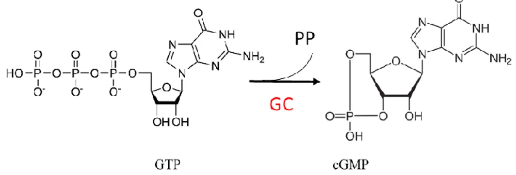
Résumé : Cette thèse est consacrée à la régulation de la guanylate cyclase soluble (sGC), le récepteur endogène du monoxyde d'azote (NO) chez les mammifères qui est impliqué dans la transduction du signal. La voie de signalisation NO/sGC/cGMP est impliquée dans de nombreux processus physiologiques et pathologiques, dont la régulation de la pression artérielle et l'angiogenèse associée aux tumeurs. L'enzyme sGC est activée par la fixation du NO sur son hème et catalyse la formation du cGMP à partir du GTP. Alors que la sGC est présente dans de nombreuses cellules de mammifères, le domaine hémique bactérien homologue (H-NOX) est impliqué dans la détection du NO et la régulation du métabolisme. Il n'existe à l'heure actuelle (janvier 2018) qu'un seul médicament qui cible la sGC : il s'agit d'un activateur utilisé pour traiter l'hypertension artérielle pulmonaire.
Le travail expérimental de cette thèse est constitué de trois parties :
1- La recherche d'inhibiteurs de la sGC.
2- L'étude du mécanisme d'activation de la sGC par la molécule médicament riociguat.
3- L'étude du contrôle de la dynamique du NO par des senseurs de NO bactériens.
_____________
Un objectif important de cette thèse est la découverte d'inhibiteurs de la sGC pour ralentir la progression tumorale. Le criblage de 300 composés naturels d'une chimiothèque, par mesure de l'activité de la sGC purifiée, a révélé six inhibiteurs actifs (IC50 = 0.2 – 1 µM). Deux autres composés naturels, l'hypéricine et l'hypocrelline, qui avaient été utilisés dans des cellules mais jamais avec la sGC purifiée, se sont avérés être des inhibiteurs. Ces huit molécules inhibent aussi la sGC cellulaire des cellules HUVEC en culture.
Par spectroscopie, nous montrons que deux inhibiteurs agissent en oxydant l'hème alors que les autres inhibiteurs sont des effecteurs allostériques qui ne se fixent ni sur l'hème, ni sur le site catalytique ou sur le site des activateurs Nous avons ainsi découvert une nouvelle classe de composés pharmacologiques ciblant la voie de signalisation NO/cGMP. Plusieurs de ces composés ont été testés sur des cultures de cellules endothéliales, modèles pour l'angiogenèse, qui développent des néo-vaisseaux. A une concentration de 10 µM l'hypéricine bloque l'angiogenèse de ces cellules modèles.
Nous avons ensuite étudié par spectroscopie d'absorption résolue en temps la transition structurale induite dans la sGC par l'activateur riociguat, un médicament indiqué pour l'hypertension artérielle pulmonaire, en synergie avec le CO. L'hypothèse de travail était que le riociguat facilite la rupture de la liaison Fe-His de l'hème, ce que nous ne pouvons pas montrer par spectroscopie d'absorption à l'équilibre du fait des spectres trop peu différenciés. L'idée directrice a été d'identifier les espèces moléculaires activées et non activées grâce à leur spectres transitoires après photo-dissociation du CO. Cette technique nous a permis de démontrer des changements de coordination de l'hème, notamment que le riociguat induit la rupture de la liaison Fe-His au niveau de l'hème, à l'instar du NO, l'activateur naturel de la sGC.
Deux états allostériques distincts de la sGC induits par le riociguat existent en présence du CO ayant les coordinations 6c-hème et 5c-hème. Nous montrons que ces activateurs allostériques possèdent une efficacité différente indépendamment de leur affinité pour la sGC. Cette efficacité peut être calculée par le rapport des espèces transitoires mesurées.
Université Paris-Saclay
Espace Technologique / Immeuble Discovery
Route de l’Orme aux Merisiers RD 128 / 91190 Saint-Aubin, France
Nous avons évalué le composé naturel isoliquiritigénine qui est commercialisé comme activateur de la sGC mais n'a jamais été testé sur la sGC purifiée. Nous montrons qu'il s'agit en réalité d'un inhibiteur de la sGC, aussi bien purifiée que dans des cellules HUVEC. Une hypothèse est l'activation de la phospho-diestérase PDE5 qui dégrade le cGMP produit par la sGC, faisant ainsi apparaître une apparente activation de la sGC.
_____________
Dans un second projet consacré au contrôle de l'affinité des gaz par les senseurs du NO, nous avons mesuré la dynamique des ligands diatomiques CO, NO et O2 sur 12 ordres de
grandeur temporelle pour le type sauvage et un mutant du transporteur bactérien du NO, le cytochrome c' de Alcaligenes xyloxosidans (AXCP). Remarquablement, la simple mutation Leu16Ala augmente l'affinité pour le CO d'un facteur 108, celle du NO 106, et rend cette
protéine réactive à O2. Ces affinités pour l'hème
de L16A-AXCP sont les plus élevées jamais mesurées.
Dans le cas de CO et NO, la recombinaison géminée après photodissociation a une amplitude de ~100 % tandis que la recombinaison bimoléculaire n'est pas détectable, ce qui indique que ces deux ligands diatomiques ne peuvent pas sortir de la protéine. Des simulations de dynamique moléculaire ont démontré que le CO dissocié est contraint de rester à 4 Å du Fe2+ dans le mutant L16A du fait
d'une rotation d'un groupe propionate de l'hème. Cette conformation est induite par la chaîne Ala16, contrairement au type sauvage Leu16, et affecte les trois ligands de l'hème CO, NO et O2.
Cette contrainte augmente considérablement la probabilité de liaison de CO et NO au fer de l'hème. Ce mécanisme constitue un nouveau mode de contrôle de l'affinité d'un ligand diatomique.
_____________
La protéine senseur H-NOX de la bactérie
Thermoanaerobacter tengcongensis (Tt
H-NOX) est homologue au domaine senseur hémique de la sGC et son rôle n'est pas connu, bien qu'elle fixe avec une très forte affinité le NO et O2. Pour comprendre son fonctionnement
en présence de ces deux ligands, nous avons mesuré la dynamique de O2 après
photo-dissociation par spectroscopie d'absorption transitoire sur 12 ordres de grandeur temporelle, en variant la concentration de O2.
Nous avons mis en évidence deux processus : la photo-oxydation de l'hème à l'échelle picoseconde après la photo-dissociation de O2
est prépondérante par rapport à la simple recombinaison de O2. Cependant l'hème revient
à son état réduit à l'échelle µs. Cette réduction a lieu simultanément à la recombinaison bimoléculaire de O2 (5 µs). L'accepteur
d'électrons dans la poche de l'hème reste à identifier. Ainsi la conformation particulière de l'hème de la protéine senseur Tt H-NOX, notamment sa distortion, lui confère des propriétés redox que ne possèdent pas les autres protéines homologues.
Ces résultats confirment l'hypothèse que Tt H-NOX n'est sans doute pas un senseur de O2 ou
de NO stricto sensu mais un senseur redox. _____________
Nous nous sommes intéressés à la régulation de la guanylate cyclase et des senseurs de NO, en nous efforçant de faire le lien entre les propriétés biochimiques des protéines et de leurs effecteurs allostériques avec leur comportement dynamique, notamment les changement de coordination de l'hème qui constituent un relai intramoléculaire à la tranduction du signal.
CHAPTER I
INTRODUCTION1 – Signal transduction
The capability of cells to receive various signals and to respond accordingly is an essential property of multicellular organisms. Signals arriving from outside the cell regulate processes that determine cell survival, their ability for division and differentiation, functional activity or death. Various molecular events and biochemical transformations occur within the cell when triggered by external chemical or physical signals.
Signals are induced by the binding of first messengers such as extracellular factors, hormones, cytokines or neurotransmitters to specific receptors that can either be allosteric transmembrane proteins or channels which, upon ligand interaction allow signals to pass in the form of small ion movement.
Complex signal transduction involves the coupling of ligand-receptor interactions at the cell surface to many intracellular events involving specialized proteins. These events imply the formation of second messengers such as (not exhaustively) 3',5'-cyclic adenosine monophosphate (cAMP), 3′,5′-cyclic guanosine monophosphate (cGMP), inositol trisphosphate, diacylglycerol, the gaseous molecules NO, CO and H2S. Second messengers
may activate phosphorylation of tyrosine kinases, serine, or threonine kinases that change conformation and activate downstream proteins in the signaling chain that leads to the formation of the primary or secondary cell response.
In the last two decades, signal transduction in pathological processes has become one of the most important subject of modern drug research due to the numerous possibilities of identifying targets. Activation or inhibition of diverse signaling pathways at different steps leads to diverse physiological responses, such as cell proliferation, death, differentiation, and metabolism [1]. One major example of the actively studied signaling pathway is NO – cGMP signaling, which is involved in the regulation of many physiological and pathological processes. Violation of at least one of the intermediate link in this signaling pathway can lead to disorders such as cardio [2], immune [3], neurodegenerative [4, 5] and metabolic diseases [6].
2 – The NO – sGC – cGMP signaling pathway
Nitric oxide (NO) is a highly reactive gas produced by organisms, ranging from bacteria to humans, and is crucial in various biological functions [7]. NO participates in intercellular signaling in the central and peripheral nervous systems. NO inhibits the growth and
multiplication of many types of pathogenic organisms [8], inhibits mitosis of human cells [9], has an anticoagulant activity [10], impedes leukocyte adhesion to the vascular endothelium and prevents platelet aggregation. NO also promotes tracheal smooth muscle relaxation of bronchi [11] and blood vessels [12].
The first known physiological target of NO is the enzyme soluble guanylate cyclase (sGC), which contains a heme iron cofactor necessary for sGC activation. NO interacts not only with metal groups but also with thiols and oxides, affects proteins, lipids, sugars, and nucleic acids [13]. The preferred reactions depend on the concentration of the NO in cells and on the reaction rates. For example, the fastest reaction rates are observed with superoxide ions forming peroxynitrite (ONOO–), a powerful oxidant that can modify proteins and lipids by nitration. For some heme proteins, reaction rates of NO with the central heme iron can be very high. NO signaling is linked to the general redox state of the cell and can affect several signaling pathways. The ubiquitous action of NO explains why it has both protective and harmful effects. For example, in some cases, NO can promote or inhibit apoptosis [14], kill tumors, or increase the potential for metastasis or vascularization [15], or trigger ischaemic preconditioning in the heart and mediate effects of preconditioning [13, 16].
The generation of NO in human cells occurs through the enzymatic oxidation of L-arginine to citrulline catalyzed by NO-synthase. The endothelial NO-synthase (eNOS) is crucial to generating NO in endothelial cells of a cardiovascular system, which is responsible for vasorelaxation, replication of smooth-muscle cells and inhibition of platelet adhesion [17]. Production of NO by neuronal NO-synthase (nNOS) [18] was first discovered in neuronal cells as a neurotransmitter, particularly in nonadrenergic noncholinergic nerves [19]. In peripheral nerves, NO is an actor of the relaxation of vascular and non-vascular smooth muscles. It relaxes sphincters in the gut, mediates relaxation of the corpus cavernosum causing penile erection, relaxes the bladder and urethra and alters responses in airways [13]. Inducible NOS (iNOS) was first detected in the mechanism of macrophage phagocitosis [20]. iNOS is not expressed in healthy quiescent cells, but only in response to inflammatory stimuli. Once expressed, it generates larger amounts of NO than eNOS and nNOS and its activity is not dependent on intracellular calcium and activation of calmodulin, contrary to eNOS and nNOS.
Although the enzyme sGC and second messenger cGMP were identified in the 1960s [21, 22], the complete outline of the NO – cGMP signaling pathway has been understood only in the last 20 years. After the discovery of cAMP in 1958 [23] and its physiological role, scientists began searching other relevant nucleotides: 6 years later, cGMP was discovered in the urine of rats [24].
Guanylate cyclase was found in mammalian cells in 1969 [21], but only in 1977, it was shown that exogenous NO gas and NO donors like nitroglycerin and nitroprusside activate sGC from rat liver and bovine tracheal smooth muscle [25, 26]. In 1980 Furchgott
and Zawadzki demonstrated the existence of a substance produced by the endothelium that was required to mediate relaxation of blood vessels, the so-called "endothelium-derived relaxing factor" (EDRF) [27]. It was later determined that the substance EDRF is endogenous nitric oxide [28, 29]. Research into its functions led to the Nobel Prize in 1998 awarded to Robert F. Furchgott, Louis J. Ignarro, and Ferid Murad for discovering the role of nitric oxide as a cardiovascular signaling molecule [30-32].
The action of NO in vasodilation starts with NO synthesis by eNOS and its diffusion from endothelial cells through the membrane of smooth muscle cells to bind to NO-specific receptor sGC, leading to synthesis of the second messenger cGMP. After sGC activation, subsequent steps of signaling pathways take place triggered by increasing the concentration of cGMP. There are three known targets for cGMP, which mediate signal transduction: cGMP-dependent protein kinase [33], cGMP-gated ion channels [34], and cGMP-regulated phosphodiesterase [35], (described in CHAPTER II).
In this work, we are interested in the NO signaling pathway involving sGC, subsequent partner proteins, and second messengers intervening especially in blood vessel relaxation. The complete biochemistry of the NO signaling pathway will be described in CHAPTER II. The functioning of the endogenous NO-receptor sGC is crucial in several diseases and this thesis is focused on the activation and deactivation mechanisms of sGC and cognate NO-sensors at the molecular level.
The present thesis is organized as follows. CHAPTER II is devoted to the description of the action and the role of sGC, both at the cellular and the molecular levels. The literature on this subject is extremely large and our overview, albeit as complete as possible, is necessarily non-exhaustive. The end of CHAPTER II is devoted to a review of existing activators and inhibitors of sGC. For a better reading, we chose to describe the experimental methods at the beginning of each chapter, but deeper descriptions of particular methods, not necessary to understand the results, are placed at the end (CHAPTER VII). The discovery of new exogenous inhibitors of guanylate cyclase will be described in CHAPTER III. CHAPTER IV is devoted to the identification of the mechanism of action of allosteric sGC activators, one of which is already commercialized as a drug. The control of NO affinity and dynamics is crucial for homologous and non-homologous bacterial NO-sensors. Thus, a complete understanding of the NO-receptors functioning requires the study of the dynamics of diatomic binding (CO, NO, O2) to their heme site, that will be discussed in CHAPTER V. As a general conclusion, we
will link the dynamic properties of these proteins to their allosteric and biochemical properties.
3 – Bibliography
1. King, M. W. Mechanisms of Cellular Signal Transduction. Medcal Biochemistry 1996.
2. Buys, E.; Sips, P. New Insights into the Role of Soluble Guanylate Cyclase in Blood Pressure Regulation. Current opinion in nephrology and hypertension 2014, 23, 135-142.
3. Nagy, G.; Clark, J. M.; Buzás, E. I.; Gorman, C. L.; Cope, A. P. Nitric oxide, chronic inflammation and autoimmunity. Immunol Lett. 2007 111, 1-5.
4. Jaffrey, S. R.; Snyder, S. H. Nitric Oxide: A Neural Messenger. Annual Review of Cell and
Developmental Biology 1995, 11, 417-440.
5. Bredt, D. S.; Snyder, S. H. Nitric oxide: a physiologic messenger molecule. Annu Rev Biochem. 1994, 63, 175-195.
6. Chin, C.-H.; Tsai, F.-C.; Chen, S.-P.; Wang, K.-C.; Chang, C.-C.; Pai, M.-H.; Fong, T.-H. YC-1, a potent antithrombotic agent, induces lipolysis through the PKA pathway in rat visceral fat cells.
European Journal of Pharmacology 2012, 689, 1-7.
7. Rőszer, T. The Biology of Subcellular Nitric Oxide. 2012.
8. Hon, W. M.; Lee, K. H.; Khoo, H. E. Nitric Oxide in Liver Diseases. Annals of the New York
Academy of Sciences 2002, 962, 275-295.
9. Murillo-Carretero, M.; Ruano, M. J.; Matarredona, E. R.; Villalobo, A.; Estrada, C. Antiproliferative effect of nitric oxide on epidermal growth factor-responsive human neuroblastoma cells. J Neurochem 2002 83, 119-131.
10. Irokawa, M.; Nishinaga, M.; Ikeda, U.; Shinoda, Y.; Suematsu, M.; Gouda, N.; Ishimura, Y.; Shimada, K. Endothelial-derived nitric oxide preserves anticoagulant heparan sulfate expression in cultured porcine aortic endothelial cells. Atherosclerosis 1997, 135, 9-17.
11. Joos, G. F. Potential usefulness of inhibiting neural mechanisms in asthma. Monaldi Arch Chest
Dis 2000 55, 411-414.
12. Ignarro, L. J.; Cirino, G.; Casini, A.; Napoli, C. Nitric Oxide as a Signaling Molecule in the Vascular System: An Overview. Journal of Cardiovascular Pharmacology 1999, 34, 879-886.
13. Vallance, P.; Leiper, J. Blocking NO synthesis: how, where and why? Nat Rev Drug Discov 2002, 1, 939-950.
14. Moncada, S.; Erusalimsky, J. D. Does nitric oxide modulate mitochondrial energy generation and apoptosis? Nat Rev Mol Cell Biol 2002, 3, 214-220.
15. Lala, P. K.; Chakraborty, C. Role of nitric oxide in carcinogenesis and tumour progression. The
Lancet Oncology 2001, 2, 149-156.
16. Nandagopal, K.; Dawson, T. M.; Dawson, V. L. Critical Role for Nitric Oxide Signaling in Cardiac and Neuronal Ischemic Preconditioning and Tolerance. Journal of Pharmacology and
Experimental Therapeutics 2001, 297, 474-478.
17. Matthys, K. E.; Bult, H. Nitric oxide function in atherosclerosis. Mediators of Inflammation 1997, 6, 3-21.
18. Alderton, W. K.; Cooper, C. E.; Knowles, R. G. Nitric oxide synthases: structure, function and inhibition. Biochemical Journal 2001, 357, 593-615.
19. Garthwaite, J.; Charles, S. L.; Chess-Williams, R. Endothelium-derived relaxing factor release on activation of NMDA receptors suggests role as intercellular messenger in the brain. Nature 1988, 336, 385-8.
20. Hibbs, J. B.; Taintor, R. R.; Vavrin, Z.; Rachlin, E. M. Nitric oxide: A cytotoxic activated macrophage effector molecule. Biochemical and Biophysical Research Communications 1988, 157, 87-94.
21. White, A. A.; Aurbach, G. D. Detection of guanyl cyclase in mammalian tissues. Biochimica et
Biophysica Acta (BBA) - Enzymology 1969, 191, 686-697.
22. Smith, M.; Drummond, G. I.; Khorana, H. G. Cyclic Phosphates. IV.1 Ribonucleoside-3',5' Cyclic Phosphates. A General Method of Synthesis and Some Properties. Journal of the American
Chemical Society 1961, 83, 698-706.
23. Rall, T. W.; Sutherland, E. W. Formation of a cyclic adenine ribonucleotide by tissue particles. J.
Biol. Chem. 1958, 232, 1065–1076.
24. Ashman, D. F.; Lipton, R.; Melicow, M. M.; Price, T. D. Isolation of adenosine 3′,5′-monophosphate and guanosine 3′,5′-3′,5′-monophosphate from rat urine. Biochemical and Biophysical
Research Communications 1963, 11, 330-334.
25. Arnold, W. P.; Mittal, C. K.; Katsuki, S.; Murad, F. Nitric oxide activates guanylate cyclase and increases guanosine 3′:5′-cyclic monophosphate levels in various tissue preparations. Proceedings of
the National Academy of Sciences of the United States of America 1977, 74, 3203-3207.
26. Schultz, K.-D.; Schultz, K.; Schultz, G. Sodium nitroprusside and other smooth muscle-relaxants increase cyclic GMP levels in rat ductus deferens. Nature 1977, 265, 750-751.
27. Furchgott, R. F.; Zawadzki, J. V. The obligatory role of endothelial cells in the relaxation of arterial smooth muscle by acetylcholine. Nature 1980, 288(5789) 373-376.
28. Moncada, S.; Palmer, R. M.; Higgs, E. A. The discovery of nitric oxide as the endogenous nitrovasodilator. Hypertension 1988, 12, 365-372.
29. Ignarro, L. J.; Buga, G. M.; Wood, K. S.; Byrns, R. E.; Chaudhuri, G. Endothelium-derived relaxing factor produced and released from artery and vein is nitric oxide. Proceedings of the National
Academy of Sciences of the United States of America 1987, 84, 9265-9269.
30. Murad, F. Discovery of some of the biological effects of nitric oxide and its role in cell signaling (Nobel lecture). Angew. Chem. Inter. Ed. 1999, 38, 1857-1868.
31. Ignarro, L. J. Nitric Oxide: A Unique Endogenous Signaling Molecule in Vascular Biology (Nobel Lecture). Angew. Chem. Inter. Ed. 1999, 38, 1882–1892.
32. Furchgott, R. F. Endothelium-Derived Relaxing Factor: Discovery, Early Studies, and Identifcation as Nitric Oxide (Nobel Lecture). Angew. Chem. Inter. Ed. 1999, 38, 1870–1880.
33. Lohmann, S. M.; Vaandrager, A. B.; Smolenski, A.; Walter, U.; De Jonge, H. R. Distinct and specific functions of cGMP-dependent protein kinases. Trends in Biochemical Sciences 1997, 22, 307-312.
34. Zagotta, W. N.; Siegelbaum, S. A. Structure and Function of Cyclic Nucleotide-Gated Channels.
Annual Review of Neuroscience 1996, 19, 235-263.
35. Degerman, E.; Belfrage, P.; Manganiello, V. C. Structure, Localization, and Regulation of cGMP-inhibited Phosphodiesterase (PDE3). Journal of Biological Chemistry 1997, 272, 6823-6826.
CHAPTER II
SOLUBLE GUANYLATE CYCLASE:
THE ENDOGENOUS NITRIC OXIDE RECEPTOR SYNOPSIS
1 – Involvement of Guanylate cyclase in physiological processes... 12
1.1 – Smooth muscle relaxation ... 12
1.2 – Platelet aggregation and disaggregation ... 14
1.3 – Apoptosis ... 15
A – NO as a pro-apoptotic inducer ... 16
B – NO as an anti-apoptotic modulator ... 16
1.4 – Tumor progression and angiogenesis ... 17
1.5 – Proliferation and differentiation of cells ... 19
A – Inhibition of cell proliferation ... 19
B – Enhanced proliferation ... 19
C – Differentiation ... 20
1.6 – Neuronal communication ... 20
1.7 – Inflammation ... 21
2 – Guanylate cyclase structure and molecular functioning ... 22
2.1 – Structure of sGC ... 22
2.2 – The heme coordination ... 26
2.3 – Electronic and chemical properties of nitric oxide ... 29
2.4 – Guanylate cyclase biochemistry ... 31
2.4.1 – cGMP-dependent protein kinases ... 32
2.4.2 – Other targets of cGMP... 33
2.4.3 – Kinetics of GTP and NO binding to sGC ... 34
3 – Guanylate cyclase pharmacology ... 35
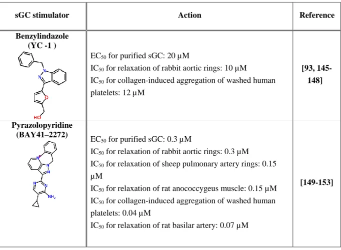
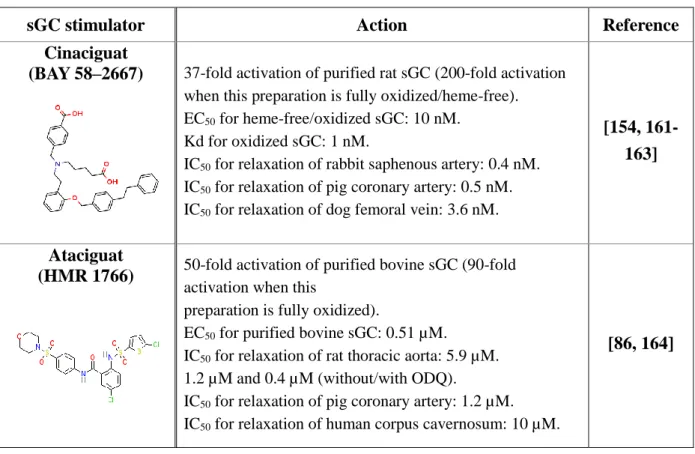
3.1 – Activators of Guanylate cyclase ... 35
3.1.1 – Heme-dependent sGC allosteric activators ... 35
3.1.2 – Heme-independent sGC activators ... 38
3.1.3 – Isoliquiritigenin ... 39
3.2 – Inhibitors of guanylate cyclase ... 40
3.2.1 – Heme-oxidizing inhibitors ... 40
3.2.2 – Inhibitors which bind to sGC catalytic domain ... 41
4 – Objectives of the present study ... 44
1 – Involvement of Guanylate cyclase in physiological processes
The soluble guanylate cyclase (sGC) is the primary nitric oxide (NO) receptor in mammals and the central component of the NO-signaling pathway. The functioning of this endogenous NO-receptor is crucial in several diseases and we focused here on the study of activation and deactivation mechanisms of sGC, which are directly linked with pathological processes.
sGC is located in many cells, tissues and organs. This fact indicates involvement of NO-sGC-cGMP signaling pathway in a wide regulation of intracellular metabolism: regulation of blood pressure (smooth muscle relaxation and vasodilation) [1, 2], platelet aggregation and disaggregation [3], apoptosis [4], signaling in tumor cell proliferation (angiogenesis) [5, 6], immune response [7], proliferation, and differentiation of cells [8], plasticity, communication between neurons [9, 10], and in inflammation [11, 12]. Because of its very wide involvement, we will describe in this first part the role of sGC in several (but not all) physiological processes, then its molecular properties, ending this part with the objectives of the present study. The experimental details are given at the beginning of each chapter.
1.1 – Smooth muscle relaxation
Smooth muscle relaxation (vasodilation) in cardiovascular system through the NO-sGC-cGMP pathway is a significant and historical feature of NO signaling [2, 13]. The research of NO as signaling molecule was conducted after the discovery of Endothelium-derived relaxing factor (EDRF) by Robert Furchgott in 1980 [14]. He showed the ability of endothelial cells to generate and release a labile substance that produced relaxation of the underlying smooth muscle in the vascular segment. Later, Ignarro and Murad conducted a series of experiments to determine whether EDRF released from artery could activate guanylate cyclase and thereby account for elevated cyclic GMP levels in response to acetylcholine or bradykinin [15, 16]. It was shown that activation of sGC by EDRF was heme-dependent, like its activation by nitric oxide. In 1988 the fact that EDRF is NO was revealed [15, 17].
Vasodilation signaling starts from the binding of first messengers (acetylcholine, bradykinin) present in blood flow to their receptors (G proteins) incorporated within the membrane of endothelial cells (EC, monocellular layer inside wall of the artery) (Figure 1). This binding causes activation of inositol trisphosphate (IP3) system, where IP3 binds to Ca2+ channels of the endoplasmic reticulum that releases Ca2+ ions to the cytosol. This event induces the binding of Ca2+ /calmodulin complex to the enzyme NO-synthase and triggers the synthesis of NO from L-arginine to L-citrulline. Then, NO very easily diffuses through the cell membrane to reach its sGC receptor in the smooth muscle cells surrounding the artery.
The sGC catalyzes the formation of cGMP from GTP leading to cGMP-dependent protein kinases (PKG) activation in smooth muscle cells. Phosphodiesterase 5 (PDE-5) is the enzyme which regulate intracellular cGMP level by catalyzing the hydrolytic cleavage of the 3′ phosphodiester bond of the cyclic nucleotides. PKG causes reuptake of Ca2+ and the opening of calcium-activated potassium channels in smooth muscle membrane. The fall in concentration of Ca2+ in the cytoplasm ensures that the myosin light-chain kinase can no longer phosphorylate the myosin molecule, thereby stopping the cross-bridge cycle and leading to relaxation of the smooth muscle cell [18].
Figure 1. Smooth muscle cell relaxation and subsequent vasodilation controlled by the NO – cGMP – sGC pathway. NO is produced in endothelial cells and diffuses through the cell membrane to the smooth-muscle cell to activate sGC. Then sGC catalysis cGMP formation from GTP, which activates in turn PKG and induces smooth muscle relaxation. IP3: inositol trisphosphate, NO: nitric oxide, sGC: soluble guanylate cyclase, PDE-5: phosphodiesterase 5. Modified scheme is taken from [19].
___________________________________________________________________________
Impairment of the NO-cGMP signaling pathway in arteries is implicated in cardiovascular diseases, including systemic arterial and pulmonary hypertension (PAH), coronary artery disease, peripheral vascular disease, and atherosclerosis [20, 21]. Pulmonary hypertension is an increase of blood pressure in lung vasculature (in the pulmonary artery, pulmonary vein, or pulmonary capillaries) due to narrowing of blood vessels connected to and within the lungs. The affected blood vessels become stiffer and thicker, in a process known as fibrosis. Unregulated vasoconstriction makes harder for the heart to pump blood through the lungs. The increased workload of the heart causes hypertrophy of the right ventricle, making the heart less able to pump blood through the lungs, and therefore, the right ventricular muscle cannot get enough oxygen to meet its needs [22].
The pathology of PAH results from pulmonary endothelial cell dysfunction or injury accompanied by dysregulation of various signaling pathways, including decreased production of NO, prostacyclin and increased levels of endothelin-1, thromboxane A2 and serotonin [23]. The oxidative stress can also lead to superoxide anions reaction with nitric oxide to generate peroxynitrite which oxidizes and uncouples eNOS thereby impairing NO synthesis and causing uncoupled eNOS to generate reactive oxygen species. Thus oxidative stress results in oxidation of heme group of sGC, which renders it less responsive to NO and can result in dissociation of heme from sGC [23]. Oxidative stress also leads to atherosclerosis, an inflammatory disease where arterial lesions are formed via a complex process involving platelet adhesion, leukocyte infiltration and activation and intimal migration and proliferation of smooth muscle cells.
1.2 – Platelet aggregation and disaggregation
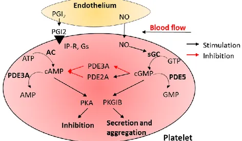
The NO-sGC-cGMP pathway in platelet activation has been investigated for more than 30 years. It appeared that NO and cGMP play both a stimulatory and an inhibitory role in platelet activation depending on cAMP activity [3, 24, 25]. The levels of free cytosolic cAMP and cGMP are controlled by their synthesis through adenylate cyclase (AC) and sGC (Figure 2).
Figure 2. NO-sGC-cGMP signaling pathway in platelets. The intact endothelium releases prostaglandin I2 (PGI2) and nitric oxide (NO) to activate adenylate cyclase (AC) and soluble guanylyl cyclase (sGC), leading to the formation of cAMP and cGMP, correspondingly. cAMP phosphorylates phosphokinase A (PKA), which inhibits granule release, adhesion, and aggregation of platelets, and cGMP phosphorylates phosphokinase G (PKG), which is responsible for secretion and aggregation of platelets. cGMP controls the activity of cAMP through inhibition of PDE3A and activation of PDE2A. Modified scheme is taken from [26].
___________________________________________________________________________
The intact endothelial cells inhibit platelet activation by producing nitric oxide, endothelial-ADPase and prostacyclin, where endothelial-ADPase degrades the platelet
activator ADP. Resting platelets maintain active calcium efflux via a cyclic AMP-activated calcium pump. Intracellular calcium concentration determines platelet activation status driving platelet conformational change and degranulation. The two signaling systems cAMP and cGMP work together for controlling inhibition or activation of granule release, adhesion, and aggregation of platelets.
Adenylate cyclase located in platelets is activated by G-protein-coupled receptor signaling. Binding of prostacyclin (prostaglandin I2) from endothelial cells to its IP receptor on the platelet surface activates the intracellular receptor-linked stimulatory G-protein (Gs). Gs is turned into its active GTP-bound form, Gs–GTP which binds to adenylate cyclase and stimulates the synthesis of cAMP from ATP. Increase of cAMP production promotes the efflux of calcium and reduces its intracellular availability for platelet activation.
Soluble guanylate cyclase is abundantly present in platelets and is activated by NO synthesized in platelets or diffused through the platelets membrane from endothelium. The second messenger cGMP synthetized by sGC from GTP activates the cGMP-dependent protein kinases G (PKG) and also inhibits phosphodiesterase 3 (PDE3), an enzyme that destroys cyclic adenosine monophosphate (cAMP). cAMP activates the protein kinase A (PKA), a known platelet aggregation inhibitor. Subsequent substrate phosphorylation results in the inactivation of small G-proteins of the Ras and Rho families, inhibition of the release of Ca2+ from intracellular stores, and modulation of actin cytoskeleton dynamics. cAMP and cGMP are both degraded by phosphodiesterases, which might restrict signaling to specific subcellular compartments. An emerging principle of cyclic nucleotide signaling in platelets is the high degree of interconnection between activating and cAMP/cGMP-dependent inhibitory signaling pathways at all levels, including cAMP/cGMP synthesis and breakdown, and PKA/PKG-mediated substrate phosphorylation. Consequently, defects in cAMP/cGMP pathways might contribute to platelet hyperactivity in cardiovascular diseases [26].
1.3 – Apoptosis
Dysregulated apoptosis plays a critical role in the development of a number of pathologies, including tumorigenesis and chemoresistance. NO may have both pro- and anti-apoptotic properties depending on cell lines, the background redox state of the cell, the amount of generated NO and the isoform of nitric oxide synthase (NOS). For instance, protective effects in stroke seem to be mediated by eNOS (vascular), whereas harmful effects are due to NO from nNOS activity (neuronal toxicity) [27, 28]. At low concentrations, NO seems to be anti-apoptotic, in part through inhibition of caspase activity by means of nitrosation, whereas at higher concentrations, it can indirectly activate caspases [29, 30].
A – NO as a pro-apoptotic inducer
NO induces biochemical characteristics of apoptosis in several cell types: macrophages, thymocytes, pancreatic islets, certain neurons, and tumor cells [31]. Although the precise mechanism that determines the cellular sensitivity to NO-induced apoptosis is not clearly understood, the pro-apoptotic effects of NO on these cells seem to be independent (but not all) of the cGMP accumulation through the activation of soluble guanylate cyclase.
Activation of NO-cGMP apoptotic signaling pathway occurs in the case of high concentrations of NO or peroxynitrite that revealed the induction of cytotoxicity against tumor cells and surrounding tissues via cytochrome c release from mitochondria, p53 accumulation and JNK/SAPK activation [31] [32, 33]. The induction of apoptosis can be blocked by the inhibition of soluble guanylyl cyclase with ODQ or the cGMP-dependent protein kinase G inhibitor KT5822 [34]. Activation of sGC induce apoptosis in cardiomyocytes, human colon cells [35], pulmonary artery smooth muscle cells [36], and neuronal cells [37].
B – NO as an anti-apoptotic modulator
The mechanism underlying the NO-mediated anti-apoptotic effects may be cell-type specific with multiple pathways in hepatocytes, human B lymphocytes, endothelial cells, splenocytes eosinophils and cells derived from rat adrenal medulla (PC12) [31]. For example, NO (5 – 100 μM) blocks apoptosis in PC12 cells via the NO/cGMP/PKG pathway [38] and inhibits hepatocyte apoptosis, both through cGMP-dependent interruption of apoptotic signaling and direct inhibition of caspase activity [39].
Inhibition of apoptosis by NO produced by sGC activation may be associated with the induction of heat shock protein 70 (Hsp 70) response, suppression of Bax expression, induction of protective pathways through the induction of heme oxygenase and cyclooxygenase. It may involve up-regulation of intracellular antioxidant systems, especially glutathione, inhibit caspase 3-like enzymes via S-nitrosylation or through a cGMP dependent mechanism, both leading to inactivation of caspases [40]. It has been shown that the signaling pathway by which NO prevents apoptosis in cardiomyocytes includes the up-regulation of an inhibitor (p21 waf1) which acts on the important cyclin-dependent kinase 2 (A/cdk2) [41]. Inhibitors of NOS down-regulate the serine/threonine kinase (Akt) survival pathway and inhibite phosphorylation of cAMP-responsive element-binding protein (CREB) via cGMP associated with decreased Bcl-2 expression in cerebellar neurons [42], whereas exposure to NO donors increases cGMP in cardiomyocytes, neural and pancreatic cell lines [40].
Figure 3 shows several possibilities of the NO–cGMP–PKG involvement in the apoptosis pathways of the ovarian cancer cells which express all key components of the NO– cGMP–PKG signaling pathway, including three isoforms of NOS, providing an endogenous source of NO [43]. Besides, cancer cells continuously produce NO at low physiological
levels, activating the sGC and elevating cGMP levels sufficiently enough to cause continuous high-level activation of PKG. Such sGC/cGMP basal activity regulates p53 expression and promotes cell survival in part through regulation of caspase-3. Another regulating mechanism goes through the cGMP/PKG-Iα signaling pathway which maintains the expression of certain inhibitors of apoptosis proteins (such as c-IAP1, livin, survivin, anti-apoptotic Bcl-2 family member Mcl-1) which decrease activity of caspase-3 and promote cell survival. In all cases, hyperactivated PKG-Iα increases inhibitors of apoptosis proteins and resistance to apoptosis [44].
Figure 3. Involvement of NO–cGMP–PKG in apoptosis pathways in ovarian cancer cells. sGC (soluble guanylate cyclase), NO (nitric oxide), cGMP (cyclic guanosine monophosphate), PKG-Ia (protein kinase G type Ia), eNOS (endothelial nitric oxide synthase), cyt c (cytochrome c), BAD (Bcl-2-associated death promoter protein), CREB (cAMP response element-binding protein, cellular transcription factor), p53 (cellular tumor antigen). Modified scheme is taken from [44].
___________________________________________________________________________
1.4 – Tumor progression and angiogenesis
The role of NO – cGMP signaling in tumor biology has been extensively studied during the last three decades and it has been shown that NO exhibits a paradoxical and diverse role in many cancer types, as we have seen for apoptosis.
The effect of NO in the regulation of cell survival and cell death in many cancer types can be either dependent or independent. The first mechanism, cGMP-dependent works through activation of soluble guanylate cyclase by NO. The second mechanism, cGMP-independent is mediated by reactive nitrogen species that are produced as a result of the interaction of NO with dioxygen (O2), oxygen reactive species or superoxide
melanoma [46], suppresses apoptosis in ovarian cancer cells [47], prostate cancer cells [48], bone cancer [49], head and neck cancer cells [50], and in some cases mediates angiogenesis with endothelial cells [51]. Endothelial NOS, sGC and PKG participate in anti-apoptotic and pro-angiogenic effects of PI3K/Akt pathway (phosphatidylinositol 3-kinase / protein kinase B). This pathway plays an essential role in promoting angiogenesis or tumor vascularization [44]. For example, the presence of active sGC in the chicken chorioallantoic membrane (CAM) was detected during the days of maximal angiogenesis [51]. Inhibition of sGC decreased neovascularization in the CAM and sGC activation promoted the formation of neovessels. In vitro, pharmacological activation of sGC or adenovirus-mediated sGC gene transfer promoted endothelial cell proliferation and migration, whereas sGC inhibition blocked tube-like network formation. In addition, sGC inhibition blocked the migratory response to vascular endothelial cell growth factor [51]. These results have directly motivated our study described in CHAPTER III.
Figure 4. Cellular model of the involvement of the NO/cGMP/PKG-Iα signaling pathway in promoting chemoresistance, tumor growth, angiogenesis, and apoptosis of ovarian cancer. The simplified scheme is taken from [44]. eNOS: endothelial nitric oxide synthase, sGC: soluble guanylate cyclase, cGMP: cyclic guanosine monophosphate, PKG-la – splice variant of phosphokinase G, C-Src – proto-oncogene tyrosine-protein kinase, VASP – vasodilator-stimulated phosphoprotein, BAD: B-cell lymphoma 2 associated death promoter protein, CREB: cyclic adenosine monophosphate response element-binding protein.
___________________________________________________________________________
A cellular model of the involvement of the NO/cGMP/PKG-Iα signaling pathway in promoting chemoresistance, tumor growth, angiogenesis, and apoptosis of ovarian cancer is shown in Figure 4. The low physiological level of NO activates sGC, elevating cGMP levels that enhance the activation of PKG-Iα splice variant of PKG. Additional NO can be produced
by endothelial cells after their invasion into the tumor by providing new blood vessels. PKG-Iα phosphorylates several downstream proteins leading to enhanced cell proliferation, contributing to chemoresistance in ovarian cancer cells and increased cell migration and invasion. We presented here a simplified scheme of the NO-sGC-cGMP signaling pathway in tumor progression and angiogenesis, although the mechanisms are more complicated and have much more intermediate steps that can be found in reference [52].
1.5 – Proliferation and differentiation of cells
The major effect exerted by NO on cell proliferation in normal and tumor cell types is the arrest of cell cycle progression and subsequent inhibition of the proliferative processes [53]. From another side, there are a large number of studies showing the stimulatory action of NO on cell proliferation. The decisive factor is the actual concentration of NO. For example, proliferation increases at low concentration of NO in the micromolar or sub-molar range but decreases for NO concentration in millimolar range [54]. An overview of the proliferation inhibition by NO in different types of normal and tumor cells is presented in [53].
A – Inhibition of cell proliferation
Soluble guanylate cyclase is the primary target of NO in cell proliferation, inducing the rise in the concentration of cGMP as demonstrated in vascular smooth muscle [55] and endothelial cells [56], and glial cerebellar cells [57]. cGMP-dependent down-regulation of proto-oncogene N-Myc is critical for the NO-induced proliferative arrest and for neuronal differentiation induced by NO in SK-N-BE neuroblastoma cells [58]. Inhibition of cell proliferation mediated by inducible NOS (through interferon IFNγ stimulation) correlates with a rise in cGMP in liver stellate cells [59] and vascular smooth muscle cells [60]. Activation of sGC stimulates production of cGMP and dependent protein kinase (PKG) leading to subsequent phosphorylation of vasodilator-stimulated phosphoprotein, inhibition of Raf1, and therefore decreases signaling by the mitogen-activated protein kinases pathway [53].
B – Enhanced proliferation
Stimulatory action of exogenous NO on the proliferation of different cultured cells is described in [54]. The molecular mechanisms underlying the proliferative action of NO• at low concentration are not yet well understood. However, it was demonstrated that NO in concentration up to 100 µM induces proliferation of bovine coronary venule endothelial cells [61], umbilical vein endothelial cells [62], cardiomyocytes [63], myoblasts [64] and cancer cells such as ovarian carcinoma [65], pancreatic tumor [66] and pheochromocytoma PC12 cells [67] via cGMP signaling pathway.
C – Differentiation
NO signaling system also participates in the differentiation of mouse and human progenitor cells in bone marrow by suppressing the activity of NO synthases [68] and embryonic stem cells (ESC) into cardiomyocytes [69]. Some studies indicate that the NOS-1 isoform is expressed in both mouse and human ESC and mRNA and protein levels fall to basal level as ESC differentiate. Stem cells express low level of protein kinase G [70] and do not express enzymatically active NO receptor sGC, but sGC α1, α2, and β1 levels increase during cell differentiation which lead to a robust increase in NO-inducible intracellular cGMP levels. Slow-release of NO donor provided a modest increase in the differentiation of mouse and human ES cells into myocardial cells [71]. Activation of sGC by BAY 41-2272 or YC-1 caused a 3- to 4-fold increase in the mRNA expression of the cardiac specific transcription factor (Nkx2.5) and cardiac markers myosin light chain 2 and major histocompatibility complex [69]. Some plant compounds have an effect on in stem cells differentiation via the NO-cGMP signaling system. For example the flavonoid icariin from Herba Epimedii [72] induces differentiation of mouse ESC into cardiomyocytes by elevating the cAMP/cGMP ratio and upregulates the endogenous generation of NO during the early stages of cardiac development. The angiogenesis inhibitor Genistein, phytoestrogen found predominantly in soy stimulates osteoblastic differentiation in bone marrow culture [73] and the polyphenol curcumin induces differentiation of stem cells via modulation of the NO pathway [74].
1.6 – Neuronal communication
Depending on the brain region and nitric oxide concentration, NO can both stimulate and inhibit the release of a particular transmitter. Being a multifunctional messenger in the central nervous system, it regulates primarily the release of glutamate, which then modulates the release of various other transmitters in several brain regions, such as the hippocampus, the striatum, the hypothalamus and the locus coeruleus.
NO signaling pathway in neuronal communication involves soluble guanylyl cyclase and cGMP, but increase of cGMP can also arise independently of NO via activation of membrane-bound particulate guanylyl cyclase by natriuretic peptides. We will only describe the NO-dependent pathway in neuronal communication presented in Figure 5. The targets of cGMP are cGMP-dependent protein kinases (PKG), cyclic nucleotide hydrolyzing phosphodiesterases and cyclic nucleotide-gated (CNG) cation channels. The neuronal NOS (nNOS) is abundantly expressed throughout the central nervous system and represents the principal source of NO in many neurons [10]. It is a Ca2+/calmodulin-regulated enzyme, which can be activated by Ca2+ influx via N-methyl-D-aspartate (NMDA) receptors [9]. Endothelial NOS expression is confined to endothelial cells but NO released within the cerebral vasculature is able to transmit signal to axons located in close proximity [75]. NO stimulates cGMP synthesis withinglutamatergic neurons (Figure 5).
Figure 5. NO – cGMP – sGC signaling pathway in a glutamatergic synapse. Modified scheme is taken from [9].
_______________________________________________
Nitric oxide (NO) generated postsynaptically by Ca2+/calmodulin-activated neuronal
NO synthase (nNOS) and derived from eNOS in nearby vessels diffuses to the presynaptic terminal, where it activates sGC. Depending on the NO concentration, glutamate release is either enhanced or decreased, resulting in either enhanced or decreased activation of postsynaptic NMDA or AMPA/kainate receptors on cholinergic neurons followed by enhanced or decreased acetylcholine release. The resulting increase of the intracellular second messenger cGMP activates various receptors, including the cGMP-dependent protein kinase I (PKGI). Through phosphorylation of its substrates, PKGI leads to an increase in presynaptic transmitter release [9]. This action is thought to involve clustering of vesicular proteins and proteins of the docking/fusion machinery in the presynaptic membrane (purple triangles) at the release sites. In addition, cGMP may modulate transmitter release by activating presynaptic ion channels regulated by cyclic nucleotides (CNG and HCN channels) [10].
1.7 – Inflammation
Inflammation involves complex signaling pathways such as increasing blood flow, cytokine interaction and the recruitment of a number of cell types that can lead to oxidative stress, cell death, angiogenesis and neurogenesis [11]. The role of NO in inflammation is complicated because at high concentration it is cytotoxic and proinflammatory but has opposite effect at low concentration. For example, inducible NOS is synthesized by macrophages and other immune cells produce NO in the micromolar range. At such
concentration, NO induces oxidative damage of DNA and modifications of protein structure and function that can lead to cell death. In contrast, NO produced by endothelial NOS, which is constantly present in the cell at nanomolar concentrations has an anti-inflammatory effect [12]. NO acts at low concentration and the level of cGMP is regulated by specific enzymes phosphodiesterases (PDEs) which convert cGMP to GMP. Thereby, intracellular level of cGMP is regulated by both sGC activity and PDE. Several PDE inhibitors have been found and used as therapeutic agents as they increase cyclic nucleotide levels, enhancing NO-cGMP signalization [76]. Selective PDE5 inhibitors sildenafil (Viagra, Pfizer) and vardenafil (Levitra, Bayer) and the PDE4 inhibitor ibudilast increase levels of cGMP/cAMP in the brain and have a protective effect, including improved memory in aged rats [77] and mice [78], decreased cell damage in the event of ischemic cerebrovascular injury [79], and protection of multiple sclerosis patients from neurodegeneration [11]. The sildenafil enhancing effect of cGMP is also useful in the treatment of pulmonary hypertension and congestive heart failure. It may promote ischemia-induced angiogenesis and immune regulation [12].
2 – Guanylate cyclase structure and molecular functioning
2.1 – Structure of sGC
Guanylate cyclase exists in the cells as a membrane-spanning form (particulate GC, pGC without heme and not activated by NO) and a soluble form (sGC activated by NO). Both pGC and sGC form cGMP but involved in different cGMP signaling pathways. The ratio between these two forms of the enzyme depends on tissues. Soluble and particulate GC encoded by separate genes and differ not only in the localization but also in activity and regulation mechanism. The 3 types of particulate guanylate cyclase are activated by different regulators: atrial natriuretic factor, the natriuretic peptide of the brain and intestinal peptide, guanylin [80]. Only sGC possess a heme cofactor, and we have only worked on this protein.
Although sGC is the endogenous NO receptor constitutively expressed in the cytoplasm of many mammalian cells, the 3D X-ray structure of the entire protein has not yet been determined, because of the difficulty of obtaining crystals. But in the last decade, several teams have been able to obtain the crystal structures of isolated sGC domains from different organisms [81, 82] and then to reconstruct the sGC full structure using a template obtained by cryo-electromicroscopy [132]. Soluble guanylate cyclase consists of two subunits (Figure 6): the catalytic α-subunit harboring the GTP binding site and the regulatory β-subunit, which contains the heme prosthetic group necessary for NO stimulation.
In humans, several isoforms of sGC exist, α1, α2, β1 and β2 [83] where subunits α1/α2 share 46% sequence identity whereas β1/β2 subunits share 41% sequence identity. Isoform α1β1 (also called GC1) is the most studied and the role of other isoforms is not well understood, although the α2β1 (GC2) complex is located in neural cells. Extensive
biochemical and kinetic analysis of both GC1 and GC2 does not reveal any significant difference especially regarding NO sensitivity and the enzymatic regulation of the isoforms appears to be similar [84]. The cloned β1 subunit of guanylate cyclase from human, bovine and rat sources contains 619 amino acids and has a molecular mass of approximately 70 kDa [85]. The α1 subunit contains 690-717 amino acids and has a molecular mass of 77-82 kDa. Each subunit comprises two modular domains: the N-terminal and the C-terminal catalytic domain (Figure 6).
B Figure 6. (A) The higher-order domain architecture of sGC obtained by electron microscopy. The individual structures of each domain were inserted in the overall envelope of sGC obtained by cryo-electron microscopy. (B) X-ray crystallographic models of sGC domains. α1 domains are shown in shades of gray, and β1 domains are shown in color. The H-NOX domain of the β1 subunit contains the heme cofactor, shown in red. The H-NOX structures are modeled from a standalone Nostoc sp. PCC 7120 H-NOX domain (PDB: 2O09). The PAS and helical domains are modeled on individual domain truncations. The PAS domain is based on the PAS domain from Manduca sexta (PDB: 4GJ4), and the helical domain is based on the β1 R. norvegicus structure (3HLS). The catalytic domain is the Homo
sapiens α1β1 crystal structure (PDB: 3UVJ). Images are taken from [80].
___________________________________________________________________________
The C-terminal domains of both subunits combine to form a heterodimeric catalytic domain. Both sGC subunits include three distinct regulatory and structural domains: N-terminal H-NOX (heme-containing NO/oxygen-binding) domain, a PAS (Per/Arnt/Sim)-like domain, and the α-helical region capable of forming coiled coils. Only the β subunit binds ferrous heme iron liganded with the histidine 105 residue (His-105). The corresponding region of the α subunit may have a similar structure, but lacks critical heme-binding residues [86].
The PAS and helical domains are responsible for heterodimerization and signal transmission [81]. The C termini of α and β subunits constitute the catalytic domain with the cyclase active site formed at the subunit interface [87]. Bradley G. Fritz [88] and William Montfort [82] first modeled domain–domain contacts among the H-NOX, PAS, and coiled-coil domains by using chemical cross-linking of a truncated form of Manduca sexta sGC (Ms sGC-NT) (Figure 7). This protein lacks the α1 H-NOX domain and both cyclase domains but responds to gaseous ligands and stimulators in a similar manner to full-length sGC. Amine– amine and amine–carboxylate chemical crosslinks were introduced into the recombinant
protein and identified by high-resolution tandem mass spectrometry [134], showing that the coiled-coil domain was indeed formed from parallel helices. The α1 H-NOX and PAS domains were in contact with the β1 H-NOX domain and with the coiled-coil domain as shown in Figure 10.
Figure 7. Model of domain contacts and intermolecular cross-links for α1 and β1 subunits of Ms sGC-NT derived from chemical cross-linking and small-angle X-ray scattering. Nine cross-links were found between the coiled-coil and other domains in Ms sGC NT protein, eight of these were between the α1 and β1 strands. Five additional cross-links were found between the other domains, four of which were between α1 and β1 subunits. The image is taken from reference [88].
___________________________________________________________________________
Interaction of NO with the sGC induces several discrete conformational changes in the enzyme, spanning from the N-terminal H-NOX domain to the C-terminal catalytic output domain. When NO binds to heme of sGC, the bond between the heme iron and the proximal histidine is broken (Figure 8). This cleavage is the very first event of the molecular switch from NO binding to cGMP release.
NH N Fe2+ O N NH N Fe2+ [+ NO] in solution Fe2+ O N NH N sGC in resting state
basal activity transient state
sGC in activated state full activity
5-coordinate with His 6-coordinate
with His and NO 5-coordinate with NO
Figure 8. Mechanism of sGC activation by NO binding to the iron in the heme of β H-NOX domain. In resting state, iron is 5 coordinate with His (Fe2+-5c-His). When NO binds to the heme of sGC, iron
forms very short-time species Fe2+-6c-NO-His as a transient state and then the covalent bond between
the heme iron and the proximal histidine is broken forming Fe2+-5c-NO leading to conformational
changes and activation of sGC.
___________________________________________________________________________
However, the entire mechanism of sGC functioning from this cleavage is not yet fully revealed. The discovery and crystal structure determination of prokaryotic homologs H-NOX
domain of sGC [141, 142] makes a valuable contribution to the understanding of sGC functioning.
The link between NO signaling in humans and bacterial NO sensing mechanism was first detected by Nioche et al. [89] and Pellicena et al. [90] independently in 2004. They proved that some bacteria have a NO sensing or NO-binding heme protein (SONO or H-NOX), which share 15 – 40% sequence identity with the human H-NOX domain of sGC. This protein was identified in bacteria across many phyla, including Proteobacteria, Firmicutes, Bacteroidetes, Cyanobacteria, and Thermotogae [91]. There are now over 250 known bacterial species that contain H-NOX proteins, but certain species, such as Actinobacteria or Bacilli lack H-NOX genes, although they produce NO [92]. Many other types of NO sensors exist and it is currently unknown what evolutionary processes govern the presence of H-NOX genes and associated signaling pathways.
The first crystal structure of H-NOX family member shown in Figure 9 was obtained from Thermoanaerobacter tengcongensis (Tt H-NOX).
Figure 9. 3D model of the NO-sensing bacterial protein from Thermoanaerobacter tengcongensis (Tt-H-NOX). His-102 residue and heme prosthetic group are displayed in purple and yellow accordingly. The figure is generated using CHIMERA.
___________________________________________________________________________
The heme cofactor is deeply buried between the two subdomains (helical N-terminal and C-terminal) and the central iron is coordinated axially to a conserved His residue on α-helix [92]. The model of heme binding domain (H-NOX) of the human sGC was created based on the crystal structure of the prokaryotic H-NOX protein of Thermoanaerobacter tengcongensis (Figure 10).
The heme prosthetic group in the ferrous Fe(II) state is capable of coordinating several diatomic gas ligands O2, NO, and CO [92]. Consequently, a variety of gas-binding
hemoproteins exist with very diverse properties toward gases. For example, the H-NOX domain of sGC is highly selective for NO and does not bind O2, whereas Tt-H-NOX has a

![Figure 4. Structure of the sGC inhibitors: (1) hypericin, (2) hypocrellin A, (3) stictic acid, (4) violastyrene [2,5-dimethoxy-4-(3-phenylallyl)phenol], (5) 2-hydroxy-3,5,8-triaceto-1,4-naphthoquinone (HTANQ), (6) 3,6-dibromo-purpuro](https://thumb-eu.123doks.com/thumbv2/123doknet/2683291.62051/72.892.186.717.255.879/structure-inhibitors-hypericin-hypocrellin-violastyrene-dimethoxy-phenylallyl-naphthoquinone.webp)