Contents lists available atScienceDirect
Food Research International
journal homepage:www.elsevier.com/locate/foodresPhytochemical pro
file of genotypes of Euterpe edulis Martius – Juçara palm
fruits
Maria E.S. Barroso
a, Bruno G. Oliveira
b, Elisângela F. Pimentel
a, Pedro M. Pereira
d,
Fabiana G. Ruas
d, Tadeu U. Andrade
a, Dominik Lenz
a, Rodrigo Scherer
a, Marcio Fronza
a,
José A. Ventura
d, Boniek G. Vaz
f, Tamara P. Kondratyuk
c, Wanderson Romão
b,e,
Denise C. Endringer
a,⁎aUniversidade Vila Velha, Rua Comissário José Dantas de Melo, 21, Boa Vista, 29102-770 Espírito Santo, Brazil
bForensic Chemistry Laboratory, Department of Chemistry, Federal University of Espírito Santo, Avenida Fernando Ferrari, 514, Goiabeiras, Vitória 29075-910, Brazil cUniversity of Hawaii at Hilo, The Daniel K. Inouye College of Pharmacy, HI, USA
dCapixaba Institute for Research, Technical Assistance and Rural Extension, R. Afonso Sarlo, 160– Bento Ferreira, Vitoria, ES 29052-010, Brazil eFederal Instituto of Espírito Santo, Av. Ministro Salgado Filho, Soteco, Vila Velha, ES 29106-010, Brazil
fFederal University of Goiás, Samambaia Campus, Chemistry Institute, Avenida Esperança, s/n Campus Universitário, 74690-900 Goiânia, GO, Brazil
A R T I C L E I N F O Keywords: Juçara Antioxidant Chemopreventive effect Sustainability A B S T R A C T
Juçara fruit (Euterpe edulis) has received attention due to its similarities to Euterpe oleracea (Açaí). The aim of this study was to evaluate the cytotoxicity, bioactive compounds, antioxidant capacities and chemopreventive ac-tivities of the fruit pulps of six populations of E. edulis (J1-J6) and one population of E. espiritosantense from
different ecological regions. ESI(−)-FT-ICR-MS was used to evaluate the pulp composition. The varieties J1 and
J4 presented higher polyphenol contents, while J2 and J5 showed higher anthocyanin contents. ESI-FT-ICR MS identified cyanidin-3-rutinoside (J1, J2, J3, J4, J5, J7), protocatechuic acid, methylhydroxybenzoate hexoside and rutin (J1 to J7) and malvidin-glicoside (J2 to J5). The J2, J3, J4, J5 and J6 samples inhibited inducible nitric
oxide synthase (iNOS). The chemoprevention biomarker quinone reductase was significantly induced by J6. Pulp
from plants J3, J4, J6 and J7 significantly reduced the inflammatory cytokine TNF-α, and J6 was selected as having the most potential for cultivation and consumption.
1. Introduction
Euterpe edulis Martius (Arecaceae), a native Atlantic tropical forest palm popularly known as white palm heart, suit palm heart or juçara, is largely distributed along the Brazilian Atlantic coast and almost reaching Argentina (Leitman, Soares, Henderson, Noblick, & Martins, 2015). juçara palm heart is economically valuable as a delicacy and is the second-most exported non-wood product in the Atlantic forest. This exploration contributes to the environment degradation with intense exploitation (Schulz et al., 2016). In addition to the palm heart, the fruit has also attracted attention because of its similarities to Euterpe oleracea (Açaí), which is widely utilized as a source of nutrition and represents 20% of the non-wood forest products sold in Brazil (de Brito et al., 2007). In addition, use of Juçara fruit has been implemented as a possibility to alleviate the exploitation, once fruit utilization increases economic income and rational use contributing to preservation since
diminishes palm cutting.
Although similarities properties as nutritional characteristics and bioactive compounds such as anthocyanins,flavonoids and phenolic acids related to potent antioxidant activity are better than those of E. oleracea indicating prospective use as food (Schulz et al., 2016). Several Juçara fruits has been related to antioxidant effects in vitro (Bicudo, Ribani, & Beta, 2014;Schulz et al., 2015, 2016) as in vivo (Cardoso et al., 2015).
The development of cancer is a complicated process in which sev-eral factors combine to disrupt normal cell growth and division. One strategy to decrease cancer incidence is cancer chemoprevention (Wattenberg, 1985), which involves the prevention, delay, or reversal of the carcinogenesis process through the ingestion of dietary or pharmaceutical agents. To determine chemopreventive potential, sev-eral short-term in vitro bioassays should be performed for all three stages of carcinogenesis (initiation, promotion and progression). In
https://doi.org/10.1016/j.foodres.2018.09.036
Received 17 June 2018; Received in revised form 26 August 2018; Accepted 12 September 2018
⁎Corresponding author at: Pharmaceutical Science Graduate Program, Universidade Vila Velha, Av. Comissário José Dantas de Melo, no 21, 29102-920– Boa
Vista, Vila Velha, ES, Brazil.
E-mail address:endringer@uvv.br(D.C. Endringer).
Available online 13 September 2018 0963-9969/ © 2018 Published by Elsevier Ltd.
addition, the samples should not present any cytotoxicity (Pezzuto et al., 2005). These chemoprevention assays should relate the cross-talk between phase II enzyme inducers and the ability to suppress in-flammatory signaling (H. Liu, Dinkova-Kostova, & Talalay, 2008). In this context, nitric oxide (NO) is a mediator of the inflammatory process and is synthesized by inducible NO synthase (iNOS). Induction of this enzyme occurs through the nuclear factor kappaB (NF-κB) pathway and is mediated by various pro-inflammatory cytokines, including inter-feron-γ, tumor necrosis factor-α, and interleukins−1 and -6 (Clancy & Abramson, 1995).
It has been reported that a diet rich in vegetables and fruits is capable of preventing approximately 20% of cancers (Terry et al., 2001). Cancer prevention associated with healthy food consumption has received government investment because it may be a successful prevention strategy and has low adverse effects. Reactive oxygen spe-cies (ROS) enhance the risks of inflammatory diseases and cancer. It is believed that antioxidants directly react with reactive radicals by ac-cepting or donating electrons to pair or remove the unpaired electron of the radical, by indirectly decreasing the formation of free radicals by inhibiting the activities or expression of free-radical-generating en-zymes, or by enhancing the activities and expression of other anti-oxidant enzymes (Lobo, Patil, Phatak, & Chandra, 2010).
Thus, the study of effective, nontoxic natural compounds with anti-oxidative and chemopreventive activities has intensified. The hypoth-esis of this study is that juçaras rich in polyphenols possess cancer chemoprevention activities. Therefore, this work is aimed at identifying and quantifying polyphenolic compounds to evaluate their antioxidant activities and to determine the chemopreventive capacity of the pulp of six genotypes of E. edulis and one variant genotype (considered a new species, E. espiritosantense) fruits produced in Espírito Santo State, Brazil as a strategy to promote sustainable consumptionn.
2. Experimental 2.1. Plant material
Juçara fruits were supplied by INCAPER (Capixaba Institute for Research, Technical Assistance and Rural Extension) and were frozen at −20 °C until use. Fruits were collected from the Biological Reserve Santa Lucia, Santa Teresa (forest fragment 1, F1), and Domingos Martins (private booking, forest fragment 2, F2) ES, Brazil. Seven Juçara palm genotypes were selected and are referred to as J1 to J7 and classified according to genotype (G) and the forest fragment (F) in which they were collected. J1 was classified as G1F2 and was collected from latitude S20°20′44.0″, longitude W040°40′37.5″, altitude of 698 m; J2 was classified as G11F1, was collected from latitude S19°58′09.4″, longitude W040°32′14.9″, altitude of 721 m; J3 was classified as G7F1 and was collected from latitude S19°58′10.3″, long-itude W040°32′15.8″, altlong-itude of 735 m; J4 was classified as G10F1 and was collected from latitude S19°58′10.8″, longitude W040°32′14.5″, altitude 721 m; J5 was classified as G9F1 and was collected from lati-tude S19°58′10.8″, longitude W040°32′14.4″, altitude 743 m; J6 was classified asG6F1 and was collected from latitude S19°58′09.8″, long-itude W040°32′15.7″, altlong-itude 728 m; and J7 was classified as G2EF1 and was collected from latitude S19°58′00.9″, longitude W040°32′12.2″, altitude 671 m. Juçara 1 through 6 are wild-type gen-otypes, while J7 is a different species, Euterpe espiritosantense.
For future cultivation and the preservation of genetic information, samples were deposited in the INCAPER Germoplasm Bank of Juçara. 2.2. Sample preparation
For pulp preparation, 1 kg of fruit circa 800 seeds per kilo of fruit were submitted to manual separation from the bunch (Fadden et al., 2008). Thereafter, fruits were selected considering as mature the fruits with black appearance separated from the green, red, and damaged or
already dried fruits. The fruits were stored at −20 °C until use. For aqueous extract obtainment fruits were transferred to a bowl with purified water and warmed, around 40 °C, for 30 to 40 min until the point where by hand-tightening, the pulp easily loosens from the seed. Then, for separation of rest of pulp solid and skin the fruits were kneaded with 250 mL of purified water until complete pulp removal was achieved. The seeds were discarded, and the aqueous extracts were lyophilized using a DIM freeze dryer line (LT, version 3.0/2014) at −80 °C and a pressure of 0.000400 mmHg.
2.3. Total phenolic content determination
The total polyphenol content was determined using the Folin-Ciocalteu method as previously described (Singleton, Orthofer, & Lamuela-Raventós, 1999), with some modifications. A gallic acid cali-bration curve was constructed for concentrations ranging from 6.25 to 150μg∙mL−1(R2= 0.9877), and the results are expressed as equiva-lents per μg of gallic acid∙g−1 in dry weight. Briefly, 30 μL of each sample were added to a 96-well plate added to 150μL of Folin-Cio-calteu reagent and 120μL of sodium carbonate. After 90 min of reac-tion, the absorption was determined at 740 nm. Samples were solubi-lized in ethanol (1.0 mg∙mL−1). The experiments were conducted in triplicate.
2.4. Evaluation of total anthocyanins content
The total monomeric anthocyanin (TMA) content of the Juçara ex-tracts was determined using a spectrophotometric pH differential pro-tocol (Giusti & Wrolstad, 2001). The anthocyanin content was ex-pressed asμg cyanidin 3-glucoside equivalents (cy-3-glu)∙g−1in a 10% solution of Juçara. The experiments were conducted in triplicate. 2.5. ESI-FT-ICR MS
To identify the molecules present in the crude portion of the aqu-eous lyophilized extract, negative ion-mode electrospray ionization Fourier transform ion cyclotron resonance mass spectrometry (ESI (−)-FT-ICR-MS) was used. For this purpose, 250 μL of the E. edulis extract and 1μL of the NH4OH peroxide-alkaline solution (Vetec Química Fina Ltda, Brazil) were injected into the FT-ICR MS spectro-meter (model 9.4 T Solarix, BrukerDaltonics, Bremen, Germany). The mass spectra were obtained in the negative mode in the mass range of m/z 200–2000. The conditions of the ESI source were: a nebulizer gas pressure of 2.0 bar, a capillary voltage of 2.5 kV, and a transfer capillary temperature of 280 °C. The accumulation time of the ions was 0.001 s. Each spectrum was acquired by the accumulation of 100 scans. The spectra were obtained at high resolution (4 M), providing unambiguous molecular formulas for singly charged molecular ions (Oliveira et al., 2016). Mass spectra were acquired and processed using Compass Data Analysis software (Bruker Daltonics, Bremen, Germany). The structural formulas of the compounds were obtained through the ChemSpider database software (www.chemspider.com).
2.6. Collision induced dissociation (CID) experiments
Two instruments were used for the CID experiments, FT-ICR MS and LCQ Fleet ion trap. For the FT-ICR MS, the quadrupole window was closed in a range of 1 Da, providing the isolation of the ion of interest. Subsequently, the ion is conducted into a collision cell (hexapole) with collision energy ranging from 3 to 20 V, with Argon being used as the collision gas. In order to increase the amount of ions in the ICR cell, the accumulation time of ions in the hexapole was 0.1 s. Each spectrum was acquired from the accumulation of 100 scans with a time domain of 4 M (mega-point). For the MS/MS experiments using an LCQ Fleet ion trap, 100μL of the samples were transferred to 1.5 mL vials which had their volume filled with methanol containing 10 μL of NH4OH. The
equipment conditions were: source voltage = 3.5 kV; vaporizer temperature = 170 °C; capillary voltage =−10 V; capillary temperature = 275 °C; and tube lens voltage =−50 V.
2.7. Antioxidant activity by ABTS assay
The antioxidant activity was evaluated by ABTS (2,2′-azinobis-(3-ethylbenzothiazoline-6-sulfonic acid) reduction colorimetric method (Re et al., 1999). A quercetin (2-(1,1-dimethylethyl)-1,4-benzenediol) standard curve was constructed for the concentration range of 4.0 to 0.5μg∙mL−1 (R2= 0.9959), and the results were expressed in μg quercetin.g−1dry weight. For the test, 20μL aliquots of the samples were added to 270μL of the ABTS working solution in a 96-well plate, and the absorption was determined at 734 nm. The samples were evaluated at eight different concentrations varying from 0.031 μg∙mL−1 to 500μg∙mL−1. Measurements were performed in triplicate, and the results are expressed as IC50.
2.8. Antioxidant potential by sodium nitroprusside (SNP) assay
The nitric oxide scavenger activity was determined indirectly using sodium nitroprusside (SNP) according to (Bates, Baker, Pharmacology, &, 1991) with modifications. The 2.5 mM NPS solution was prepared in water and mixed with different sample concentrations ranging of 3 to 75μg.mL−1, which was dissolved in ethanol and incubated at room temperature for 10 min. Nitrite formation was determined by Griess reaction (Green et al., 1982) adding 150μL of Griess reagent in mixture, (equal volumes of 1% sulphanilamide in 5% phosphoric acid and 0.1% N-(1-naphthyl) ethylenediamine solution). The 1.5 mM gallic acid was used as positive control. Absorbance was determined using a microplate reader (Multi-Mode, Filter Max F5, Molecular Devices Spectra, EUA) at 540 nm. The NO concentration was determined against a calibration curve of sodium nitrite. The quantification of nitrite was determined by regression analysis from a standard curve of sodium nitrite (R2 0.996) and the percentage of the NO inhibition was calculated by using the nitrite level of SNP induced group as control. Determinations were performed in triplicate. The quantity of nitrite was determined by re-gression analysis from a standard curve of sodium nitrite (2500–2.44 μg∙mL−1). The percentage of NO inhibition was calculated using the nitrite level induced by the control.
2.9. Assessment of in vitro cytotoxicity
The cytotoxicity was evaluated in vitro using murine hepatoma (Hepa 1c1c7) (ATTC® CRL-2026TM) and the mouse macrophage cell line RAW 264.7 (ATCC® TIB-71™). The colorimetric MTT (3-(4,5-di-methylthiazol−2-yl)-2,5-diphenyl tetrazolium bromide) assay was carried out as described (Mosmann, 1983). These experiments were carried out in triplicate.
2.10. Inhibition of nitric oxide (NO) production in murine macrophage RAW 264.7 cells assay
The level of nitrite, the stable end product of NO, was estimated as described previously (Park et al., 2011). Briefly, RAW 264.7 cells were seeded at a density of 1 × 10⁵ cells/well and incubated in 96-well culture plates at 37 °C, 5% CO2in humidified air for 24 h. The cultured medium was replaced with phenol red-free medium containing various concentrations of compounds for 15 min prior to 1μg/ml of LPS ex-posure for 20 h. The amount of nitrite in the cultured media was measured by using Griess reagent. Under the same experimental con-ditions, sulforhodamine B assay was performed to evaluate the cyto-toxic effect of compounds toward RAW 264.7 cells. L-NG-monomethyl arginine citrate (L-NMMA), a positive control of this assay, showed an IC50value of 25.1μM.
2.11. Nuclear factor-κB (NF-κB) luciferase assay
NF-κB inhibition assay was conducted using luciferase reporter assay as previously described (SudaratHomhual et al., 2006). Briefly, the cell line 293NFκB (human renal derived 293 cell line, 293.12 -PTA5554) transfected with NF-κB-luciferase reporter gene were seed into a sterile 96-well plate at 2 × 10⁴ cells per well and allowed to achieve approximately 80% confluence for 48 h incubation. Pulp ex-tracts were tested at the concentration of 20μg. mL−1. After treatment with tested compounds for 10 min, cells were incubated for additional 6 h with or without TNF-α (5 ng. mL−1). Thereafter, cells were washed with PBS and luciferase assay was performed using the Luc assay system from Promega®, as previously described (Sudarat Homhual et al., 2006). Luciferase activity was monitored (Mults-Mode Microplate Reader, Filter Max F5, Molecular Devices Spectra, USA). The results were expressed as percentage of NF-κB inhibitory activity. Na-tosyl-L-phenylalanine chloromethyl ketone (TPCK) was used as a positive control; IC50= 3.8 nM. The experiments were carried out in triplicate. 2.12. Aromatase
Aromatase activity was assayed as previously reported byPezzuto et al. (2005). Briefly, the NADPH regenerating system (90 μL) was pre-incubating with 20μg. mL-1 of aqueous extract of E. edulis. Narigenin curve was constructing with concentrations ranging of 74.46μM to 0.57μM as positive control. Dimetilsulfoxide (DMSO) 0.5% was used as negative control. After adding test compound and controls, incubation for 10 min at 37 °C was performed. Then, enzyme and substrate mixture were added (100μL) and incubation 30 min at 37 °C realized. After that, the reaction was stopped by the addition of 75μL of 5 M NaOH, the reaction was shaking for 5 min, and further incubated for 2 h at 37 °C. This last step allows signal ratio enhancing over background. Finally, fluorescence was measured at 485 nm (excitation) and 530 nm (emis-sion). The experiments were conducted in triplicate.
2.13. Determination of the induction of NAD(P)H: quinone reductase The activity of quinone reductase (QR) in the phenolic fractions was assessed in 96-well plates using Hepa 1c1c7 (murine hepatoma cells, ATCC CRL-2026), as previously described (Pezzuto et al., 2005). Briefly, cells were grown to a density of 2 × 10⁴ cells. mL−1
in 200μL of Dulbecco's modified Eagle's medium (DEMEM containing 100 IU. mL−1penicillin, and 100μg. mL−1streptomycin at 37 °C in a humidi-fied atmosphere containing 5% CO2(all Sigma–USA). After a 24 h pre-incubation period, the media were renovated. The cells were incubated for an additional 48 h. Enzyme activity was determined using aqueous extract of E. edulis at the concentration of 20μg mL−1as a function of the NADPH-dependent menadiol-mediated reduction of 3-(4,5-di-methylthiazo-2-yl)-2,5-diphenyltetrazolium bromide (MTT) to for-mazan. Absorption was determined at 595 nm. A total protein assay using crystal violet staining was run in parallel. The induction ratio (IR) of QR activity represents the specific enzyme activity of agent-treated cells compared with a DMSO-treated control. Data presented are the result of three independent experiments run in duplicate. 40-Bromo-flavone (CD = 53.71 nM) was used as a positive control. The experi-ments were carried out in triplicate.
2.14. Measurement of cytokines
Quantification of TNF-α and IL1-α were carried out on the super-natant of RAW 264.7murine macrophages activated with LPS (1μg∙mL−1). The enzyme-linked immunosorbent assay (ELISA) was performed using specific antibodies (purified and biotinylated) and cytokine patterns according to the manufacturer's instructions (eBioscience, San Diego, California, USA). Optical densities were de-termined at 450 nm using a microplate reader (Mults-Mode Microplate
Reader, Filter Max F5, Molecular Devices Spectra, USA). Cytokine levels were expressed in picograms (pg), and sensitivities were considered as > 10 pg∙mL−1. Dexamethasone (IC
50= 3.92μg∙mL−1) was used as the positive control. Concentrations were tested in the range of 10 to 100μg∙mL−1, and all samples were run in triplicate.
2.15. Statistical analysis
Results were expressed as the mean ± standard deviation. Analysis of variance (one-way ANOVA) followed by Tukey's post-test for mul-tiple comparisons were performed using GraphPad Prism 5.0(2007) software for Windows. The results were considered significant when p < .05.
3. Results and discussion
3.1. Identification of bioactive compounds
The different genotypes of Juçara fruits analyzed were labelled J1 through J7 and classified according to genotype (G) and fragment (F) in which it was collected. Total polyphenols and anthocyanins contents are displaced byTable 2. Genotypes J1 and J4 presented higher poly-phenol contents compared to the other analyzed samples, but less than the values reported previously for E. oleracea (Rufino et al., 2010).
The anthocyanin contents of Juçara varieties are depicted in Table 2, Fig. 1. J2 and J5 had anthocyanin contents lower than pre-viously described for E. edulis (4.25 ± 0.08 mg∙g−1cyanidin− 3-O-glucoside) (Inada et al., 2015). J1, J3 and J7 had low content of an-thocyanins compared to the values reported for E. oleracea, which varied from 2.13 ± 0.12 mg∙g−1 fruit to 2.68 ± 0.93 mg∙g−1 fruit when using cyanidin-rutinoside and cyanidin-glucoside standards for the calibration curves (Pompeu, Silva, & Rogez, 2009). Statistical analysis confirmed that J2 and J5 samples had elevated levels of
anthocyanins and differed significantly (p < .0001) from the other samples analyzed in the present study.
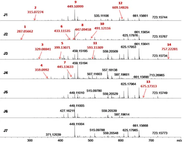
The chemical compositions of E. edulis fruits were studied using ESI (−)FT-ICR-MS,Fig. 1.The mass error acquired were even lower than 4.5 ppm, which, among the 117 signals detected (Table 1S), 14 organic compounds were identified and classified into four phytochemical classes: phenolic acids (40%), flavonoids (33.33%), anthocyanins (20%), and stilbenes (6.67%) (Fig. 1andFig. 2). It was possible to note that with the increase of their molar weight (Mw), the DBE values ranged from 6 to 20 corresponding to compounds containing from 1 to 4 phenolic rings. In addition, CID experiments (ESI(-)MS/MS) allowed the identification of the chemical connectivity of 14 proposed poly-phenolic structures in work (Table 1and Fig. 1S, supplementary ma-terial).
The compound 1, m/z 287, [C15HO6]−ion, was assigned as dihy-drokaempferol. The CID experiment produces fragments of m/z 259 (loss of CO,− 28 Da) (Fig. 1S-a), corroborating with the reported in the literature (Garzón, Narváez-Cuenca, Vincken, & Gruppen, 2017;Kang et al., 2010). Compound 2, m/z 315, [C13H15O9]−ion, was assigned as protocatechuic acid hexoside., where its MS2spectrum shows fragments of m/z 271 (− 44 Da), resulting from loss of CO2, and of m/z 153 (− 163 Da), resulting from the loss of the hexoside fraction (Fig. 1S-b1). The MS3 experiments for ion of m/z 271 shows losses of 73, 45 and 30 Da that occur in the hexoside ring structure, whereas the MS3spectra of ion of m/z 153 show a loss of 44 Da, CO2(Fig. 1S-b2 and b3). Pro-tocatechuic acid hexoside had already been reported in Juçara fruit samples (Garzón et al., 2017). Compound 3, m/z 329, [C14H17O9]−ion, was assigned as methylhydroxybenzoate hexoside. Its fragmentation profile produces signals of m/z 314 (−15 Da), resulting from loss of the methyl radical of the ester group, m/z 270 (−60 Da), due to loss of the ester group attached to the phenolic ring, and m/z 242 (−59 Da and− 29 Da) resulting from loss of the ester group attached to the phenolic ring and in hexoside fraction, respectively (Fig. 1S-c1). MS3
Table 1 Chemical compounds identi fi ed by ESI( − )FT-ICR MS in aqueous extracts of juçara fruits from di ff erent genotypes (J1 to J7). m/z measured (M-H +) − or (M-2H + ) − DBE Erro (ppm) MS n Juçaras genotypes Proposed compound Phytochemical class J1 J2 J3 J4 J5 J6 J7 1 287.05662 C15 H11 O6 10 -1,76 (b) MS 2: 259, 243, 201 ND D ND D ND ND ND Dihydrokaempferol Flavonoid 2 315.07274 C13 H15 O9 6 -1,87 (b) MS 2: 287, 283, 279, 277, 271, 153 DDDDD DD Protocatechuic acid hexoside Phenolic Acid MS 3(271): 243, 227, 199, 173 ms 3 (153) 109 3 329.08841 C14 H17 O9 6 -1,85 (b) MS 2: 314, 301, 285, 269, 258, 242, 201, 167 DDDDD DD Methylhydroxybenzoate hexoside Phenolic Acid derivative 4 359.0992 C15 H19 O10 6 -2,31 (b) MS 2: 340, 322, 312, 301, 279, 257, 237, 197, 191, 181, 153, 151, 135, ND D ND D ND ND ND Syringic acid hexoside Phenolic Acid 5 399.13071 C18 H23 O10 7 -2,61 (b) MS 2: 382, 363, 341, 331, 309, 297, 281, 263, 236, 223, 205, 189 ND ND D D D D ND Derived from Phenolic Acid Feruloyl sinapic acid 6 433.11535 C21 H21 O10 11 − 3,06 (b) MS 2:413, 401, 352, 325, 300, 269, 179, 151 MS 3(269): 225, 197, 151, 107 D D ND ND D D D Flavonoid Glycoside Flavonoid 7 445.13633 C19 H25 O12 7 -2,64 (b) MS 2: 427, 401, 387, 384, 360, 341, 301, 283 ND ND D D D D D Derived from Sinapoyl hexoside Phenolic Acid 8 447.09458 C21 H19 O11 12 -2,90 (b) MS 2:321, 285 ND D D ND D ND D Orientin Flavonoid 9 449.11007 C21 H21 O11 11 -2,53 MS 2:269 MS 3(269): 225, 197, 183, 151, 149 DDDDD DD dihydrokaempferol glucoside Flavonoid 10 491.12116 C23 H23 O12 12 − 3,39 (b) MS 2: 473, 459, 447, 445, 402, 387, 361, 329, 299, 280, 258, 225, 179, 167 N D DDDD N D N D Malvidin-glucoside Anthocyanins 11 593.15309 C27 H29 O15 13 -3,19 (b) MS 2:574, 503, 438, 327, 299, 285 DDDDD N D D Cyanidin-3-rutinoside Anthocyanins 12 609.14826 C27 H29 O16 13 -3,54 (a) MS 2:523, 300, 283, 271, 257, 215 DDDDD DD Rutin Flavonoid 13 675.17313 C35 H31 O14 20 − 1,78 (b) MS 2: 643, 507 ND ND ND ND D ND ND Flavonoid Flavonoid 14 757.22281 C33 H41 O20 13 − 4,15 (a) MS 2 727, 625, 611, 593, 479, 447, 285, 202 DDDDN D DN D Flavonoid Flavonoid *D: detected **ND: non-detected. *** (a): Fragmentation experiment using FT ICR-MS ****(b):Fragmentation experiment using LCQ Fleet.
experiments of ion of m/z 242 generates the fragment of m/z 213 (~ 31 Da) due to loss of hexoside fraction (Fig. 1S-c2). This phenolic acid had not been described in of Juçara palm fruit samples. Compound 4, m/z 359, [C 15H19O10]−ion, was assigned as syringic acid hexoside. The fragmentation of ion of m/z 359 generated signals with m/z 197 (−162 Da) and m/z 182 (−181 Da), resulting from cleavage of hexo-side fraction (Fig. 1S-d). This phenolic acid had already been reported (Rezaire et al., 2014). Compound 5, m/z 399, [C18H23O10]−ion, was assigned as a derivative of feruloyl sinapic acid (Garzón et al., 2017). The fragmentation of compound generated signals of m/z 382 (−17 Da) from the radical loss of hydroxyl group in the hexoside fraction, m/z 236 (−163 Da) and m/z 223 (−177 Da), both from hexoside fraction losses (Fig. 1S-e). Compound 6, m/z 433, [C21H21O10]−ion, was as-signed as aflavonoid glycoside (Mulabagal & Calderón, 2012), in which its ESI(−)MS2spectrum generates a signal of m/z 269 (−164 Da) from loss of the glycoside fraction (Fig. 1S-f1), whereas the fragmentation profile of the MS3 spectrum for this ion produces fragment of m/z 225 (−44 Da) from the loss of C2H4O (Fig. 1S-f2) (Galaverna, Sampaio, Barata, Eberlin, & Fidelis, 2015).
Compound 7, m/z 445, [C19H25O12]−ion, was identified as a de-rivative of sinapoyl hexoside (Garzón et al., 2017). Its CID spectrum generated ions of m/z 427 (−18 Da) relative to the loss of H2O, m/z 383 (−62 Da) and m/z341 (−104 Da), both resulting from fragmen-tations in the hexoside group, and m/z 283 (−162 Da) corresponding to loss of synapoyl fraction (Fig. 1S-g). Compound 8, m/z 447, [C21H19O11]−ion, is identified as orientin, being reported in several works(Garzón et al., 2017; Gordon et al., 2012; Kang et al., 2010; Mulabagal & Calderón, 2012; Pacheco-Palencia, Hawken, & Talcott, 2007). Its fragmentation profile produces the ion of m/z 285 (−162 Da) from loss of hexoside group (Fig. 1S-h). Compound 9, m/z 449, [C21H21O11]−ion, was assigned as dihydrokaempferol glucoside and had already been reported (Kang et al., 2010). The MS2experiment of the m/z 449 generates the fragment of m/z 269 (−180 Da), resulting from the loss of the glucoside fraction (Fig. 1S-i1), whereas the MS3 experiment for the ion of m/z 269 generates the fragment of m/z 225 (−44 Da), resulting from the neutral loss of C2H4O (Fig. 1S-i2), as
reported in the literature (Galaverna et al., 2015). Compound 10, m/z 491, [C23H23O12]−ion, was identified as malvidin-glucoside (Poulose et al., 2014). Its fragmentation profile generated ions of m/z 473 (−18 Da), m/z 459 (−32 Da), m/z 447 (−44 Da) and m/z 387 (−104 Da) from fragmentations in the glucoside fraction. Finally, the fragment of m/z 179 (−312 Da) is derived from the loss of the gluco-side fraction (Fig. 1S-j). Compound 11, m/z 593, [C27H29O15]−ion, is identified as cyanidin-rutinoside, which is reported in several works in literature (Bicudo et al., 2014;Garzón et al., 2017;Gordon et al., 2012; Mulabagal & Calderón, 2012;Pacheco-Palencia et al., 2007; Poulose et al., 2014;Rezaire et al., 2014).Its fragmentation profile produces ion of m/z 285 (−308 Da), being corresponding to loss of rutinoside frac-tion(Fig. 1S-k). Compound 12, m/z 609, [C27H29O16]−ion, was iden-tified as Rutin and had already been reported (Garzón et al., 2017). Their fragmentation profile showed ions of m/z 300 (−309 Da) and m/ z 285 (−310 Da) resulting from losses of the hexoside fraction (Fig. 1S-l). Compound 13, m/z 675, [C35H31O14]−ion, has been identified as a flavonoid, which has not yet been reported in the literature. Their fragmentation profile showed fragments of m/z 643 (−32 Da) and m/z 507 (−168 Da), characteristic of loss of methoxyl and trimethoxylated benzene rings, respectively (Fig. 1S-m). Compound 14, m/z 757, [C33H41O20]−ion, is identified as a flavonoid, not being reported in Juçara fruit samples. Their fragmentation profile shows ions of m/z 611 (−146 Da) and m/z 593 (−164 Da), being characteristic of loss of hexoside, and m/z 447 (−310 Da) characteristic of loss of two hexoside rings (Fig. 1S-n).
Fig. 3 shows a histogram of class distribution composed of 116 chemical species, proving that six genotypes of Juçara fruit (samples J1-J3 and J5-F7) are mainly rich in phenolic compounds, being abundant in species containing 13 and 14 oxygen atoms whereas the sample J4 is rich in phenolics species containing 11 oxygen atoms.
3.2. Determination of the antioxidant capacities of Juçara genotypes The in vitro antioxidant capacities determined by ABTS and SNP are shown inTable 2. Indeed, the ABTS method could better quantify the
Fig. 2. Classification of chemical compounds identified inTable 2into of the phytochemical classes. The size of the bubbles is directly proportional to the relative
antioxidant activities for all Juçara genotypes analyzed.
The presented ABTS antioxidant capacities varied from 5.88 ± 2.06 mg∙g−1 (J2) to 88.51 ± 13.56 mg∙g−1 (J6) and agree well with those determined by ABTS for E. oleraceae of 55.79 ± 1.12 mg∙g−1(Rufino et al., 2010). SNP antioxidant activities were detected for J3 (22.77 ± 0.15 mg∙g−1) and for J4 (13.52 ± 2.52 mg∙g−1) but were not significantly different.
Among the seven genotypes of Juçara analyzed, J6 presented a higher antioxidant capacity and a significantly higher content of total polyphenols compared to the analyzed samples. The consumption of J6 fruits is highly recommended because foods with high polyphenol contents and antioxidant activities have been shown to prevent several diseases associated with oxidative stress, such as cancer and cardio-vascular and neurodegenerative diseases (Sotgia, Martinez-Outschoorn, & Lisanti, 2011).
3.3. Determination of the chemopreventive capacity of Juçara genotypes None of the Juçara genotypes evaluated were cytotoxic against murine hepatoma (Hepa 1c1c7) (ATTC® CRL-2026TM), mouse macro-phage cell line RAW 264.7 (ATCC® TIB-71™) until the highest evaluated concentration was tested (1–1000 μg∙mL−1). Therefore, further experi-ments on the chemoprevention approach could be performed.
The J2, J3, J4, J5 and J6 Juçara genotypes showed significant NO synthesis inhibition, in RAW264.7 macrophages, with percentages of 64.67 ± 5.43%, 53.90 ± 7.91%, 50.55 ± 2.72% and 61.46 ± 6.32%, respectively (Table 2). The NF-κB inhibition levels detected for J3 (28.95 ± 6.22%) and J6 (10.11 ± 3.99%) were quite low. All Juçara genotypes presented statistically similar NF-κB inhibi-tion abilities (Table 2). All The analyzed Juçara samples presented antioxidant activity in the ABTS assay and only J3, J4 and J5 in the in vitro NO production (SNP). The inhibition of NO synthesis in the RAW264.7 macrophages could be related with the antioxidant activity. The inhibition of NO production by antioxidant compounds has been demonstrated, especially through the inhibition of inducible nitric oxide synthase (iNOS) and the inhibition of the nuclear factor kappa B (NF-κB). This approach is an important mechanism of chemoprevention associated with the second stage in cancer evolution and is also known as the cancer promotion phase (Bonavida & Baritaki, 2011;Kleinert, Euchenhofer, Ihrig-Biedert, & Förstermann, 1996; Pascual & Glass, 2006;Tedeschi et al., 2003).
The induction of phase II enzyme NAD(P)H:QR can offer protection against toxic and reactive chemical species (Pezzuto et al., 2005). Therefore, phase II enzyme NAD(P)H is considered an important che-moprevention biomarker. In addition, antioxidant activity can be as-sayed indirectly in Hepa1c1c7 cells by using the induction of NAD(P)H
Fig. 3. Histogram of class distribution of compounds (CcHhOo) classified as function of number of oxygen atoms present in each molecular formula.
Table 2
Antioxidant capacities and chemopreventive in vitro analyses.
Antioxidant activities Chemopreventive activities
Pulp extract Total phenolic (μg.g−1) Anthocyanins (μg.g−1) ABTS (mg∙g −1) SNP (mg∙g−1) (NO in vitro production) NO (%inhibition of NO synthesis) NF-κB (% inhibition) Aromatase (% inhibition) Quinone reductase (IR**) J 1 167.92 ± 5.52a 204.62 ± 2.27b 22.83 ± 3.44b ND* ND* 6.20 ± 3.65b −8.5 1 J 2 115.15 ± 11.03b,c 378.51 ± 1.39a 5.88 ± 2.06c,d ND* 52.34 ± 19.29a ND* 25.4 1 J 3 125.84 ± 5.34b 191.26 ± 7.18c 33.69 ± 3.01b 22.77 ± 0.15a,b 64.67 ± 5.43a 28.95 ± 6.22a 24.5 1 J 4 166.26 ± 4.08a 48.43 ± 2.31d 17.65 ± 2.18b,d 13.92 ± 2.52b 53.90 ± 7.91a ND* 23.6 1 J 5 134.15 ± 8.45b 392.54 ± 3.66a 29.42 ± 7.00c,d 3.81 ± 0.63b 50.55 ± 2.72a ND* 32.0 2 J 6 128.55 ± 6.91b 41.19 ± 3.38d 88.51 ± 13.56a ND* 61.46 ± 6.32a 10.11 ± 3.99b 21.1 3 J 7 110.62 ± 0.65c 243.25 ± 3.98b,c 31.45 ± 11.98b,d ND* ND* ND* 14.2 2
Results expressed as the mean ± SD for triplicates. Different letters indicate significant differences between fractions (One-way ANOVA followed by Tukey post hoc
test, p < .05). *ND-not detected **IR induction ratio. Cell lines used: Quinone reductase-Hepa 1c1c7 (murine hepatoma cells, ATCC CRL-2026), 293-NF-κB (human
renal derived 293 cell line, 293.12 - PTA5554), RAW 264.7 cells (iNOS). QR results are considered when the IR is 2. For inhibition of NF-κB, results are considered
quinone oxidoreductase 1 activity (Pezzuto et al., 2005). The anti-oxidant activity found for J6 is consistent with its quinone reductase induction (2.8 ± 0.9) observed in this study (Table 2) and can be re-lated to the polyphenol content and protocatechuic acid hexoside and rutin levels found in J6. These compounds are known to have anti-inflammatory, anti-tumor and anti-genotoxic effects (Babich, Sedletcaia, & Kenigsberg, 2002; Liu, Wang, Chu, Cheng, & Tseng, 2002). Quercetin, the aglycone of rutin, inhibited the production of nitric oxide (NO) in a concentration-dependent manner in lipopoly-saccharide (LPS)-induced RAW 264.7 macrophage cells, and these compounds also exhibited QR induction in cultured Hepa 1c1c7 cells (Nitteranon, Zhang, Darien, & Parkin, 2011).The varieties J5 and J7 induced quinone reductase and J6 inhibited NO synthesis, in RAW264.7 macrophages, demonstrating that these three varieties, principally J6, have high chemopreventive potential and are strongly recommended for agricultural cultivation.
To further evaluate the chemopreventive potential of the Jucaras genotypes, their ability to prevent aromatase activity was evaluated since its inhibitors have been reported to effectively treat hormone receptor-positive breast cancer in postmenopausal women (Lukong, 2017). However, the results for aromatase were not significant. Thus, aromatase inhibition is not the route utilized by Juçara fruits to elicit chemoprevention (Table 2).
3.4. In vitro cytokines evaluation
The anti-inflammatory capacity of Juçara fruit was evaluated using macrophages RAW 264 stimulated by LPS. The cytokines TNFα and IL-1α were detected by ELISA using specific antibodies. The Juçara fruits did not present significant inflammatory inhibition for the pro-in-flammatory cytokine IL-1α in the evaluated concentration range (0 to 100μg∙mL−1) (Fig. 2S – A and C). Dexamethasone, a potent gluco-corticoid that was used as a positive control, inhibited IL-1α at levels of 99.3 ± 1.2% and at a concentration of 3.92μg∙mL−1.
Extracts J3, J4, J6 and J7 significantly reduced the TNF-α levels (Fig. 2S– B and D). The anti-inflammatory effects elicited by J3 and J4 (both at 100μg∙mL−1) for J6 (10μg∙mL−1) and for J7 (all concentra-tions tested ranging from 0 to 100μg∙mL−1) were similar to those of the positive control dexamethasone. However, no difference was present between the samples. Concomitantly, J3 and J4 reduced TNF-α levels by 37.8 ± 18.8% and 29.4 ± 10.9%, respectively. J6 (10μg∙mL−1) significantly reduced the TNF-α level by 28.00 ± 9.6%. Among the extracts, J7 presented the highest inhibition of TNF-α, reducing its level by 46.4 ± 7.3% at a concentration of 10μg∙mL−1. This inhibition was similar to the inhibitory effect of dexamethasone, the positive control, at a concentration of 3.92μg∙mL−1(49.1 ± 10.6%). Previous studies with species of the genus Euterpe have demonstrated that the extracts of acai (Euterpe oleracea) are involved in the down regulation of cytokine TNF-α expression (Kang et al., 2010).
4. Conclusion
The six evaluated genotypes of E. edulis and one E. espiritosantense had different polyphenol, flavonoid and anthocyanin compositions. Consequently, the specimens demonstrated different antioxidant and chemopreventive profiles. The high antioxidant activity in vitro de-tected in J6 supported the induction of quinone reductase in cell culture assays. J6 also inhibited TNFα and NO synthesis, in RAW264.7 mac-rophages. TNF-α inhibition could be a possible cross-talk effect between the induction of QR and the inhibition of NO synthesis, in RAW264.7 macrophages. E. edulis genotype J6 should be included in the daily diet as in natura consumption or as a nutraceutical. In addition, the culti-vation of this genotype is a feasible and sustainable option for rational palm use instead of palm cutting. Further studies should be conducted understand the potential health effects of the consumption of Juçara fruit (J6) as food or as a supplement.
Acknowledgments
The authors thank the Foundation for Support to Research and Innovation of Espírito Santo e SEAG/FAPES, National Council for Scientific Technological Research (CNPq– PVE 2014 process #401409/ 2014-7, PQ- process # 310680/2016-6) for theirfinancial support and Coordination for the Improvement of Higher Level Personnel (CAPES). Funding
This work was supported by the Foundation for Support to Research and Innovation of Espírito Santo-FAPES [grant numbers 65835131/ 0010-2013, TO # 241/2016]; the Secretaria de Estado da Agricultura, Abastecimento, Aquicultura e Pesca- SEAG [grant number TO # 665/ 2016]; and the United States Institutes of Peace [grant number aaaa]. Conflicts of interest
The authors declare no conflicts of interest. Appendix A. Supplementary data
Supplementary data to this article can be found online athttps:// doi.org/10.1016/j.foodres.2018.09.036.
References
Babich, H., Sedletcaia, A., & Kenigsberg, B. (2002). In vitro cytotoxicity of protocatechuic acid to cultured human cells from oral tissue: involvement in oxidative stress. Pharmacology and Toxicology, 91(5), 245–253.https://doi.org/10.1034/j.1600-0773. 2002.910505.x.
Bates, J., Baker, M., & Pharmacology, R. G. J.-B (1991). Nitric oxide generation from nitroprusside by vascular tissue: evidence that reduction of the nitroprusside anion and cyanide loss are required. Elsevier. Retrieved fromhttps://www.sciencedirect. com/science/article/pii/000629529190406U.
Bicudo, M. O. P., Ribani, R. H., & Beta, T. (2014). Anthocyanins, phenolic acids and antioxidant properties of juçara fruits (Euterpe edulis M.) along the on-tree ripening process. Plant Foods for Human Nutrition, 69(2), 142–147.https://doi.org/10.1007/ s11130-014-0406-0.
Bonavida, B., & Baritaki, S. (2011). Dual role of NO donors in the reversal of tumor cell resistance and EMT: Downregulation of the NF-κB/Snail/YY1/RKIP circuitry. Nitric Oxide, 24(1), 1–7.https://doi.org/10.1016/J.NIOX.2010.10.001.
de Brito, E. S., de Araújo, M. C. P., Alves, R. E., Carkeet, C., Clevidence, B. A., & Novotny, J. A. (2007). Anthocyanins present in selected Tropical Fruits: Acerola, Jambolão, Jussara, and Guajiru. Journal of Agricultural and Food Chemistry, 55(23), 9389–9394.
https://doi.org/10.1021/jf0715020.
Cardoso, A. L., Di Pietro, P. F., Vieira, F. G. K., Boaventura, B. C. B., Liz, S., Silva Campelo Borges, G., ... Silva, E. L. (2015). Acute consumption of juçara juice (Euterpe edulis) and antioxidant activity in healthy individuals. Journal of Functional Foods, 17, 152–162.https://doi.org/10.1016/J.JFF.2015.05.014.
Clancy, R. M., & Abramson, S. B. (1995). Nitric Oxide: A Novel Mediator of Inflammation. Experimental Biology and Medicine, 210(2), 93–101.https://doi.org/10.3181/ 00379727-210-43927AA.
Fadden, J., Seoane, C., Paolinetti, V., Lima, A., Zanatta, R., Amêndola, D., & Foufre, L. (2008). Extração caseira de polpa de juçara: Euterpe edulis Martius. - Portal Embrapa. In EMBRAPA (Ed.). Embrapa Florestas (pp. 8). Ministério da Agricultura. Retrieved fromhttps://www.embrapa.br/busca-de-publicacoes/-/publicacao/ 315624/extracao-caseira-de-polpa-de-jucara-euterpe-edulis-martius.
Galaverna, R. S., Sampaio, P. T. B., Barata, L. E. S., Eberlin, M. N., & Fidelis, C. H. V. (2015). Differentiation of two morphologically similar Amazonian Aniba species by mass spectrometry leaffingerprinting. Analytical Methods, 7(5), 1984–1990.https:// doi.org/10.1039/C4AY02598A.
Garzón, G. A., Narváez-Cuenca, C.-E., Vincken, J.-P., & Gruppen, H. (2017). Polyphenolic composition and antioxidant activity of açai (Euterpe oleracea Mart.) from Colombia. Food Chemistry, 217, 364–372.https://doi.org/10.1016/J.FOODCHEM.2016.08.107.
Giusti, M. M., & Wrolstad, R. E. (2001). Characterization and measurement of antho-cyanins by UV-visible spectroscopy. Current Protocols in Food Analytical Chemistry, F1.2.1–F1.2.13.
Gordon, A., Cruz, A. P. G., Cabral, L. M. C., de Freitas, S. C., Taxi, C. M. A. D., Donangelo, C. M., ... Marx, F. (2012). Chemical characterization and evaluation of antioxidant properties of Açaí fruits (Euterpe oleraceae Mart.) during ripening. Food Chemistry, 133(2), 256–263.https://doi.org/10.1016/J.FOODCHEM.2011.11.150.
Green, L. C., Wagner, D. A., Glogowski, J., Skipper, P. L., Wishnok, J. S., & Tannenbaum, S. R. (1982). Analysis of nitrate, nitrite, and [15N]nitrate in biologicalfluids. Analytical Biochemistry, 126(1), 131–138.https://doi.org/10.1016/0003-2697(82) 90118-X.
Pezzuto, J. M., ... Zhang, H.-J. (2006). Bioactive Dammarane Triterpenes from the Mangrove Plant Bruguiera gymnorrhiza.https://doi.org/10.1021/NP058112X. Inada, K. O. P., Oliveira, A. A., Revorêdo, T. B., Martins, A. B. N., Lacerda, E. C. Q., Freire,
A. S., ... Monteiro, M. C. (2015). Screening of the chemical composition and occurring antioxidants in jabuticaba (Myrciaria Jaboticaba) and Jussara (Euterpe edulis) fruits and their fractions. Journal of Functional Foods, 17, 422–433.https://doi.org/10. 1016/J.JFF.2015.06.002.
Kang, J., Li, Z., Wu, T., Jensen, G. S., Schauss, A. G., & Wu, X. (2010). Anti-oxidant capacities offlavonoid compounds isolated from acai pulp (Euterpe oleracea Mart.). Food Chemistry, 122(3), 610–617.https://doi.org/10.1016/J.FOODCHEM.2010.03. 020.
Kleinert, H., Euchenhofer, C., Ihrig-Biedert, I., & Förstermann, U. (1996). Glucocorticoids inhibit the induction of nitric oxide synthase II by down-regulating cytokine-induced activity of transcription factor nuclear factor-kappa B. Molecular Pharmacology, (1), 49.
Leitman, P., Soares, K., Henderson, A., Noblick, L., & Martins, R. (2015). Lista do Brasil -Arecaceae Schultz Sch. Retrieved June 17, 2018, fromhttp://floradobrasil.jbrj.gov.br/ jabot/FichaPublicaTaxonUC/FichaPublicaTaxonUC.do?id=FB53.
Liu, C.-L., Wang, J.-M., Chu, C.-Y., Cheng, M.-T., & Tseng, T.-H. (2002). In vivo protective effect of protocatechuic acid on tert-butyl hydroperoxide-induced rat hepatotoxicity. Food and Chemical Toxicology, 40(5), 635–641. https://doi.org/10.1016/S0278-6915(02)00002-9.
Liu, H., Dinkova-Kostova, A. T., & Talalay, P. (2008). Coordinate regulation of enzyme markers for inflammation and for protection against oxidants and electrophiles. Retrieved fromhttp://www.pnas.org/content/pnas/105/41/15926.full.pdf.
Lobo, V., Patil, A., Phatak, A., & Chandra, N. (2010). Free radicals, antioxidants and functional foods: Impact on human health. Pharmacognosy Reviews, 4(8), 118–126.
https://doi.org/10.4103/0973-7847.70902.
Lukong, K. E. (2017). Understanding breast cancer– The long and winding road. BBA Clinical, 7, 64–77.https://doi.org/10.1016/J.BBACLI.2017.01.001.
Mosmann, T. (1983). Rapid colorimetric assay for cellular growth and survival: Application to proliferation and cytotoxicity assays. Journal of Immunological Methods, 65(1–2), 55–63. Retrieved fromhttp://www.ncbi.nlm.nih.gov/pubmed/6606682. Mulabagal, V., & Calderón, A. I. (2012). Liquid chromatography/mass spectrometry based
fingerprinting analysis and mass profiling of Euterpe oleracea (açaí) dietary supple-ment raw materials. Food Chemistry, 134(2), 1156–1164.https://doi.org/10.1016/J. FOODCHEM.2012.02.123.
Nitteranon, V., Zhang, G., Darien, B. J., & Parkin, K. (2011). Isolation and synergism of in vitro anti-inflammatory and quinone reductase (QR) inducing agents from the fruits of Morinda citrifolia (noni). Food Research International, 44(7), 2271–2277.https:// doi.org/10.1016/J.FOODRES.2010.11.009.
Oliveira, B. G., Costa, H. B., Ventura, J. A., Kondratyuk, T. P., Barroso, M. E. S., Correia, R. M., ... Romão, W. (2016). Chemical profile of mango (Mangifera indica L.) using electrospray ionisation mass spectrometry (ESI-MS). Food Chemistry, 204, 37–45.
https://doi.org/10.1016/j.foodchem.2016.02.117.
Pacheco-Palencia, L. A., Hawken, P., & Talcott, S. T. (2007). Phytochemical, antioxidant and pigment stability of açai (Euterpe oleracea Mart.) as affected by clarification, ascorbic acid fortification and storage. Food Research International, 40(5), 620–628.
https://doi.org/10.1016/J.FOODRES.2006.11.006.
Park, E.-J., Kondratyuk, T. P., Morrell, A., Kiselev, E., Conda-Sheridan, M., Cushman, M., ... M, J. (2011). Induction of retinoid X receptor activity and consequent upregulation of p21WAF1/CIP1 by indenoisoquinolines in MCF7 cells. Cancer Prevention Research (Philadelphia, Pa.), 4(4), 592–607. https://doi.org/10.1158/1940-6207.CAPR-10-0004.
Pascual, G., & Glass, C. K. (2006). Nuclear receptors versus inflammation: Mechanisms of
transrepression. Trends in Endocrinology & Metabolism, 17(8), 321–327.https://doi. org/10.1016/J.TEM.2006.08.005.
Pezzuto, J. M., Kosmeder, J. W., Park, E.-J., Lee, S. K., Cuendet, M., Gills, J., ... Kinghorn, A. D. (2005). Characterization of natural product chemopreventive agents. Cancer Chemoprevention (pp. 3–37). Totowa, NJ: Humana Press.https://doi.org/10.1007/ 978-1-59259-768-0_1.
Pompeu, D. R., Silva, E. M., & Rogez, H. (2009). Optimisation of the solvent extraction of phenolic antioxidants from fruits of Euterpe oleracea using Response Surface Methodology. Bioresource Technology, 100(23), 6076–6082.https://doi.org/10.1016/ J.BIORTECH.2009.03.083.
Poulose, S. M., Fisher, D. R., Bielinski, D. F., Gomes, S. M., Rimando, A. M., Schauss, A. G., & Shukitt-Hale, B. (2014). Restoration of stressor-induced calcium dysregulation and autophagy inhibition by polyphenol-rich açaí (Euterpe spp.) fruit pulp extracts in rodent brain cells in vitro. Nutrition, 30(7–8), 853–862.https://doi.org/10.1016/J. NUT.2013.11.011.
Re, R., Pellegrini, N., Proteggente, A., Pannala, A., Yang, M., & Rice-Evans, C. (1999). Antioxidant activity applying an improved ABTS radical cation decolorization assay. Free Radical Biology and Medicine, 26(9–10), 1231–1237.https://doi.org/10.1016/ S0891-5849(98)00315-3.
Rezaire, A., Robinson, J.-C., Bereau, D., Verbaere, A., Sommerer, N., Khan, M. K., ... Fils-Lycaon, B. (2014). Amazonian palm Oenocarpus bataua (“patawa”): Chemical and biological antioxidant activity– Phytochemical composition. Food Chemistry, 149, 62–70.https://doi.org/10.1016/J.FOODCHEM.2013.10.077.
Rufino, M.d. S. M., Alves, R. E., de Brito, E. S., Pérez-Jiménez, J., Saura-Calixto, F., & Mancini-Filho, J. (2010). Bioactive compounds and antioxidant capacities of 18 non-traditional tropical fruits from Brazil. Food Chemistry, 121(4), 996–1002.https://doi. org/10.1016/J.FOODCHEM.2010.01.037.
Schulz, M., Borges, G., Da, S. C., Gonzaga, L. V., Seraglio, S. K. T., Olivo, I. S., Azevedo, M. S., ... Fett, R. (2015). Chemical composition, bioactive compounds and antioxidant capacity of juçara fruit (Euterpe edulis Martius) during ripening. Food Research International, 77, 125–131.https://doi.org/10.1016/J.FOODRES.2015.08.006. Schulz, M., Campelo, S., Valdemiro, L., Carolina, A., Costa, O., & Fett, R. (2016). Juçara
fruit (Euterpe edulis Mart.): Sustainable exploitation of a source of bioactive compounds. vol. 89, FRIN14–26.https://doi.org/10.1016/j.foodres.2016.07.027.
Singleton, V. L., Orthofer, R., & Lamuela-Raventós, R. M. (1999). Analysis of total phenols and other oxidation substrates and antioxidants by means of folin-ciocalteu reagent. Methods in Enzymology, 299, 152–178.https://doi.org/10.1016/S0076-6879(99) 99017-1.
Sotgia, F., Martinez-Outschoorn, U. E., & Lisanti, M. P. (2011). Mitochondrial oxidative stress drives tumor progression and metastasis: Should we use antioxidants as a key component of cancer treatment and prevention? BMC Medicine, 9(1), 62.https://doi. org/10.1186/1741-7015-9-62.
Tedeschi, E., Menegazzi, M., Margotto, D., Suzuki, H., Förstermann, U., & Kleinert, H. (2003). Anti-inflammatory actions of St. John's wort: Inhibition of human inducible nitric-oxide synthase expression by down-regulating signal transducer and activator of transcription-1alpha (STAT-1alpha) activation. The Journal of Pharmacology and Experimental Therapeutics, 307(1), 254–261.https://doi.org/10.1124/jpet.103. 054460.
Terry, P., Giovannucci, E., Michels, K. B., Bergkvist, L., Hansen, H., Holmberg, L., & Wolk, A. (2001). Fruit, vegetables, Dietary Fiber, and risk of Colorectal Cancer. JNCI Journal of the National Cancer Institute, 93(7), 525–533.https://doi.org/10.1093/jnci/93.7. 525.
Wattenberg, L. W. (1985). Chemoprevention of cancer. Cancer Research, 45(1), 1–8. Retrieved fromhttp://www.ncbi.nlm.nih.gov/pubmed/3880665.