The role of the hippocampus in the semantic variant of primary progressive aphasia: A resting-state fcMRI study
The role of the hippocampus in the semantic variant of primary progressive aphasia: A resting-state fcMRI study
Authors: Marianne Chapleau, B.Sc.1,2, Maxime Montembeault, B.Sc.1,2, Mariem Boukadi, M.A. 1,2 Christophe Bedetti, M.Sc. 1,2 Robert Jr. Laforce, Ph.D./M.D.3, Maximiliano Wilson, Ph.D.4, Simona M. Brambati, Ph.D.1,2
1 Département de Psychologie, Université de Montréal, Québec, Canada
2 Centre de Recherche de l'Institut Universitaire de Gériatrie de Montréal, Québec,Canada
3 Clinique Interdisciplinaire de Mémoire (CIME), Centre Hospitalier Universitaire de Québec; Faculté de Médecine, Université Laval, Québec, Canada.
4 Institut universitaire en santé mentale de Québec (CRIUSMQ), Université Laval, Québec, Canada.
Search Terms: Semantic Variant of Primary Progressive Aphasia, Resting-State Functional Connectivity, CONN Toolbox, Semantic Memory, Hippocampus
Submission Type: Article (Rapid Communication) Number of Tables: 6
Number of Figures: 5
Word Count of Abstract: 160 Word Count of Paper: 2218 Corresponding author: Simona Maria Brambati
Centre de Recherche de l’IUGM, 4565, Chemin Queen Mary, Montréal QC H3W 1W5, Canada; Tel.: (514) 340-3540, ext. 4147; Fax: (514) 340-3548
ABSTRACT
The goal of the study was to determine whether the semantic variant of primary progressive aphasia (svPPA) affects the intrinsic connectivity network anchored to left and right anterior
hippocampus, but spares the posterior hippocampus. A resting-state functional connectivity MRI (rs-fcMRI) study was conducted in a group of patients with svPPA and in controls, using a seed-to-voxel approach. In comparison to controls, massively reduced connectivity was found in the anterior
hippocampus, mainly the left one, for svPPA patients but not in the left or right posterior hippocampus. In svPPA, the anterior hippocampus showed reduced functional connectivity with regions implicated in the semantic memory network. Significant correlation was also found between the functional
connectivity strength of the left anterior hippocampus and the ventromedial cortex, and performance in semantic tasks. These findings indicate that the functional disconnection of the anterior hippocampus may be a promising in vivo biomarker of svPPA and illustrate the role of this hippocampal subregion in the semantic memory system.
INTRODUCTION
The semantic variant of primary progressive aphasia (svPPA) is a neurodegenerative disease characterized by progressive multimodal loss of semantic knowledge (Gorno-Tempini et al., 2011; Hodges, Patterson, Oxbury, & Funnell, 1992; Mesulam, 2003; Snowden, 1989), mainly targeting the anterior temporal regions of the left hemisphere. Early atrophy of the hippocampus, bilaterally but predominantly in the left side, has been recently reported as one of the early anatomical characteristics of svPPA (Chapleau, Aldebert, Montembeault, & Brambati, 2016).
In studies of neurodegenerative diseases, atrophy of the hippocampus has been generally linked to episodic memory consolidation deficits (Petersen et al., 2000). Thus, early hippocampal atrophy in svPPA is allegedly in contradiction with the fact that svPPA patients present partially or completely preserved episodic memories compared to other neurodegenerative patients with comparable
hippocampal atrophy, such as in amnestic Alzheimer’s disease (Gorno-Tempini et al., 2011; Hodges et al., 1992; Hornberger & Piguet, 2012). In addition, mild deficits in episodic memory tests often
manifested by svPPA are secondary to possible perceptual and semantic deficits rather than the consequence of consolidation deficits as observed in AD.
One possible explanation for this paradox is based on the functional specialization of the hippocampus along an antero-posterior axis (Poppenk, Evensmoen, Moscovitch, & Nadel, 2013). Evidence on healthy brains shows that the anterior hippocampus (aHip) is mainly functionally connected to regions of the semantic memory network whereas the posterior hippocampus (pHip) is mainly functionally connected to the episodic memory network (La Joie et al., 2014; Poppenk et al., 2013; Ranganath & Ritchey, 2012) (Figure 1). Based on this model, it is thus conceivable that svPPA patients without prominent episodic memory deficits present lower functional connectivity, principally
between the aHip and the regions of the semantic memory network, and relatively spared functional connectivity between the pHip and regions of the episodic memory network.
To test this hypothesis, we used resting-state functional connectivity magnetic resonance imaging (rs-fcMRI) to measure and compare the patterns of functional connectivity within the intrinsic
connectivity networks (ICNs) anchored to the aHip and pHip in the left and right hemispheres, between svPPA patients and healthy controls (CTRLs). Rs-fcMRI allows the investigation of functional
connectivity within ICNs, by measuring and correlating the spontaneous fluctuation of the blood oxygen level-dependent (BOLD) signal. Rs-fcMRI studies have demonstrated that various
neurodegenerative diseases distinctively deteriorate the pattern of functional connectivity within the ICNs anchored to brain regions showing most prominent syndrome-specific atrophy (Seeley, Crawford, Zhou, Miller, & Greicius, 2009). Therefore, measures of rs-fcMRI are considered emerging in vivo biomarkers of neurodegenerative diseases. Confirmation of the dissociation between the patterns of rs-fcMRI in the ICNs anchored to the aHip and pHip holds promise for supporting differential diagnosis and tracking disease progression, notably in svPPA.
METHODS
The study included 12 patients with a clinical diagnosis of svPPA (Gorno-Tempini et al., 2011) (3 women, 9 men; mean age of 65.9±10.8), and 11 cognitively unimpaired CTRLs (4 women, 7 men; mean age of 65.7±8.1) matched for age, sex and education. SvPPA patients were recruited at relatively mild stages of the disease (Mini-Mental State Examination 25.0 ±2.6). Demographic and
neuropsychological characteristics of the sample are displayed in Table 1.
MRI images were obtained using a 3T Philips Achieva TX scanner at IRM Québec-Mailloux in Quebec City, in order to obtain: 1) a volumetric magnetization prepared rapid gradient echo
(MP-RAGE) sequence (TR/TE = 8.2/3.7 ms, FoV= 250 mm, flip angle = 8◦, 180 slices/volume, slice thickness = 1mm, no gap), 2) echo-planar imaging (EPI) images while subjects were instructed to rest quietly (TR/TE = 2110/30 ms, FoV=224 mm, flip angle = 70◦, 40 transverse slices, slice thickness=3.5 mm, no gap, 300 volumes).
MRI images were pre-processed using SPM12 (www.fil.ion.ucl.ac.uk/spm/software/spm12/) running on Matlab R2017 (v. 9.3.0). Image pre-processing included: 1) study-specific
Diffeomorphic Anatomical Registration Through Exponentiated Lie Algebra (DARTEL) template creation; 3) warping of all realigned and slice-timing corrected EPI images to DARTEL template and 4) 8-mm Gaussian kernel smoothing.
CONN functional connectivity toolbox (www.conn-toolbox.org) was then used to extract BOLD timeseries from the seed regions and to correlate them with the time series for all other brain voxels (seed-to-voxel analysis). Four-mm radius spheres centered on the coordinates -26 -6 -22 (left aHip), 26 -6 -22 (right Hp), -28 -35 3 (left pHip) and 28 -35 3 (right pHip) were used as seed regions.
The coordinates of the spheres’ centers were based on our previous meta-analysis, in which we pooled together all the voxel-based morphometry studies investigating gray matter atrophy in patients with svPPA and Alzheimer’s disease (Chapleau et al., 2016). The coordinate in the aHip sphere’s center represents the peak coordinate of hippocampal atrophy in svPPA compared to controls. The coordinate of the pHip sphere’s center represents the maximal hippocampal atrophy difference between
Alzheimer’s disease patients characterized by prominent episodic memory deficits (amnestic profile) and svPPA patients. The positions of the coordinates were verified using Anatomical Toolbox, version 2.2b (Eickhoff et al., 2005). This program allows showing MNI coordinates on probabilistic
cytoarchitecture maps of the hippocampus projected on a reference brain (colin27, i.e. MNI single
subject template)(Amunts et al., 2005). Based on these probabilistic cytoarchitecture maps, we verified
the position of the selected coordinates in the hippocampus. Six motion correction parameters were
included as regressors in the first-level analysis, and age and sex were included in the second-level analysis. The resulting statistical parametric map (SPM) was thresholded at a primary threshold of voxel-wise P≤0.001, and then corrected for False Discovery Rate at p<0.05 based on cluster extent and Gaussian Random Field (GRF) theory (Woo, Krishnan, & Wager, 2014). Between-group comparison maps were inclusively masked for CTRLs main effects.
RESULTS
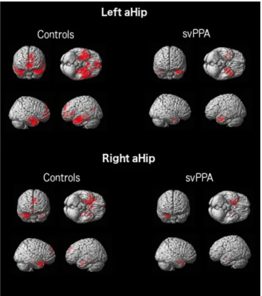
The pattern of rs-fcMRI in the ICN anchored to the aHip and pHip in the CTRL group generally replicate previous findings (Libby, Ekstrom, Ragland, & Ranganath, 2012; Poppenk & Moscovitch, 2011). In fact, we observed that the aHip (both left and right) is functionally connected mainly to the anterior semantic network, while the pHip is connected to a more medial posterior episodic network (see Supplementary Material, Tables 3-6 and Figure 3-4). The pattern of rs-fcMRI in svPPA shows generally reduced functional connectivity in the ICN anchored to the aHip, especially in the left
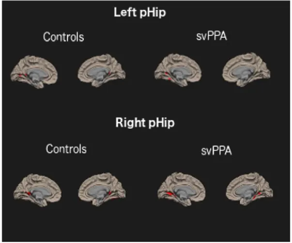
hemisphere. On the other hand, the pattern of rs-fcMRI in the ICNs anchored to the pHip (both left and right) is largely comparable to the CTRLs (see Supplementary Material, Tables 3-6 and Figure 3-4). Results of the direct comparison between svPPA and CTRLs
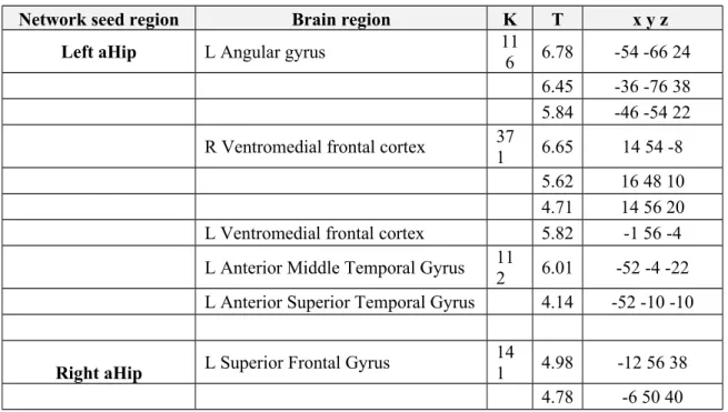
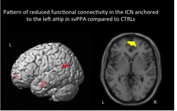
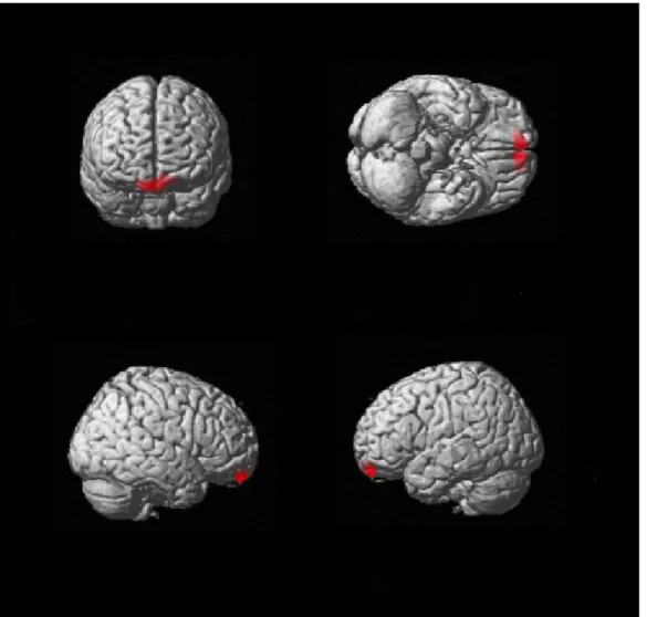
Reduced functional connectivity in the ICNs anchored to the left and right aHip in svPPA (see Table 2 and Figure 2)
SvPPA, compared to CTRLs, showed significant lower functional connectivity between the left aHip and the left angular gyrus, bilateral ventromedial frontal cortex and the left anterior temporal lobe.
SvPPA patients also showed significant lower functional connectivity between the right aHip and the left superior frontal gyrus. No increased functional connectivity was observed in svPPA compared to CTRLs. All these results persisted when we included hippocampal volume as covariate in the model, although at a predictable lower level of significance (p<0.005 uncorrected)
Comparable functional connectivity in the ICNs anchored to both the left and right pHip in svPPA and CTRLs
There were no significant differences between CTRLs and svPPA in the pHip-anchored ICNs. Post-hoc correlation analysis
Our results raised the question whether the rs-fcMRI strength, in the ICN anchored to the aHip with regards to other brain voxels, was associated with semantic memory performance in svPPA. Thus, we performed a second-level correlational analysis using CONN toolbox to test the association
between the accuracy scores in semantic tests (Boston Naming Test, Pyramid and Palm Tree Test and
Similarities Subtest of the WAIS III) and our left and right aHip seed regions’ connectivity strength in
relation to other brain voxels. In order to investigate the correlational effect that was unique to
semantics and not confounded by the effect of episodic memory, we included the verbal and non-verbal episodic memory performances (recognition scores of the Rey Auditory Verbal Learning Test and delayed recall scores of the Rey Complex Figure Test) as confounding variables in the model.
Composite scores of the neuropsychological tests were used in the present analysis. The results showed that the rs-fcMRI between the seed region in the left aHip and the bilateral ventromedial frontal cortex correlated with semantic performance (p<0.05 FDR corrected) (peak in the left hemisphere: x=-1, y=56, z=-4, peak in the right hemisphere: x=14, y=54, z=-8) (Supplementary Material, Figure 5). No significant result was observed when the right aHip was used as a seed region.
DISCUSSION
Here we demonstrated that svPPA leads to a deterioration of the rs-fcMRI in the ICNs anchored to the left and right aHip, and that the functional disconnection from mainly the left aHip could represent a possible substrate for semantic deficits. This study has several important strengths. First, it provides direct confirmation of the vulnerability of the aHip-anchored ICNs in svPPA. Previous support to this hypothesis was mostly based on compelling but more indirect evidence, such as results showing that the atrophy in svPPA mainly involves the anterior portion of the hippocampus (Chapleau et al., 2016), and that decreased regional metabolism in svPPA involves regions that are functionally connected to the aHip in healthy subjects (La Joie et al., 2014). Furthermore, consistently with our hypothesis, the group of regions showing decreased functional connectivity with the aHip is remarkably overlapping to the brain network previously associated with the semantic memory system (Binder & Desai, 2011). In fact, we found decreased connectivity between the left aHip and the following brain regions: left angular gyrus, left and right ventromedial frontal cortex and left anterior middle and superior temporal gyrus. All these brain regions have all been previously included in the neuroanatomical semantic memory network (Binder & Desai, 2011). The right aHip only showed decreased connectivity with the left medial superior frontal gyrus. A meta-analysis of 120 functional neuroimaging studies focusing on semantic processing have linked the activation of the dorsomedial frontal cortex, including the medial superior frontal gyrus, to semantic processing (Binder, Desai, Graves, & Conant, 2009). The specific role of this region in semantic is poorly understood. Some authors propose a key role of this region in goal-directed knowledge retrieval, i.e. top-down retrieval of semantic information to solve a given task (Binder & Desai, 2011). Altogether, these findings confirm that svPPA mainly targets the functional connectivity between the aHip and key regions of the semantic network. Consistently, we observed that
the severity of functional connectivity damage between the left aHip and the bilateral ventromedial prefrontal cortex is associated with the severity of semantic deficits. Supporting the results of the correlational analysis, previous work has shown a key role of the ventromedial prefrontal cortex (vmPFC) in the semantic memory system (Binder & Desai, 2011). More specifically, a recent fMRI study in healthy young participants demonstrated that the vmPFC and the anterior hippocampus contribute to concept generalization by representing abstract category information (Bowman & Zeithamova, 2018), which is a necessary process for the semantic representation of concepts. In addition, a resting-state fMRI analysis of 1000 subjects, performed using Neurosynth meta-analysis tool (Yarkoni, Poldrack, Nichols, Van Essen, & Wager, 2011), revealed that the activity of the aHip correlates with that of the vmPFC (Zeidman & Maguire, 2016), confirming that these two regions are functionally connected. In conclusion, these studies provide critical evidence about the implication of the vmPFC in the semantic memory network and its functional connection to the aHip. Finally, in agreement with previous reports on the asymmetrical damage in svPPA, our results suggest that svPPA predominantly targets the left hemisphere, consistently with its role in verbal semantic tasks.
Future studies should include larger samples of participants and should determine whether the aHip’s connectivity represents a more sensitive predictor of semantic deficits than volume. In addition, future complementary studies should investigate the possible opposite rs-fcMRI pattern in AD (pHip ICN disconnection more severe than that of the aHip). Finally, in order to provide critical support to the hypothesis that a decreased rs-fcMRI in the ICN anchored to the aHip is related to the semantic profile in svPPA patients, this group should be compared to other clinical variants of PPA showing no
predominant semantic deficits, such as logopenic and non-fluent variants. To the best of our knowledge, no study has addressed this issue to date.
In conclusion, our results indicate that the functional disconnection of the aHip, mainly the left one, could be a promising in vivo biomarker of svPPA. Moreover, we suggest that studying the functional connectivity patterns separately for the anterior and posterior hippocampus could help in the diagnosis and follow-up of different neurodegenerative diseases.
Acknowledgments
The Alzheimer’s Society of Canada supported this study [grant number: 1420]. MC is supported by Fonds de Recherche du Québec Santé (FRQ-S) doctoral award. MM is supported by Alzheimer Society of Canada and Fonds de Recherche du Québec Santé (FRQ-S) doctoral awards. MB is supported by a Canadian Institutes of Health Research (CIHR) and FRQ-S doctoral awards. RL is supported by Fondation du CHU de Québec and Société Alzheimer du Québec. MAW is supported by Fonds de Recherche du Québec Société et Culture (FRQ-SC) and Réseau Québécois de Recherche en
Vieillissement (RQRV). SMB is supported by a Chercheur Boursier Award obtained from the Fonds de la Recherche du Québec Santé (FRQ-S). Imaging data collection was partially supported by the
“Consortium d’imagerie en neuroscience et santé mentale de Québec” (CINQ) via a platform support grant from the Brain Canada Foundation
Data sharing
The data that support the findings of this study are available from the corresponding author upon reasonable request.
References
Amunts, K., Kedo, O., Kindler, M., Pieperhoff, P., Mohlberg, H., Shah, N. J., . . . Zilles, K. (2005). Cytoarchitectonic mapping of the human amygdala, hippocampal region and entorhinal cortex: intersubject variability and probability maps. Anat Embryol (Berl), 210(5-6), 343-352.
doi:10.1007/s00429-005-0025-5
Binder, J. R., & Desai, R. H. (2011). The neurobiology of semantic memory. Trends Cogn Sci, 15(11), 527-536. doi:10.1016/j.tics.2011.10.001
Binder, J. R., Desai, R. H., Graves, W. W., & Conant, L. L. (2009). Where is the semantic system? A critical review and meta-analysis of 120 functional neuroimaging studies. Cereb Cortex, 19(12), 2767-2796. doi:10.1093/cercor/bhp055
Bowman, C. R., & Zeithamova, D. (2018). Abstract Memory Representations in the Ventromedial Prefrontal Cortex and Hippocampus Support Concept Generalization. J Neurosci, 38(10), 2605-2614. doi:10.1523/JNEUROSCI.2811-17.2018
Chapleau, M., Aldebert, J., Montembeault, M., & Brambati, S. M. (2016). Atrophy in Alzheimer's Disease and Semantic Dementia: An ALE Meta-Analysis of Voxel-Based Morphometry Studies. J Alzheimers Dis, 54(3), 941-955. doi:10.3233/JAD-160382
Eickhoff, S. B., Stephan, K. E., Mohlberg, H., Grefkes, C., Fink, G. R., Amunts, K., & Zilles, K. (2005). A new SPM toolbox for combining probabilistic cytoarchitectonic maps and functional imaging data. Neuroimage, 25(4), 1325-1335. doi:10.1016/j.neuroimage.2004.12.034
Gorno-Tempini, M. L., Hillis, A. E., Weintraub, S., Kertesz, A., Mendez, M., Cappa, S. F., . . . Grossman, M. (2011). Classification of primary progressive aphasia and its variants. Neurology, 76(11), 1006-1014. doi:10.1212/WNL.0b013e31821103e6
Hodges, J. R., Patterson, K., Oxbury, S., & Funnell, E. (1992). Semantic dementia. Progressive fluent aphasia with temporal lobe atrophy. Brain, 115 ( Pt 6), 1783-1806. doi:10.1093/brain/115.6.1783 Hornberger, M., & Piguet, O. (2012). Episodic memory in frontotemporal dementia: a critical review. Brain, 135(Pt 3), 678-692. doi:10.1093/brain/aws011
La Joie, R., Landeau, B., Perrotin, A., Bejanin, A., Egret, S., Pelerin, A., . . . Chetelat, G. (2014). Intrinsic connectivity identifies the hippocampus as a main crossroad between Alzheimer's and semantic dementia-targeted networks. Neuron, 81(6), 1417-1428. doi:10.1016/j.neuron.2014.01.026 Libby, L. A., Ekstrom, A. D., Ragland, J. D., & Ranganath, C. (2012). Differential connectivity of perirhinal and parahippocampal cortices within human hippocampal subregions revealed by high-resolution functional imaging. J Neurosci, 32(19), 6550-6560. doi:10.1523/JNEUROSCI.3711-11.2012 Mesulam, M. M. (2003). Primary progressive aphasia--a language-based dementia. N Engl J Med, 349(16), 1535-1542. doi:10.1056/NEJMra022435
Petersen, R. C., Jack, C. R., Jr., Xu, Y. C., Waring, S. C., O'Brien, P. C., Smith, G. E., . . . Kokmen, E. (2000). Memory and MRI-based hippocampal volumes in aging and AD. Neurology, 54(3), 581-587. doi:10.1212/wnl.54.3.581
Poppenk, J., Evensmoen, H. R., Moscovitch, M., & Nadel, L. (2013). Long-axis specialization of the human hippocampus. Trends Cogn Sci, 17(5), 230-240. doi:10.1016/j.tics.2013.03.005
Poppenk, J., & Moscovitch, M. (2011). A hippocampal marker of recollection memory ability among healthy young adults: contributions of posterior and anterior segments. Neuron, 72(6), 931-937. doi:10.1016/j.neuron.2011.10.014
Ranganath, C., & Ritchey, M. (2012). Two cortical systems for memory-guided behaviour. Nat Rev Neurosci, 13(10), 713-726. doi:10.1038/nrn3338
Seeley, W. W., Crawford, R. K., Zhou, J., Miller, B. L., & Greicius, M. D. (2009). Neurodegenerative diseases target large-scale human brain networks. Neuron, 62(1), 42-52.
doi:10.1016/j.neuron.2009.03.024
Snowden, J. S. G., P.J.; Neary, D. (1989). Semantic dementia: a form of circumscribed cerebral atrophy. Behavioral Neurology, 2(3), 167-182.
Woo, C. W., Krishnan, A., & Wager, T. D. (2014). Cluster-extent based thresholding in fMRI analyses: pitfalls and recommendations. Neuroimage, 91, 412-419. doi:10.1016/j.neuroimage.2013.12.058
Yarkoni, T., Poldrack, R. A., Nichols, T. E., Van Essen, D. C., & Wager, T. D. (2011). Large-scale automated synthesis of human functional neuroimaging data. Nat Methods, 8(8), 665-670.
doi:10.1038/nmeth.1635
Zeidman, P., & Maguire, E. A. (2016). Anterior hippocampus: the anatomy of perception, imagination and episodic memory. Nat Rev Neurosci, 17(3), 173-182. doi:10.1038/nrn.2015.24
TABLES
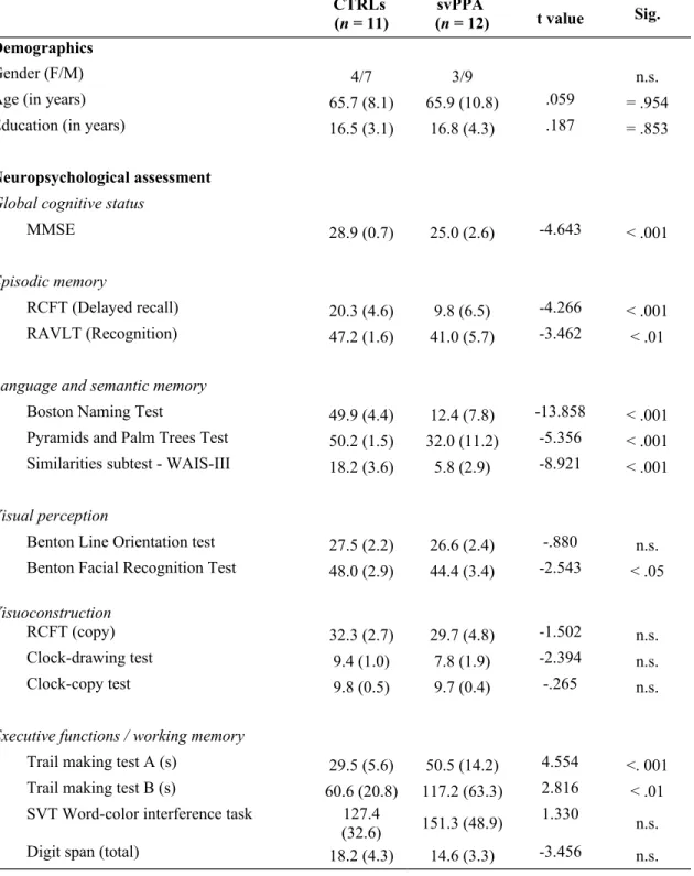
Table 1: Demographic and neuropsychological characteristics of CTRL and svPPA patients
CTRLs
(n = 11) (n = 12)svPPA t value Sig.
Demographics
Gender (F/M) 4/7 3/9 n.s.
Age (in years) 65.7 (8.1) 65.9 (10.8) .059 = .954
Education (in years) 16.5 (3.1) 16.8 (4.3) .187 = .853
Neuropsychological assessment
Global cognitive status
MMSE 28.9 (0.7) 25.0 (2.6) -4.643 < .001
Episodic memory
RCFT (Delayed recall) 20.3 (4.6) 9.8 (6.5) -4.266 < .001
RAVLT (Recognition) 47.2 (1.6) 41.0 (5.7) -3.462 < .01
Language and semantic memory
Boston Naming Test 49.9 (4.4) 12.4 (7.8) -13.858 < .001
Pyramids and Palm Trees Test 50.2 (1.5) 32.0 (11.2) -5.356 < .001 Similarities subtest - WAIS-III 18.2 (3.6) 5.8 (2.9) -8.921 < .001
Visual perception
Benton Line Orientation test 27.5 (2.2) 26.6 (2.4) -.880 n.s. Benton Facial Recognition Test 48.0 (2.9) 44.4 (3.4) -2.543 < .05
Visuoconstruction
RCFT (copy) 32.3 (2.7) 29.7 (4.8) -1.502 n.s.
Clock-drawing test 9.4 (1.0) 7.8 (1.9) -2.394 n.s.
Clock-copy test 9.8 (0.5) 9.7 (0.4) -.265 n.s.
Executive functions / working memory
Trail making test A (s) 29.5 (5.6) 50.5 (14.2) 4.554 <. 001 Trail making test B (s) 60.6 (20.8) 117.2 (63.3) 2.816 < .01 SVT Word-color interference task 127.4
Abbreviations: MMSE = Mini-Mental State Examination; RCFT: Rey Complex Figure Test; RAVLT: Rey Auditory Verbal Learning Test; SVT = Stroop-Victoria Test
Table 2. Between-group differences (CTRLs>svPPA) in the rs-fcMRI network anchored to the left and right anterior hippocampus (p ≤ .05 FDR corrected at cluster level)
Network seed region Brain region K T x y z
Left aHip L Angular gyrus 11
6 6.78 -54 -66 24
6.45 -36 -76 38
5.84 -46 -54 22
R Ventromedial frontal cortex 371 6.65 14 54 -8
5.62 16 48 10
4.71 14 56 20
L Ventromedial frontal cortex 5.82 -1 56 -4
L Anterior Middle Temporal Gyrus 11
2 6.01 -52 -4 -22
L Anterior Superior Temporal Gyrus 4.14 -52 -10 -10
Right aHip L Superior Frontal Gyrus 141 4.98 -12 56 38
FIGURES
Figure 1. Schematic representation of the functional specialization of the hippocampus along an antero-posterior axis in the semantic and episodic memory networks (La Joie et al., 2014; Poppenk et al., 2013; Ranganath & Ritchey, 2012)
Abbreviations. SvPPA: semantic variant of primary progressive aphasia; AD: Alzheimer’s disease; ATL: Anterior temporal lobe; VMPC: Ventromedial prefrontal cortex; TPJ: Temporoparietal junction; PCC: Posterior cingulate cortex; LPC: Lateral parietal cortex; aHip: Anterior hippocampus; pHip: Posterior hippocampus
Figure 2. Pattern of decreased rs-fcMRI in the network anchored to the left aHip in svPPA compared to CTRLs (p ≤ .05 FDR corrected at cluster level).
SUPPLEMENTARY FIGURES
Figure 3. Above: rs-fcMRI network anchored to the left anterior hippocampal seed in CTRLs and svPPA. Below: rs-fcMRI network anchored to the right anterior hippocampal seed in CTRLs and svPPA (p ≤ .05 FDR corrected at cluster level).
Figure 4. Above: rs-fcMRI network anchored to the left posterior hippocampal seed in CTRLs and svPPA. Below: rs-fcMRI network anchored to the right posterior hippocampal seed in CTRLs and svPPA (p ≤ .05 FDR corrected at cluster level).
Figure 5. Correlational analysis between the rs-fcMRI strength of the left aHip and the bilateral ventromedial frontal cortex and semantic performance (Boston Naming Test, Pyramid and Palm Tree Test and Similarities Subtest of the WAIS III, p<0.05 FDR corrected at cluster level).