Identification of the Functions of Liver X Receptor-

in Sertoli Cells Using a Targeted Expression-Rescue
Model
Salwan Maqdasy,* Fatim-Zohra El Hajjaji,* Marine Baptissart, Emilie Viennois, Abdelkader Oumeddour, Florence Brugnon, Amalia Trousson, Igor Tauveron, David Volle, Jean-Marc A. Lobaccaro, and Silvère Baron
Department of Génétique Reproduction et Développement (GReD) (S.M., F.-Z.E.H., M.B., A.O., F.B., A.T., I.T., D.V., J.-M.A.L., S.B.), Université Blaise Pascal, Centre de Recherche en Nutrition Humaine d’Auvergne (S.M., F.-Z.E.H., M.B., A.O., F.B., A.T., D.V., J.-M.A.L., S.B.), and Department of Assistance Médicale à la Procréation (F.B.), CECOS, Centre Hospitalier Universitaire Clermont Ferrand, Centre Hospitalier Universitaire Estaing, F-63000 Clermont-Ferrand, France; Centre National de la Recherche Scientifique (S.M., F.-Z.E.H., M.B., A.O., F.B., A.T., I.T., D.V., J.-M.A.L., S.B.) and INSERM (S.M., F.-Z.E.H., M.B., A.O., F.B., A.T., I.T., D.V., J.-M.A.L., S.B.), Unité Mixte de Recherche 6293, GReD, F-63177 Aubiere, France; Center for Diagnostics and Therapeutics (E.V.), Georgia State University, Atlanta, Georgia 30302– 4010; Veterans Affairs Medical Center (E.V.), Decatur, Georgia 30033; Service d’Endocrinologie, Diabétologie, et Maladies Métaboliques (S.M., I.T.), Hôpital Gabriel Montpied, F-63003 Clermont-Ferrand, France; and Service de Médecine Nucléaire (S.M.), Centre Jean Perrin, F-63011 Clermont-Ferrand, France
Liver X receptors (LXRs) are key regulators of lipid homeostasis and are involved in multiple
tes-ticular functions. The Lxr␣⫺/⫺;Lxr⫺/⫺mice have illuminated the roles of both isoforms in
main-tenance of the epithelium in the seminiferous tubules, spermatogenesis, and T production. The
requirement for LXR in Sertoli cells have been emphasized by early abnormal cholesteryl ester
accumulation in the Lxr⫺/⫺and Lxr␣⫺/⫺;Lxr⫺/⫺mice. Other phenotypes, such as germ cell loss and
hypogonadism, occur later in life in the Lxr␣⫺/⫺;Lxr⫺/⫺mice. Thus, LXR expression in Sertoli cells
seems to be essential for normal testicular physiology. To decipher the roles of LXR within the
Sertoli cells, we generated Lxr␣⫺/⫺;Lxr⫺/⫺:AMH-Lxr transgenic mice, which reexpress Lxr in
Sertoli cells in the context of Lxr␣⫺/⫺;Lxr⫺/⫺mice. In addition to lipid homeostasis, LXR is
nec-essary for maintaining the blood-testis barrier and the integrity of the germ cell epithelium. LXR
is also implicated in the paracrine action of Sertoli cells on Leydig cells to modulate T synthesis. The
Lxr␣⫺/⫺;Lxr⫺/⫺and Lxr␣⫺/⫺;Lxr⫺/⫺:AMH-Lxr mice exhibit lipid accumulation in germ cells after
the Abcg8 down-regulation, suggesting an intricate LXR-dependentcooperationbetweentheSertoli
cells and germ cells to ensure spermiogenesis. Further analysis revealed also peritubular smooth muscle defects (abnormal lipid accumulation and disorganized smooth muscle actin) and spermatozoa
stag-nation in the seminiferous tubules. Together the present work elucidates specific roles of LXRinSertoli
cell physiology in vivo beyond lipid homeostasis. (Endocrinology 156: 4545– 4557, 2015)
L
iver X receptors (LXR␣ and LXR, NR1H3, and NR1H2, respectively) belong to a subclass of nuclear receptors that are activated after binding to their ligands, the oxysterols. They are classically implicated inintracel-lular cholesterol homeostasis (1). Many physiological roles have been discovered using the Lxr␣⫺/⫺;Lxr⫺/⫺
mice, from lipid homeostasis to the regulation of glucose metabolism or immunity (for review see reference 2). Both
ISSN Print 0013-7227 ISSN Online 1945-7170 Printed in USA
Copyright © 2015 by the Endocrine Society
Received April 30, 2015. Accepted September 18, 2015. First Published Online September 24, 2015
* S.M. and F.-Z.E.H. contributed equally to this work.
Abbreviations: ABCG8, ATP-binding cassette transporters G8; BTB, blood-testis barrier; LXR, liver X receptor; PLIN1, perilipin; PTSM, peritubular smooth muscle cells; RT-qPCR, real-time quantitative PCR; SMA, smooth muscle actin; SOX9, Sry-type high-mobility-group box transcription factor 9.
doi: 10.1210/en.2015-1382 Endocrinology, December 2015, 156(12):4545– 4557 press.endocrine.org/journal/endo 4545
isoforms are expressed in the testis and play a crucial role in fertility (3–5). During adulthood, each isoform is pre-dominant in a specific testicular cell type: LXR␣ in Leydig cells, LXR in Sertoli cells, and both isoforms in germ cells. The Lxr␣⫺/⫺mice are characterized by the decreased steroidogenic activity of Leydig cells and increased germ cell apoptosis; the Lxr⫺/⫺mice display cholesteryl ester accumulation in the Sertoli cells, which is associated with a decreased germ cell proliferation rate. Mice deficient for both isoforms, Lxr␣⫺/⫺;Lxr⫺/⫺, exhibit progressive tes-ticular degeneration with hypogonadism, germ cell deple-tion. and definitive infertility by 7–9 months old (3, 5). LXR␣ and LXR have complementary and/or redundant roles in the testis because the absence of one isoform is partially compensated by the other; thus, the single knock-out mice are fertile with few obvious defects (3, 6).
Sertoli cells represent the central cell type of the testis that support germ cell differentiation and modulate the androgens that are necessary for this purpose (7). LXR expression in Sertoli cells is presumed to be essential for normal testicular physiology. Indeed, lipid accumulation in Sertoli cells is the earliest phenotype and appears by 1.5 months of age in the Lxr⫺/⫺and Lxr␣⫺/⫺;Lxr⫺/⫺mice; it has been suggested as the central dysfunction in the testis of these mice (3, 4). Likewise, a study of men with non-obstructive azoospermia showed a significant decrease of
LXR expression within the testis, which was associated
with fewer proliferating germ cells; this study further sup-ports the pivotal role of LXR within the testis (8).
To gain further insights into the roles of LXR in Sertoli cells, we generated a mouse line that reexpresses Lxr only in Sertoli cells in the Lxr␣⫺/⫺;Lxr ⫺/⫺mice, using the human AMH promoter (antimullerian hormone) (9). This strain of mice, named Lxr␣⫺/⫺;Lxr⫺/⫺:AMH-Lxr, was compared with the wild-type and Lxr␣⫺/⫺;Lxr⫺/⫺mice. Here we note the various fundamental roles of LXR in Sertoli cell physiology and its implication in the structural integrity and endocrine functioning of the testicular tissue. Reexpression of LXR in the Sertoli cells of the
Lxr␣⫺/⫺;Lxr⫺/⫺mice restores the structural architecture of the seminiferous tubules; the large lipid vacuoles dis-appear and the integrity of the blood-testis barrier is re-established. Furthermore, LXR expression in Sertoli cells exerts a paracrine function on androgen synthesis by the Leydig cells. Together these beneficial effects support the germ cell population and only a few degenerated tubules are found with aging. In addition, we show that germ cells display persistent lipid vacuoles in the later stages of dif-ferentiation, and thus, they require Abcg8 expression as an essential LXR target gene to reestablish lipid homeostasis. This mouse model also reveals a peritubular smooth mus-cle phenotype with lipid accumulation and actin
disorga-nization. These new mice provide a relevant model for deciphering the additional roles of LXRs in the testis.
Materials and Methods Animals
The Lxr␣⫺/⫺;Lxr⫺/⫺mice were kindly supplied by
labora-tory of Repa and Mangelsdorf (10) and were maintained on a
mixed strain background (C57BL/6:129Sv). The Lxr␣⫺/⫺;
Lxr⫺/⫺:AMH-Lxr mice were generated in the local transgenic
facility of Génétique Reproduction et Développement. All strains were fed ad libitum with the Global diet 2016S from Harlan and maintained on a 12-hour light, 12-hour dark cycle. For the experiments, some mice were gavaged with 25 mg/kg T0901317 (Sigma-Aldrich) or vehicle (methyl cellulose), as de-scribed previously (3). Other mice were injected with 20 mg/kg busulfan and euthanized 4 or 8 weeks later (11). All of the pro-tocols and experiments were approved by the regional ethics committee and have already been published (3, 11).
Generation of transgenic mice
The Lxr␣⫺/⫺;Lxr⫺/⫺:AMH-Lxr mice were generated by
additive transgenesis. As described inSupplemental Figure 1A,
the transgene construct was linearized by HindIII/XhoI digestion
and microinjected into Lxr␣⫺/⫺;Lxr⫺/⫺-fertilized mouse
oocytes. The eggs were retrieved from 3-month-old Lxr␣⫺/⫺;
Lxr⫺/⫺females. Before the eggs were harvested, the females
were superovulated using a 48-hour delayed stimulation with pregnant mare serum gonadotropin (7.5 IU) and human chori-onic gonadotropin (5 IU) and mated with 3-month-old
Lxr␣⫺/⫺;Lxr⫺/⫺males. The purified DNA was microinjected
using the Zeiss Transgenesis station Zeiss AXIOVERT 135 M. The oocytes that were microinjected with the transgene construct were transferred into pseudopregnant DBA/2 females. Southern blotting was used to detect the transgenic founders and the copy numbers of the transgene, as depicted in Supplemental Figure 1, B and C. Two founders, F0 –1 and F0 –2, transmitted the
AMH-Lxr transgene. Based on real-time quantitative PCR
(RT-qPCR) and western blotting analyses for transgene expression, the F0 –1 line was selected for further phenotypic investigations. Histology, immunohistochemistry, and
immunofluorescence
Staining with hematoxylin/eosin, Oil red O (Sigma-Aldrich), Ki67, and Azure 2 dye (Sigma-Aldrich and Agar Scientific) was performed on semithin sections, as previously described (3). Briefly, the tissues were fixed using 4% paraformaldehyde (Sig-ma-Aldrich) and embedded in paraffin. For semithin sections, the testes were fixed and postfixed as previously described (3). Terminal deoxynucleotidyl transferase-mediated deoxyuridine triphosphate nick end labeling assays were performed as de-scribed (3). Antibodies against Flag (clone M2, F1804; Sigma-Aldrich), Sry-type high-mobility-group box transcription factor 9 (SOX9; AB5535; Millipore), Ki67 (SP6, M3064; Spring Bio-science), smooth muscle actin (SMA; M0851; DAKO), perilipin (GP29; Progen Biotechnik), and ATP-binding cassette transport-ers G8 (ABCG8; NB400 –110B; Novus Biological) were used according to the manufacturers’ recommendations. The
iments were performed at least twice, with five to six mice per
group, except for the Azure 2 dye staining (n⫽ 3).
Blood-testis barrier (BTB) integrity assay
The integrity of the BTB was analyzed in the wild-type,
Lxr␣⫺/⫺;Lxr⫺/⫺, and Lxr␣⫺/⫺;Lxr⫺/⫺:AMH-Lxr mice by an
intratesticular infusion of a biotin tracer, as previously reported (11, 12). Briefly, the testes of anesthetized mice were injected with the qualitative TJ functional tracer biotin (10 mg/mL; EZ-Link Sulfo-NHS-LC-Biotin; Pierce) in a volume of 10% of the weight of the testis. They were then removed 30 minutes after the injection and directly fixed in 4% paraformaldehyde (Sigma-Aldrich). The pen-etration of the biotin tracer into the seminiferous lumen was visu-alized by fluorescence microscopy using Alexa-488 streptavidin-horseradish peroxidase (Invitrogen). The total number of tubes with biotin penetration into the lumen of the seminiferous tubules (defective tubes) and the number of tubes with biotin retained near the basement membrane (intact tubes) were counted in a
cross-section of the entire testis (n⫽ 6/group).
Percentage of histological destruction
For each animal (n⫽ 6), a complete histological analysis of
three entire sections of the testis (9 mo old) was performed by hematoxylin and eosin staining. Microscopic destruction in-cluded vacuolated tubules, thin seminiferous epithelium, empty tubules, and spermatozoa stagnation and were quantified for each genotype. The percentage of each anomaly was evaluated in relation to the total number of tubules per section.
Lipid measurements
The lipids were extracted as previously described (3). Briefly, lipid separation was performed by high-performance thin-layer chromatography using silica plates (Silica gel 60; Merck). The plates were analyzed by densitometry (Sigma Scan Pro; Sigma-Aldrich) using standards and migration solutions.
Hormone measurements
The steroids were extracted from the testes as previously de-scribed (13). The intratesticular T levels were measured using commercial kits (Diagnostic Biochem). The plasma FSH and LH levels were measured by a Milliplex MAP mouse pituitary mag-netic bead panel (96-well plate assay MPTMAG-49K) based on the Luminex xMAP technology as previously described (14). Western blot analysis
The proteins were extracted using HEPES 20 mM, NaCl 0.42
M, MgCl21.5 mM, EDTA 0.2 mM, and Nonidet P40 1%
sup-plemented with phenylmethylsulfonyl fluoride 1 mM
(Sigma-Aldrich), Complete protease inhibitors 1⫻ (Roche Molecular
Biochemicals), NaF 0.1 mM, and Na2VO30.1 mM
(Sigma-Al-drich). The lysates were subjected to 10% SDS-PAGE and blot-ted onto a nitrocellulose membrane (Amersham Pharmacia Bio-tech). Antibodies against FLAG, which were used to detect the
transgene (F7425, Sigma-Aldrich) or-actin (A2066;
Sigma-Al-drich) were used as previously described (15). Quantitative PCR
The testis RNA was isolated using the RNeasy kit (QIAGEN) and quantifications were performed by RT-qPCR as previously
de-scribed (3). The primer sequences are reported in the Supplemental Information (Supplemental Table 1). The RT-qPCR products were
analyzed using the⌬⌬cycle threshold method for relative
quantifi-cation. The values were normalized to the 36b4 gene levels. Statistical analysis
The data are expressed as the means ⫾ SEM. A one-way
ANOVA was performed to determine differences between the various groups, and statistical analysis is indicated as follows: *,
P⬍ .05; **, P ⬍ .01; and ***, P ⬍ .001.
Results
Lxr is specifically expressed in the Sertoli cells of
Lxr␣⫺/⫺;Lxr⫺/⫺AMH-Lxr mice
To examine the effect of rescued expression of Lxr in the Sertoli cells of the Lxr␣⫺/⫺;Lxr⫺/⫺mice, Lxr␣⫺/⫺; Lxr⫺/⫺:AMH-Lxr transgenic mice were generated by
additive transgenesis using a linear construct, which con-tains the Flag-tagged Lxr cDNA driven by the human
AMH promoter (Supplemental Figure 1A). This promoter
has been already used to successfully target Sertoli cell-specific expression (9). Despite the altered hormone stim-ulation for egg retrieval in the Lxr␣⫺/⫺;Lxr⫺/⫺females (16), two transgenic mouse founders were engineered (Supplemental Figure 1B). One strain was selected based on the criteria of successful construct insertion, Lxr re-expression, and transgene transmission (Figure 1A and Supplemental Figure 1C). The subsequent analyses on the second strain revealed the absence of the transgene ex-pression, and thus, these mice were no longer used for the phenotypic investigations. LXR expression and activity was confirmed in the Lxr␣⫺/⫺;Lxr⫺/⫺:AMH-Lxr
mouse testis by the accumulation of the 55-kDa Flag-tagged protein (Figure 1B) as well as by induced transcrip-tion of Srebp1c and Abca1 (known LXR target genes) in response to T0901317 treatment (a synthetic LXR ago-nist) (Figure 1C) (2, 17). The specific accumulation of LXR in Sertoli cells was verified by coimmunolocaliza-tion of the Flag epitope and SOX9 (Figure 1D), a Sertoli cell-specific marker (18). A careful analysis of the immu-nohistochemistry in the Lxr␣⫺/⫺;Lxr⫺/⫺:AMH-Lxr
testis sections demonstrated Flag-LXR-positive staining in many or all of the Sertoli cells, suggesting that LXR expression was completely restored in this cell compart-ment (Supplecompart-mental Figure 1D). Flag-LXR labeling was restricted to the testis and ovary, consistent with the en-dogenous AMH promoter activity (Figure 1E). A devel-opmental analysis of transgene expression in the testis showed that it was expressed upon 14.5 days post coitum, which was later than endogenous Lxr but was robustly expressed (7 and 15 days post partum) and persisted
throughout adulthood (75 dpp), the temporal window in which the phenotype occurs (Supplemental Figure 2). To-gether these results confirmed the specific expression of a functional transgene within the Sertoli cells.
Targeted expression of Lxr in the Lxr␣⫺/⫺;
Lxrⴚ/ⴚ:AMH-Lxr mice restores lipid homeostasis
in Sertoli cells
Lipid accumulation in Sertoli cells is the hallmark of the
Lxr␣⫺/⫺;Lxr⫺/⫺testicular phenotype; cholesteryl esters
are the predominant lipids that ac-cumulate inside the testicular tissue (3, 4). Histological and biochemical analyses of the Lxr␣⫺/⫺;Lxr⫺/⫺: AMH-Lxr mice testis showed
nor-mal cholesteryl ester levels (Oil Red-O Staining and thin-layer chro-matography analysis) (Figure 2, A and B) compared with the Lxr␣⫺/⫺; Lxr⫺/⫺ and wild-type mice. This phenotype occurs in the Lxr␣⫺/⫺; Lxr⫺/⫺mice with aging. Here we showed that LXR expression was sufficient to maintain cholesterol ho-meostasis in the Sertoli cells of the
Lxr␣⫺/⫺;Lxr⫺/⫺:AMH-Lxr mice
and prevented even long-term cho-lesteryl ester accumulations as shown in the 9-month-old mice (Supplemental Figure 3, A–C). The quantitative representation of the Sertoli cells in the testicular tissue re-mained unchanged in the Lxr␣⫺/⫺; Lxr⫺/⫺:AMH-Lxr mice
com-pared with the wild-type mice and
Lxr␣⫺/⫺;Lxr⫺/⫺ mice because the
Sox9 transcript accumulation
exhib-ited a similar profile (Figure 2C). This suggests that the excessive lipid accumulation within the Sertoli cells did not affect the number of Sertoli cells. One of the main functions of the Sertoli cells in the testis is to maintain the BTB to protect the germ cells from the immune system. This function is sustained by the presence of a tight junction network between Sertoli cells. We postulated that the lipid droplet accumulation could mechanically impair the Sertoli cell cytoskeleton by disrupting adjacent Sertoli junctions and Sertoli-germ cell adhesions and interfering with the paracrine actions of the Sertoli cells in the
Lxr␣⫺/⫺;Lxr⫺/⫺mice (for review see reference 19). Bi-otin EZ, a sensitive marker of barrier alterations/disrup-tions, was used to study the BTB in vivo (11, 20). The wild-type seminiferous tubules efficiently prevented tracer penetration, whereas the 4-month-old Lxr␣⫺/⫺;Lxr⫺/⫺
mice had significant alterations of their BTBs (Figure 2D). Reexpression of Lxr abolished Biotin EZ penetration in the tubules of the Lxr␣⫺/⫺;Lxr⫺/⫺:AMH-Lxr mice,
thus indicating a restoration of BTB integrity in these an-Flag β ACTIN Flag SOX9 Flag SOX9 0 Relative expression ***
A
*** *** * Nr1h3 Nr1h2 1 0.5 1.5 2 +/+ Lxrα-/-;Lxrβ-/-:AMH-Lxrβ Lxrα-/-;Lxrβ-/-B
C
0Relative fold induction
Srebp1c Abca1 2 1 3 4 *** * ** *** ***
D
- + - + - + - + - + - + T09testis ovary epididymis caput seminal vesicle prostate adrenal liver kidney lung brain
testis uterus
E
+/+ Flag β actin epididymis cauda * Lxr α-/-Lxrβ-/- Lxr α-/-Lxr β-/-AMH-Lxrβ +/+ HE Lxrα-/-;Lxrβ-/-:AMH-LxrβFigure 1. LXR is specifically expressed in the Sertoli cells of the Lxr␣⫺/⫺;Lxr⫺/⫺:AMH-Lxr mice. A,
The relative accumulation of the Lxr␣ (Nr1h3) and Lxr (Nr1h2) mRNAs in the 4-month-old wild-type
(white bars), Lxr␣⫺/⫺;Lxr⫺/⫺(black bars), and Lxr␣⫺/⫺;Lxr⫺/⫺:AMH-Lxr (gray bars) testis samples
(n⫽ 6–9) were quantified by RT-qPCR. B, Western blot analyses were performed on the protein
lysates from 4-month-old wild-type, Lxr␣⫺/⫺;Lxr⫺/⫺, and Lxr␣⫺/⫺;Lxr⫺/⫺:AMH-Lxr mouse testes
using antibodies against the Flag epitope (sequence incorporated with the Lxr transgene). -Actin was used as a loading control. C, The relative accumulation of the Srebp1c and Abca1 mRNAs in testes extracts from 4-month-old mice of each genotype that were treated with vehicle
(methylcellulose) or T0901317 (25 mg/kg) were analyzed by RT-qPCR. D, Immunolocalization of the Flag epitope and SOX9 (Sertoli cell specific marker) and HE staining of testes sections from
4-month-old Lxr␣⫺/⫺;Lxr⫺/⫺:AMH-Lxr mice. The arrowheads indicate the colocalization of both proteins in
the Sertoli cells. The scale bar represents 100m. E, Immunoblot analyses from the wild-type and
Lxr␣⫺/⫺;Lxr⫺/⫺:AMH-Lxr testes and other Lxr␣⫺/⫺;Lxr⫺/⫺:AMH-Lxr organs indicate that
Flag-LXR accumulation was restricted to the Lxr␣⫺/⫺;Lxr⫺/⫺:AMH-Lxr testis and ovary. The data are
expressed as the means⫾ SEM. Statistical analysis: *, P ⬍ .05,**, P ⬍ .01, ***, P ⬍ .001, compared
with the wild-type mice. HE, hematoxylin/eosin.
Percent 60 20 80 100 * 40 0 BTB Integrity
C
2. 1 0 0.4 0.2 0.6 0.8 Relative expression Sox9 1.0B
Biotin EZ HE Azur blue OROA
0 *** *** 2.0 1.6 1.2 0.8 0.4Cholesterol esters (nmol/ng tissue)
D
+/+ Lxrα-/-;Lxrβ-/-:AMH-Lxrβ Lxrα-/-;Lxrβ-/- +/+ Lxr α -/-;Lxr β -/-: AMH-Lxr β Lxr α -/-;Lxr β -/- +/+ Lxr α -/-;Lxr β -/-: AMH-Lxr β Lxr α -/-;Lxr β -/-Figure 2. Targeted expression of LXR in the Lxr␣⫺/⫺;Lxr⫺/⫺:AMH-Lxr mice restores lipid homeostasis and the BTB in Sertoli cells. A,
Histological sections of 4-month-old wild-type, Lxr␣⫺/⫺;Lxr⫺/⫺, and Lxr␣⫺/⫺;Lxr⫺/⫺:AMH-Lxr testes showing the disappearance of lipid
vacuoles in the Lxr␣⫺/⫺;Lxr⫺/⫺:AMH-Lxr mouse testis. The left vertical panel represents frozen sections stained with Oil Red-O (ORO) and
hematoxylin. The right panel represents semithin cross-sections of testes stained with Azure 2 dye. The arrowheads indicate the lipid vacuoles
within the Sertoli cell cytoplasm. Scale bar, 50m. B, Biochemical measurements of cholesteryl ester (CE) accumulation in the testis of
4-month-old wild-type (white bars), Lxr␣⫺/⫺;Lxr⫺/⫺(black bars), and Lxr␣⫺/⫺;Lxr⫺/⫺:AMH-Lxr (gray bars) mice. C, The relative accumulation of the Sox9
mRNA in the wild-type (white bars), Lxr␣⫺/⫺;Lxr⫺/⫺(black bars), and Lxr␣⫺/⫺;Lxr⫺/⫺:AMH-Lxr (gray bars) mouse testis samples (n ⫽ 6–9) was
measured by RT-qPCR. D, Visualization of the Biotin-EZ tracer and hematoxylin/eosin (HE) staining and quantification of the BTB integrity in the
wild-type (white bars), Lxr␣⫺/⫺;Lxr⫺/⫺(black bars) and Lxr␣⫺/⫺;Lxr⫺/⫺:AMH-Lxr (gray bars) mice. Biotin is retained near the basement
membrane when the BTB is intact; biotin diffusion into the seminiferous tubules (arrowheads) indicates a defective BTB. Scale bar, 100m. The
data are expressed as the means⫾ SEM. Statistical analysis: *, P ⬍ .05, ***, P ⬍ .001, compared with the wild-type mice.
imals (Figure 2D). These results indicate that rescued ex-pression of a functional LXR in the Lxr␣⫺/⫺;Lxr⫺/⫺
Sertoli cells in vivo corrects abnormal cholesteryl ester accumulation, leading to a proper BTB.
LXR expression in Sertoli cells participates in the endocrine function of Leydig cells
Leydig cells are responsible for T production, which is necessary for germ cell differentiation, libido, and the maintenance of secondary sexual characteristics (6). The
Lxr␣⫺/⫺and Lxr␣⫺/⫺;Lxr⫺/⫺mice exhibit a defect in T production, which underlies abnormal Leydig cell steroid-ogenesis (3). To decipher the specific role of LXR within Sertoli cells in the regulation of Leydig cell function, the intratesticular T levels were measured in the wild-type,
Lxr␣⫺/⫺;Lxr⫺/⫺, and Lxr␣⫺/⫺;Lxr⫺/⫺:AMH-Lxr
mice. Although the T levels were significantly lower in the
Lxr␣⫺/⫺;Lxr⫺/⫺mice, as already described, they were restored in the Lxr␣⫺/⫺;Lxr⫺/⫺:AMH-Lxr mice
(Fig-ure 3A). This effect was evident in the mating capacity of these males (Figure 3B).
An analysis of the transcripts encoding steroidogenic enzymes demonstrated the rescued regulation of Star and
3hsd expression after Sertoli cell-specific LXR
expres-sion (Figure 3C). The Srb1 transcript levels (Figure 3C) and perilipin (PLIN1) staining (Supplemental Figure 4) did not show anomalies in the Leydig cells, suggesting that cholesterol was available for T synthesis. Scavenger re-ceptor I is involved in cholesterol supply for steroidogen-esis by high-density lipoprotein uptake PLIN1 and is a marker of lipid droplet formation that is indirectly related to cholesterol storage in Leydig cells (21). We next inves-tigated the potential involvement of the hypothalamo-pi-tuitary axis in regulating T production in the Lxr␣⫺/⫺; Lxr⫺/⫺:AMH-Lxr mice. By monitoring of Lhr
expression in the testis, we identified an equivalent situ-ation with a minor but persistent up-regulsitu-ation of the
ex-C
E
D
0. 2 0 0.6 1.5 Relative expressionStar Cyp11a1 Hsd3b1 Cyp17
1.0 * * 0.05 * 0 Testosterone Percent 60 20 80 100 * 40 0 Vaginal plugs 2. 1 0 0.4 0.2 0.6 0.8 Relative expression Lhb Fshb 1.0 *** Pituitary 2 12 ** 1 0 Lhr Relative expression 10 14 *** Testicular steroidogenesis 0.15 0.20 0.25 0.30 0.35 0.10 T estosterone (ng/mg) p=0.055 p=0.1 1
B
A
F
+/+ Lxrα-/-;Lxrβ-/-:AMH-Lxrβ Lxrα-/-;Lxrβ-/- Srb1 0 2 1 0 40 20 60 80 Plasmatic LH (% of control) 100 ** ** 0 2 1 04 0 20 60 80 Plasmatic FSH (% of control) 100 ** * *Figure 3. LXR expression within the Sertoli cells assists in regulating the endocrine function of the Leydig cells. A, Intratesticular T levels from the
testes of 4-month-old wild-type (white bars), Lxr␣⫺/⫺;Lxr⫺/⫺(black bars), and Lxr␣⫺/⫺;Lxr⫺/⫺:AMH-Lxr (gray bars) mice (n ⫽ 10). B, The
mating capacity of 6- to 7-month-old mice was evaluated by the percentage of vaginal plugs. Twelve matings per group were performed in two independent experiments. C, The relative expression levels of the genes involved in the steroidogenesis pathway within the testis was analyzed by RT-qPCR. D, The relative Lhr mRNA levels within the testis were quantified by RT-qPCR. E, Plasma LH and FSH levels in the blood from each genotype. F, The relative Fshb and Lhb mRNA levels in the pituitary gland of 4-month-old male mice was quantified by RT-qPCR. The data
represent the relative expression between the 4-month-old wild-type (white bars), Lxr␣⫺/⫺;Lxr⫺/⫺(black bars), and Lxr␣⫺/⫺;Lxr⫺/⫺:AMH-Lxr
(gray bars) mice (n⫽ 6–9). The data are expressed as the means ⫾ SEM. Statistical analysis: *, P ⬍ .05, **, P ⬍ .01, ***, P ⬍ .001, compared
with the wild-type mice.
pression of this gene in the Lxr␣⫺/⫺;Lxr⫺/⫺:AMH-Lxr
mouse testis (Figure 3D) compared with the wild-type and
Lxr␣⫺/⫺;Lxr⫺/⫺mice. Given that Lhr expression is di-rectly influenced by the plasma levels of LH, we monitored the plasma LH levels and showed that the Lxr␣⫺/⫺; Lxr⫺/⫺mice exhibited a decrease in circulating LH that is not restored to the wild-type levels in the Lxr␣⫺/⫺; Lxr⫺/⫺:AMH-Lxr mice (Figure 3E). In contrast, the
decrease in the plasma FSH concentrations in the Lxr␣⫺/
⫺;Lxr⫺/⫺mice was partially restored in the transgenic mice (Figure 3E), consistent with changes in the Fshr and
Inh␣ transcript levels in the mouse testis from each
geno-type (Supplemental Figure 5).
In line with the plasma FSH and LH levels, the Fshb levels were reduced in the Lxr␣⫺/⫺;Lxr⫺/⫺mice and re-stored in the Lxr␣⫺/⫺;Lxr⫺/⫺:AMH-Lxr mice,
sup-porting the hypothesis that the Sertoli cell-mediated reg-ulation of inhibin is enough functional in the transgenic mice to drive endocrine Sertoli cell regulation (Figure 3F). The Lhb levels remained altered (Figure 3F). The persis-tent central defect in LH production in the Lxr␣⫺/⫺; Lxr⫺/⫺:AMH-Lxr mice clearly demonstrated that the
control of Leydig cell steroidogenesis by LXR expression in Sertoli cells is independent of the hypothalamo-pitu-itary axis and did not involve the regulation of pituhypothalamo-pitu-itary LH secretion. Thus, it could be hypothesized that LXR expression in Sertoli cells regulates the production of para-crine factors that modulate the production of steroids in Leydig cells. Together we demonstrated that LXR ex-pression in Sertoli cells participated in an intratesticular paracrine factor network to sustain T production.
Lipid homeostasis in germ cells is independent of LXR expression in Sertoli cells
Previous Lxr␣⫺/⫺;Lxr⫺/⫺testicular phenotype inves-tigations highlighted lipid inclusion in spermatids, sug-gesting an alteration during spermiogenesis. It still unclear whether the initiation of this phenotype is cell autono-mous. Given that germ cells are known to express both LXR isoforms in the wild-type mice, we took advantage of the Sertoli cell-specific LXR expression in the Lxr␣⫺/⫺; Lxr⫺/⫺:AMH-Lxr mice to address this question. We
investigated the presence of lipid accumulation in the germ cells by semithin histology analysis (Azur blue) (Figure 4A)
120 % of Perilipin-positive spermatids 100 80 60 40 20 0 ** ** PLIN1 +/+ 1 N I L P 1 N I L P
B
A
+/+ Tub Tub Tub Tub Tub Tub Tub Tub Tub Int Int Int Int Tub Lxrα-/-;Lxrβ-/-:AMH-Lxrβ Lxrα-/-;Lxrβ-/- +/+ Lxrα-/-;Lxrβ-/-:AMH-Lxrβ Lxrα-/-;Lxrβ-/- Lxrα-/-;Lxrβ-/-:AMH-Lxrβ Lxrα-/-;Lxrβ-/-Figure 4. Lipid homeostasis in germ cells is independent of LXR expression in Sertoli cells. A, Azure 2-stained semithin cross-sections from the
wild-type, Lxr␣⫺/⫺;Lxr⫺/⫺, and Lxr␣⫺/⫺;Lxr⫺/⫺:AMH-Lxr mouse testes exhibited persistent lipid accumulation in the germ cells (black squares
and arrowheads) in the Lxr␣⫺/⫺;Lxr⫺/⫺and Lxr␣⫺/⫺;Lxr⫺/⫺:AMH-Lxr genotypes. B, PLIN1 immunostaining of wild-type, Lxr␣⫺/⫺;Lxr⫺/⫺, and
Lxr␣⫺/⫺;Lxr⫺/⫺:AMH-Lxr mouse testis sections. The dashed lines separate the seminiferous tubules (Tub) from the interstitial tissue (Int). The
white open arrowheads indicate PLIN1-positive staining in the Leydig cells, the white filled arrowheads indicate PLIN1-positive staining of spermatids, and the white empty arrowheads indicate the spermatocyte-negative staining. C, Quantification of the PLIN1-positive staining for
spermatids in each genotype. Scale bar, 50m. The data are expressed as the means ⫾ SEM. Statistical analysis: **, P ⬍ .01. compared with the
wild type mice.
and PLIN1 immunolocalization (Figure 4B), which stains the lipid droplet surface. As already described by Chen et al (21), we showed that Leydig cells were significantly stained, which represented strong lipid metabolic activity that is consistent with their steroidogenic activity. Inter-estingly, Leydig cells exhibit constitutive perilipin stain-ing, regardless of genotype (Figure 4B and Supplemental Figure 4). We observed persistent lipid inclusions (Figure 4A) as well as PLIN1 staining (Figure 4B) in both the
Lxr␣⫺/⫺;Lxr⫺/⫺ and Lxr␣⫺/⫺;Lxr⫺/⫺:AMH-Lxr
mouse germ cells, particularly in the spermatids within the tubules (Figure 4B). The PLIN1 staining in the Lxr␣⫺/⫺; Lxr⫺/⫺and Lxr␣⫺/⫺;Lxr⫺/⫺:AMH-Lxr mouse germ
cells appeared to be restricted to the spermatid differen-tiation stage and suggested this deregulation could impact
spermiogenesis. Hence, germ cell lipid homeostasis ap-peared to be independent of LXR activity in Sertoli cells. To further investigate this phenomenon, we wondered which of the altered genes in the LXR target gene panel could explain these observations. Nonexhaustive screen-ing of the LXR target genes involved in both fatty acid synthesis and cholesterol metabolism revealed that Abcg8 exhibited a persistent down-regulation in either the
Lxr␣⫺/⫺;Lxr⫺/⫺ or Lxr␣⫺/⫺;Lxr⫺/⫺:AMH-Lxr
mouse testes (Figure 5, A and B). Experiments using busul-fan treatment were performed to demonstrate germ cell
Abcg8 expression in vivo. Busulfan is an alkylating agent
that results in a reversible loss of germ cells. At 4 weeks after treatment, the pool of germ cells disappears, but it regenerates after 8 weeks (11). The Sox9 and Smad6
ac-2. 1 0 0.4 0.2 0.6 0.8 Relative expression
Abca1 Abcg1 Abcg8
**
Cholesterol metabolism
**
1.0
Fatty acid synthesis
2. 1 0 0.4 0.2 0 .6 0.8 Relative expression Srebp1c Fasn Scd1 Scd2 * *** * 2.5 2.0 1.0 2. 1 0 0.4 0.2 0.6 0.8 Relative expression
Sox9 Smad6 Abcg8
Control 8 weeks Busulfan 4 weeks Busulfan 10.0 6.0 1.0 14.0 *** * ** 2. 1 0 0.4 0.2 0.6 0.8 Relative expression 1.0 Cell compartment
A
C
**B
1.4D
ABCG8 ABCG8 ABCG8 ABCG8
+/+ Lxrα-/-;Lxrβ-/-:AMH-Lxrβ Lxrα-/-;Lxrβ-/- +/+ Lxrα-/-;Lxrβ-/-:AMH-Lxrβ Lxrα-/-;Lxrβ-/-
Figure 5. LXR-dependent expression of Abcg8 is coordinated by the germ cells in the seminiferous tubules. A and B, Relative mRNA levels of the LXR target genes involved in fatty acid synthesis and cholesterol metabolism, respectively, in the 4-month-old wild-type (white bars),
Lxr␣⫺/⫺;Lxr⫺/⫺(black bars), and Lxr␣⫺/⫺;Lxr⫺/⫺:AMH-Lxr (gray bars) testes. C, Relative expression of the mRNA levels of known Sertoli
cell-and germ cell-specific genes (Sox9 cell-and Smad6, respectively) cell-and Abcg8 in the testes of wild-type mice (n⫽ 6–7) treated with busulfan; at 4 weeks
after the busulfan treatment, there was a 90% loss of germ cell mass as confirmed by the low Smad6 levels and high Sox9 levels. At 8 weeks after the busulfan treatment, the germ cells were regenerated, as demonstrated by the restored Smad6 and Sox9 levels. D, ABCG8 immunostaining
analyses performed on wild-type (white bars), Lxr␣⫺/⫺;Lxr⫺/⫺(black bars), and Lxr␣⫺/⫺;Lxr⫺/⫺:AMH-Lxr (gray bars) testis sections. ABCG8
staining in the wild-type testis exhibited canonical Sertoli cell (left panel) as well as germ cell staining (right panel). The white arrowheads indicate
constitutive ABCG8 accumulation in Leydig cells. Scale bar, 20m. The data are expressed as the means ⫾ SEM. Statistical analysis: *, P ⬍ .05,
**, P⬍ .01, ***, P ⬍ .001, compared with the wild-type mice.
cumulation profiles allow us to follow the enrichment of Sertoli cells and the depletion of germ cells, respectively (11, 18). Busulfan exposure in wild-type mice prompted us to conclude that Abcg8 was mainly expressed in germ cells because its expression profile mimics Smad6 expression (Figure 5C). Unexpectedly, the immunolocalization of ABCG8 in testis sections revealed an accumulation in germ cells but also in Leydig and Sertoli cells (Figure 5D). Moreover, the Sertoli and germ cell accumulation of ABCG8 was clearly decreased in both the Lxr␣⫺/⫺; Lxr⫺/⫺and Lxr␣⫺/⫺;Lxr⫺/⫺:AMH-Lxr mice,
with-out any change in Leydig cell staining, regardless of ge-notype (Figure 5D). These results are in agreement with the Abcg8 mRNA accumulations that were decreased in the Lxr␣⫺/⫺;Lxr⫺/⫺and Lxr␣⫺/⫺;Lxr⫺/⫺:AMH-Lxr
mice (Figure 5B). Both tubule cell types exhibited de-creased accumulation in the Lxr␣⫺/⫺;Lxr⫺/⫺ and
Lxr␣⫺/⫺;Lxr⫺/⫺:AMH-Lxr mice. This observation
demonstrated that LXRs control Abcg8 expression in this compartment and that the rescued expression of LXR in Sertoli cell is not sufficient to restore Abcg8 expression. Together these observations suggest that the LXR-depen-dent expression of Abcg8 in the seminiferous tubules oc-curred primarily in germ cells. This last idea is in accord
with the busulfan experiments and allows us to conclude that Abcg8 was expressed in germ cells but that ABCG8 accumulation in Sertoli cells was highly dependent on germ cells. Thus, we proposed that the LXR-driven ex-pression of Abcg8 in the seminiferous tubules is essential for lipid homeostasis in the later stages of spermatogenesis of germ cells.
Rescue of LXR expression in Sertoli cells reveals
an accumulation of neutral lipids in the myoid peritubular cells that is correlated with abnormal spermatozoa transit
The Lxr␣⫺/⫺;Lxr⫺/⫺mouse testis has been described to develop progressive testicular degeneration that is char-acterized by vacuoles and thin desquamated epithelium that could progress to empty seminiferous tubules (Figure 6A) (3). An evaluation of seminiferous tubules in 9-month-old Lxr␣⫺/⫺;Lxr⫺/⫺:AMH-Lxr mice showed
a global improvement of histological integrity that is char-acterized by the disappearance of vacuoles, fewer thin ep-ithelial tubules, and normalized testicular weight (Figure 6B and Supplemental Figure 6A). Strikingly, in mice that reexpress LXR, an additional phenotype appears with aging, which is characterized by stagnant spermatozoa in
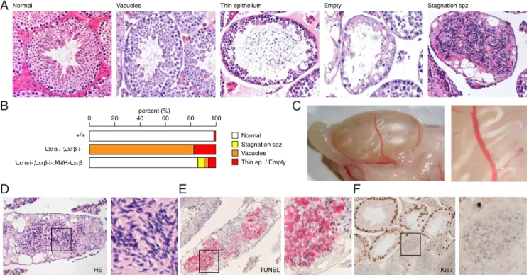
0 0 1 0 20 40 60 80 percent (%) Normal
Thin ep. / Empty Vacuoles Stagnation spz s e l o u c a V l a m r o
N Thin epithelium Empty Stagnation spz
TUNEL
A
B
HE Ki67C
F
E
D
+/+ Lxrα-/-;Lxrβ-/-:AMH-Lxrβ Lxrα-/-;Lxrβ-/-Figure 6. Rescue of Lxr expression in Sertoli cells suppresses seminiferous epithelium degeneration and reveals abnormal spermatozoa transit. A, Microscopic histological alterations (vacuolated tubules, thin seminiferous epithelium, empty tubules, and spermatozoa stagnation) were identified
in HE staining of the wild-type, Lxr␣⫺/⫺;Lxr⫺/⫺, and Lxr␣⫺/⫺;Lxr⫺/⫺:AMH-Lxr testes. B, Relative quantification of the histological alterations in
each genotype. C, The macroscopic image represents the entire testis and shows the whitish seminiferous tubules filled with stagnating
spermatozoa in the Lxr␣⫺/⫺;Lxr⫺/⫺:AMH-Lxr testis. The hematoxylin/eosin (HE) (D), terminal deoxynucleotidyl transferase-mediated deoxyuridine
triphosphate nick end labeling (TUNEL) (E) and Ki67 immunostaining (F) analyses of corked tubules demonstrated apoptotic spermatozoa in the
Lxr␣⫺/⫺;Lxr⫺/⫺:AMH-Lxr testis.
the lumen of the seminiferous tubules (Figure 6C,D). Mac-roscopically, these tubules appear as whitish, thick tubules (Figure 6C); microscopically, they are surrounded by a thick peritubular layer, they lose their epithelial structure and are filled with a large accumulation of spermatozoa (Figure 6D). The spermatozoa within these tubules even-tually become apoptotic (Figure 6E), and the bulk of the luminal cells do not proliferate (Figure 6F).
In light of this unexpected phenotype, we raised two hypotheses: either a distal obstruction or an impaired tu-bular motility could lead to spermatozoa stagnation in the
Lxr␣⫺/⫺;Lxr⫺/⫺:AMH-Lxr mice. We carefully
exam-ined the rete testis (Figure 7A), which did not reveal any obstructions because they harbored spermatozoa in their lumen. We next investigated the layer surrounding the seminiferous tubules. We noticed lipid accumulations in this layer, as shown by semithin Azure 2 dye and PLIN1 staining (Figure 7B). The peritubular smooth muscle cells (PTSM) surrounding the tubules are necessary for tubular contractility and spermatozoa propulsion.␣SMA is a spe-cific differentiation marker of these cells (22). In the
Lxr␣⫺/⫺;Lxr⫺/⫺:AMH-Lxr mice, spermatozoa
stagna-tion is tightly associated with disorganized␣SMA depo-sition in the PTSM, as manifested by an enlarged smooth muscle layer in the Lxr␣⫺/⫺;Lxr⫺/⫺ and Lxr␣⫺/⫺; Lxr⫺/⫺:AMH-Lxr mice (Figure 7C). The stagnation
was rarely present in the Lxr␣⫺/⫺;Lxr⫺/⫺mouse testis due to the degeneration of seminiferous epithelium and the loss of spermatogenesis. In contrast, seminiferous epithe-lium maintenance, which is rescued in the Lxr␣⫺/⫺; Lxr⫺/⫺:AMH-Lxr mice, allows germ cell
differentia-tion and spermatozoa producdifferentia-tion and would unmask the motility defects of the PTSM. Together the rescued Lxr expression in the Lxr␣⫺/⫺;Lxr⫺/⫺:AMH-Lxr mice
re-stored the seminiferous epithelium thickness, revealing PTSM defects, a masked phenotype of the Lxr␣⫺/⫺; Lxr⫺/⫺mice. The question of whether the spermatozoa transit defect is the cause or the consequence of such phe-notype persists. In addition to abnormal lipid accumula-tion within the germ cells (defective spermiogenesis), the defective PTSM phenotype could also participate in the persistent infertility in the Lxr␣⫺/⫺;Lxr⫺/⫺:AMH-Lxr
male mice (Supplemental Figure 6, B–D).
HE SMA rete testis Distal Distal Proximal
A
Azur blue Perilipin HE spz HE SMA HE HE SMAC
SMAB
+/+ Nr1h3-/-:Nr1h2-/-:Amh-Nr1h2 Nr1h3-/-:Nr1h2-/-: Amh-Nr1h2 Nr1h3-/-:Nr1h2-/-: Amh-Nr1h2 +/+ Nr1h3 -/-: Nr1h2-/-: Amh-Nr1h2 Nr1h3 -/-:Nr1h2-/-Figure 7. Description of an LXR-linked phenotype of the peritubular cells. A, hematoxylin/eosin (HE) and␣SMA immunostaining of the rete testis
of the Lxr␣⫺/⫺;Lxr⫺/⫺:AMH-Lxr mice confirmed the presence of spermatozoa in rete testis distal region. B, Semithin cross-sections of
6-month-old wild-type and Lxr␣⫺/⫺;Lxr⫺/⫺:AMH-Lxr testes stained with Azure 2 dye; the white arrowheads indicate lipid accumulations. PLIN1 staining
confirmed the presence of lipid accumulations (white arrowheads) within the PTSM. C, HE and␣SMA immunostaining of wild-type, Lxr␣⫺/⫺;Lxr⫺/
⫺and Lxr␣⫺/⫺;Lxr⫺/⫺:AMH-Lxr testicular tubules exhibited spermatozoa stagnation. The high-magnification image emphasizes the thickness of
the perimuscular cell layer surrounding seminiferous tubules.
Discussion
By generating an Lxr␣⫺/⫺;Lxr⫺/⫺:AMH-Lxr mouse,
we provided an original in vivo model to decipher the physiological roles of LXR in Sertoli cells. LXR regu-lates the levels of lipids in Sertoli cells by controlling the activation of specific genes. Interestingly, LXR is funda-mentally required in Sertoli cells to maintain a functional and effective BTB and to sustain the germ cell pool. In addition, it modulates the endocrine activity of Leydig cells.
As expected, LXR reexpression in Sertoli cells re-stored lipid homeostasis. This is due in part to the in-creased ATP-binding cassette transporter A1/ABCG1 lev-els. This increased gene expression promotes the prompt export of cholesterol from Sertoli cells, preventing vacuole formation. Moreover, Abca1⫺/⫺mice exhibit lipid accu-mulation similar to that of the Lxr␣⫺/⫺;Lxr⫺/⫺ mice (23). Many other mouse models targeting genes that en-code nuclear receptors or proteins that are implicated in lipid metabolism suffered from altered fertility with aging, such as the Abca1⫺/⫺, Rxr⫺/⫺and Ncoa2⫺/⫺mice, for which explanations have been rarely found (4, 5, 23–25). It could be supposed that the large lipid vacuoles alter Sertoli cell physiology due to deformations. Indeed, lipid droplet accumulation could mechanically impair the Ser-toli cell cytoskeleton by disrupting adjacent SerSer-toli-SerSer-toli junctions and Sertoli-germ cell adhesion and interfere with the paracrine actions of Sertoli cells (for review see refer-ence 19). Notably, BTB alterations were found in the
Lxr␣⫺/⫺;Lxr⫺/⫺ mice but not in wild-type and
Lxr␣⫺/⫺;Lxr⫺/⫺:AMH-Lxr mice. The BTB is formed
by tight junctions between neighboring Sertoli cells and protects the germ cells in meiosis from the immune system. The rupture of this barrier could lead to altered spermato-genesis and infertility (for review see reference 26). We demonstrated the necessity of LXR within Sertoli cells to maintain BTB integrity in vivo. This effect could be ex-plained by the disappearance of lipid vacuoles and im-proved T levels within the testis because this barrier is sensitive to androgens.
During maturation, many modifications in the lipid and protein contents of germ cells occur to render them easily motile and fertile. When spermatozoa are released from the seminiferous epithelium, they are believed to be highly loaded with cholesterol, and a progressive loss takes place to acquire the typical membrane fluidity of spermatozoa. Therefore, the regulation of cholesterol me-tabolism in germ cells represents a critical step in sperma-tozoa differentiation. Volle et al (3) have previously de-scribed putative inclusions in the germ cells of the
Lxr␣⫺/⫺;Lxr⫺/⫺ mouse testis. We have demonstrated
that one of the reasons for the lipid accumulation could be the absence of Abcg8 regulation. Indeed, our data show that this gene is expressed in both germ cells and Sertoli cells. Interestingly, the Abcg8⫺/⫺ mice have a very low fertility rate (27). Observations of the Lxr␣⫺/⫺;Lxr⫺/⫺: AMH-Lxr testes support the idea that the intrinsic
ex-pression of LXRs in germ cells plays a pivotal role in final processes of spermiogenesis, which are necessary to obtain fully competent spermatozoa. The intrinsic germ cell de-fects would affect germ cell maturation and even later stages of capacitation, which take place in an apparently abnormal epididymis in the Lxr␣⫺/⫺;Lxr⫺/⫺mice (28). Moreover, Abcg8 is expressed in Sertoli cells and is not under the control of LXR in this compartment because the Lxr␣⫺/⫺;Lxr⫺/⫺:AMH-Lxr mice exhibited lower
levels of this transcript compared with the wild-type mice. The results obtained from the immunolocalization of ABCG8 (Figure 5D) in the three genotypes as well as the busulfan experiments (Figure 5C) suggested that LXR reg-ulation in germ cells was essential for the expression of
Abcg8 in the entire tubule, even in Sertoli cells. Consistent
with the idea that ABCG8 is important during spermio-genesis, we observed that different patterns of accumula-tion of this protein (Figure 5D) from one tubule to another could suggest that Abcg8 expression is associated with waves of spermatogenesis across the seminiferous tubules. Together these results encourage the development and analysis of new mouse models with germ cell-specific de-letions of LXRs to decipher the role of LXR in spermiogenesis.
Our in vivo strategy using the Lxr␣⫺/⫺;Lxr⫺/⫺mice instead of the Lxr⫺/⫺mice to restore Lxr expression was motivated by the opportunity to identify the role of Sertoli cell-specific expression of Lxr regarding the other testicular cell types. This approach leads us to identify the essential role of LXR in Sertoli cells to control steroid-ogenesis in Leydig cells. We previously showed that LXR␣ could activate T synthesis in Leydig cells by modulating the star and 3hsd levels (3). The Lxr␣⫺/⫺;Lxr⫺/⫺: AMH-Lxr strain demonstrated that LXR expression
within the Sertoli cells is able to regulate T synthesis through a putative paracrine effect on Leydig cell steroid-ogenesis. Indeed, both star and 3hsd expressions are re-stored in the Lxr␣⫺/⫺;Lxr⫺/⫺:AMH-Lxr mouse testis.
The secreted mediator of Sertoli cells that is able to regu-late steroidogenesis under the control of LXR needs to be identified. The Lxr␣⫺/⫺;Lxr⫺/⫺:AMH-Lxr mice
pro-vide a unique in vivo model to answer this question. Finally, one of the most striking facts is an unexpected phenotype that was revealed in both the Lxr␣⫺/⫺;Lxr⫺/⫺
and Lxr␣⫺/⫺;Lxr⫺/⫺:AMH-Lxr mice. This phenotype
is characterized by PTSM defects due to lipid
tion and smooth muscle actin disorganization. Interest-ingly, PTSM defects have been associated with altered Ser-toli cell functions and oligoazoospermia or even azoospermia and infertility (7, 29). This phenotype re-mained hidden in the Lxr␣⫺/⫺;Lxr⫺/⫺mice, which may be explained by the quantity of germ cells to be propelled because germ cells are scarce in Lxr␣⫺/⫺;Lxr⫺/⫺mice testis. This observation could be correlated to cholesteryl ester accumulation, as previously described in the muscle compartment of the myometrium from females lacking LXR, who display contractility defects (30).
In conclusion, we present further evidence supporting the critical roles of LXR in Sertoli cells beyond lipid ho-meostasis. As previously postulated by Volle et al (3), the
Lxr␣⫺/⫺;Lxr⫺/⫺:AMH-Lxr mouse model confirmed
that the defective function of the Lxr␣⫺/⫺;Lxr⫺/⫺mouse testis resulted from a combination of phenotypes that emerged from distinct compartments of this gland. The focus on Sertoli cell activity in the Lxr␣⫺/⫺;Lxr⫺/⫺: AMH-Lxr mice demonstrated that paracrine and
endo-crine signals from these cells actively participate in Leydig and germ cell homeostasis. This model allowed us to dis-cover a new role for LXRs in the PTSM compartment.
Acknowledgments
We warmly thank Dr Jean-Yves Picard and Dr Charlotte Lécu-reuil for their helpful discussion and AMH promoter plasmid construct. We thank Dr David Mangelsdorf (Howard Hughes Medical Institute, Department of Pharmacology and Biochem-istry, University of Texas Southwestern Medical Center, Dallas, Texas) for his fruitful collaboration and the availability of LXR-deficient mouse models. We also thank Dr Ivan Wawrzyniak for the semithin sections and Professor Vincent Sapin for the go-nadotropin measurements. This study has been performed with the assistance of Christelle Damon-Soubeyrand for her histology technical assistance using the “Anipath” Platform and Sandrine Plantade, Keredine Ouchen, and Philippe Mazuel for the animal facilities and transgenesis facilities.
Address all correspondence and requests for reprints to: Sil-vère Baron, PhD, Génétique Reproduction et Développement, 24 Avenue des Landais, F-63177 Aubiere, France. E-mail: silvere.baron@univ-bpclermont.fr.
Author contributions include the following: S.M. and F.-Z.E.H. performed in vivo studies and molecular investigations. M.B. assisted with the blood-testis barrier investigation and per-formed the busulfan treatment. E.V. and A.O. assisted with the molecular investigations. F.B., A.T., D.V., and I.T. contributed to the data analyses and the experimental design. J.-M.A.L. and S.B. conceived the study. S.M. and S.B. wrote the paper.
This work was supported by the Fondation pour la Recherche Médicale (to J.-M.A.L.) and Région Auvergne “Nouveau Cher-cheur” (to S.B.).
Disclosure Summary: The authors have nothing to disclose.
References
1. Peet DJ, Turley SD, Ma W, et al. Cholesterol and bile acid metab-olism are impaired in mice lacking the nuclear oxysterol receptor LXR alpha. Cell. 1998;93(5):693–704.
2. Tontonoz P, Mangelsdorf DJ. Liver X receptor signaling pathways in cardiovascular disease. Mol Endocrinol. 2003;17(6):985–993. 3. Volle DH, Mouzat K, Duggavathi R, et al. Multiple roles of the
nuclear receptors for oxysterols liver X receptor to maintain male fertility. Mol Endocrinol. 2007;21(5):1014 –1027.
4. Mascrez B, Ghyselinck NB, Watanabe M, et al. Ligand-dependent
contribution of RXR to cholesterol homeostasis in Sertoli cells.
EMBO Rep. 2004;5(3):285–290.
5. Robertson KM, Schuster GU, Steffensen KR, et al. The liver X
re-ceptor- is essential for maintaining cholesterol homeostasis in the
testis. Endocrinology. 2005;146(6):2519 –2530.
6. Maqdasy S, Baptissart M, Vega A, Baron S, Lobaccaro J-MA, Volle DH. Cholesterol and male fertility: what about orphans and ad-opted? Mol Cell Endocrinol. 2013;368(1–2):30 – 46.
7. Wang R-S, Yeh S, Tzeng C-R, Chang C. Androgen receptor roles in spermatogenesis and fertility: lessons from testicular cell-specific androgen receptor knockout mice. Endocr Rev. 2009;30(2):119 – 132.
8. Rondanino C, Ouchchane L, Chauffour C, et al. Levels of liver X receptors in testicular biopsies of patients with azoospermia. Fertil
Steril. 2014;102(2):361–371.e5.
9. Lécureuil C, Fontaine I, Crepieux P, Guillou F. Sertoli and granulosa cell-specific Cre recombinase activity in transgenic mice. Genesis. 2002;33(3):114 –118.
10. Repa JJ, Mangelsdorf DJ. The liver X receptor gene team: potential new players in atherosclerosis. Nat Med. 2002;8(11):1243–1248. 11. Baptissart M, Vega A, Martinot E, et al. Bile acids alter male fertility
through TGR5 signaling pathways. Hepatology. 2014;60(3):1054 – 1065.
12. Meng J, Holdcraft RW, Shima JE, Griswold MD, Braun RE. An-drogens regulate the permeability of the blood-testis barrier. Proc
Natl Acad Sci USA. 2005;102(46):16696 –16700.
13. Volle DH, Duggavathi R, Magnier BC, et al. The small heterodimer partner is a gonadal gatekeeper of sexual maturation in male mice.
Genes Dev. 2007;21(3):303–315.
14. Pitetti J-L, Calvel P, Zimmermann C, et al. An essential role for insulin and IGF1 receptors in regulating sertoli cell proliferation, testis size, and FSH action in mice. Mol Endocrinol. 2013;27(5): 814 – 827.
15. Pommier AJC, Dufour J, Alves G, et al. Liver x receptors protect from development of prostatic intra-epithelial neoplasia in mice.
PLoS Genet. 2013;9(5):e1003483.
16. Mouzat K, Volat F, Baron S, et al. Absence of nuclear receptors for oxysterols liver X receptor induces ovarian hyperstimulation syn-drome in mice. Endocrinology. 2009;150(7):3369 –3375. 17. El-Hajjaji F-Z, Oumeddour A, Pommier AJC, et al. Liver X
recep-tors, lipids and their reproductive secrets in the male. Biochim
Bio-phys Acta. 2011;1812(8):974 –981.
18. Koopman P, Bullejos M, Bowles J. Regulation of male sexual de-velopment by Sry and Sox9. J Exp Zool. 2001;290(5):463– 474. 19. Pelletier R-M. The blood-testis barrier: the junctional permeability,
the proteins and the lipids. Prog Histochem Cytochem. 2011;46(2): 49 –127.
20. McCabe MJ, Allan CM, Foo CFH, Nicholls PK, McTavish KJ,
ton PG. Androgen initiates Sertoli cell tight junction formation in the hypogonadal (hpg) mouse. Biol Reprod. 2012;87(2):38.
21. Chen M, Wang H, Li X, Li N, Xu G, Meng Q. PLIN1 deficiency affects testicular gene expression at the meiotic stage in the first wave of spermatogenesis. Gene. 2014;543(2):212–219.
22. Tung PS, Fritz IB. Characterization of rat testicular peritubular
myoid cells in culture:␣-smooth muscle isoactin is a specific
differ-entiation marker. Biol Reprod. 1990;42(2):351–365.
23. Selva DM, Hirsch-Reinshagen V, Burgess B, et al. The ATP-binding cassette transporter 1 mediates lipid efflux from Sertoli cells and influences male fertility. J Lipid Res. 2004;45(6):1040 –1050. 24. Kastner P, Mark M, Leid M, et al. Abnormal spermatogenesis in
RXR mutant mice. Genes Dev. 1996;10(1):80–92.
25. Gehin M, Mark M, Dennefeld C, Dierich A, Gronemeyer H, Cham-bon P. The function of TIF2/GRIP1 in mouse reproduction is distinct
from those of SRC-1 and p/CIP. Mol Cell Biol. 2002;22(16):5923– 5937.
26. Lui W-Y, Mruk D, Lee WM, Cheng CY. Sertoli cell tight junction dynamics: their regulation during spermatogenesis. Biol Reprod. 2003;68(4):1087–1097.
27. Solca C, Tint GS, Patel SB. Dietary xenosterols lead to infertility and loss of abdominal adipose tissue in sterolin-deficient mice. J Lipid
Res. 2013;54(2):397– 409.
28. Frenoux JM, Vernet P, Volle DH, et al. Nuclear oxysterol receptors, LXRs, are involved in the maintenance of mouse caput epididymidis structure and functions. J Mol Endocrinol. 2004;33(2):361–75. 29. Welter H, Kampfer C, Lauf S, et al. Partial loss of contractile marker
proteins in human testicular peritubular cells in infertility patients.
Andrology. 2013;1(2):318 –324.
30. Mouzat K, Prod’homme M, Volle DH, et al. Oxysterol nuclear
re-ceptor LXR regulates cholesterol homeostasis and contractile
function in mouse uterus. J Biol Chem. 2007;282(7):4693– 4701.