Electrical stimulation for reducing trapezius muscle dysfunction in cancer patients: traditional treatment protocols also work
RE: Baldwin ERL, Baldwin TD, Lancaster JS, et al. Neuromuscular electrical stimulation and exercise for reducing trapezius muscle dysfunction in survivors of head
and neck cancer: A case-series report. Physiother Can. 2012;64:317-24.
Simon Charest-Hallée,* PT; Guillaume Léonard, PT, PhD*†
*Clinique de physiothérapie Physio Atlas;
†Research Center on Aging, University of Sherbrooke, Sherbrooke, QC
Correspondence to: Guillaume Léonard, Research Center on Aging, University of Sherbrooke, 1036, Belvédère Sud, Sherbrooke (Québec) J1H 4C4;
Dear Editor,
We read Baldwin et al.’s report1 in the summer 2012 issue of Physiotherapy
Canada with great interest. The topic raised by Baldwin et al. is of particular interest to us because we recently treated a patient who was suffering from oral cancer. Following his neck dissection surgery, the patient developed a neuropathy of the XIth cranial nerve,
with paralysis of the trapezius and sternocleidomastoid muscles. The encouraging results obtained by Baldwin et al. led us to try electrical stimulation to promote strength gain in the trapezius muscle and to foster recovery. Unfortunately, the advanced technology used by the authors (allowing, for instance, the triggering of the electrical stimulation via electromyographic feedback) was not available at our clinic. Although the technology used by Baldwin et al. was interesting, we believed that similar effects could be achieved even without such a feedback system.
We therefore started a 6-week treatment program that included electrical
stimulation (ES) and therapeutic exercises (ergocycle, active range of motion [AROM], strengthening). The ES component used a Gymna apparatus designed to stimulate denervated muscles (galvanic current, 1 ms pulse width). The patient came to the clinic three times per week for 6 weeks. The treatment sessions always began with a warm-up period consisting of ergocycle pedaling with moderate resistance for 5 minutes. The patient then completed an exercise routine with the assistance of ES. Specifically, the patient was asked to actively flex both shoulders at the onset of ES and to maintain the contraction as long as he felt the electric current in his dorsal (trapezius) area. The electrodes were positioned on the affected right lower trapezius muscle, based on our physical examination,2 during which we observed marked weakness of the inferior
portion of the trapezius. We began with 2 sets of 10 repetitions, then gradually progressed to 3sets of 12 repetitions at the end of the second week. ES duration was initially set at 5 seconds, progressing to 10 seconds (ON:OFF ratio = 1:3).
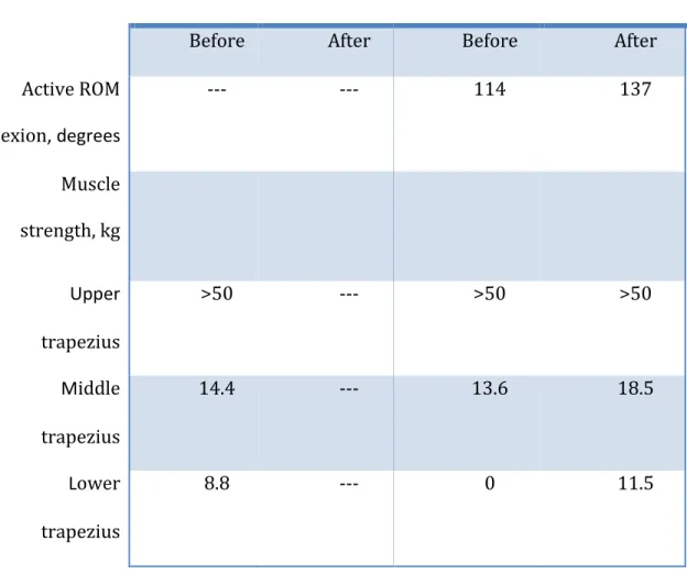
As Table 1 shows, the strength of the affected trapezius significantly improved after treatment. This improvement was associated with an increased AROM and, presumably, increased shoulder function. The most important change in muscle strength was observed in the lower trapezius, consistent with our electrode placement.
The treatment protocol we used significantly improved the strength of the patient’s middle and lower trapezius muscle. We cannot exclude the possibility that the treatment also had beneficial effects on the upper portion of the trapezius, but because we were unable to break the muscle contraction during our evaluation, we cannot fully appreciate its effect on upper trapezius strength. The absence of upper trapezius weakness at initial evaluation surely raises important questions. Given the patient’s history and the structure probably affected by the surgery (the spinal accessory nerve), we would have expected weakness in the whole trapezius muscle. We believe that the strength measured during shoulder shrug might in fact be due to compensation by the levator scapulae muscle. Perhaps a more specific evaluation technique (i.e., with slight arm abduction to inhibit the levator scapulae muscle) would have produced a different pattern of results.
As mentioned above, the protocol reported here is simple and easy to apply in a standard physiotherapy clinic setting. Unlike Baldwin et al., we used no feedback system to initiate ES of the affected trapezius. We also placed the electrodes on the muscle itself rather than on the spinal accessory nerve, to avoid the negative impact that ES might have
on nerve recovery.3, 4 We also believed that such electrode placement would enhance the
reliability of the protocol, as the trapezius muscle is easier to locate than the spinal accessory nerve).
In conclusion, our experience suggests that the treatment protocol described in Baldwin et al.’s research can be slightly modified yet remain effective. Of course, we believe that physiotherapy practice must evolve and take advantage of the technological breakthroughs and new equipment that are emerging in the medical and paramedical field. Nevertheless, those who have access to a more modest array of equipment can still use ES effectively to treat patients with peripheral nerve lesions.
Table 1. Active shoulder range of motion (ROM) and shoulder strength before and after treatment
Unaffected shoulder Affected shoulder
Before After Before After
Active ROM flexion, degrees --- --- 114 137 Muscle strength, kg Upper trapezius >50 --- >50 >50 Middle trapezius 14.4 --- 13.6 18.5 Lower trapezius 8.8 --- 0 11.5
Active shoulder range of motion was measured with a standard goniometer with the patient in a standing position (average of three measures); muscle strength was measured by isolating each muscle section individually, using the method advocated by Hislop & Montgomery.2 The greatest change in muscle strength was observed in the lower
References
1. Baldwin ERL, Baldwin TD, Lancaster JS, et al. Neuromuscular electrical stimulation and exercise for reducing trapezius muscle dysfunction in survivors of head and neck cancer: A case-series report. Physiother Can. 2012;64:317-24.
2. Hislop HJ, Montgomery J. Daniels and worthingham's muscle testing: Techniques of manual examination. Philadelphia: Saunders; 2002.
3. Enes J, Langwieser N, Ruschel J, et al. Electrical activity suppresses axon growth through ca(v)1.2 channels in adult primary sensory neurons. Curr Biol. 2010;20:1154-64. 4. Fields RD, Neale EA, Nelson PG. Effects of patterned electrical activity on neurite outgrowth from mouse sensory neurons. J Neurosci. 1990;10:2950-64.