Université de Montréal
Genetic association study of plasma creatine kinase levels in the Montreal Heart
Institute Hospital Cohort
Par Rosa Zea Zetler
Unité académique Sciences Biomédicales, Université de Montréal Faculté Médicine
Mémoire présenté à la Faculté Médicine en vue de l’obtention du grade de M.Sc
en Sciences Biomédicales option générale
Juin, 2014
i
1 RÉSUMÉ
Il a déjà été démontré que les statines (ou inhibiteurs de la HMG-CoA réductase) sont efficaces pour réduire le LDL-cholestérol et elles se sont depuis établies comme étant le pilier dans le traitement de la dyslipidémie. Toutefois, environ 10 pourcent des utilisateurs de statines souffrent d'effets indésirables, généralement sous forme de myopathie qui est souvent accompagnée d’un taux élevé de la créatine kinase (CK) plasmatique. Il est fréquent que les patients doivent arrêter les statines à cause d’un taux de CK dépassant un seuil de référence. Nous avons examiné le taux de CK de près de 6000 participants de la biobanque de l’ICM, qui ont récemment été génotypés à l'aide de la micropuce d'ADN ExomChip d'Illumina. Des études antérieures ont démontré une association significative entre le taux de CK plasmatique et des polymorphismes génétiques et nous avons cherché à répliquer ces résultats par association génétique et à l'aide du test SKAT pour les polymorphismes rares. Nous avons répliqué les résultats dans le gène CKM (rs11559024, p=1.59x10⁻²³) et le gène LILRB5 (rs12975366, p=1.44x10⁻²⁶) dans le chromosome 19. Nous espérons que ces résultats seront éventuellement utilisés en clinique pour la prédiction des taux de référence de CK personnalisés selon le profil génétique des patients utilisateurs de statines.
ii
2 ABSTRACT
Statins (HMG-CoA reductase inhibitors) have been shown to reduce LDL-cholesterol and are undoubtedly the mainstay in the treatment of hyperlipidemia. Approximately 10 percent of statin users suffer from adverse side effects, the most common being muscle myopathy. Muscle myopathy is often accompanied by elevated levels of plasma creatine kinase (CK). Oftentimes, patients are taken off statins after their CK levels surpass a reference threshold. We looked at CK levels in the MHI Biobank, which have recently been genotyped in over 6000 participants with the Illumina ExomChip. Prior studies have found significant association between plasma CK levels and genetic variants and we aimed to replicate these findings using a genome wide association and a SKAT burden test for rare variants. We were able to replicate findings in the
CKM gene (rs11559024, p=1.59x10⁻²³) and LILRB5 gene (rs12975366, p=1.44x10⁻²³) in chromosome 19. We hope that these results will eventually be utilized in clinical statin care by aiding in the prediction of personalized reference CK levels based on genetic information for patients using statins.
iii
TABLE OF CONTENTS
1 RÉSUMÉ ... i
2 ABSTRACT ... ii
3 LIST OF TABLES... v
4 LIST OF FIGURES ... vii
5 LIST OF ABBREVIATIONS ... ix
6 ACKNOWLEDGEMENTS... xi
7 INTRODUCTION ... 1
7.1 Background on statins ... 1
7.1 Creatine kinase levels as a biomarker ... 5
7.2 Patient management through evaluation of CK levels ... 6
7.3 Predictors of serum creatine kinase levels ... 9
7.4 Adverse effects of statins ... 11
7.5 Compliance with statins ... 13
7.6 Loci associated with statin-induced myotoxicity ... 13
7.7 Myotoxicity reports in clinical trials ... 18
7.8 Summary of prior project ... 20
7.9 Reason for replication in a larger cohort ... 22
7.10 Discovery Study with the MHI Biobank ... 24
8 RATIONAL AND HYPOTHESIS ... 26
9 METHODS ... 27 9.1 Study Design ... 27 9.2 Study population ... 27 9.3 Creatine kinase ... 29 9.4 Statins ... 30 9.5 Physical activity ... 33
9.6 Ethnicity and principal components ... 33
9.7 Hardy-Weinberg equilibrium and linkage disequilibrium ... 34
9.8 Genetic data and cleanup... 34
9.9 Summary of available variants for replication study ... 36
9.10 Statistical analysis ... 39
9.11 Regression model for replication study ... 39
9.12 Statistical analysis of multiple variants at a time for replication study ... 40
iv
9.14 GWAS for discovery study ... 42
9.15 SKAT for discovery study... 44
10 RESULTS ... 45
10.1 Participants ... 45
10.2 Descriptive statistics from MHI Biobank... 46
11 RESULTS FOR REPLICATION STUDY - Objective 1 ... 51
11.1 Results of replication model ... 51
11.2 Linkage disequilibrium analysis... 59
11.3 Statistical analysis of multiple variants ... 61
11.4 Results from imputation analysis ... 68
12 RESULTS FOR THE DISCOVERY STUDY - Objective 2 ... 72
12.1 GWAS Results ... 72
12.2 Results of SKAT ... 76
12.3 Candidate genes of statin-induced myotoxocity ... 83
13 CONCLUSION ... 87 13.1 Key results ... 87 14 DISCUSSION ... 91 14.1 Limitations ... 91 14.2 Study strengths ... 92 14.3 Interpretations... 93 14.4 Generalizability ... 94 14.5 Future Studies ... 95
14.6 Relevance of the genes ... 95
v
3 LIST OF TABLES
TABLE I. OTHER RARE ADVERSE EFFECTS OF STATINS ARE DEMONSTRATED IN THE FOLLOWING TABLE17 ... 12
TABLE II.DEFINITIONS OF STATIN-INDUCED MYALGIA ... 7
TABLE III.TABLE OF EQUIVALENT DOSAGE OF STATINS ... 32
TABLE IV.TABLE OF VARIANTS IN CKM AND LILRB5 GENES AVAILABLE FOR REPLICATION STUDY ... 36
TABLE V.DESCRIPTIVE STATISTICS OF PARTICIPANTS FROM THE MHIBIOBANK ... 47
TABLE VI.OVERVIEW OF THE 3516 PATIENTS CURRENTLY TAKING STATINS, WITH STATIN DOSAGE AVAILABLE, AND WITH A CREATININE LEVEL UNDER 200 MMOL/L ... 48
TABLE VII.ASSOCIATION ANALYSIS BETWEEN LOG(CK LEVELS) AND SNPS FOR ALL PARTICIPANTS IN THE MHIHOSPITAL COHORT.THE MULTIVARIATE ANALYSIS WITH A GENERAL LINEAR MODEL OF CK IS PERFORMED FIRST WITH ADDITIVE EFFECT ONLY.P-VALUES SURPASSING THE BONFERONI THRESHOLD ARE HIGHLIGHTED. ... 52
TABLE VIII.ASSOCIATION ANALYSIS BETWEEN LOG(CK LEVELS) AND SNPS FOR PARTICIPANTS IN THE MHIHOSPITAL COHORT TAKING STATINS.THE MULTIVARIATE ANALYSIS WITH A GENERAL LINEAR MODEL OF CK IS PERFORMED FIRST WITH ADDITIVE EFFECT ONLY AND SECOND WITH ADDITIVE EFFECT, GENDER, STATIN DOSE, AGE AND PHYSICAL ACTIVITY.P-VALUES SURPASSING THE BONFERONI THRESHOLD ARE HIGHLIGHTED. ... 55
TABLE IX.ASSOCIATION ANALYSIS BETWEEN LOG(CK LEVELS) AND SNPS FOR PARTICIPANTS IN THE MHIHOSPITAL COHORT NOT TAKING STATINS.THE MULTIVARIATE ANALYSIS WITH A GENERAL LINEAR MODEL OF CK IS PERFORMED FIRST WITH ADDITIVE EFFECT ONLY AND SECOND WITH ADDITIVE EFFECT, GENDER, AGE AND PHYSICAL ACTIVITY.P-VALUES SURPASSING THE BONFERONI THRESHOLD ARE HIGHLIGHTED. ... 56
TABLE X.MEAN SERUM CK VALUES BY GENOTYPE OF THE ASSOCIATED VARIANTS IN THE CKM AND LILRB5 GENES IN MHIHOSPITAL COHORT ... 58
TABLE XI.ASSOCIATION ANALYSIS BETWEEN LN(CK LEVELS) AND SNPS RS11559024 AND RS12975366 GENOTYPES STRATIFIED BY STATIN/NO STATIN/ALL PARTICIPANTS IN THE MHIHOSPITAL COHORT.THE MULTIVARIATE ANALYSIS WITH A GENERAL LINEAR MODEL OF CK IS PERFORMED FIRST WITH GENOTYPES ONLY AND SECOND WITH ADDITIVE EFFECT, GENDER, STATIN DOSE, AGE AND PHYSICAL ACTIVITY. ... 62
TABLE XII.VARIANCE EXPLAINED FOR EACH OF THE STRATIFIED MODELS IN TABLE XII SUMMARIZED BY R-SQUARED. ... 63
TABLE XIII.ASSOCIATION ANALYSIS BETWEEN LOG(CK LEVELS) AND SNPS RS11559024 AND RS12975366 GENOTYPES STRATIFIED BY STATIN/NO STATIN/ALL PARTICIPANTS IN THE MHIHOSPITAL COHORT.THE MULTIVARIATE ANALYSIS WITH A GENERAL LINEAR MODEL OF CK IS PERFORMED FIRST WITH GENOTYPES ONLY AND SECOND WITH ADDITIVE EFFECT, GENDER, STATIN DOSE, AGE AND PHYSICAL ACTIVITY. ... 64
TABLE XIV.VARIANCE EXPLAINED FOR EACH OF THE STRATIFIED MODELS IN TABLE XIV SUMMARIZED BY R-SQUARED. ... 65
TABLE XV.ASSOCIATION ANALYSIS BETWEEN LOG(CK LEVELS) AND SNPS RS11559024, RS12975366 AND EXM 1480239 GENOTYPES STRATIFIED BY STATIN/NO STATIN/ALL PARTICIPANTS IN THE MHIHOSPITAL COHORT.THE MULTIVARIATE ANALYSIS WITH A GENERAL LINEAR MODEL OF CK IS PERFORMED FIRST WITH GENOTYPES ONLY AND SECOND WITH ADDITIVE EFFECT, GENDER, STATIN DOSE, AGE AND PHYSICAL ACTIVITY. ... 66
TABLE XVI.VARIANCE EXPLAINED FOR EACH OF THE STRATIFIED MODELS IN TABLE XVI SUMMARIZED BY R-SQUARED. ... 67
TABLE XVII.INFORMATION ON IMPUTATION OF SNP RS2361797 ... 69
TABLE XVIII.THIS TABLE EXPANDS UPON REGIONS THAT SURPASSED THE SIGNIFICANT THRESHOLD LEVEL IN THE SKAT ANALYSIS WHERE ALL PARTICIPANTS WERE LOOKED AT, ADJUSTED FOR GENETIC COMPONENTS 1 AND 2 ... 78
TABLE XIX.THIS TABLE EXPANDS UPON REGIONS THAT SURPASSED THE SIGNIFICANT THRESHOLD LEVEL IN THE SKAT ANALYSIS WHERE STATIN USERS WERE LOOKED AT, ADJUSTED FOR GENETIC COMPONENTS 1 AND 2 ... 80
TABLE XX.THIS TABLE EXPANDS UPON REGIONS THAT SURPASSED THE SIGNIFICANT THRESHOLD LEVEL IN THE SKAT ANALYSIS WHERE NON -STATIN USERS WERE LOOKED AT, ADJUSTED FOR GENETIC COMPONENTS 1 AND 2. ... 82
TABLE XXI.TABLE OF CANDIDATE GENES ASSOCIATED WITH STATIN-INDUCED MYOTOXICITY FOUND IN A LITERATURE REVIEW.THE TABLE INCLUDES P-VALUES FROM THE GWAS; P-VALUES BELOW 0.05 ARE HIGHLIGHTED.VARIANTS WERE ONLY INCLUDED IN THE CHART IF THEY HAD A MAF>0.05 IN THE MHIHOSPITAL COHORT. ... 84
TABLE XXII.TABLE OF CANDIDATE GENES ASSOCIATED WITH STATIN-INDUCED MYOTOXICITY FOUND IN A LITERATURE REVIEW.THE TABLE INCLUDES GENE-BASED P-VALUES FROM THE SKAT ANALYSIS; P-VALUES BELOW 0.05 ARE HIGHLIGHTED.VARIANTS WERE ONLY INCLUDED IN THE CHART IF THEY HAD A MAF>0.0001 AND <0.05 IN THE MHIHOSPITAL COHORT. ... 86
vi
TABLE XXIII.SUMMARY TABLE OF SNP RS12975366 LOCATED IN THE LILRB5 GENE IN CHROMOSOME 19 IDENTIFIED IN THE GWAS
ANALYSIS ... 89
TABLE XXIV.SUMMARY TABLE OF SNP VARIANTS IDENTIFIED IN THE SKAT ANALYSIS VARIANTS SURPASSING THE SIGNIFICANCE THRESHOLD
vii
4 LIST OF FIGURES
FIGURE 1.MANHATTAN PLOT SHOWING THE RESULTS OF A GENOME-WIDE ASSOCIATION STUDY FOR GENETIC DETERMINANTS OF CK LEVELS MEASURED IN STATIN USERS IN THE MHI STATIN STUDY.A GLM REGRESSION WITH THE NATURAL LOGARITHM OF CK WAS USED, WITH ADJUSTMENT FOR THE CASE-CONTROL STATUS, THE LAB WHERE THE CK MEASURES WERE TAKEN, GENDER, PHYSICAL ACTIVITY LEVEL, AGE, DIABETES AND BMI. THERE WERE 3388 PARTICIPANTS IN THE STATIN MYOTOXICITY CASE-CONTROL STUDY THAT WERE USING STATINS AT THE TIME OF CK MEASUREMENT.GENETIC VARIANTS IN THE CKM GENE (P=5.03X10⁻¹⁶) AND THE
LILRB5 GENE (P=5.71X10⁻¹¹) WERE IDENTIFIED. ... 21
FIGURE 3.FLOWCHART OF PARTICIPANTS FROM MHIBIOBANK THAT TRACKS WHERE PARTICIPANTS WERE EXCLUDED ... 46 FIGURE 4.THE PRINCIPAL COMPONENTS FROM THE MHI COHORT DATA USING PCA AS A PROXY FOR ETHNICITY.CHARTS, FROM LEFT TO
RIGHT, REPRESENT DAL-OUTCOMES (ROCHE) AND PROPORTIONAL CUMULATIVE EXPLAINED VARIANCE OF THE FIRST 10
COMPONENTS.THE THIRD PRINCIPAL COMPONENT SEEMS TO BE AN INFLECTION POINT ON THE SCREEN PLOT, SO IN FURTHER ANALYSIS ONLY THE FIRST TWO PRINCIPAL COMPONENTS WERE USED. ... 50 FIGURE 5.PRINCIPAL COMPONENTS 1 AND 2, MARKED BY SELF REPORTED ETHNICITY ... 50 FIGURE 6.LINKAGE DISEQUILIBRIUM OF AVAILABLE SNPS IN THE CKM AND LILRB5 GENE OBTAINED FROM HAPLOVIEW.THE ABOVE
CHART LOOKS AT THE R2BETWEEN VARIANTS OF THE 5093 PARTICIPANTS FROM THE MHIHOSPITAL COHORT.ALL SNPS ARE IN
HARDY-WEINBERG EQUILIBRIUM. EACH TWO DIFFERENT SNPS ARE IN LINKAGE DISEQUILIBRIUM. ... 60
FIGURE 7.MANHATTAN PLOT SHOWING THE RESULTS FOR GENETIC DETERMINANTS OF CK LEVELS MEASURED IN ALL PARTICIPANTS IN THE
MHI COHORT STUDY ON CHROMOSOME 19 BETWEEN POSITION 52272131 AND 57182204(BUILD 37) INCLUDING VARIANTS FROM THE IMPUTATION ANALYSIS.AGLM REGRESSION WITH THE NATURAL LOGARITHM OF CK WAS USED, WITH ADJUSTMENT FOR
COMPONENTS 1 AND 2. A GENETIC VARIANTS IN THE LILRB5 GENE (P= 6.01X10⁻¹⁵) WAS IDENTIFIED ON RS12975366. ... 69
FIGURE 8.QQ PLOT CORRESPONDING TO FIGURE 1 FOR ALL PARTICIPANTS IN THE MHI COHORT STUDY.AGLM REGRESSION WITH THE NATURAL LOGARITHM OF CK WAS USED WITH ADJUSTMENT FOR PRINCIPAL COMPONENTS 1 AND 2. ... 70
FIGURE 9.MANHATTAN PLOT SHOWING THE RESULTS OF A GENOME-WIDE ASSOCIATION STUDY FOR GENETIC DETERMINANTS OF CK LEVELS MEASURED IN ALL PARTICIPANTS (N=5809) IN THE MHI COHORT STUDY.ONLY VARIANTS WITH MAF>5% WERE USED.AGLM
REGRESSION WITH THE NATURAL LOGARITHM OF CK WAS USED, WITH ADJUSTMENT FOR COMPONENTS 1 AND 2. THE GENETIC
VARIANT RS12975366 IN THE LILRB5 GENE (P=1.44X10⁻²³) WAS IDENTIFIED. ... 73
FIGURE 10.QQ PLOT CORRESPONDING TO FIGURE 8 AND COMPARED WITH A QQ PLOT OF ALL VARIANTS, COMMON SNPS WITH GREATER THAN 5% ALLELE FREQUENCY FOR ALL PARTICIPANTS IN THE MHI COHORT STUDY.AGLM REGRESSION WITH THE NATURAL LOGARITHM OF CK WAS USED WITH ADJUSTMENT FOR PRINCIPAL COMPONENTS 1 AND 2. ... 73
FIGURE 11.MANHATTAN PLOT SHOWING THE RESULTS OF A GENOME-WIDE ASSOCIATION STUDY FOR GENETIC DETERMINANTS OF CK
LEVELS MEASURED IN STATIN USERS (N=3673) IN THE MHI COHORT STUDY.ONLY VARIANTS WITH MAF>5% WERE USED.A GLM REGRESSION WITH THE NATURAL LOGARITHM OF CK WAS USED, WITH ADJUSTMENT FOR COMPONENTS 1 AND 2.THE GENETIC VARIANT RS12975366 IN THE LILRB5 GENE (P=3.87X10⁻¹⁴) WAS IDENTIFIED. ... 74
FIGURE 12.QQ PLOT CORRESPONDING TO FIGURE 10 AND COMPARED WITH A QQ PLOT OF ALL VARIANTS, COMMON SNPS WITH GREATER THAN 5% ALLELE FREQUENCY FOR STATIN USERS IN THE MHI COHORT STUDY.AGLM REGRESSION WITH THE NATURAL LOGARITHM OF CK WAS USED WITH ADJUSTMENT FOR PRINCIPAL COMPONENTS 1 AND 2. ... 74
FIGURE 13.MANHATTAN PLOT SHOWING THE RESULTS OF A GENOME-WIDE ASSOCIATION STUDY FOR GENETIC DETERMINANTS OF CK
LEVELS MEASURED IN NON-STATIN USERS (N=2134) IN THE MHI COHORT STUDY.ONLY VARIANTS WITH MAF>5% WERE USED.A
GLM REGRESSION WITH THE NATURAL LOGARITHM OF CK WAS USED, WITH ADJUSTMENT FOR COMPONENTS 1 AND 2. THE GENETIC VARIANT RS12975366 IN THE LILRB5 GENE (P=7.9X10⁻¹¹) WAS IDENTIFIED. ... 75
FIGURE 14.QQ PLOT CORRESPONDING TO FIGURE 12 AND COMPARED WITH A QQ PLOT OF ALL VARIANTS, COMMON SNPS WITH GREATER THAN 5% ALLELE FREQUENCY FOR NON-STATIN USERS IN THE MHI COHORT STUDY.AGLM REGRESSION WITH THE NATURAL LOGARITHM OF CK WAS USED WITH ADJUSTMENT FOR PRINCIPAL COMPONENTS 1 AND 2. ... 75
FIGURE 15.MANHATTAN GRAPH SHOWING THE RESULTS FOR A BETA WEIGHTED SKAT ANALYSIS FOR ALL PARTICIPANTS (N=5349) FROM THE MHI HOSPITAL COHORT, RARE SNPS ONLY, WITH ADJUSTMENT FOR C1 AND C2, WITH INTERGENIC GENES. A GENETIC VARIANT IN THE CKM GENE (P=1.59X10⁻²³) WAS FOUND. ... 77 FIGURE 16.QQ PLOT CORRESPONDING FIGURE 14 FOR THE BETA WEIGHTED SKAT ANALYSIS FOR ALL PARTICIPANTS FROM THE MHI
viii
FIGURE 17.MANHATTAN GRAPH SHOWING THE RESULTS FOR A BETA WEIGHTED SKAT ANALYSIS FOR STATIN USERS (N=3403) FROM THE
MHI HOSPITAL COHORT, RARE SNPS ONLY, WITH ADJUSTMENT FOR C1 AND C2, WITH INTERGENIC GENES.A GENETIC VARIANT IN
THE CKM GENE (P=2.55X10⁻¹⁶) WAS FOUND. ... 79
FIGURE 18.QQ PLOT CORRESPONDING TO FIGURE 16 FOR THE BETA WEIGHTED SKAT ANALYSIS FOR STATIN USERS FROM THE MHI
HOSPITAL COHORT, RARE SNPS ONLY, WITH ADJUSTMENT FOR C1 AND C2, WITH INTERGENIC GENES. ... 79 FIGURE 19.MANHATTAN GRAPH SHOWING THE RESULTS FOR A BETA WEIGHTED SKAT ANALYSIS FOR NON STATIN USERS (N=1944) FROM THE MHI HOSPITAL COHORT, RARE SNPS ONLY, WITH ADJUSTMENT FOR C1 AND C2, WITH INTERGENIC GENES.GENETIC VARIANTS IN THE CKM GENE (P=1.46=X10-7) AND THE KLHL18 GENE (P=8.82X10⁻⁸) WERE FOUND. ... 81
FIGURE 20.QQ PLOT FOR BETA WEIGHTED SKAT ANALYSIS FOR NON STATIN USERS FROM THE MHI HOSPITAL COHORT, RARE SNPS ONLY,
ix
5 LIST OF ABBREVIATIONS
ABCB1 – ATP-binding cassette, subfamily B (MDR/TAP), member 1 Transporter ABCG2 – ATP-binding cassette, subfamily G (WHITE), member 2 Transporter ADP – adenosine diphosphate
AGTR1 – angiotensin II receptor, type 1 ApoB – apolipoprotein B
ATP – adenosime tripohsphate
ATP2B1 – ATPase, Ca++ transporting, plasma membrane 1 CK – Creatine kinase
CKM – creatine kinase muscle gene
COQ2 – Coenzyme Q2 4-hydroxybenzoate polyprenyltransferase CYP2C9 – Cytochrome P450, family 2, subfamily C, polypeptide 9 CYP3A4 – Cytochrome P450, family 3, subfamily A, polypeptide 4 CYP3A5 – cytochrome P450, family 3, subfamily A, polypeptide 5 CYP2D6 – cytochrome P450, family 2, subfamily D, polypeptide 6 ER – Exertional rhabdomyolysis
GATM – Glycine amidinotransferase GLM – general linear model
GWAS – Genome wide association study
HMG-CoA – Hydroxy-methylglutaryl-coenzyme A HTR3B – 5-hydroxytryptamine (serotonin) receptor 3B
HTR7 – 5-hydroxytryptamine (serotonin) receptor 7,adenylate cyclase-coupled LDL – Low density lipoprotein
MAF – minor allele frequency
NOS3 – nitric oxide synthase 3, endothelial cell
OATP1B1 – Organic anion-transporting polypeptide, member 1B1 PCA – principal component analysis
RYR2 – ryanodine receptor 2
SKAT – Sequence kernel association test
SLCO1B1 – Solute carrier organic anion transporter family, member 1B1 SNP – single nucleotide polymorphism
SNV – single nucleotide variant ULN – upper limit of normal
x
I dedicate this work to Copernicus, for never being afraid to question science.
xi
6 ACKNOWLEDGEMENTS
I would like to thank everyone at the Pharmacogenomics lab; your patience and guidance made this paper possible. A special thanks to my supervisor Marie-Pierre Dubé – your support was unwavering.
To my family and friends, thank you for making me laugh. Through this experience, I have learnt that it is not how we deal with our success, but with our adversity that defines us. In the words of the late Pete Seeger, “we’re waist deep in the Big Muddy and the big fool says to push on!”
1
7 INTRODUCTION
7.1 Background on statins
Statins (HMG-CoA (hydroxy-methylglutaryl-coenzyme A) reductase inhibitors) have been shown to reduce LDL-cholesterol and are undoubtedly the mainstay in the treatment of hyperlipidemia. Statins have revolutionized primary and secondary prevention of coronary atherosclerotic disease due to their lipid-lowering properties and other pleiotropic effects that affect atherosclerotic plaque stability2. Statins work as an inhibitor as they share an HMG-CoA like moiety that acts as a HMG-CoA reductase by competing with the HMG-CoA at the binding site.
Among persons with coronary artery disease, there is an evident decrease in mortality in statin users which persists in all age groups. One study found that when non-statin users were compared to statin users, mortality decreased from 29.5% to 8.5% in participants greater than or equal to 80 years of age, 18.7% to 6.9% in participants 65 to 79 years old and 8.9% to 3.1% in participants younger than 653. The efficacy of statins as primary prevention on patients with high cholesterol but no history of heart disease is unclear; one meta-analysis found no significant mortality benefits4. When used in secondary prevention, statins have been found to decrease cardiovascular endpoints in patients with pre-existing cardiovascular disease and lower LDL-cholesterol by an average of 1.8 mmol (70 mg/dl), preventing approximately 60% of cardiac events5.
The synthesis of a cholesterol lowering drug began in 1971 with Akira Endo, a Japanese biochemist. She speculated that fungi such as molds and mushrooms would produce antibiotics
2
that inhibited HMG-CoA reductase. Endo’s team identified mevastatin, a molecule produced by the fungus Penicillium citrinum. Although mevastatin is thought to be the first known statin, it was never deemed safe for human use6 as adverse effects included tumors, muscle deterioration and death. In 1982, lovastatin was identified in Pleurotus ostreatus, and by 1987 it was approved by the FDA.
There are currently six statins available for patients in Canada: atorvastatin, fluvastatin, lovastatin, pravastatin, rosuvastatin and simvastatin. Lovastatin, simvastatin and pravastatin are derived from fungi while the other three are synthetic. Their relative potency, from highest to lowest, is: rosuvastatin, atorvastatin, simvastatin, lovastatin, pravastatin, and fluvastatin7. Relative risk rates when comparing statins are considered to be highest in rosuvastatin, then atorvastatin and simvastatin and lastly pravastatin and lovastatin8. Relative potency of statins seems to correlate with relative risk, with the exception of fluvastatin8.
Mechanism of statins. HMG-CoA reductase is a catalyst in the early biosynthesis of cholesterol
which derives mevalonate by a 4-electron reduction of HMG-CoA. All statins work by effectually inhibiting this step by competing for the catalytic binding domain for the HMG-CoA in the HMG-CoA reductase molecule. Statins are competitive inhibitors of HMG-CoA reductase and, when competing with HMG-CoA, have a significantly higher binding affinity with the reductase molecule. The HMG-CoA-like moiety of a statin monopolizes the HMG-CoA binding pocket in the HMG-CoA reductase molecule and links via its O5 hydroxy group9. When the HMG-CoA moiety is in the binding pocket, the remainder of the statin structure undergoes a conformational change in order to maximize contact with, and binding to, amino acid residues of
3
the reductase. The number and strength of the bonds vary between statins, dictating the length and degree of inhibition and ultimately resulting in the level of LDL cholesterol-lowering efficacy. Rosuvastatin forms nine bonding interactions with the reductase, making it the most potent statin, followed by atorvastatin, which forms eight bonds. The fungal statins, lovastatin, simvastatin and pravastatin, each have six bonding interactions9.
Through the inhibition of HMG-CoA reductase, there is a reduction of cholesterol synthesis in the hepatocyte, a reduction in the cholesterol pool and subsequent up-regulation of the nuclear transcription factors sterol regulatory element-binding proteins, resulting in an increase in the transcription of LDL receptors on the hepatic cell surface. The LDL receptors link with apolipoprotein (apo) B and apoE on the surface of circulating LDL and very-low density lipoprotein particles are integrated, along with their lipid content, into the hepatocyte10. In addition, when hepatic cholesterol is converted to bile acids the LDL receptors are up-regulated to help with food absorption in the gastrointestinal tract.
Metabolism of statins. Lipid-soluble statins are converted to water-soluble salts and glucuronide
conjugates for elimination from the body while simvastatin, lovastatin and, to a lesser extent, atorvastatin are metabolized by cytochrome P450 3A4 (CYP3A4) enzymes. Numerous drugs are either inhibitors or substrates for this enzyme system and can result in drug-drug interactions, leading to increased plasma concentrations of the statin. Approximately 10% of fluvastatin and rosuvastatin are metabolized primarily by CYP2C911, the rest remaining fairly unchanged until excretion. These two statins are excreted from the body largely unchanged as parent compounds by transporter-mediated excretion mechanisms in the liver into the feces via bile and into the
4
urine. Pravastatin is not metabolized by the P450 enzyme system. The open acid forms of several statins often undergo glucuronidation followed by lactonization12. In their lactone form, statins
may be converted to their open acid form by an esterase enzyme and metabolized, excreted by bile or urine, or directly metabolized by the cytochrome P450 system. Excretion of statins through bile or feces is mediated by transport proteins such as OATP1B1 and multidrug resistance-associated protein-2.
Drug interactions in statin users must be closely monitored, as drugs often affect metabolism time, which in turn affects the systematic concentration of statin. Drugs inhibiting the CYP3A4 enzyme can interfere with the metabolism of atorvastatin, simvastatin, and lovastatin causing an increase in the area under the statin concentration-time curve. Drugs that inhibit CYP2C9 should theoretically affect the metabolism of fluvastatin, although in practice there are very few clinical observations of this. OATP1B1 is an inhibitor that, alongside other inhibitors of CYP3A4, inhibits a vital transporter by which hydrophilic statins are transported into the liver.
Certain fibrates, a class of drugs that lowers blood triglyceride levels, and Warfarin, an anticoagulant mainly used in the prevention of thrombosis and thromboembolism, have the potential to interact with statin therapy. The fibrate gemfibrozil can interact with the OATP1B1-mediated transport resulting in an increase of plasma statin concentration13. Additionally, gemfibrozil interferes with the glucuronidation and lactonization of statins, promoting a higher concentration of open acid statins. To avoid these drug interactions, it is recommended that the fibrate fenofibrate is used instead to help avoid adverse effects, or the statins atorvastatin or
5
fluvastatin should be used. Warfarin-statin interactions may result in a displacement from plasma protein binding sites by the statin, resulting in a minor increase of prothrombin times and bleeding. While adverse effects are uncommon, close monitoring of the patient is recommended for a patient using Warfarin when lovastatin, rosuvastatin or simvastatin is added or removed from a regiment.
7.1 Creatine kinase levels as a biomarker
Creatine kinase is the most commonly used enzyme utilized in the diagnosis of muscle disease. Serum levels of CK are the most sensitive known biomarker and the best known indicator of the course of muscle injury14. In extreme cases, this muscle myopathy can manifest into rhabdomyolysis, showcasing a correlated increase in plasma CK levels. Using CK as a biomarker is an effective and non-invasive sensitivity test for various levels of muscle myopathy as well as myocardial infractions.
CK is an enzyme found predominantly in skeletal muscle, the myocardium and the brain. CK catalyzes the reversible reaction of creatine, consuming ATP resulting in the production of phosphocreatine and ADP. CK is expressed as any of the following three isoenzymes: CK-MM, CK-BB, CK-MB where B represents the brain type subunit and M the muscle type subunit.
Disruption of cell membranes can release CK from the cellular cytosol into the systemic circulation. This elevation of plasma CK levels can be used as a biomarker for myocardial infarction, rhabdomyolysis, muscle dystrophy, the autoimmune myositides and in acute renal failure.
6
Serum CK is a commonly used biomarker for myocardial infarction. 40% of CK in cardiac muscles is CK-MB. Levels of CK-MB tend to increase within 3 to 4 hours of myocardial necrosis, peak within a day and return to normal within 36 hours. Subsequent reinfarctions can be diagnosed if CK-MB levels do not begin to decline after approximately 24 hours15. Although highly effective in diagnosing acute MI, it is important not to rely solely on CK-MB as a biomarker, as an electrocardiogram should ultimately be used as an assessment of the patients risk and stability16.
7.2 Patient management through evaluation of CK levels
Clinicians often measure serum CK levels as a rough proxy for severity of statin-induced myotoxicity, but the correlation between symptoms and CK level remains incomplete. The clinical interpretation of CK level is complex and there is not yet a consensus on the definition of statin myopathy. The American College of Cardiology17, American Heart Association (AHA), National Heart, Lung and Blood Institute (NHLBI)18, the FDA19 and National Lipid Association (NLA)20 have each proposed different definitions for statin-related muscle effects (Table II).
7 Table I. Definitions of statin-induced myalgia
Clinical entity ACC/AHA/NHLBI 2002 NLA 2006 FDA
Myopathy General term referring to any disease of muscles
Complaints of myalgia (muscle pain or soreness), weakness, and/or cramps, plus elevation in serum CK > 10 × upper limit of normal (ULN)
CK ≥ 10 × ULN
Myalgia Muscle ache or weakness
without CK elevation
NA NA
Myositis Muscle symptoms with
increased CK
NA NA
Rhabdomyolysis Muscle symptoms associated with marked CK elevations, typically substantially > 10 × ULN and with creatinine elevation
CK > 10,000 IU/l or CK > 10 × ULN plus an elevation in serum creatinine or medical intervention with i.v. hydration CK > 50 × ULN and evidence of organ damage, such as renal compromise
The lack of consensus on the definition of statin myopathy hinders estimation of its true incidence. Elevations less than threefold the upper limit of normal (ULN) are typically considered of little consequence. Conversely, clinicians often intervene in statin therapy (change dose or change drug) when CK levels exceed threefold the ULN. At present, the literature supports three diagnostic strata: (i) incipient myopathy (CK above 3-fold the ULN, less than 10-fold the ULN), (ii) myopathy (CK above 10-10-fold the ULN, less than 50-10-fold the ULN), and (iii) rhabdomyolysis (CK above 50-fold the ULN)21. CK levels are not routinely measured before statin therapy begins. Patients are encouraged to report myalgia by a physician warning or the product information sheet. When CK levels are elevated, the statin is usually withdrawn,
8
although, largely due to the lack of baseline CK measurement, it is difficult to determine whether statin therapy or another cause is to blame.
Disagreement in the definition of statin-induced muscle events impedes the precise estimation of its true prevalence. Because patients with a considered high susceptibility to statin toxicity are generally excluded from clinical trials of statins, reported adverse event rates from controlled trials may underestimate the true rate of these adverse effects in an unselected patient population. As genetic variants may play a role in elevated levels of plasma CK, it is valuable to analyze the SNPs in the hopes of finding a new biomarker to better diagnose CK levels in patients. There has been a preliminary study done at the Pharmacogenomics center, and strong association was found between variants in the muscle CK gene (CKM) and elevated plasma levels of CK.
A meta-analysis of 21 clinical trials providing 180,000 person-years of follow-up found that myopathy, defined by muscle symptoms and CK levels above a 10-fold ULN, occurs in five patients per 100,000 person-years20. Rhabdomyolysis, defined by either CK levels above 10,000 IU/L, CK levels above a 10-fold ULN with an elevation in serum creatinine or requirement for hydration therapy, occurs in 1.6 patients per 100,000 person-years22. Less severe manifestations are much more common. Although CK has long been used as a diagnostic biomarker, CK measurement is not always accurate for the clinical management of statin-induced myotoxicity23, and therefore more accurate tools and/or novel sensitive biomarkers are required.
9
7.3 Predictors of serum creatine kinase levels
Although elevated levels of CK are often used as a biomarker for muscle myopathy, the expected level of CK in a patient is often unclear. Elevated CK levels tend to be higher in certain populations, and as baseline CK levels are not routinely measured due to cost and time limitations, it is hard to gauge what the actual effects of statins are on CK levels in a patient. Covariates such as gender, ethnicity, age and physical activity levels have been found to be highly correlated with plasma CK levels.
High intensity exercise will spike CK levels for a limited amount of time, usually for a few days following exercise24. Females tend to have lower baseline levels of CK, although it has been suggested that females might undergo a greater spike in CK levels with physical activity25,26. It has been refuted that this increased spike in CK levels may be attributed to poor study design, and not actually account for the differences in genders exercising27,28. Although there is a clear correlation between serum CK levels and recent physical activity, it is unclear whether this spike is a good predictor of muscle damage29. When completing a routine measure of a patient’s serum CK levels, it is hard to account for the effect of physical activity in the measurement and whether this increase in serum CK levels is leading to muscle myopathy.
Exertional rhabdomyolysis (ER) is the degradation of skeletal muscle cells caused by strenuous exercise. Common indicators of ER include muscle pain, myoglobinuria and an elevated level of CK after exercise. It is unclear why certain individuals experience ER while others, who participate in comparable amounts of physical activity, do not. Confounders include, but are not limited to, fasting, hypokalemia, certain dietary supplements, dehydration, concurrent
10
illnesses, low baseline fitness levels and extreme or repetitive exercise30. Although CK is often used to monitor ER, correlation is unclear. Some patients with CK levels greater than 10,000 U/L have no overt clinical symptoms, while others with a CK level less than 5,000 U/L exhibit symptoms of ER31. One study that looked at genetic polymorphisms associated with ER found three noteworthy SNPs in the CKMM, ACTN3 and MYLK2 genes32. Another study that looked at association between serum CK and ER in military recruits found the SNPs IL-6 and MLCK 37885 to be positively associated, and furthermore these SNPs were more common in participants of African-American descent33. It was noted that this association may have been due to genetic differences in race as opposed to differences in CK levels.
Age and gender have an effect on expected serum CK levels. The average male’s baseline CK level is higher than the average females. Additionally, there is an age-dependant decrease of CK levels, seen predominantly in males34.
Individuals of African American ancestry have been shown to have higher levels of creatine kinase when compared to Caucasian, Hispanic and Asian people34. Current standards for measuring muscle myopathy through CK levels disregard biological and environmental differences in patients. Serum CK levels are reported to be approximately 70% higher in a healthy Black person when compared to a healthy Caucasian35. A young African American male can be expected to experience higher than normal levels of CK levels without muscle damage, which is not accounted for in the above definitions of myopathy and rhabdomyolysis.
11
7.4 Adverse effects of statins
Most patients tolerate statins well; however, 10% of patients develop muscle-related adverse effects36. The most commonly reported adverse effect of statins is muscle myotoxicity, which can, in the worst cases, progress to rhabdomyolysis. Other adverse effects include toxicity in the kidneys and liver, and may be heightened by age or concomitant medications. Accurate diagnosis of statin-induced myopathy is important given the large and expanding numbers of patients eligible for statin therapy37 and the fact that myalgia, or muscle pain, is one of the most frequent causes of discontinuation of therapy. Statins can cause a wide range of muscular adverse effects with no specific clinical characteristics, from non-specific myalgias to rhabdomyolysis38, with symptoms usually developing within four weeks but can be delayed up to four years after statin initiation.
Advocates of statin therapy believe benefits greatly outweigh risks. Myopathy and rhabdomyolysis are estimated to affect 1/100,000 patient-years and by monitoring altered CK levels these adverse effects can be minimized39. Monitoring and measuring statin myopathy includes watching for elevated levels of CK, statin dosage reduction, discontinuation, or switching of statin and alternative treatments, such as low-dose or alternative day (as opposed to daily) rosuvastatin21.
Hepatotoxicity, or chemically driven liver damage, manifesting in statin users as asymptomatic elevation of serum transaminases, hepatits, cholestasis and acute liver failure, has been observed in a small percentage of patients39. Serious liver damage is extremely rare in statin
12
users, one study finding that low to moderate dosages of pravastatin, lovastatin and simvastatin are not associated with liver function test abnormalities40.
Nephrotoxocity, toxic chemicals or medications to the kidneys, is a rare adverse effect of statins. Studies have reported moderate proteinuria, seen in 1-2% of potent statins given at recommended dosages41. Patients with end stage renal disease are more susceptible to nephrotoxicity. Patients with renal dialysis see an increase in cardiovascular events but statins have little or no observed beneficial effects on mortality and cardiovascular events, despite a reduction in serum cholesterol levels42. The Study of Heart and Renal Protection (SHARP) found success in reducing major atheroscleroticevents when ezetimibe 10 mg was used in addition to simvastatin 20 mg daily in patients with chronic kidney disease43.
Table II. other rare adverse effects of statins are demonstrated in the following table39
13
7.5 Compliance with statins
Despite reducing clinical cardiovascular end points by 30%44 and having a positive benefit to risk ratio, statins are underutilized. A recent European study estimated that 20% of patients with coronary heart disease do not use statins45. The primary reason for discontinuation of statins is due to muscle pain, followed by cost and then by perceived lack of efficacy46. If physician-patient communication were to increase, there may be an improvement in physician-patients adherence to a statin47. Through personalized medication and pharmacogenomics, we aim to eventually increase patient-doctor insight in statin therapy, henceforth increasing patient wellbeing and adherence while minimizing muscle adverse effects.
Patient adherence to statins is low, and even more so in geriatric patients. In elderly patients, most of the statin discontinuation occurs within six months of commencement and long term use remains low48. As a result, elderly patients who discontinue may have little to no benefit from receiving statin therapy49. In order to gain insight on discontinuation, studies have compared two specific statins, in order to better gauge each of their adherence rates. Discontinuation was found to be much lower in atorvastatin than simvastatin, although it is unclear whether this was due to effectiveness, cost, adverse effects or another confounder50. It is difficult to gauge adherence of prescription medicine, as WHO estimates that 50% of patients do not take their medication as prescribed51.
7.6 Loci associated with statin-induced myotoxicity
Patient-related risk factors for statin-related myotoxicity include female gender, low body mass index, concomitant treatment with certain cytochrome P450 inhibitors, a decline in renal and
14
hepatic function, and changes in albumin and -1 glycoprotein levels with subsequent changes in free concentration levels of statins21. Statin myopathy is dose-related. An increase in statin dose and statin systemic exposure magnifies the risk of muscle toxicity21. It is speculated that there may be a genetic link to a patient’s susceptibility to statin adverse effects. Below is a review of genetic variants found in prior studies that are associated with an increase in adverse muscle related effects in statin users.
SLCO1B1. A genome-wide study has identified common genetic variants in SLCO1B1 that are
associated with substantial alterations in the risk of simvastatin-induced myopathy52. The finding of an association between the variant rs4149056 in the SLCO1B1 gene and statin-induced myotoxicity has since been replicated in both an independent trial and a practice-based longitudinal cohort53,54. Recently, the Clinical Pharmacogenomics Implementation Consortium published a guideline paper that discusses the relationship between rs4149056 and the clinical outcome for simvastatin55. The mechanism of statin-induced muscle injury is not completely understood, although several mechanisms have been suggested including isoprenoid depletion, decreased sarcolemmal membrane cholesterol, inhibition of ubiquinone or coenzyme Q10 (CoQ10) synthesis, disturbed calcium metabolism or an autoimmune occurrence21.
Other variants, such as rs4363657 located in SLOC1B1, have been found to be
associated with a higher risk of statin induced myopathy56. This variant is located within intron 11 of SLCO1B1 on chromosome 12. These two SNPs identified to be associated with statin
15
rs2306283 has been found to be borderline associated with risk of myopathy in statin users, validated in the SEARCH trial among the HPS subjects52.
COQ2. 2 SNPs in the COQ2 gene that are in linkage disequilibrium were identified in a study
that is associated with adverse muscle symptoms in statin users57. Statins reduce CoQ10 levels by inhibiting the HMG-CoA reductase and diminishing the CoQ10 transport capacity by decreasing LDL levels58 which may hinder statin mechanisms and somehow increase the frequency of muscle adverse affects in statin users.
Another variant associated with statin induced myopathy, rs4693570, was identified in the COQ2 gene59.
GATM. GATM is an enzyme required for the synthesis of creatine that encodes glycine
aminotransferase. Since the phosphorylation of creatine is the primary downstream product of
GATM activity, it was hypothesized that variants in GATM may affect muscle adverse effects in
statin users. In a primary case control study, and then again in a secondary replication study, it was found that statin users with the minor allele at the GATM differential eQTL locus had a reduced incidence of myopathy60. rs9806699 was identified, along with 5 other loci, a cis-eQTL for the gene glycine amidinotransferase (GATM) that encodes the rate limiting enzyme in creatine synthesis60. Although the mechanism behind this association is unclear, a possible explanation could be that diminished capacity for phosphocreatine storage modifies cellular energy storage and adenosine monophosphate-activated protein kinase signalling61,62 in a manner that is protective against cellular stress as induced by glucose deprivation62 or, potentially, by
16
cholesterol depletion. It is possible that statin users may be at high risk for muscle toxicity partially through metabolic effects in the liver, the primary site of statin’s pharmacologic actions.
ATP2B1. ATP2B1, located in chromosome 12, aids in intracellular calcium regulation and may
play a role in statin induced myalgia. Variant rs17381194 was identified in the Statin Induction and Neuro-Myopathy study as being associated with intracellular calcium regulation, and thereby may be associated with myalgia in statin users59.
DMPK. Statins have been found to expose mild forms of muscle dystrophy, caused by certain
genes. DMPK is thought to be related to muscle dystrophy, and variant rs672348 in the DMPK gene, located in chromosome 12, may play a part in muscle related adverse effects in statin users59.
ABCB1. The SNP rs2235046 (C1236T) located in the ABCB1 gene, in chromosome 7, has been
found to be associated with elevated plasma CK levels in statin users63,64.
ABCG2. The ABCG2 gene, located in chromosome 4, helps to limit the absorption of statins in
the gut. The SNP rs2231142 (genotype ABCG2 c.421C>A) SNP was found to significantly affect the risk of atorvastatin-induced myopathy65.
CYP2D6. The SNP rs3892097 (CYP2D6*4), located in chromosome 22, has been found to be
associated with muscle adverse events caused by at least two structurally dissimilar HMG-CoA reductase inhibitors, and may be related to statin induced myopathy66.
17
CYP3A5. The CYP3A5 gene, located in chromosome 7, is involved in drug metabolism and
synthesis of cholesterol. One study found the variant rs776746 (CYP3A5*3) to be associated with atorvastatin-induced myalgia67. It was found that persons who were homozygous for CYP3A5*3 had a greater risk for increased plasma CK levels than those that were heterozygous for the SNP67.
RYR2. Mutations in RYR2, an intronic gene located in chromosome 1, may indicate an increased
risk for muscle myopathy68. One study noted an increased risk for cerviastatin-induced rhabdomyolysis which correlated with a mutation in the SNP rs2819742.
HTR3B and HTR7. People who have a lower tolerance for pain can be expected to experience
muscle pain at a higher frequency. Two SNPs, rs2276307 SNP in the HTR3B gene, located in chromosome 11, and rs1935349 SNP in the HTR7 gene, located in chromosome 10, were found to be associated with the myalgia score in statin users68,70. This is thought to be due to these mutations which affect an individual’s pain tolerance.
NOS3 and AGTR1. SNPs associated with endothelial homeostasis may be correlated to plasma
CK levels in statin users. One study identified two variants, rs1799983 in the NOS3 gene, located in chromosome 7, and rs12695902 in the AGTR1 gene, located in chromosome 368,71.
18
7.7 Myotoxicity reports in clinical trials
Data from systematic reviews, meta-analyses, clinical trials, and post marketing surveillance indicate that statin-associated myalgia typically affects around 5.0% of patients, as myopathy in 0.1% and as rhabdomyolysis in 0.01%. The frequency of muscle symptoms associated with statin therapy was evaluated in the Prediction of Muscular Risk in Observational Conditions study, an observational study conducted in an unselected population of 7,924 hyperlipidemic patients receiving high-dose statins in an outpatient setting in France36. Muscle-related symptoms were reported by 832 patients (10.5%), which is a rate at least 2 times higher than that observed in clinical trials involving statins (1–5%)72. The number of patients reporting muscle-related symptoms was the highest in those receiving simvastatin (18.2%), followed by atorvastatin (14.9%) and pravastatin (10.9%). Myalgia is among the leading reasons patients discontinue statins. It is thought that hyroliphic statins such as fluvastatin and pravastatin are less likely to result in muscle adverse events. The number of muscle complaint incidences varies between studies is mainly due to the contradictory definitions of myotoxicity.
One study that assessed the benefits and harms of early administered statins to patients with acute coronary syndrome found that there were no short term benefits (less than four months) although there were long term favourable benefits. Therefore, statins are more useful as a long term preventative measure against cardiovascular incidences. It was found that severe muscle toxicity (found in 0.13% of participants) was mostly limited to those taking 80 mg of simvastatin73. When compared to atorvastatin, cerivastatin had a higher rate of muscle myopathy and rhabdomyolysis74, which was at times fatal, and was taken off the market in 2001. Since statin induced myotoxocity is dose dependant, there may be some success with intermittent statin
19
dosing regimens in patients with previous intolerance due to myopathy, particularly with atorvastatin and rosuvastatin75. The efficacy of this method is questionable, as the benefits of statin may go down as dosage is lowered.
A study from the University of Toronto noted that there has been a shift towards intensive versus moderate statin therapy76. This shift has primarily taken place within atorvastatin users, and is not generalizable to other statins. This shift has led some to believe that statins are oftentimes excessively administered. These studies call for a more personalized approach to statin care77.
Because hypercholesterolemia is largely asymptomatic, any unpleasant effects of pharmacologic agents used to manage it can undermine adherence. However, patients often underestimate the degree to which elevated cholesterol increases their risk of coronary events. Given these perceptions, adverse effects, such as myalgia, can assume an important role in a patient’s decision to discontinue a much-needed lipid-modifying medication. Indeed, several studies document a significant decrease in adherence to lipid-lowering treatments over time among outpatients, and some indicate that patient’s perception of adverse effects is a primary reason for discontinuation48,49. Statin intolerance may substantially reduce treatment compliance and statin efficacy in preventing cardiovascular endpoints. Physicians, pharmacists and patients are often hasty to assume that a variety of common musculo-skeletal aches and pains are related to statin use.
20
7.8 Summary of prior project
The Pharmacogenomics Center at the MHI conducted a case-control study on statin myotoxicity evaluating genetic determinants of statin-induced muscle pain. 4391 participants were recruited, all taking statins and half reporting muscle adverse events. 979 participants were removed for the following reasons: they were recruited into the study because of a history of high CK levels, they had a missing CK measure or they were not currently on a statin. An additional 3 outliers were removed.
The objective of this project was to perform a genome wide association study evaluating the association between CK measures and SNPs.
It was found that patients with ongoing muscle pain had, on average, slightly higher plasma CK levels than those without a prior history of statin-induced muscle pain who generally tolerated high-dose statins well. In a multivariate analysis, when adjusting for factors known to influence CK levels including age, sex, physical activity, statin and statin dose, a significant difference in plasma CK levels between cases and controls (p-value = 9.90x10⁻⁵) was found, despite the fact that the majority of CK values were within the “normal” clinical range.
To follow up on this observation, a genome-wide search for genetic determinants of CK levels in all statin users in the study population was performed. The 610quad and iselect were used to identify 584509 SNPs for the project. A strongly significant association signal between the muscle CK gene (CKM) variants and plasma levels of CK (p = 10-16; Figure 1) was found.
21
Figure 1. Manhattan plot showing the results of a genome-wide association study for genetic determinants of CK levels measured in statin users in the MHI statin study. A GLM regression with the natural logarithm of CK was used, with adjustment for the case-control status, the lab where the CK measures were taken, gender, physical activity level, age, diabetes and BMI. There were 3388 participants in the statin myotoxicity case-control study that were using statins at the time of CK measurement. Genetic variants in the CKM gene (p=5.03x10⁻¹⁶) and the LILRB5 gene (p=5.71x10⁻¹¹) were identified.
The CKM variant rs11559024 encodes a missense variant (421A/G) in the 3rd exon of the gene causing an amino acid change at GLU83GLY, a polymorphism with a population frequency of 2%. This gene encodes creatinine-kinase-M which reversibly catalyzes the transfer of phosphate between ATP and creatine phosphate and functions as a homodimer in striated muscle. The two other associated variants (rs2361797 and rs406231) are located in the LILRB5 gene, over 5M base pairs away from the CKM gene. The LILRB5 gene encodes a leukocyte immunoglobulin-like receptor which binds to MHC class I molecules and is involved in immune response. Genetic association of the LILRB5 variants by conditioning on the CKM variant rs11559024 provides a strongly significant signal (p=2.55x10⁻¹¹) and is therefore expected to function as an independent factor from CKM. As the findings in this study are issued from a genome-wide investigation, it is likely that the associations found are indirect associations and
22
that the identified variants are in linkage disequilibrium with other neighbouring and potentially causal variants.
7.9 Reason for replication in a larger cohort
The prior project warrants a follow up study that will more finely characterize the genetic factors in the CKM and LILRB5 genes. Pharmacogenetics has the potential to optimize patient benefice from drugs by removing the ‘fixed dosage’ method and instead looking at variants in the DNA in order to gauge the patient’s potential benefits and adverse effects of a drug78. Replication on a larger cohort will help to ensure that prior finding were credible associations and not a chance finding or due to uncontrolled biases79. The current replication study on 5091 patients will validate prior association found with variants in the CKM and LILRB5 genes, and possibly lead to further discoveries, helping to pave the way towards better statin care.
Bias in a GWAS can occur for a number of reasons. One major source of bias when conducting genetic association tests in a population is heterogeneity, especially if the population has a similar ethnic background. A GWAS done for a continuous trait using a population of a certain ethnic background can not necessarily be applied to a population of a different ethnic background. Bias in a study can produce an outcome that differs from the truth, resulting in false negatives, false positives or inaccurate effect sizes. Hardy-Weinberg disequilibrium, genotyping quality, how you arrive at a population, incorrect confounders, and Winner’s curse can all cause bias in a GWAS. Replication studies and careful choosing of a population, as well as being aware of all limitations can help to reduce bias.
23
Conclusive evidence should not be drawn from one study, but rather from a study and the replication studies it spawns. When a regression model shows an association with a p-value below 0.05, this is an indicator that there may be significant correlation, but by no means a complete argument80. The probability of a study yielding true results is based on the probability of truth before the study was done, the statistical power of the study and the statistical significance of the study81,82. Therefore it is best if replication is shown before publishing findings, in order to minimize the number of contradictory publications and better further studies in the area.
In a preliminary genome wide association study, a phenomenon called the winner’s curse can arise. This refers to the regression toward the mean. Since numerous tests are done at once, a preliminary GWAS will report the most significant findings, or the winners, which tend to be inflated. When association of a rare variant is discovered, the power can be less than 1%, grossly inflating the effects and creating a need for further replication before confirmation of association83.
Preliminary, hypothesis testing studies are a first step, and are not sufficient on their own. Carefully conducted replication studies are necessary, followed by a sensible meta-analysis that incorporates not only statistical analysis but also provides a cautious interpretation of the results84. The prior study done at the Pharmacogenomics Center of the MHI warrants a replication project in a larger cohort. Before confirmation of the genetic variants previously identified, it is necessary to test for replication in order to increase confidence that identified variants are not false negatives.
24
7.10 Discovery Study with the MHI Biobank
The MHI Biobank study uses the ExomeChip from Illumina to provide genetic data of the coding regions of genes in participants, which allows for the testing of rare genetic variants in a large cohort. We propose to use this wealth of genetic data to examine these rare SNPs and their relation to plasma CK levels in order to potentially further validate or discover novel variants that were not necessarily covered in the prior study. The discovery study will ultimately pinpoint any genetic variants in this population that are associated with plasma CK levels, providing a deeper comprehension of genetic variants related to statin adverse muscle effects.
The MHI Cohort is diverse in terms of age and sex and is thorough in its questionnaire regarding alcohol intake, dietary habits, tobacco, medication and depression. Vital signs (heart rate, blood pressure, weight, height, waist circumference) are obtained by a nurse, and blood, DNA, and plasma are collected. The self-reported physical activity is lacking in comparison to the WHO physical activity standards, but this pitfall has been amended for any new participants in the continuation of the study. Participants were recruited from MHI and at its affiliated EPIC centre. Over 11,000 participants have been genotyped with the Illumina ExomeChip. The MHI Biobank is a robust cohort that can be used for the analysis of a multitude of studies. As in any study, it must be noted that the cohort population was located in Montreal, so this population can be expected to differ in genetic makeup than a population residing elsewhere.
The ExomeChip from Illumina tests for over 270,000 genome-wide variants located in the exon of genes. The ExomeChip aims to create an intermediate platform which will enable examination of coding variants, down to singletons. Both relatively common variants currently
25
used in genotyping arrays, and exome sequencing of a very large numbers of samples are looked at using the ExomeChip.
26
8 RATIONAL AND HYPOTHESIS
We hypothesize that there exist genetic variants in the coding regions of the exome that would regulate plasma CK levels. We further hypothesize that we would be able to detect those genetic variants by genetic association tests with the Illumina ExomeChip.
Objective 1. We aim to test the association of genetic variants in the CKM and the LILRB5 genes with plasma CK levels in order to replicate the results of a prior study where those genes were identified.
Objective 2. We aim to search for novel genetic variants in an exome-wide discovery study. The study will consist of two major parts:
1. GWAS: This will look at common genetic variants (MAF>5%) 2. SKAT: This will look at rare genetic variants (MAF<5%)
27
9 METHODS
9.1 Study Design
The goal of this study is to provide information that may, in the future, help to better clinical statin care by improving the expected CK level algorithm for a patient. Along with covariates already known to affect CK levels, genetic factors and the role they played were looked at that may help to ultimately create a new method for calculating expected CK levels of a patient. In doing this, one can hope to better future statin care by creating a more realistic threshold for CK levels and decreasing the number of patients taken off statins unnecessarily.
This study is a cohort study for a continuous trait. Data on participants was collected from the MHI Biobank, including both descriptive statistics and genetic data. The study was limited to those who had an available CK measure and participants were stratified by three subgroups: statin users, non statin users and all participants. The study consists of two phases: a replication study using 17 variants from the CKM and LILRB5 genes, both areas identified in a previous study conducted at the Pharmacogenomics center followed by a discovery study. The discovery study was genome wide, genotyping 584,509 SNPs, consisting of both a GWAS for common variants and a SKAT burden test for rare variants (MAF < 5%).
9.2 Study population
The MHI Hospital Cohort participants came from the MHI Biobank. This is a longitudinal cohort study led by Dr. Jean-Claude Tardif which was initiated in 2005 with the aim to recruit 30,000 patients of the MHI for clinical and genetic research. Participants are recruited from different departments within the MHI and its affiliated EPIC centre; the largest fitness centre in Canada
28
for coronary patients and research in primary and secondary prevention. The MHI Cohort collects data by using a 35-page questionnaire administered by a research nurse at baseline and questions about demographics, personal and family medical history, physical activity, diet, tobacco, medication use, as well as an additional depression and hostility questionnaire. Vital signs (heart rate, blood pressure, weight, height, waist circumference) are obtained by the nurse and blood, DNA, and plasma are collected and stored at the Beaulieu-Saucier Pharmacogenomics Center. Patient’s health status is confirmed by the nurse by using the Hospital's health record and with governmental databases (RAMQ, MedEcho) for retrospective and prospective follow up until patient’s death. The Cohort's database is updated daily with patient's medical information from the hospital's electronic records and a follow-up study questionnaire is administered every three years. The MHI Biobank comprises of 17,000 participants as of August 2012 with a mean follow up period of 2.8 years including over 5000 participants who are using or have used a statin during the follow-up period.
The association of genetic factors with plasma CK levels in both the replication (objective 1) and discovery studies (objective 2) was investigated in a subset of the MHI Biobank. The MHI Biobank is a large hospital-based longitudinal population of over 11,000 patients with a wide spectrum of cardiovascular diseases who have recently had their DNA genotyped using the ExomeChip from Illumina. The MHI Biobank was initiated in 2005 with the aim of clinical and genetic research.
In the present study, a sub-cohort of the original Biobank consisting of 7821 participants with available genotyping and CK levels was chosen of which the data of 5875 participants had
29
usable CK levels. All participants that had a creatinine level exceeding 200 mmol/L (n=85) taken within a month of the CK measurement used were excluded from the study. The reasoning behind this is that patients with severe renal impairment or muscle disease not related to statins should be removed from the study. To control for this, patients with a creatinine level greater than 200 umol/L were removed. Additional outliers were also removed using jack-knife residuals. Therefore, discovery study was performed on 5875 available participants including 3677 on a statin and 2198 not on a statin.
9.3 Creatine kinase
Creatine kinase data collection. From the cohort, we used a subset of participants with available
plasma CK levels and genetic data. CK is an enzyme and can be assayed from the blood serum with a detection kit. This was done in the MHI laboratory with a blood sample taken from each participant.
Creatine kinase measurements. The most recent available CK measurement was used, using a
selection of total CK measures (variables CKi1 and CK1 from softlab, n=43518). We have opted to exclude all measurements that were requested for hospitalized patients and emergency patients in order to exclude CK measurements. This is because measurements would be inflated by elevation of CK-MB, which is typically the case in patients who have suffered a recent myocardial infarction. Specifically, measurements requested from the following clinics were excluded:
Code Clinic
30
4EST 4ieme est
5EST 5ieme est
3CEN 3ieme centre
PSERP projet serp-1
PBARI projet barath
PPRIM projet primo
ANDG introuvable
PPGXS génomie
PREPA projet repare
URG urgence
CLDIA cl.dialyse
4CEN 4ieme centre
URGAM urgence ambulatoire
5CEN 5ieme centre
CLPGR clinique de pré greffe
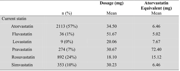
9.4 Statins
For patients taking a statin, their dosage is related to the type of statin used. The equivalent dosage to atorvastatin was calculated for each participant in order to allow for comparisons between patients on applicable statins. The statin dosage was transformed to the equivalent dosage as shown in table III.
31
Different statins use varying pathways, and therefore different statins affect statin induced muscle adverse effects differently. We used one all-inclusive category of statin, instead of stratifying by each statin type, which could be a limitation of our study. The majority of participants in the MHI cohort with available CK levels used atorvastatin. It has been found that the most hydrophilic statins, such as pravastatin and fluvastatin, are less likely to cause myalgia, while lipophilicone statins such as simvastatin are more likely to induce muscle adverse effects85. We did not stratify the users by statin type because most of the subsets were too small to preserve power.
32 Table III. Table of equivalent dosage of statins Atorvastatin equivalent Atorva- statin Fluva-statin Lova-statin Prava-statin Rosuva- statin Simva- statin 5 mg -- 40 mg 20 mg 20 mg -- 10 mg 10 mg 10 mg 80 mg 40 mg 40 mg -- 20 mg 20 mg 20 mg -- 80 mg 80 mg 5 mg 40 mg 40 mg 40 mg -- -- -- 10 mg 80 mg 80 mg 80 mg -- -- -- 20 mg -- 160 mg -- -- -- -- 40 mg --
33
9.5 Physical activity
Muscle damage, often accompanied with a spike in CK levels, is commonplace after intense physical activity. The increase in CK levels that occurs after physical activity varies in relation to the type (low impact verse high impact) and the intensity of exercise. Participants in the MHI cohort completed a survey of their physical activity which was based off the World Health Organization (WHO) guidelines. The two surveys differed only in that the MHI Cohort survey did not take into account unpaid labour, such as gardening and household chores, that are commonly accepted as physical activity. This is an expected pitfall of the survey, as we have no way of adjusting for this and may lose some accuracy in the analysis. The MHI Cohort survey is being amended to adjust for this in future studies.
In order to clean up data from the survey and split the participants into three classes of physical activity, guidelines from the WHO survey were followed. The procedure included calculating the metabolic minutes (a measure to combine the counts of physical and moderate activity) of physical activity. Using the metabolic minutes and the days of activity per week, the participants were split into three classes of physical activity: elevated, moderate or limited.
9.6 Ethnicity and principal components
In the MHI Hospital Cohort survey, participants were asked to choose between Caucasian, Hispanic, Black, Native American, Asian or other as their principal ethnicity. The vast majority of participants identified as Caucasian. Although self-reported ancestry is sometimes a viable way to stratify data in a large cohort86, it can have limited reliability, especially when an individual has more than one country of origin87. As the vast majority of participants identified as Caucasian, principal component analysis (PCA) was used. In a GWAS, a small percentage of