Université de Montréal
Cholinergic enhancement of perceptual learning:
behavioral, physiological, and neuro-pharmacological
study in the rat primary visual cortex
par Jun-Il Kang
Programme de sciences neurologiques, Ph.D. Faculté de médecine
Thèse présentée à la Faculté des études supérieures en vue de l’obtention du grade de Philosophiae Doctor (Ph.D.)
en sciences neurologiques
June 2014
Faculté des études supérieures et postdoctorales
Cette thèse intitulée:
Cholinergic enhancement of perceptual learning: behavioral, physiological, and neuro-pharmacological study in the rat primary visual cortex
Présentée par :
Jun-Il Kang
A été évaluée par un jury composé des personnes suivantes :
Dr Franco Leporé, président-rapporteur
Dre Elvire Vaucher, directrice de recherche
Dr Jean-François Bouchard, membre du jury
Dr Gregor Rainer, examinateur externe
Résumé
Les cortices sensoriels sont des régions cérébrales essentielles pour la perception. En particulier, le cortex visuel traite l’information visuelle en provenance de la rétine qui transite par le thalamus. Les neurones sont les unités fonctionnelles qui transforment l'information sensorielle en signaux électriques, la transfèrent vers le cortex et l'intègrent. Les neurones du cortex visuel sont spécialisés et analysent différents aspects des stimuli visuels. La force des connections entre les neurones peut être modulée par la persistance de l'activité pré-synaptique et induit une augmentation ou une diminution du signal post-synaptique à long terme. Ces modifications de la connectivité synaptique peuvent induire la réorganisation de la carte corticale, c’est à dire la représentation de ce stimulus et la puissance de son traitement cortical. Cette réorganisation est connue sous le nom de plasticité corticale. Elle est particulièrement active durant la période de développement, mais elle s’observe aussi chez l’adulte, par exemple durant l’apprentissage. Le neurotransmetteur acétylcholine (ACh) est impliqué dans de nombreuses fonctions cognitives telles que l’apprentissage ou l’attention et il est important pour la plasticité corticale. En particulier, les récepteurs nicotiniques et muscariniques du sous-type M1 et M2 sont les récepteurs cholinergiques impliqués dans l’induction de la plasticité corticale.
L’objectif principal de la présente thèse est de déterminer les mécanismes de plasticité corticale induits par la stimulation du système cholinergique au niveau du télencéphale basal et de définir les effets sur l’amélioration de la perception sensorielle.
Afin d’induire la plasticité corticale, j’ai jumelé des stimulations visuelles à des injections intracorticales d’agoniste cholinergique (carbachol) ou à une stimulation du télencéphale basal (neurones cholinergiques qui innervent le cortex visuel primaire). J'ai analysé les potentiels évoqués visuels (PEVs) dans le cortex visuel primaire des rats pendant 4 à 8 heures après le couplage. Afin de préciser l’action de l’ACh sur l’activité des PEVs dans V1, j’ai injecté individuellement l’antagoniste des récepteurs muscariniques, nicotiniques, α7 ou NMDA avant l’infusion de carbachol. La stimulation du système cholinergique jumelée avec une stimulation visuelle augmente l’amplitude des PEVs durant plus de 8h. Le blocage des
récepteurs muscarinique, nicotinique et NMDA abolit complètement cette amélioration, tandis que l’inhibition des récepteurs α7 a induit une augmentation instantanée des PEVs. Ces résultats suggèrent que l'ACh facilite à long terme la réponse aux stimuli visuels et que cette facilitation implique les récepteurs nicotiniques, muscariniques et une interaction avec les récepteur NMDA dans le cortex visuel. Ces mécanismes sont semblables à la potentiation à long-terme, évènement physiologique lié à l’apprentissage.
L’étape suivante était d’évaluer si l’effet de l’amplification cholinergique de l’entrée de l’information visuelle résultait non seulement en une modification de l’activité corticale mais aussi de la perception visuelle. J’ai donc mesuré l’amélioration de l’acuité visuelle de rats adultes éveillés exposés durant 10 minutes par jour pendant deux semaines à un stimulus visuel de type «réseau sinusoïdal» couplé à une stimulation électrique du télencéphale basal. L’acuité visuelle a été mesurée avant et après le couplage des stimulations visuelle et cholinergique à l’aide d’une tâche de discrimination visuelle. L’acuité visuelle du rat pour le stimulus d’entrainement a été augmentée après la période d’entrainement. L’augmentation de l’acuité visuelle n’a pas été observée lorsque la stimulation visuelle seule ou celle du télencéphale basal seul, ni lorsque les fibres cholinergiques ont été lésées avant la stimulation visuelle. Une augmentation à long terme de la réactivité corticale du cortex visuel primaire des neurones pyramidaux et des interneurones GABAergiques a été montrée par l’immunoréactivité au c-Fos. Ainsi, lorsque couplé à un entrainement visuel, le système cholinergique améliore les performances visuelles pour l’orientation et ce probablement par l’optimisation du processus d’attention et de plasticité corticale dans l’aire V1.
Afin d’étudier les mécanismes pharmacologiques impliqués dans l’amélioration de la perception visuelle, j’ai comparé les PEVs avant et après le couplage de la stimulation visuelle/cholinergique en présence d’agonistes/antagonistes sélectifs. Les injections intracorticales des différents agents pharmacologiques pendant le couplage ont montré que les récepteurs nicotiniques et M1 muscariniques amplifient la réponse corticale tandis que les récepteurs M2 muscariniques inhibent les neurones GABAergiques induisant un effet excitateur. L’infusion d’antagoniste du GABA corrobore l’hypothèse que le système inhibiteur est essentiel pour induire la plasticité corticale. Ces résultats démontrent que l’entrainement
visuel jumelé avec la stimulation cholinergique améliore la plasticité corticale et qu’elle est contrôlée par les récepteurs nicotinique et muscariniques M1 et M2.
Mes résultats suggèrent que le système cholinergique est un système neuromodulateur qui peut améliorer la perception sensorielle lors d’un apprentissage perceptuel. Les mécanismes d’amélioration perceptuelle induits par l’acétylcholine sont liés aux processus d’attention, de potentialisation à long-terme et de modulation de la balance d’influx excitateur/inhibiteur. En particulier, le couplage de l’activité cholinergique avec une stimulation visuelle augmente le ratio de signal / bruit et ainsi la détection de cibles. L’augmentation de la concentration cholinergique corticale potentialise l’afférence thalamocorticale, ce qui facilite le traitement d’un nouveau stimulus et diminue la signalisation cortico-corticale minimisant ainsi la modulation latérale. Ceci est contrôlé par différents sous-types de récepteurs cholinergiques situés sur les neurones GABAergiques ou glutamatergiques des différentes couches corticales. La présente thèse montre qu’une stimulation électrique dans le télencéphale basal a un effet similaire à l’infusion d’agoniste cholinergique et qu’un couplage de stimulations visuelle et cholinergique induit la plasticité corticale. Ce jumelage répété de stimulations visuelle/cholinergique augmente la capacité de discrimination visuelle et améliore la perception. Cette amélioration est corrélée à une amplification de l’activité neuronale démontrée par immunocytochimie du c-Fos. L’immunocytochimie montre aussi une différence entre l’activité des neurones glutamatergiques et GABAergiques dans les différentes couches corticales. L’injection pharmacologique pendant la stimulation visuelle/cholinergique suggère que les récepteurs nicotiniques, muscariniques M1 peuvent amplifier la réponse excitatrice tandis que les récepteurs M2 contrôlent l’activation GABAergique. Ainsi, le système cholinergique activé au cours du processus visuel induit des mécanismes de plasticité corticale et peut ainsi améliorer la capacité perceptive. De meilleures connaissances sur ces actions ouvrent la possibilité d’accélérer la restauration des fonctions visuelles lors d’un déficit ou d’amplifier la fonction cognitive.
Mots-clés : électrophysiologie, système cholinergique, amélioration cognitive, plasticité corticale, récepteur nicotinique, récepteur muscarinique, apprentissage perceptuel, cortex visuelle
Abstract
Sensory cortex is an essential area where sensory perception occurs. Especially visual cortex processes visual information transmitted from the retina through the thalamus. By different neuronal activation the information is segregated and sent to diverse visual area for interpretation. Neurons are the basic unit that transform sensory information into electrophysiological signal, transfer to the cortex and integrate it. Connection between neurons can be modulated depending on the persistent presynaptic activity inducing either a long-term increase or decrease of the post-synaptic activity. Modification in synaptic strength can affect large area and induce reorganization of cortical map (i.e. cortical plasticity) which changes the representation of the visual stimulus and its weight in visual processing. Cortical plasticity can occur during juvenile while forming developmental connection or in adult while acquiring novel information (i.e. learning). The neurotransmitter ACh is involved in many cognitive functions, such as learning or attention and it was demonstrated that lesioning or blocking cholinergic system diminishes cortical plasticity. It was shown that nicotinic, M1 subtype and M2 subtype muscarinic receptors are the major cholinergic receptors abundant in the cortex and implicated during cortical plasticity induction.
In a first part, I analyzed visual evoked potentials (VEPs) in V1 of rats during a 4-8h period after coupling visual stimulation to an intracortical injection of ACh agonist carbachol or stimulation of basal forebrain. To clarify the action of ACh on VEP activity in V1, we individually injected muscarinic, nicotinic, α7, and NMDA receptor antagonists just before carbachol infusion. Stimulation of the cholinergic system paired with visual stimulation significantly increased VEP amplitude for long-term. Pre-inhibition of muscarinic, nicotinic and NMDA receptor completely abolished this long-term enhancement, while α7 inhibition induced an instant increase of VEP amplitude. This suggests a role of ACh in facilitating visual stimuli responsiveness which involves nicotinic and muscarinic receptors with an interaction of NMDA transmission in the visual cortex. These mechanisms were similar to long-term potentiation, a neurobiological mechanism of learning.
In a second step, I evaluate whether cholinergic modulation of visual neurons results in cortical activity and visual perception changes. Awake adult rats were exposed repetitively for two weeks to an orientation-specific grating with coupling visual stimulation to an electrical stimulation of the basal forebrain. The visual acuity, as measured using a visual water maze before and after coupling visual/cholinergic stimulation was increased. The increase in visual acuity was not observed when visual or basal forebrain stimulation was performed separately nor when cholinergic fibers were selectively lesioned prior to the visual stimulation. There was a long-lasting increase in cortical reactivity of the primary visual cortex shown by c-Fos immunoreactivity of both pyramidal and GABAergic interneuron. These findings demonstrate that when coupled with visual training, the cholinergic system improves visual performance for the trained orientation probably through enhancement of attentional processes and cortical plasticity in V1 related to the ratio of excitatory/inhibitory inputs.
Finally, I also investigated the different pharmacological mechanisms involved in the visual enhancement. Pre- and post-pairing visual/cholinergic stimulation VEP were compared with selective administered agonist/antagonist during the pairing. Awaken adult rats were exposed during 10 minutes per day for 1 week to an orientation specific grating with an electrical stimulation of the basal forebrain. Intracortical injection of different pharmacological agents during pairing demonstrated that nicotinic and M1 muscarinic receptors are used to amplify cortical response while M2 muscarinic receptor suppresses GABAergic neurons to disinhibit excitatory neurons. Infusion of GABAergic antagonist supported that inhibitory system is crucial to induce cortical plasticity. These findings demonstrate that visual training coupled with the cholinergic stimulation enhances the cortical plasticity mediated by nicotinic, M1 and M2 muscarinic receptors, which the latter induces a disinhibition by suppressing GABAergic neuron.
The cholinergic system is a potent neuromodulatory system. Boosting this system during perceptual learning robustly enhances the sensory perception. Especially, pairing a cholinergic activation with a visual stimulation increases the signal-to-noise ratio, cue detection ability in the primary visual cortex. This cholinergic enhancement increases the strength of thalamocortical afferent to facilitate the treatment of a novel stimulus while decreasing the
cortico-cortical signaling to minimize recurrent or top-down modulation. This is mediated by different cholinergic receptor subtypes located in both glutamatergic and GABAergic neurons of the different cortical layers. The mechanisms of cholinergic enhancement are closely linked to attentional processes, long-term potentiation and modulation of the excitatory/inhibitory balance.
The present thesis shows that electrical stimulation of the basal forebrain has similar effect with cholinergic agonist release and pairing visual/cholinergic stimulation induces cortical plasticity. Repetitive pairing of visual/cholinergic increases visual discrimination capacity and enhances perceptual ability. This enhancement is followed by an augmentation of neuronal activity demonstrated by c-Fos immunohistochemistry. Immunoreactivity also shows difference in glutamatergic and GABAergic neurons activities between layers. Pharmacological injection during visual/cholinergic pairing suggests that nicotinic and M1 muscarinic receptor can amplify excitatory response while M2 receptor controls GABAergic activation. Altogether cholinergic system activated during visual process induces cortical plasticity and can enhance perceptual ability. Further understanding of this training has the potential to accelerate visual recovery or boost cognitive function.
Keywords: electrophysiology, cholinergic system, cognitive enhancement, cortical plasticity, nicotinic receptors, muscarinic receptors, perceptual learning, visual cortex
Table des matières
Résumé ... i
Abstract ... iv
Table des matières... vii
Liste des tableaux ... x
Liste des figures ... xi
Abbreviations ... xiii
Remerciements ... xvii
CHAPTER I: General Introduction ... 1
I.1 Prologue ... 2
I.2 Introduction to the cortical function ... 2
I.2.1 Sensory perception ... 2
I.2.2 Neurons ... 4
I.2.3 The visual system ... 8
I.3 Plasticity in the brain ... 16
I.3.1 Synaptic plasticity... 17
I.3.2 Cortical plasticity ... 23
I.4 Cholinergic system ... 29
I.4.1 Cholinergic pathways in the brain ... 30
I.4.2 Nicotinic system ... 31
I.4.3 Muscarinic system ... 33
I.4.4 Cholinergic modulation of cortical plasticity ... 33
I.5 Objectives of the study ... 36
CHAPTER II: Article 1 ... 39
Abstract ... 41
Introduction ... 42
Materials and methods ... 45
Results ... 52
CHAPTER III: Article 2 ... 66
Abstract ... 68
Introduction ... 70
Materials and Methods ... 72
Results ... 83
Discussion ... 95
Conclusion ... 103
CHAPTER IV: Manuscript 3 ... 105
Abstract ... 107
Introduction ... 108
Materials and Methods ... 111
Results ... 120
Discussion ... 128
CHAPTER V: Article 4 ... 134
CHAPTER VI: General Discussion ... 163
Technical aspect: Cortical response measurement by electrophysiological method ... 164
VI.1 Discussion of the objective 1: Does the visual stimulation paired with HDB stimulation (VS/HDB) induce cortical plasticity? ... 167
VI.2 Discussion of the objective 2: Does HDB stimulation have a similar effect to ACh in V1? ... 169
VI.2.1 Pairing visual and basal forebrain stimulation... 169
VI.2.2 The basal forebrain stimulation has similar effect to ACh release ... 170
VI.3 Discussion of the objective 3: Does a repetitive VS/HDB pairing improve behavioral performance similar to perceptual learning? ... 171
VI.3.1 Measuring visual discrimination capacity in the water maze ... 172
VI.3.2 VS/HDB stimulation induced perceptual learning related to cortical plasticity .. 173
VI.4 Discussion of the objective 4: Does repetitive VS/HDB pairing increase neuronal activity? ... 175
VI.4.1 Increase of neuronal activity shown by c-fos immunoreactivity ... 175
VI.4.2 Excitatory/inhibitory ratio is modulated differently between layers by visual training ... 176
VI.5 Discussion of the objective 5: What is the pharmacological mechanism of visual
training? ... 177
VI.5.1 VS/HDB stimulation induced cortical response enhancement transfers to untrained stimulus ... 177
VI.5.2 Presynaptic enhancement and disinhibition by nAChR ... 179
VI.5.3 Postsynaptic amplification by M1 and presynaptic disinhibition by M2 mAChR ... 180
VI.5.4 Long-term effect of cholinergic stimulation ... 182
VI.6 Future perspective ... 185
VI.6.1 Cortical function enhancement ... 185
VI.6.2 Visual restoration by perceptual learning mechanism ... 186
VI.7 Conclusion ... 187
Liste des tableaux
CHAPTER II
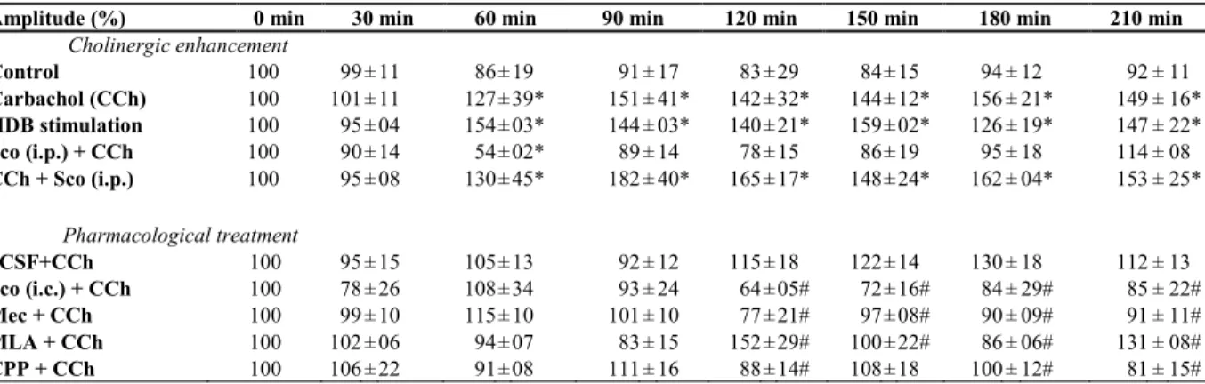
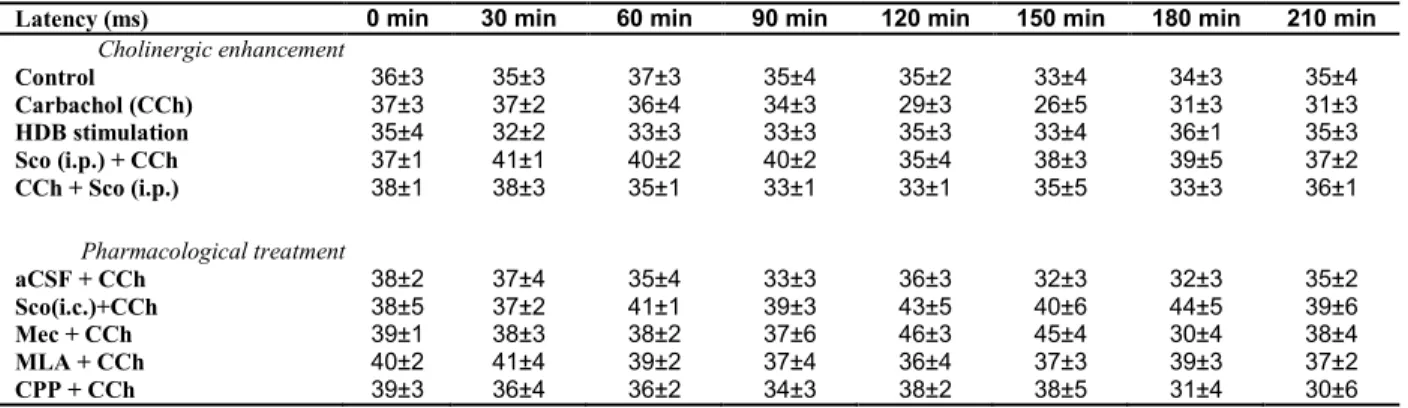
Table 1. Amplitude of VEP normalized after CCh injection or HDB stimulation and drug treatment………... 54 Table 2. Latency of VEP after CCh injection or HDB stimulation and drug treatment……... 57
CHAPTER III
Table 1. Number and nature of activated neurons in monocular area of the activated visual cortex………... 91
CHAPTER IV
Table 1. Experimental groups ……… 116
Liste des figures
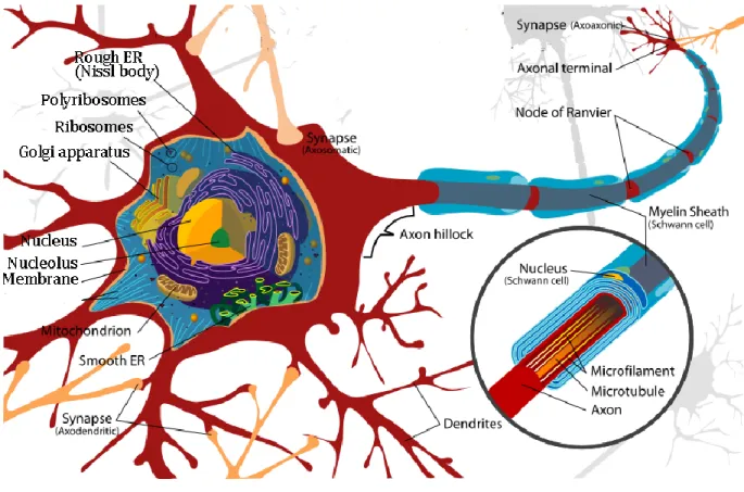
Figure I.1. Structure of a neuron. ... 5
Figure I.2. Anatomy of the retina... 10
Figure I.3. Visual projection from the retina to the thalamus and visual cortex (V1) ... 12
Figure I.4 Connections between visual cortical areas. ... 13
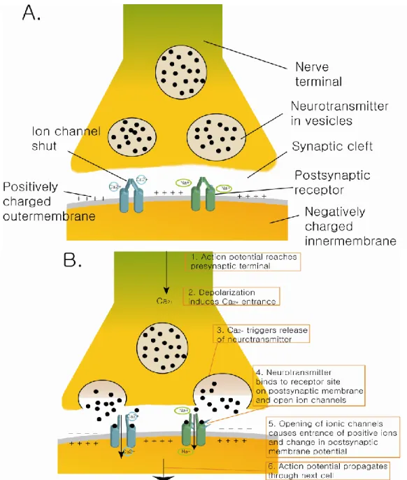
Figure I.5. Synapse and action potential. ... 18
Figure I.6 Comparison of LTP in hippocampus and LTP in V1. ... 25
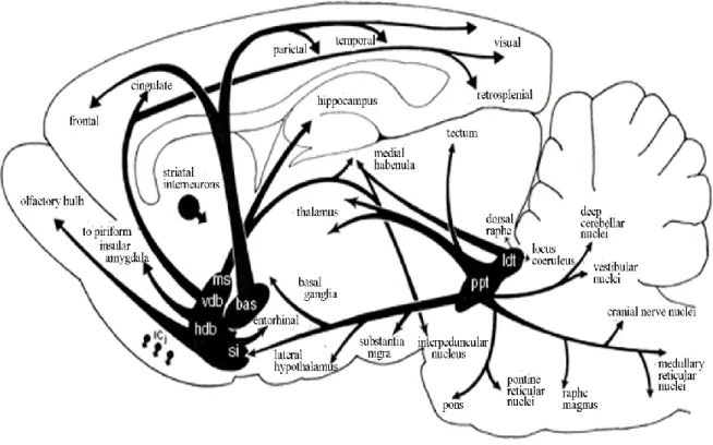
Figure I.7. Rat cholinergic central pathway. ... 32
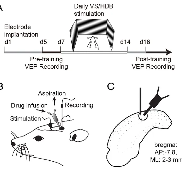
Figure II.1. Design of the experiment. ... 49
Figure II.2. Effects of cholinergic system activation through pharmacological injection and electrical stimulation paired with visual stimuli at different time points... 53
Figure II.3. VEP amplitude changes after pharmacological infusion of different drugs in the V1 with aCSF+CCh injected animals as control group. ... 56
Figure III.1. Timeline of the experimental procedure and stimulus parameter for the different groups. ... 74
Figure III.2. Measurement of visual acuity in the different groups pre- and post- training. .... 84
Figure III.3. Localization of the electrode implantation sites on coronal sections of the HDB. ... 86
Figure III.4. Microphotographs of ChAT immunolabelling in V1. ... 87
Figure III.5. VEP recording in the different groups after the training. ... 90
Figure III.6. Neurochemical phenotype of activated c-Fos neurons in V1. ... 93
Figure III.7. Schematic representation of the effects after VS ... 100
Figure IV.1 . Design of the experimental procedure. ... 112
Figure IV.2 Effects of repetitive VS/HDB stimulation on VEP amplitudes. ... 121
Figure IV.3 Changes in VEP amplitudes following pharmacological modulation during visual/cholinergic stimulation. ... 123
Figure IV.4 Microphotographs of parvalbumin and M2 mAChR double-immunostaining of coronal brain sections. ... 125
Figure V.1 Hypothesis of the effect of the cholinergic system on visual perception. ... 139 Figure V.2 Schematic representation of the primary visual cortex (V1) and its cholinergic
modulation on cortical processing. ... 141 Figure V.3 Neuronal connectivity within the primary visual cortex (V1). ... 144 Figure V.4 Summary of the effect of acetylcholine on neuronal transmission of the visual
inputs. ... 158 Figure VI.1. Hypothetical mechanism of perceptual learning and visual recovery. ... 189
Abbreviations
ACh acetylcholine
AMPA α-Amino-3-hydroxy-5-methyl-4-isoxazolepropionic acid BDNF brain-derived neurotropic factor
BF basal forebrain
CaMKII calcium/calmodulin-dependent protein kinases II CCh carbachol
ChAT choline acetyltransferase CNS central nervous system CPD cycle per degree
CR calretinin
CRE cAMP response element
CREB cAMP response element binding E/I excitation/inhibition ratio
ERK extracellular-signal-regulated kinase FP field potential
GABA γ-amino butyric acid
HDB horizontal limb of the diagonal band of Broca LFP local field potential
LGN lateral geniculate nucleus LPZ lesion projection zone LTD long-term depression
LTP long-term potentiation
M1 mAChR M1 subtype muscarinic receptor M2 mAChR M2 subtype muscarinic receptor mAChR muscarinic receptors
Mec mecamylamine
MLA methyllycaconitine nAChR nicotinic receptors NMDA N-methyl-D-aspartate
NSF N-ethylmaleimide-sensitive factor
OD ocular dominance
PK protein kinase
PNS peripheral nervous system PSD power spectral density PTX picrotoxin
PV parvalbumin
PZP pirenzepine
RBPC rat brain pyramidal cell marker RF receptive field
Sch Schaffer-commissural
sco scopolamine
SOM somatostatin
STDP spike-timing dependent plasticity TBS theta-burst stimulation
V1 primary visual cortex VEP visual evoked potential VS visually stimulated rats
Remerciements
Most of anyone I would like to thank you my director, Dr Elvire Vaucher for her support during my Ph.D. degree, providing a pleasant environment of study, giving abundant advices and encouraging me to enlarge my knowledge in neuroscience. And after all, I also would like to thank you for accepting me as a member of laboratory that gave me the reason to fly all over from Korea to here Montreal, which is such a beautiful place.
I would like to thank all my team members, colleagues, co-workers and friends, whose generous help, valuable advice and enjoying activities altogether gave lots of encouragement in my Ph.D. research. I especially thank you Florence for all your technical advices and motivating counsel from the beginning of my study.
I would like to thank the school of Optometry in the University of Montreal, for providing me not only a great research environment but also generous scholarships during my graduate studies. I could not have finished my studies without it.
I want to say thank you to all the members of my church, New Life Presbyterian, for all the moral support. Especially pastor Lee and Song who are always praying for me.
I also thank to the member of Les Incroyables, FC Desormeaux, and FC Jager, who played soccer with me. You helped to keep my body in good health and relieve from stress during the whole study.
I also would like to thank to my wife, Eunjung, whose unconditional love and support encourages me to have faith in myself. My two daughters, Laël and Joanna with their beautiful smiles always restored me after exhausted day. You were the reason that I could continue during all those years.
For the last, I would like to thank God in heaven, who gave me strength and faith. Your unfailing love holds me until these days.
I.1 Prologue
Pablo Picasso once said “If only we could pull out our brain and paint using only our eyes.” Although nowadays it is a common sense that without the brain we could not see anything, the famous painter probably wanted to describe the world without brain’s interpretation. As matter of fact, like a computer that transform electrical signal (e.g. from keyboard) into a letter, the brain interprets the outer information that is transferred from sensory organs so that our body could understand it. Sometimes the information is saved to be retrieved whenever needed or it could permanently affect the whole system. Based on previous and novel data, the process that our body understands the environment through the brain, we call it “perception”. For a living organism perception is critical to survive since based on the information it could make a decision and hence avoid dangerous situation.
The improvement of perception is nominated as perceptual learning since it appears only after repetitive training. This enhancement could occur through life time and recently it was observed that it is possible to accelerate the process by specific training. This study will be focused on the function and mechanism of the brain to increase its perceptual ability by presenting relevant concepts.
I.2 Introduction to the cortical function
I.2.1 Sensory perception
Perception of sensory information is analyzed in a hierarchical manner. Different organs convey different types of information and each sensory modality reaches different
cortical areas according to their specificity. The first cortical area where sensory signals are entered is called the primary sensory cortex. Fingers (somatosensory cortex), eyes (visual), ears (auditory) and nose (olfactory) send signals to their own primary cortex. As the raw information is transferred from lower to higher sensory cortex, it is segregated based on its property (e.g. shape for visual or frequency for auditory) with more precision (Kandel et al., 2013).
Compared to sensory cortices, associative areas process and integrate the information. For example in primate, the ventral intraparietal area in the parietal lobe receives input from visual, somatosensory and auditory senses (Avillac et al., 2005). Also the frontal lobe is suggested to have the ability of planning (Shallice and Burgess, 1991) or motivation (Eslinger and Damasio, 1985). Memory retrieval could occur during the perception by limbic system such as hippocampus and influence the interpretation of the stimulus. Cortical area which receives sensory information from primary sensory cortex and is involved in cognitive function (e.g. attention, learning or planning), it is called higher order sensory area.
The process of sensory perception is bidirectional and changeable. The flow of information from periphery to lower sensory cortex and to higher cortical area is called bottom-up. The reverse action from higher area modulating the response of lower area is called top-down. Top-down effect could influence the selectivity of stimulus in lower cortical area and facilitate the perception of a specific target. Bottom-up and top-down effect occur simultaneously and experience could refine the process. Repetitive exposures to similar environment reduce the process time by strengthening the cortical connection and increase the acuity by expanding the cortical area that treat the information. All those ameliorations of
perception is achieved through perceptual learning. Perceptual learning can influence in global, which means it affects both bottom-up and top-down process from neuronal to systematic change. To understand better how the brain perceive the world, it will be necessary to look into the function of its basic unit; the neuron.
I.2.2 Neurons
I.2.2.1 Neurons anatomy
Neuron is the basic unit of the brain composed of cell bodies (soma), axon and dendrite which transmit information through electrical and chemical signals. The neuron receives outer signal through their dendrites and transfer it to neighboring neuron by the axon (Figure I.1). The junctions between two neurons are called synapses. Generally synapses are established between axons to a dendrite, to another axons or a cell body. Sensory information is transported under form of electrical signal and at the end of the axon it is transferred by releasing chemical molecules (neurotransmitters) from presynaptic neuron delivered by the synaptic vesicles. At the dendritic membrane of neuron receiving neurotransmitters (postsynaptic neuron) there are proteins (receptors) which bind specifically the released neurotransmitters. Receptor, when coupled with ligand-gated channel, opens the ion gate to transfer the electrical signal to the soma.
There are numerous ways to classify neurons (e.g. shape, size, discharge pattern, or function) but probably the simplest way is its action on postsynaptic neurons. Neuron that release neurotransmitters permitting positive ion (Na+, Ca2+) entrance is called excitatory (e.g. glutamate). Neuron that release neurotransmitters permitting negative ion (Cl-) (e.g. GABA: γ-
Figure I.1. Structure of a neuron.
The neuron is composed by a soma (cell body), axon and dendrite. Myelin sheath is a phospholipid layer surrounding axon in most of the neurons, although some short projecting or interneurons do not have myelin. Adapted from http://wikimedia.org
amino-butyric-acid) admission is called inhibitory and is generally released by interneuron although spiny stellate cell in sensory cortex is glutamatergic (Kandel et al., 2013). Pyramidal neuron is excitatory neuron that has long axons to send information from one cortical area to another and in contrast interneuron has short axons to locally process signals (Rockland and Lund, 1982).
I.2.2.2 Single Neuronal activity
In general, neurons communicate each other through synaptic transmission which evokes action potential. During the rest state, the extracellular fluid contains excessive positive ion and maintains -75mV of difference (resting potential) between the extracellular and intracellular compartment. This membrane potential is maintained by ionic pump which are constantly pumping out Na+ and intruding K+ ions. Binding of an excitatory neurotransmitter induces conformational change of ionic channel and allows positive ions to enter within the neuron and initiates action potential. An action potential is generally composed of three sequences of events. (1) A depolarization of membrane causes entrance of Na+ through voltage-gated-channel. (2) A repolarization, when the inward Na+ induces an outward of K+ current to repolarize the membrane. (3) And hyperpolarization state by overwhelmed entrance of K+ before voltage-gated K+ channels shut (Hille, 2001). To measure such electro-potential activation Huxley and Hodgkin used voltage clamp where an intracellular electrode measured transmembrane voltage while a current electrode maintained the cell at a constant voltage (Hodgkin and Huxley, 1952). The action potential is said to be all-or-none signal since either it occurs fully independent of the amount of stimulating current or it does not occur at all.
This change of polarity on the membrane by depolarization propagates along the axon until the axon terminal. When the action potential reaches axon terminal or en passant varicosities it opens the voltage-sensitive calcium channels in the presynaptic membrane. This triggers synaptic vesicles filled with neurotransmitter to migrate to the synaptic cleft and release their content (Rusakov, 2006, Suudhof, 2008).
Neuronal activation can also be measured extracellularly. Compared to intracellular method which inserts electrode through the cell membrane, extracellular recording places electrode close to the neuron. By inserting close enough so that a single trans-membrane current (associated with an action potential) dominates the signal, it is possible to isolate the neuronal activity. Although compared to intracellular method (e.g. voltage clamp or patch clamp) this cannot provide information about postsynaptic potentials. Extracellular method can detect neuronal activation without cell damage and for longer period (Boulton et al., 1990). For example, Hubel and Wiesel showed by this method how single unit activities respond to very specific aspects of a visual stimulus (Hubel and Wiesel, 1965).
I.2.2.3 Neuronal activity (Multiple neurons)
The sensation of an external stimulus generally induces a simultaneous activation of a massive number of neurons. Multiple neuronal activations (i.e. multi-unit activities) can be measured either by inserting multiple electrodes or by enlarging electrode tip. With a large tip electrode it is possible to record the sum of multiple action potentials in the range of 50-350μm (Legatt et al., 1980, Gray et al., 1995).
Another method to measure multiple actions potential is the use of local field potentials (LFP). LFP is obtained by recording signals using extracellular low impedance microelectrode
inserted in the cortex. This signal is then filtered at ~300 Hz. With such filtering the action potential near the electrode which activates at high frequency range has less contribution to LFP signal. Compared to the action potentials which are visible only for adjacent electrodes, the synaptic events may be recorded in a distant area through the extracellular space. Moreover, since LFP records multiple activities on a large range, it is less attenuated with a small positional change of the electrode compared to single-unit activity recording. The LFP also differs from electroencephalogram which is recorded at the surface of the scalp or electrocorticogram which is recorded from the surface of the brain. LFP is believed to represent the synchronized input such as the synaptic current within 0.5-3 mm from the electrode tip (Juergens et al., 1999)(but see also (Katzner et al., 2009)). Despite its utility, physiological origin of LFP is still incompletely understood. Many attempts were made to understand its source although the current view is that synchronized synaptic currents on cortical pyramidal neurons generate LFPs (Niedermeyer and Lopes da Silva, 2005, Nunez and Srinivasan, 2006). In sum, LFP is useful to record synchronized synaptic events in a large area.
I.2.3 The visual system
Among sensory systems, the visual system is probably the most studied and well determined. Vision is provided through highly complex and organized interconnected processes among various parts of the brain. Although this mechanism is still not completely revealed and differs from species to species the visual function in primate is introduced to accommodate a better comprehension and comparison.
I.2.3.1 Primate visual system
Neurons in sensory system have their own receptive field (RF), an area of space where stimulation will cause the firing of that neuron. Light projected in the RF triggers activation of two different classes of photoreceptor in the retina: the rods which are sensitive to light’s intensity and the cones sensitive to the wavelength of the afferent light which allow color vision. In order to reach the photoreceptors the light must go through different layers in the retina: ganglion cell layer, inner plexiform layer, inner nuclear layer, outer plexiform layer, and outer nuclear layer (Fig I.2). The visual information adapted by the photoreceptors is transformed into electrical signals, delivered to the bipolar cells and to the retinal ganglion cells. The axons from the retinal ganglion cells project to the optic chiasm where they are distributed to major subcortical targets: the superior colliculus, the pretectum and the Lateral Geniculate Nucleus (LGN). The visual information from the retina reaching LGN via optic nerves and optic tracts, are transferred via the optical radiations mainly to the cortex in the occipital lobe, called the primary visual cortex (V1). To the other hand, information is received by the superior colliculus and the pretectum which have an essential role in visual ability by controlling saccade movement and pupillary reflexes (Kandel et al., 2013).
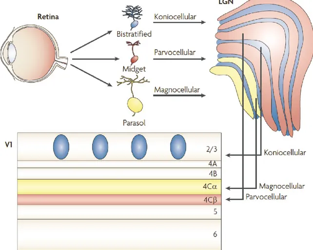
The visual signals are normally processed in two pathways: the dorsal and the ventral pathways. The dorsal pathway begins in the retina with ganglion cells of M type (M for Magnus, meaning large because of the large RF of these cells). These cells respond transiently to sustained illumination. M cell projections going through magnocellular layers (I and II) of LGN reach the layer IV in the V1. Layer IV is divided into three sublayers nominated IV A, IV B, and IV C. Layer IV C is itself subdivided into IV Cα and IV Cβ, and axons in
Figure I.2. Anatomy of the retina
Photoreceptors (rod and cone) are situated in the deep layer. Light must pass through ganglion cell layer, inner plexiform layer, inner nuclear layer, outer plexiform layer and outer nuclear layer to reach photoreceptors. (See text for details)
magnocellular layers of LGN project into IV Cα (Fig I.3) (Callaway, 1998, Nassi and Callaway, 2009). The majority of neurons in the layer IV are spiny stellate cells. Their axons transfer information to the dendrites of the pyramidal cells in layers IV B and III, which are excitatory and use glutamate as their neurotransmitters. Pyramidal neuron projections in V1 reach the dorsal area of brain, the middle temporal area (also referred area V5) and extend to the posterior parietal cortex. Neurons in this area are relatively insensitive to color or to stationary objects (Corbetta et al., 1991).
For the ventral pathway, P cells (for parvus meaning small) in the retina send visual response to parvocellular layers in LGN (3 to 6) that project to layer IV Cβ and IV A of V1. This information is sent to V4 that is linked with the inferior temporal cortex where neurons are sensitive to the outline of images or orientation, color and shape. It is suggested that dorsal pathway is related with “where” objects are, and ventral pathway with “what” the objects are (Mishkin et al., 1983). Several studies suggest that each system is specialized for different visual functions (Zeki, 1978, DeYoe and Van Essen, 1988, Livingstone and Hubel, 1988, Zeki and Shipp, 1988). The visual information transferred in different area is used differently i.e. visuospatial recognition for dorsal pathway and recognition of complex objects for ventral pathway (Fig I.4).
Between layers of LGN there is koniocellular cell (K cell) located in koniocellular layer (Fig I.3). Koniocellular layers of LGN receive projection from bistratified retinal ganglion cells. It seems that koniocellular layer supplement the color information by transmitting to the blobs in layer II/III of V1 (Hendry and Yoshioka, 1994).
Figure I.3. Visual projection from the retina to the thalamus and visual cortex (V1)
The retinal ganglion cells, midget, parasol and bistratified remain separate through LGN and into V1. Parvocellular layers of the LGN receive projection from midget cells and project onto layer IV Cβ of V1 (red). Magnocellular layers of the LGN receive projection from parasol cells and project onto layer IV Cα of V1 (yellow). Koniocellular layers of the LGN receive projection from bistratified cells and project to the cytochrome oxidase-expressing batches (or blobs) of layer II/III (blue). See text for details (Abbreviations: LGN, lateral geniculate nucleus; V1, primary visual cortex) (Nassi and Callaway, 2009).
Reprinted by permission from Macmillan Publishers Ltd: Nature neuroscience reviews (Nassi and Callaway) © 2009
Figure I.4 Connections between visual cortical areas.
The visual areas are organized into two pathways: a ventral pathway that is important for object recognition heading toward temporal lobe, and a dorsal pathway important for spatial vision leading to the parietal lobe.
In the primate’s visual cortex, specific features of the organization of V1 are orientation columns, blobs and ocular dominance columns. Tangential penetrations with microelectrodes, neurons in the same orientation column usually respond to same oriented light bars (Hubel and Wiesel, 1968). Each column contains cells in layer IVC and permits cortical cells to produce linear RF properties from the information generated by cells of LGN. Blobs, mostly situated in layer II and III, respond to different color stimuli but have no preferred orientation. Transferring monocular visual information optic nerves cross each other in optic chiasm and most of them (~60%) reach contralateral visual cortex. In the result according to their input source those two separate tracts compose ocular dominance columns.
I.2.3.2 Rodents visual system
Comparing to primate, the rat’s visual system has some distinctive features. First, laterally placed eyes provide a large panoramic visual field with a small binocular overlap. Moreover only a small proportion (~5%) of retinal ganglion cells project ipsilaterally. Secondly, the rat photoreceptors are different from those of primate. Not only has the density of cone in rat retina appeared to be lesser than other mammals but also the photoreceptor cones had shown to be sensitive at ultraviolet light (Jacobs et al., 1991).
Although in the rat system the visual pathway also includes LGN, superior colliculus and pretectum, our main interest is on LGN afferent pathway, so the physiological description in the present section will be focused on the structure of LGN. Precisely, the retinal ganglion inputs reach the dorsal area of LGN (dLGN). Despite the lack of lamination in dLGN of the rat, many studies have discriminated different regions in the distribution of cells of different sizes, in the composition of afferent axons and different patterns of degeneration after lesions
(Martin, 1986, Land, 1987, Reese, 1988). According to Reese (1988) discerning caudodorsally located nucleus as “outer shell” and “inner core” for ventromedial located nucleus, regions of dLGN were observed to be innervated by different classes of retinal ganglion cells. Variety in the regional distribution of inputs signifies that different cell types within dLGN are located in different broad regions. Cells in the outer shell mostly project to V1 and to cortical area Oc2L (occipital cortex; cytoarchitectonic area 18a). Even though the homology between Oc2L with V2 region of primate is still controversial.
In the visual cortex, most of the innervation from dLGN reaches the layer IV of V1. But dLGN projections were also observed in lower layer III and layer VI. It was observed that geniculocortical axons form asymmetrical synapses in layer IV (sparsely spined stellate cells, spiny nonpyramidal cells with perikarya and dendritic spines of pyramidal cells) and the lower part of layer III (dendritic spines of basal dendrites of pyramidal cells) (Peters et al., 1976, Peters and Feldman, 1976, 1977, Feldman and Peters, 1978). Prominent projection from layer IV cells exists in a precise manner to lower layers II/III while weaker projection extends laterally and diffusely in layer II/III. There are also a vertical projection toward the layers V and VI. The vertical intracortical connections convey the information to layers above and below to provide RF properties such as binocularity (Gilbert, 1983).
Pyramidal neurons in the layer V receive projections from the layer II/III and from the layer VI. Whereas the lower layer V make clustered projections in a diffuse manner to the layer I, the bottom of the layers II/III, and the top of the layer IV and V. Finally, neurons in the layer VI make clustered projections to the boarder of layer III and IV. Additional projections
were also observed from the layer VI to the boarder of layer V and VI and layer I/II (Burkhalter, 1989).
Nonpyramidal neurons, which are about 15% of the entire neuronal population in rat’s visual cortex, are GABAergic. Three major families of GABAergic neurons are distinguished according to their immunoreractivity: parvalbumin (PV), calretinin (CR), and somatostatin (SOM). The PV-immunoreactive neurons are present in all layers except the layer I and constitute about 51% of GABAergic neurons. The SOM-immunoreactive neurons are also absent in the layer I but mainly located in the infragranular layers V and VI. Finally, the CR coexpressing neurons account for 17% of GABAergic neurons and they are abundant in the layer I (Gonchar and Burkhalter, 1997). The function of GABAergic neurons during visual stimulation will be discussed further.
Although the rodent functional organization is similar to that of primate, in the rat’s visual cortex there is no evidence of orientation column (Girman et al., 1999). However, most neurons show a sharp adjusted selectivity about the direction of stimuli presented with a tendency for horizontal stimuli (Burne et al., 1984, Girman et al., 1999). This implies a distinctive mechanism for neurons in the visual cortex of the rat toward orientation selectivity.
I.3 Plasticity in the brain
In one of the episode of Sherlock Holmes, in a scene he said “I consider that a man’s brain originally is like a little empty attic, … It is a mistake to think that that little room has elastic walls and can distend to any extent.”. Sherlock Holmes was probably the greatest
detective character of the century, but obviously, he was not aware of plasticity of the brain. Plasticity is the ability of the brain to adapt to environmental change by reorganizing its neural circuits. Plasticity permits the brain to expand its capacity, to optimize its function or even to delete unused connection. It is estimated that instead of changing genetic code, as was chosen during evolution, our physiological system chose to change brain network to adapt to the environment. For this reason, plasticity is considered to occur as a result of long and continuous mechanism instead of spontaneous changes. Two kinds of plasticity are presented in this study: synaptic plasticity and cortical plasticity.
I.3.1 Synaptic plasticity
Synaptic plasticity is the ability of a synapse between presynaptic and postsynaptic neurons to modify its strength by changing the efficacy of receptor response and/or changing postsynaptic transduction. Earlier works in laboratories such as the one of Eric Kandel in aplysia had revealed part of the molecular mechanisms for synaptic plasticity (Castellucci et al., 1978).
Plasticity at the presynaptic level is likely to be a result of Ca2+ influx that activate calcium/calmodulin-dependent protein kinases II (CaMKII). These kinases phosphorylate synaptic vesicle associated proteins, synapsin and detach them from cytoskeleton. Direct entrance through voltage-gated Ca2+ channels or modulation of presynaptic K+ channels can both induce an increase of intracellular Ca2+. This facilitation can occur autonomously by a homosynaptical transmitter release from the terminal itself or heterosynaptically by a modulatory neuron at axo-axonic synapses.
Figure I.5. Synapse and action potential.
(A) Resting potential. Neurotransmitters are synthesized at nerve terminals and transported to the synaptic cleft by vesicles. Outer membrane of neuron is positively charged due to the sodium and calcium ion. (B) Synaptic transmission. The action potential is propagated through synapse and delivered to next cell. When ionic channel is opened those ions enter in the neuron and convert the polarity. This signal is transmitted through the cell.
The most common mechanism of postsynaptic plasticity results from the direct phosphorylation of an ionotropic receptor by serine/threonine or tyrosine protein kinases. Typically when modification of existing synaptic proteins, mostly protein kinases (i.e. PKA, PKC), is involved, it alters the synaptic function (Shi et al., 1999). However a second long lasting mechanism which is triggered by protein phosphorylation depends on second messenger neurotransmitters and involves changes in the levels of key protein as well as gene transcription (Kaang et al., 1993). This second mechanism provides the mechanism for long-lasting memory storage.
I.3.1.1 Hebbian rule
Among many models of synaptic plasticity that were introduced, Hebbian rule is summarized as “Cells that fire together, wire together”. Synaptic plasticity that follows Hebbian theory (or Hebbian plasticity) is induced by a continuous activation of presynaptic neuron stimulating postsynaptic cell. The repetitive and simultaneous activation increases the synaptic efficacy in the hippocampus. Representative aspects of Hebbian plasticity are long-term potentiation (LTP) and long-long-term depression (LTD).
I.3.1.1.a LTP
Discovered in the rabbit hippocampus (Andersen et al., 1966), LTP is the long-lasting enhancement of connection. Postsynaptic neuron shows a persistent increase in synaptic strength after high frequency stimulation of a chemical synapse. Since LTP and long-term memory possess common features it has been suggested as the most attractive candidate for cellular mechanism for learning.
LTP is usually induced with presynaptic tetanic stimulation (100 Hz during 1 sec) followed by an increase of excitatory postsynaptic potential (EPSP) lasting more than an hour (Fig I.6)(Huang and Kandel, 1994). Non-tetanic stimulation causes release of glutamate in presynaptic axon terminal which binds to AMPA (α-amino-3-hydroxy-5-methylisoxazole-4- propionic acid) receptors embedded in the postsynaptic membrane. Glutamate binding causes EPSP and can results in depolarization. However, when repetitive stimuli at high frequency are given to the presynaptic fiber this can cause prolonged EPSP in postsynaptic cell. The expression of stronger EPSP will remove magnesium ion blocking NMDA (N-methyl-D-aspartate) receptors and allow calcium influx during glutamate binding. The rise of Ca2+ triggers the activation of several protein kinase enzymes, such as CaMKII, protein kinase C (PKC) and cAMP-dependent protein kinase A (PKA) (Sweatt, 1999).
I.3.1.1.b LTD
As the name imply, LTD shows a depression of EPSP after a prolonged presynaptic stimulation with low frequency (0.5-10 Hz) (Dudek and Bear, 1992). Comparable with LTP, induction of LTD through low frequency stimulation (LFS) could be blocked by NMDAR antagonists (Lee et al., 1998, Kamal et al., 1999).
LTD is correlated with dephosphorylation of an AMPAR subunit (GluR1) containing serine 831 and 845. Serine 831 in GluR1 can be phosphorylated by CaMKII and PKC while serine 845 is phosphorylated by PKA. The latter has higher basal phosphorylation rate than former. Compared to LTP which is induced by phosphorylation of the CaMKII-PKC site LTD is induced by dephosphorylation of the PKA site (Barria et al., 1997a, Lee et al., 2000).
Reversing the LTD process by phosphorylation of PKA complements that dephosphorylation of AMPAR is necessary for LTD in hippocampal CA1.
Besides the phosphorylation regulation, several lines of evidence suggest that AMPAR expression in the postsynaptic membrane is subject to mechanism in LTD; (1) Prior saturation of LTD yields the AMPARs at the synapse and insensitive to inhibitors of NSF-GluR2 interaction (Luthi et al., 1999). (2) A low-frequency presynaptic stimulation induced an NMDAR dependent depression of miniature excitatory postsynaptic current amplitude and a decrease of GluR1 expressed in surface (Carroll et al., 1999). Altogether these results suggest that AMPAR internalization is an intermediate mechanism for LTD. It was proposed that another subtype of AMPAR GluR2 binds with N-ethylmaleimide-sensitive factor (NSF), which is an important protein during membrane fusion events (Nishimune et al., 1998). Blocking this operation causes the process of rapid internalization of receptors and decrease of AMPAR currents. Since LFS had no effect after receptor internalization, it is estimated that LTD requires the pool of NSF-regulated AMPARs (Luscher et al., 1999, Luthi et al., 1999).
Recently another form of Hebbian synaptic plasticity theory is introduced: spike-timing dependent plasticity (STDP). This theory highlights the precise spike-timing of firing between presynaptic and postsynaptic neurons. Since the depolarization is followed by a hyperpolarization due to K+ efflux, it is obvious that neuronal activation at the same moment could not induce synaptic plasticity. The precise timing of the perisomatic inhibition alter the backpropagation which is critical in STDP (Dan and Poo, 2004). Two connected neurons stimulated with varying interval confirmed the importance of precise timing to induce Hebbian plasticity (Caporale and Dan, 2008).
I.3.1.2 Homeostatic plasticity
Homeostatic plasticity refers to the compensatory mechanism of neuron to maintain its normal electrical activity. It is suggested that this self-adjustment is required to balance Hebbian plasticity. For instance, chronic neuronal firing could induce continuous LTP and quickly saturating plasticity. Instead of reaching an extreme level, the receptors are desensitized and the stability of network activity is preserved. On the other side, when a prolonged sensory deprivation occurs (e.g. light-deprivation or damage by stroke) the sensitivity of the synapses is increased to maintain the overall activity.
Homeostatic plasticity was observed in the rat’s pyramidal neuron in the visual cortex after a blockade of GABAergic synapses to increase neuronal activity. Turrigiano et al. observed that such stimulation results in decrease of excitatory postsynaptic currents by down-regulating postsynaptic glutamate receptors (Turrigiano et al., 1998). Comparatively, the injection of TTX that decreases the neuronal activity induced (1) an increase of the intrinsic excitability (Gibson et al., 2006), (2) a spontaneous activity enhancement via synaptic plasticity (Maffei and Turrigiano, 2008), or (3) an up-regulation of presynaptic Ca2+ influx (Zhao et al., 2011). Homeostatic plasticity can occur either by modulating ion channel expression (intrinsic homeostasis) or by modulating the synaptic input (synaptic homeostasis). During the development of the postnatal visual cortex, for example, dark rearing reduces the ratio of NMDAR subunits NR2A/NR2B (Quinlan et al., 1999). Compared to NR2A the NR2B which subunit remains open longer and reduces the thresholds for LTP and LTD probably by facilitating Ca2+ influx (Erreger et al., 2005). On the other hand, blocking the excitatory synaptic transmission for several days increases the synapse size and thereby enhances the
balance mechanism to maintain electrophysiological homeostasis in synapse allowing neuron to be modified by a strong activity exceeding the threshold.
I.3.2 Cortical plasticity
While synaptic plasticity occurs between two neurons, the cortical plasticity refers to the changes occurring in the organization of the cortex according to the experience. Brain activity transferring from a given function to a different location which results from a normal experience or a brain damage is the remarkable consequence of cortical plasticity. The cortical plasticity involves changes in multiple neuronal connections that are represented by alternation of broad range cortical response.
There are many similarities between synaptic plasticity and cortical plasticity. For example when a rat whisker of postnatal day 12-14 is stimulated it expresses recombinant AMPAR subunit GluR1 into synapses of the somatosensory cortex (Takahashi et al., 2003). In the visual cortex, similarly with LTD, monocular deprivation shows alternation in the GluR1 phosphorylation level (Heynen and Bear, 2001). An LTP is also induced in V1 after tetanic stimulation in the LGN (Heynen and Bear, 2001). The cortical plasticity is also shown to be NMDAR dependent. The inhibition of NMDAR in developing visual cortex blocks the effects of monocular deprivation suggesting a crucial role of NMDAR (Bear et al., 1990). Involvement of NMDAR implies that Ca2+ dependent enzymes are implicated in cortical plasticity. Indeed, similarly with synaptic plasticity PKA (Beaver et al., 2001), extracellular-signal-regulated kinase (ERK (Di Cristo et al., 2001)) and αCaMKII (Taha et al., 2002) are active during cortical plasticity process. The role of those kinases are to phosphorylate substrates like synapsin (Hosaka et al., 1999), AMPAR (Barria et al., 1997a, Barria et al.,
1997b, Benke et al., 1998), GABAR (Brandon et al., 2003), or actin (Matus, 2000). Those molecules are used in synaptic transmission, neuronal excitability and morphological stabilization.
Changes in synaptic molecules can activate transcription factor, such as CREB (cAMP response element binding) (Liao et al., 2002). CREB protein that binds to a specific DNA sequence called cAMP response elements (CRE) is also involved in LTP (Martin and Kandel, 1996). Starting by the postsynaptic receptor activation, the production of a second messenger such as cAMP or Ca2+ activates in turn the protein kinase which then induces CREB protein to bind to a CRE region. With successive binding of CREB-binding protein, CREB regulates other transcription factors such as c-fos, c-jun or egr-1 (Boutillier et al., 1992, Masquilier and Sassone-Corsi, 1992). Gene transcription synthesizes new proteins, a process critical for both ocular dominance plasticity (Taha et al., 2002) and long-term changes in synaptic strength (Silva et al., 1998).
Although cortical and synaptic plasticity share numerous common aspects there is no direct evidence that they are correlated. For example, some essential molecules that are used in synaptic plasticity (e.g. BDNF or type 2 metabotropic glutamate receptor) were shown to be unnecessary to induce a shift in ocular dominance (Bartoletti et al., 2002, Renger et al., 2002). Moreover, the continuous induction of LTP in synapse fails to render in cortical plasticity (Hensch, 2003).
Although many models of cortical plasticity in V1 were proposed, two classes of cortical plasticity are well documented: ocular dominance shift and lesion-induced plasticity.
Figure I.6 Comparison of LTP in hippocampus and LTP in V1.
(A) Schematic illustration showing extracellular recording and tetanic stimulation on the Sch projection to CA1 (Top). Tetanus stimulation (a brief, high-frequency train of electrical stimuli) induces an enhanced synaptic response for many hours (i.e. LTP) (Bottom). (Neves et al., 2008)(B) Schematic illustration showing the position of stimulating electrode in the LGN and recording electrode in V1 (Top). TBS in the LGN evoked LTP of FPs in V1. After 30 minutes of baseline recording, application of TBS to the LGN elicits LTP of FPs in the V1 (Bottom). (Abbreviations: Sch, Schaffer-commissural; TBS, theta-burst stimulation; LGN, lateral geniculate nucleus; V1, primary visual cortex; FP, field potential)
Different models of cortical plasticity which are neurotransmitter-dependent and learning-induced plasticity will be introduced later.
I.3.2.1 Cortical plasticity in juvenile: ocular dominance shift during critical period Few decades ago when the neocortex was considered unmodifiable, Hubel and Wiesel had demonstrated that during development and before the critical period, ocular dominance (OD) columns in V1 were highly plastic (Wiesel and Hubel, 1963). Exclusively, monocular vision during the critical period results in an expansion of the columns serving the open eye and the columns that were responding to the deprived eye becomes reduced in size and afferent connectivity. Following this result of Hubel and Wiesel, the cortical plasticity had been shown to occur in the somatosensory cortex (Van der Loos and Woolsey, 1973), the auditory cortex (Moore, 1985) and in diverse experimental and natural conditions.
It is suggested that GABAergic system is essential for the initiation of critical period. In mice with genetically deprived GABA-synthesizing enzyme (glutamic acid decarboxylase: GAD65), the critical period was delayed until treatment with the GABAA receptor agonist diazepam (Hensch et al., 1998, Fagiolini and Hensch, 2000). Also overexpression of brain-derived neurotropic factor (BDNF) shows a premature onset of the critical period. This is probably because BDNF promotes the maturation of GABAergic system in the visual cortex (Hanover et al., 1999, Huang et al., 1999).
On the other hand, recent study suggests that inhibitory system maturation closes the critical period. It was found that the disruption of the perineuronal nets can re-induce OD plasticity during adulthood (Carulli et al., 2010). This extracellular matrix (ECM) structure is known to surround densely the PV interneurons (Carulli et al., 2006). Also, it was
demonstrated that restoration of deprived eye vision or OD plasticity in adult rat can be induced by a reduction of GABAergic inhibition (Baroncelli et al., 2010, Harauzov et al., 2010). Those studies suggest that inhibitory system maturation affects the potential of OD plasticity.
Among theories about how inhibitory innervation can regulate plasticity of excitatory connections (e.g. glutamate and AMPAR), one convincing mechanism is by altering STDP. Parvalbumin (PV) expressing GABAergic interneurons innervate excitatory neurons and other interneurons on the soma and dendrites. Such connection allows PV neurons to modulate backpropagation (Tsubokawa and Ross, 1996) which is crucial for STDP. Indeed, increased perisomatic inhibition can disrupt STDP while blocking it facilitates (Pouille and Scanziani, 2001). In a broader range, PV interneurons can form groups of 30-50 cells and synchronize their activations through gap junctions (Galarreta and Hestrin, 1999, 2002). Such coordination alters the firing timing of excitatory neurons and hence PV interneurons can control large sets of excitatory neurons. This functioning also supplies the underlying mechanism of gamma-band oscillations (30-90 Hz) (Tamas et al., 2000). The gamma-gamma-band oscillation is suggested to be involved in sensory input processing and reflects the synchronized neural activity (Cardin et al., 2009). Overall excitatory system is affected by inhibitory input via STDP and PV interneurons create network to organize coherent firing of neurons in the visual cortex.
In summary, the initiation or the closure of critical period is affected by GABAergic system. It seems that the maturation of the GABAergic system regulate the cortical plasticity. The regulation on excitatory synapse is achieved through STDP rule and PV interneurons
network can synchronize neuronal firing. Long-term alternation of synaptic strength can shift OD response.
I.3.2.2 Cortical plasticity in adult: lesion-induced plasticity
Under normal conditions in the adult primary sensory cortex, the gross structure of neurons and their circuitry cannot be modified dramatically (Grutzendler et al., 2002, Trachtenberg et al., 2002). However, following a partial retinal lesions structural plasticity was found in the adult primary visual cortex (Darian-Smith and Gilbert, 1994, 1995). A binocular lesion at the retinal level produces a silenced region in the corresponding retinotopic zone (the lesion projection zone: LPZ). During restoration, neurons in the LPZ recover their sensitivities to visual input from the undamaged retinal regions surrounding LPZ (Darian-Smith and Gilbert, 1995, Eysel et al., 1999).
Reorganization of the cortical topography was proposed to explain the changes in RF properties, circuitry and molecular mechanism during restoration. It was shown with fMRI that reorganization of V1 retinotopic map occurs after a partial damage of the input fibers caused by macular degeneration (Baker et al., 2005) or stroke (Dilks et al., 2007). Rearrangement of cortical map is mediated by the long-range horizontal connections (Rockland and Lund, 1982). Normally, these connections are used for propagation of information and to integrate information of large area on the visual field (Albright and Stoner, 2002). Such a stimuli placed outside of RF can still influence neuronal response by the global context (Hubel and Wiesel, 1965, Crist et al., 2001). Strengthening of these horizontal connections enables neurons surrounding LPZ to innervate lesion-affected neurons and thereby shifting RFs.
It is documented that cortical reorganization in LPZ is accompanied by growth or degradation of synaptic branches. With the use of in vivo two-photon imaging Yamahachi et al. observed that the horizontal axons in V1 rapidly sprout their branches while removing older connections (Yamahachi et al., 2009). Also, retinal lesions induce an upregulation in the rate of turnover of dendritic spines (Keck et al., 2011) which is considered to be a sign of cortical plasticity. These changes of connections are observed both in excitatory and inhibitory system (Keck et al., 2011, Marik et al., 2014) probably to balance inputs in reorganizing cortex (Priebe and Ferster, 2012). Altogether those studies indicate that sprouting and pruning of synaptic branches is the underlying mechanism of the remapping of cortical topography following retinal lesions.
The cortical map reorganization after a partial retinal lesion opens the possibility that the cortical plasticity could also occur in mature cortex. However, the distinction in properties and connections should be made from that of juvenile cortex (e.g. OD plasticity). The nature of the cortical plasticity after lesion is similar with the mechanism of experience-dependent plasticity in adulthood. Especially, it was proposed that cortical plasticity during perceptual learning is mediated by strengthening horizontal connections and change of cortical topography (Recanzone et al., 1992, Ramalingam et al., 2013). It is possible that both changes use the same cortical circuits (Gilbert and Li, 2012).
I.4 Cholinergic system
The cholinergic system is widely distributed in the cortex and carries out the complex function with region specific manner. The two main intrinsic systems in the cortex are glutamatergic and GABAergic systems which have excitatory and inhibitory action,
respectively (DeFelipe et al., 2002). Compared to those two transmitters which roles are mainly to transfer information, other transmitters such as acetylcholine, norepinephrine, serotonin, dopamine, histamine, or adenosine have modulatory effect and arise from neurons located outside of the cortex. A single modulator may be coupled to diverse postsynaptic receptors and produce distinct postsynaptic effect and hence launching different second messenger cascade (McCormick, 1992). Such functional diversities allowed neuromodulator to be involved during control of cortical state (e.g. arousal, attention, slow-wave sleep, etc) and its network (McCormick, 1992, Briand et al., 2007). Among various neuromodulator, recently acetylcholine (ACh) attracted high interest for its implication in attention and learning (Sarter et al., 2005).
I.4.1 Cholinergic pathways in the brain
Cholinergic forebrain innervations are divided into six distribution pathways; Ch1-Ch6 (Mesulam et al., 1983). Cholinergic nuclei from the medial septum (Ch1), the vertical and horizontal limb of the diagonal band (Ch2 and Ch3), project to the hippocampus and prefrontal and occipital cortex, the nucleus basalis of Meynert (Ch4) project to the entire cerebral cortex, and the cholinergic neurons in the pedunculopontine tegmental nucleus (Ch5) and laterodorsal tegmental nucleus (Ch6) project to superior colliculus, thalamus, basal forebrain and substantia nigra. According to their activating agonist cholinergic receptors are categorized as muscarinic (activated by muscarine) and nicotinic (activated by nicotine) receptors.