HAL Id: tel-01037941
https://tel.archives-ouvertes.fr/tel-01037941
Submitted on 23 Jul 2014HAL is a multi-disciplinary open access archive for the deposit and dissemination of sci-entific research documents, whether they are pub-lished or not. The documents may come from teaching and research institutions in France or abroad, or from public or private research centers.
L’archive ouverte pluridisciplinaire HAL, est destinée au dépôt et à la diffusion de documents scientifiques de niveau recherche, publiés ou non, émanant des établissements d’enseignement et de recherche français ou étrangers, des laboratoires publics ou privés.
Study of immunological properties of sperm and seminal
plasma antigens : anti-seminal and anti-sperm antibodies
in female immune infertility : characterization of
targeted proteins
Andrea Brazdova
To cite this version:
Andrea Brazdova. Study of immunological properties of sperm and seminal plasma antigens : anti-seminal and anti-sperm antibodies in female immune infertility : characterization of targeted proteins. Immunology. Université Pierre et Marie Curie - Paris VI; Vysoká škola chemicko-technologická (Praze), 2014. English. �NNT : 2014PA066084�. �tel-01037941�
Université Pierre et Marie Curie
Institut des technologies chimiques
Ecole doctorale 394 : Physiologie et physiopathologie
Centre de Recherche des Cordeliers / Equipe 16 Immunopathologie et immunointervention thérapeutique
Etude des propriétés immunologiques des antigènes de sperme et
de liquide séminal :
L’infertilité féminine due à des anticorps anti-protéines de liquide
séminal et de spermatozoïdes. Caractérisation des protéines cibles.
Par Andrea Brázdová
Thèse de doctorat de Sciences de la vie
Dirigée par Dr. Srinivas Kaveri et Dr. Jarmila Zídková
Présentée et soutenue publiquement le 29 Avril 2014
Devant un jury composé de :
Carole Elbim Présidente Gérard Chaouat Rapporteur Markéta Sedmíková Rapporteur Pascal Poncet Examinateur
Jan Káš Examinateur
Srinivas Kaveri Directeur de thèse Jarmila Zídková Co-directeur de thèse Gabriel Peltre Membre invité
Jointly supervised thesis
STUDY OF IMMUNOLOGICAL PROPERTIES OF SPERM AND
SEMINAL PLASMA ANTIGENS:
ANTI-SEMINAL AND ANTI-SPERM ANTIBODIES IN FEMALE
IMMUNE INFERTILITY. CHARACTERIZATION OF TARGETED
PROTEINS.
Author: Ing. ANDREA BRÁZDOVÁ
Supervisors: Dr. Srinivas Kaveri
RNDr. Jarmila Zídková, CSc.
ICT Prague
Study program: Chemistry
Field of study: Biochemistry
UPMC Paris
Training and research unit UFR 927: Life Sciences
Doctoral school 394: Physiology and Pathophysiology
Paris 2014
Declaration
The jointly supervised thesis was worked out at the Department of Biochemistry and Microbiology, Institute of Chemical Technology, Prague, Czech Republic and at the Immunopathology and Therapeutic Immuno-Intervention, Cordelier Research Center, Paris, Pierre and Marie Curie University, Paris, France from September 2010 to March 2014.
I hereby declare that I have worked out this thesis independently while noting all resources used, as well as all co-authors. I consent to publication of this thesis under Act No. 111/1998, Coll., the Higher Education Act of the Czech Republic, as amended by subsequent regulations. I have been informed of all duties and obligations applicable under Act No. 121/2000, Coll., the Copyright Act, as amended by subsequent regulations.
Paris, March 14th, 2014
Acknowledgement
I thank, first of all, my supervisors Dr. Srinivas Kaveri and RNDr. Jarmila Zídková, CSc. who accepted me for this interesting topic, in which they are specialists. Special gratitude belongs to Dr. Srinivas Kaveri and Dr. Pascal Poncet as without them and their support, this jointly supervised thesis would have been but a dream. I am immensely grateful to my mentors Dr. Gabriel Peltre and M.Ed. Charlotte Doubravová who were my scientific as well as moral support, who have never stopped to believe in me and who guided me as mentors through thick and thin. I also appreciate professional approach of Dr. Hélène Sénéchal, Dr. Pascal Poncet, prof. Ing. Martin Fusek, CSc. who showed me the way forward when I found myself to be in the blind spot and who always knew how to improve the value of presented research. I wish to convey my thanks to prof. MUDr. Zdenka Ulčová-Gallová, CSc. for sample donation from infertile and fertile women, for her professional attitude, which was of immense help to publish the obtained results. Of course, I appreciate willingness of voluntary anonymous male donors to provide repeatedly semen samples and to undergo the semen quality examination.
I would like to express my gratitude to the Allergy team, Department of Biochemistry and Molecular Biology, Armand-Trousseau Children Hospital, Paris, France and the team 16 - Immunopathology and Therapeutic Immuno-Intervention, Cordelier Research Center, Paris, France, both of which I belonged to and where I obtained valuable knowledge and experience. I would like to thank my home laboratory at the Department of Biochemistry and Microbiology, Institute of Chemical Technology, Prague, Czech Republic.
This jointly supervised thesis would not have been carried out without fellowships awarded by the Department of Foreign Affairs, Institute of Chemical Technology, Prague, Czech Republic, in particular by Ing. Hana Opatová, CSc., and by the French Embassy in Prague, Czech Republic, in particular by Mr. Rachid Makhloufi, Mr. Jonathan Banigo, as well as prof. Ing. Kateřina Demnerová, CSc., whose personal approach was of great value to extend my internship in Paris among great scientists from the Allergy team. I would like to express my immense gratitude to Mr. Frédéric Huynh who always found a solution of intractable pitfalls and rigours concerning the French system. My thanks belong to Ms. Patricia Zizzo, Department of Foreign Affairs, Pierre and Marie Curie University, Paris, France who kindly and patiently guided me through the administrative process at the French university.
Sincere gratitude has to be also expressed to grant agencies and projects, by which the thesis was co-financed. In particular, this work was supported by the grant project from the
Ministry of Education, Youth and Sport of the Czech Republic (0021620812, 6044613705), by specific university (Institute of Chemical Technology, Prague, Czech Republic) research projects no. 21/2011, 21/2012, 20/2013, 20/2014 and by the Czech Academy of Sciences grant IAA 600110902.
I would like to thank prof. Carole Elbim, prof. Gérard Chaouat, prof. Jan Káš, prof. Markéta Sedmíková, Dr. Srinivas Kaveri, Dr. Pascal Poncet, Dr. Jarmila Zídková and Dr. Gabriel Peltre for accepting the invitation to be the members of the jury.
I am grateful to prof. Corinne Mitchell and Ms. Ashley Truxal for the English language correction and proofreading of the thesis.
I am immensely indebted to Ing. Tomáš Durda for his presence during the entire time of my doctorate studies and then, together with Ing. Alena Keprová, for being an endless fountain for my lamentation. By the end and right from the heart, I express my deepest gratitude to my beloved mother for her never-ending support, help and confidence placed into myself.
Summary
The World health Organization reports infertility as a disease and a failure of reproductive tract to achieve pregnancy after 12 months or more of regular unprotected sexual intercourse. Nowadays, infertility has become a common life phenomenon affecting 1 out of 5 couples at reproductive age. Idiopathic cause is mostly associated with active immune system which may produce high levels of anti-seminal and/or anti-sperm antibodies. Auto-immunization as well as iso-immunization has a significant role in up to 30% of reported cases of infertility. Semen that is defined as a complex fluid containing sperm, cellular vesicles and other cells and components, could immunize the female genital tract.
This thesis is related to female immune infertility, in particular to female iso-immunization. The better understanding of this pathophysiological event consists of (1) the determination of antibody isotype playing a significant role in this disease, then (2) the characterization and identification of semen antibody-binding proteins, seminal and/or sperm, (3) the proposal of potential diagnostic markers to adapt specific therapy and, in addition, the design of miniaturized diagnostic tool based on the selected markers, (4) the suggestion of potential immuno-intervention.
Based on the distribution of seminal/sperm-specific antibody isotypes, we suggest that immunoglobulins E, M, A1,2, G3 are not involved in the primary pathophysiological female sensitization. IgG4 appears to be the major subclass interacting with sperm proteins. On a contrary, IgG1 seems to be the one mainly involved in the reactivity towards seminal proteins. We have also extended the existing group of IgG-binding sperm proteins, among which heat shock protein 70 1A/1B, heat shock cognate protein 71 kDa and alpha-enolase have been shown, for the first time, to be related to female iso-immunization. We have put the emphasis on the role of seminal proteins in iso-immunization and not only in the IgE-mediated semen hypersensitivity as known so far. In particular, prostate-specific antigen, prostatic acid phosphatase and zinc finger protein 778 have been determined as immunodominant among IgG-binding seminal proteins.
The determination of female serum seminal/sperm-specific IgG subclasses could make the patient diagnoses more comprehensive. Anti-seminal/sperm IgG1,4 might be of interest for immunotherapy. Furthermore, the herein described proteins could be useful biomarkers of such pathology. The miniaturized chip could be a lateral flow immunoassay-based device acting on the immunochemical detection of specific antibodies. The intended immuno-intervention could consist of the effect of intravenous immunoglobulins.
Résumé
L‟Organisation Mondiale de la Santé définit l‟infertilité comme une maladie et un échec de l‟appareil reproducteur à parvenir à une grossesse après 1β mois ou plus de rapports sexuels réguliers non protégés. De nos jours, l‟infertilité est devenue un phénomène commun affectant 1 couple en âge de procréer sur 5. Une origine idiopathique est le plus souvent associée à un système immunitaire actif qui pourrait produire des niveaux élevés d‟anticorps anti-liquide séminal ou anti-sperme. L‟auto-immunisation, aussi bien que l‟iso-immunisation, joue un rôle significatif dans jusqu‟à γ0% des cas signalés. Le liquide séminal, qui est défini comme un fluide complexe contenant le sperme, les vésicules cellulaires et autres cellules et composantes, pourraient immuniser l‟appareil génital féminin.
Cette thèse est liée à l‟infertilité féminine immune, en particulier à l‟iso-immunisation féminine. Une meilleure compréhension de cette manifestation physiopathologique consiste en (1) la détermination des isotypes d‟anticorps jouant un rôle significatif dans cette maladie, puis en (β) la caractérisation et l‟identification des antigènes de liquide séminal ou de sperme reconnus par ces anticorps, (3) la proposition de marqueurs diagnostiques potentiels afin d‟adapter des thérapies spécifiques, et, en outre, la conception d‟un outil de diagnostic miniaturisé basé sur les marqueurs sélectionnés, (4) la suggestion d‟une éventuelle immuno-intervention.
En se fondant sur la distribution des isotypes d‟anticorps spécifiques au liquide séminal/sperme, nous suggérons que les immunoglobulines E, M, A1,2, et G3 ne sont pas impliquées dans la sensibilisation physiopathologique chez les femmes. Les IgG4, semblent constituer la sous classe majeure interagissant avec les protéines de sperme. A l‟inverse, les IgG1 semblent être principalement impliquées dans la réactivité vis-à-vis des protéines séminales. Nous avons également élargi le groupe déjà existant d‟IgGs liés aux protéines de sperme à d‟autres protéines, parmi lesquelles la protéine de choc thermique 70 1A/1B, la protéine apparentée aux protéines de choc thermique 71kDa et l‟alpha-énolase ont été reconnues, pour la première fois, être liés à l‟iso-immunisation féminine. Nous avons mis en évidence le rôle des protéines séminales dans l‟iso-immunisation et pas seulement dans l‟hypersensibilité au sperme par l‟intermédiaire d‟IgE. En particulier, l‟antigène spécifique de la prostate, la phosphatase acide prostatique et la protéine à doigt de zinc 778 ont été décrites comme immuno-dominants parmi les protéines séminales reconnues par des IgGs liés aux protéines de sperme.
La détermination des sous classes d‟IgG de sérum féminin, spécifiques au liquide séminal/sperme, pourrait rendre le diagnostic des patients plus complet. Les IgG1 et IgG4 anti-liquide séminal/sperme pourraient présenter un intérêt pour l‟immunothérapie. Par ailleurs, les protéines décrites, dans notre étude, pourraient se révéler être des bio marqueurs utiles pour de telles pathologies. Le dispositif miniaturisé pourrait être de type LFIA (Lateral Flow Immuno Assay), se basant sur la détection immuno-chimique d‟anticorps spécifiques. L‟immuno-intervention envisagée pourrait reposer sur l‟effet d‟immunoglobulines intraveineuses.
Aims of the thesis
1. Determine the distribution of serum semen-specific antibodies, which immunoglobulin classes, in particular which subclasses, play a significant role in female immune infertility within the patient group selected
a. Quantify the anti-seminal/sperm antibodies in the sera of infertile female patients
b. Compare the observed distribution of anti-seminal/sperm antibodies in the sera of fertile healthy women
2. Propose a method for the characterization of semen antigens associated with antibody formation involved in the pathophysiological iso-immunization
a. Characterize the immunodominant seminal and sperm patterns in order to keep as much as possible detectable epitopes of potential seminal/sperm antigens b. Identify the immunodominant antigen repertoire detected by the sera of female
patients diagnosed with the selected fertility disorder
3. Verify suggested protocols
a. Verify the results in order to generalize the knowledge of antigens detected by the sera of infertile women
b. Detect both seminal and sperm antigenic structures by the sera of fertile women in order to confirm the relevance of previous results
4. Propose the potential use of obtained results
a. Suggest potential diagnostic markers to adapt specific therapeutic treatments b. Based on the selected markers, design a miniaturized diagnostic tool that could
refine, simplify and hasten the diagnosis in order to make patient profiling more precise and to improve the diagnosis that may differentiate between seminal- and sperm-sensitivity
CONTENT
1 INTRODUCTION 1
2 MALE FACTOR IN REPRODUCTION 2
2.1 Seminal fluid 4 2.2 Spermatozoa 8 2.2.1 Spermatogenesis 10 2.2.2 Capacitation 13 2.2.3 Acrosome reaction 15 2.2.4 Fertilization 18
3 FEMALE FACTOR IN REPRODUCTION 20
3.1 Oocyte 20 3.1.1 Oogenesis 21 3.2 Embryo development 23 4 INFERTILITY 25 4.1 Unexplained infertility 25 4.2 Immune infertility 26 4.2.1 Anti-sperm antibodies 27
4.2.2 Association of seminal components with female sensitization 30
4.2.3 Auto-immune aspects in infertility 31
4.3 Mucosal immunity of the female genital tract 32
4.3.1 Cervical Mucus 34
4.4 Treatment of female infertility 36
4.4.1 Fertility drugs 37
4.4.2 Reproductive assistance 38
4.4.3 Intravenous immunoglobulins 39
4.4.3.1 IVIg and infertility related disorders 40
5 RESULTS 42
5.1 Publications 42
5.1.1 Anti-sperm antibodies 43
5.1.2 Disintegration of human sperm and characterization of its antigen 48 5.1.3 IgG, IgA and IgE reactivities to sperm antigens in infertile women 54 5.1.4 Female serum immunoglobulins G, A, E and their immunological reactions
to seminal fluid antigens 60
5.1.5 Immunodominant semen proteins I: New patterns of sperm proteins related
to female immune infertility 66
5.1.6 Immunodominant semen proteins II: Contribution of seminal proteins to
female immune infertility 73
5.1.7 Immunodominant semen proteins III: IgG1 and IgG4 linkage in female
immune infertility 80
5.1.8 Pre-eclampsia: a life-threatening pregnancy syndrome 95
6 FUTURE ASPECTS 101
6.1 Design of a miniaturized diagnostic tool 101
7 DISCUSSION 104
7.1 Antibody recognition 104
7.2 Protein markers 105
7.3 Immunoassay to screen female semen sensitivity 107
7.4 Immuno-Intervention strategies 108 8 CONCLUSION 111 9 PERSPECTIVES 114 10 REFERENCES 115 11 LIST OF FIGURES 123 12 LIST OF TABLES 124 13 LIST OF ABBREVIATIONS 125 14 ANNEXES 128
14.1 Publications not related to the topic of female immune infertility 128 14.1.1 Tumor markers and their use in clinical practice 129 14.1.2 Indoor long-term persistence of cypress pollen allergenic potency: a
10-month study 134
14.1.3 Complementarity between microarray and immunoblot for the comparative evaluation of IgE repertoire of French and Italian cypress pollen allergic
1
1
INTRODUCTION
The World Health Organization (WHO) defines infertility as a disease of the reproductive tract characterized by the failure to achieve pregnancy after 12 months or more of regular unprotected sexual intercourse. Infertility has increased over the last 30 years, WHO announced that the number of infertile couples has been increasing worldwide, up to 2 million per year. In 2003, infertility was reported as the most prevalent chronic health disorder concerning couples regardless of age. The causes of infertility are diagnosed at side of a man as well as a woman, about 40% of the time per each. The rest, about 20% of the time, the fertility problems are equally shared by both of them. An undeniable fact is the reproductive age, to which infertility is related. The maximal female fertility is reached at the age of 19 to 25. On a contrary, male fertility potential is not limited. However, the quality of semen decreases. Besides, the issues of conceiving are associated with worsened state of the environment and stress as well. The background of infertility is based on congenital, hormonal, morphological and immunological disorders. It may also be related to other diseases. Nevertheless, a specific, 100% sure, cause has never been diagnosed. In 15% of all cases, the idiopathic cause is suggested, further linked to immune infertility. The failure of natural tolerance leads to local/systematic immune system response resulting in the immune rejection of male sperm. Female immune infertility has become a serious health issue at the level of reproductive immunology.
The presented thesis is focused on female immune infertility. The better understanding of this pathophysiological event consists of the characterization and identification of semen proteins, seminal and/or sperm, which are involved in the iso-immunization avoiding the fertilization. At the same level, the experimental part reveals which antibody isotype, even which antibody subclass plays a significant role in this disease. Despite the fact that female immune infertility is not, nowadays, considered as an auto-immune disease, the effect of intravenous immunoglobulins could be of therapeutic significance.
The results of this doctoral thesis contribute to a detailed patient diagnosis and an improved therapy. The data shown suggests the potential diagnostic markers that might be of value for the following treatment and might lead the design of a diagnostic tool that could refine, simplify and hasten the diagnosis to differentiate between seminal- and sperm-sensitivity.
2
2
MALE FACTOR IN REPRODUCTION
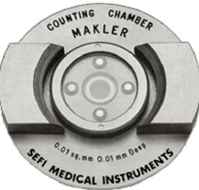
Human semen is produced as a concentrated suspension of spermatozoa stored in the paired epididymides. During ejaculation it is mixed with, and diluted by, fluid secretions from the accessory sex organs. It is present as a fluid conglomerate emitted in several boluses. Semen has two major quantifiable attributes: (1) the total number of spermatozoa reflecting sperm production by the testes and the potency of the post-testicular duct system, (2) the total fluid volume reflecting the secretory activity of the glands. Semen reflects a heterogeneous cell suspension that differs among individuals in protein content, sperm number, pH, volume etc. These factors defined by the World Health Organization (WHO) are crucial when judging semen quality. Semen quality (Tab. 1, 2) is taken into account to measure male fertility in clinical andrology, male fertility, reproductive toxicology, epidemiology and pregnancy risk assessments (WHO, 2010). The quality of semen is verified by the presence of several components. For instance, the abundance of proteolytic fragments, decreased seminal level of albumin, poor testicular contribution characterize the oligozoospermic ejaculates. Seminal glycoprotein deficiency, the increased level of acid phosphatase and increased prostatic secretory activity are associated with asthenozoospermia (Autiero et al., 1991). Since specific substances can be assigned to specific reproductive compartments, chemical substances serve as diagnostic markers. Neutral -glucosidase, carnitine, glycerolphosphocholine represent the indicators of epididymal functions. Prostate function is evaluated according to the levels of fructose, prostaglandins, citrate, zinc and prostatic acid phosphatase (Rowe et al., 1993). Semen also contains several immunoregulatory factors as well as immunogenic agents that represent the possible targets of activated inflammatory cytokines, leukocytes and complement cascade in any part of the female genital tract (Chung et al., 1994; Sharkey et al., 2007; Rodriguez-Martinez et al., 2011). One of the most common devices to evaluate spermiogram is the Makler counting chamber. It is composed of a metallic base unit, semi-circular ring, cover glass with surface graticule (Fig. 1, Makler, 1980).
The inability to achieve penile erection or to maintain an erection until ejaculation, caused by the congenital, neurological and metabolic disorders or abnormalities, is known as erectile dysfunction (impotentia coeundi). In addition, if spermatogenesis is disrupted, it results in the inability to procreate (impotentia generandi) (Ulcova-Gallova, 2006).
3 Fig. 1 Makler counting chamber, metallic base tool to evaluate sperm amount. (Makler, 1980)
Tab. 1 Semen characteristics (WHO, 2010)
Semen type Description
aspermia no semen/no mature sperm in semen no/retrograde ejaculation
asthenozoospermia percentage of progressively motile sperm below the lower reference limit
asthenoteratozoospermia percentages of both progressively motile and morphologically normal sperm below the lower reference limit
azoospermia no sperm in the ejaculate, activity of seminal enzymes retarded cryptozoospermia
less than 1 million of sperm in the ejaculate or none are seen on an initial evaluation, but only after centrifugation and concentration of the sample
haemospermia presence of erythrocytes in the ejaculate leukospermia presence of leukocytes in the ejaculate
necrozoospermia low percentage of live sperm, high percentage of immotile/dead sperm in the ejaculate
normozoospermia total number of sperm and percentages of progressively motile and morphologically normal sperm equal to/above the lower reference limit oligoasthenozoospermia total number of sperm and percentage of progressively motile sperm
less than 20 million
oligoasthenoteratozoospermia total number of sperm and percentages of both progressively motile and morphologically normal sperm below the lower reference limit
oligoteratozoospermia total number of sperm and percentage of morphologically normal sperm below the lower reference limit
oligozoospermia total number of sperm below the lower reference limit
teratozoospermia percentage of morphologically normal sperm below the lower reference limit
4 Tab. 2 Reference limits for semen characteristics (Cooper et al., 2010)
Semen characteristics Reference limit
volume >1.5 ml
pH 7.1 – 7.8
sperm concentration > 20 mil/ml total sperm amount > 40 mil
vitality > 75%
motility > 50% motile
morphology > 50% normal morphology agglutination < 2 (scale 0 - 3)
viscosity < 3 (scale 0 - 4) liquefaction within 30 min leukocytes < 1 mil/ml
2.1
Seminal fluid
Seminal fluid (SF), also cited as seminal plasma, represents a part of the semen containing a range of organic/inorganic substances (e.g. neutral α-glucosidase, hyaluronidase, carnitine, glycerolphosphocholin, fructose, prostaglandins, citrate, zinc, selenium) that are necessary for the physiological metabolism of sperm. The seminal complex mixture of secretions originates in the testis, epididymis and accessory glands including the prostate, seminal vesicles and Cowper‟s gland. It also acts as a nutritive, transport and buffering medium of pH 7.35 - 7.5 that defines the main SF functions: sperm protection from the acidic environment of the vagina, metabolic support and competence, liquefaction and clot formation. SF composition is similar to blood plasma. However, it differs in saccharide content (Kumar et al., 2009; Rodriguez-Martinez et al., 2011; Brazdova et al., 2012a).
Semen liquefaction is caused by the fibrinolytic activity of some proteinases and peptidases present in prostatic SF. The key role of seminal enzymes (kallikrein-like protease 3, prostate-specific antigen) consists of a clot digestion formed immediately after ejaculation. It is said that SF containes hundreds of proteins of molecular or catalytic activity including additional proteins classified as their regulators (Yousef and Diamandis, 2001; Pilch and Mann, 2006).
In general, seminal proteins can be divided into three different groups. The first group contains the extracellular and intracellular proteins supporting basic SF function. The second group, originating in proteasomes and membrane-enclosed structures, is mainly involved in oocyte-sperm fusion. The third group is known as the potential biomarkers of testis/prostate cancer and male infertility as they represent the abraded epithelial cells from tissue surface (Pilch and Mann, 2006).
5 SF is in routine examined to evaluate pathological spermiogram and to monitor the progression of either testis or prostate cancer. In particular, prostate cancer is diagnosed by the seminal level of prostate-specific antigen (PSA), prostatic acid phosphatase (PAP), prostate stem-cell antigen or glutamate carboxypeptidase II that is a prostate-specific membrane antigen (Jones, 1991; Ostrowski and Kuciel, 1994; Cao et al., 2003). Prostate cancer can be diagnosed by the serum PSA level. The serum concentration of 4 ng/ml means a 20-30% risk of prostate cancer. The risk increases approximately to 60% with a PSA level higher than 10 ng/ml (Fung et al., 2004). An early elevated level of serum zinc alpha-2-glycoproteins (ZAG), originally secreted by the prostate, indicates tumor growth as well. PSA, PAP and prostate-specific protein-94 (PSP-94) belong to the prostate secretion that accounts for about 30% of the total SF volume. This SF fraction is in direct contact with sperm and is the first to confront the cervical tissues. PSA ( -seminoprotein, seminin, kallikrein-3, P30 antigen, semenogelase) is a 33 kDa member of the glandular kallikrein subfamily of serine proteases. In particular, it is a member of the kallikrein subgroup of the (chymo)-trypsin serine protease family. It differs from the prototype member of this subgroup, tissue kallikrein, by possessing specificity more similar to that of chymotrypsin than trypsin. PSA is known as a zinc-binder and the most common protease in human semen. It is mainly released in proteasomes. It hydrolyzes semenogelin-1, thus leading to the liquefaction of the seminal coagulum. Its activity is strongly inhibited by zinc ions. This inhibition is relieved by exposure to semenogelins, which are avid zinc binders (Espana et al., 1991; Utleg et al., 2003; Pilch and Mann, 2006). PAP belongs to histidine acid phosphatase family, is stored in lysosomes and has an acid pH optimum below 7.0. It is a non-specific tyrosine phosphatase that dephosphorylates macromolecules and inactivates lysophosphatidic acid in SF. Its isoform 2 acts as a tumor suppressor of prostate cancer through the deactivation of mitogen-activated protein kinases. Decreased PAP level exhibits poor liquefaction (Autiero et al., 1991; Tanaka et al., 2004).
It contains the membrane-enveloped secretory vesicles. The fructose level is a marker of seminal vesicle function. High concentration of fructose is essential for sperm survival in the female reproductive tract. Fructose and other sugars are a source of energy for mitochondria-rich sperm. A decreased fructose level means a lower intensity of fructose oxidation. It leads to a lactate accumulation, which results in reduced sperm motility (Anderson et al., 2004). SF is rich in proteins secreted by seminal vesicles. Semenogelin I and II, fibronectin and fibronectin-related derivates belong to the gel-forming proteins. They are active after the cleavage by kallikrein-like protease and then have a role in entrapping sperm into a viscous
6 gel immediately after ejaculation. They are also involved in sperm capacitation and sperm-oocyte interaction. Another SF element is lactoferrin, thanks to which SF may have an antimicrobial activity. SF content also involves albumin, serum and testis derived, that is a predominant protein participating in cholesterol removal from the sperm membrane during capacitation. The decreased seminal level of albumin and the increased seminal level of cholesterol are found in oligozoospermic men (Lilja et al., 1987; Autiero et al., 1991; Kosanovic and Jankovic, 2010).
Seminal components that bind to the acrosomal sperm head region protect sperm and are then carried together with it into the higher female genital tract. SF plays an important role in moving the sperm into the female reproductive tract due to its high content of transforming growth factor beta (TGF ) and prostaglandin E (PGE), both of which inhibit the function of natural killer (NK) cells and neutrophils that are recruited into the superficial epithelial layers of the cervical tissues. SF is rich in PGE of 19-hydroxy form that promotes an expression of chemotactic interleukin 8 (IL-8). TGF is synthesized in the prostate and is testosterone-dependent. This glycoprotein belongs to cell-secreted molecules and occurs in 75% in the latent form in SF. It is further activated in the female reproductive tract by either the enzymes of male/female origin, acidic vaginal pH or through conformational change after an interaction with epithelial cells. The remaining proportion of TGF , β5%, exists in an active form (Denison et al., 1999; Robertson et al., 2002). TGF acting may result in the immune tolerance of seminal antigens, for which TGF I is, most likely, responsible. It is a cytokine of TGF family. A divergent member of this family is growth/differentiation factor 15 (GDF 15), which is highly abundant in SF. Surprisingly, its level is not related to semen quality but its expression serves as a cancer marker, often in combination with PSA. However, GDF 15 has antitumorigenic activity. In contrast, the high level of GDF 15 in female serum corresponds to spontaneous abortion as it is expressed in placenta as well. It has been suggested that thanks to the presence of seminal antigens on a conceptus, TGF facilitates the female immune tolerance to a fetus (Robertson, 2005; Soucek et al., 2010).
SF includes a repertoire of signaling molecules interacting with epithelium in the female reproductive tract. SF may modulate the chemotactic and phagocytic response of the female reproductive tract. Phagocytes serve to filter out the morphologically abnormal sperm. Sperm selection is based on morphological or antigenic structures. Mainly, the immune modulating properties are mediated by the prostaglandins of the E series, complement inhibitors, cytokines and proteins capable to bind IgG antibodies (Tomlinson et al., 1992; Kelly and Critchley, 1997). Local reactions may lead to an inflammation (Robertson, 2005).
7 However, SF has a built-in mechanism preventing an immunological sensitization of the female against sperm as well as seminal structures. This protective system exists due to the presence of immune inhibitors originating in the male sex accessory glands (Prakash, 1981). SF has been suggested to be the modulator of sperm-induced inflammation that leads to sperm elimination from the female genital tract (Troedsson et al., 2005).
SF elicits endometrial changes by inducing pro-inflammatory cytokines and cyclooxygenase-2. Their presence leads to macrophage and dendritic cell recruitment into the female reproductive tract. Seminal components activate the income of neutrophils into the endometrial stroma (Robertson, 2005; Bronson, 2011; Morrell et al., 2012). However, it has been proved that the influx of neutrophils is higher and faster when the washed sperm inseminated (Rozeboom et al., 1999). This fact proves the protective and signaling activity of SF. The immuno-suppressive activity prevents the iso-immunization of the female reproductive tract and suppresses the cell-mediated cytotoxicity (Lord et al., 1977). Seminal prostaglandin D2 is known for its immuno-suppressive effect, by which the anti-sperm antibody formation is avoided in the female genital tract. The immuno-modulating properties are mediated by prostaglandin E, complement inhibitors, cytokines and proteins capable of binding the Fc region of IgG. These IgG-binding proteins are Fc receptor-like proteins. In general, seminal antibody-binding proteins contribute to sperm protection against immune-mediated damage by enabling successful sperm passage in the female reproductive tract and by blocking an interaction with immune effectors. For instance, prolactin-inducible protein (PIP), which is a secretory glycoprotein located in seminal vesicles, binds to immunoglobulin G via its Fc fragment, it may therefore be involved in immune regulation by trapping anti-sperm antibodies (ASA) and neutralizing them (Chiu and Chamley, 2002; Chiu and Chamley, 2003).
SF has been considered to be linked to the IgE-mediated rare reaction to semen (Weidinger et al., 2006). This rare phenomenon was firstly reported by James (1945). Human seminal plasma allergy (HSPA) or the so-called hypersensitivity to semen is defined by localized and/or systemic symptoms after exposure to seminal fluid. The symptoms occur immediately after contact with semen or even within several hours after intercourse. The local symptoms include vulvar/vaginal itching, burning, redness and swelling. Local reaction can appear on any semen contact site. Local symptoms can be misdiagnosed as chronic vulvo-vaginitis caused by bacteria, yeasts, viruses and other parasites. Systemic features include generalized urticaria, angioedema (face, tongue, lips, throat), dyspnea, wheezing, cough, chest tightness, rhinorrhea, nausea, vomiting, diarrhea. Generalized malaise may result in an
8 anaphylactic shock, which is a life-threatening reaction. The symptoms can manifest after the first time intercourse in up to 50% of cases. Response mediated by IgE antibodies is then the most common mechanism. It has been suggested (Basagana et al., 2008) that female patients experiencing any allergic symptoms after/during the first time intercourse might be sensitive to other antigens/allergens that cross-react with SF. Basagana et al. (2008) has already proved the IgE cross-reactivity among proteins from dog epithelium and PSA. Patients diagnosed with HSPA have difficulties conceiving but infertility has not been demonstrated, so far (Shah and Panjabi, 2004; Weidinger et al., 2006; Bernstein, 2011).
2.2
Spermatozoa
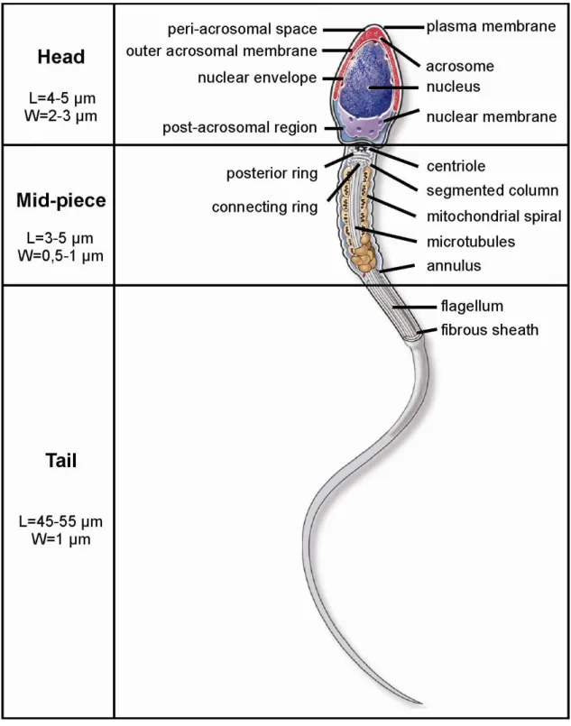
Mature male gamete is commonly known and cited as sperm but specifically called as spermatozoa. It was firstly described in 1677 by Anthony van Leeuwenhoek. Sperm is a male reproductive cell of approximately 55-65 µm, containing genetic information and participating in the fertilization of an ovum (150-300 µm). Sperm consists of a head, middle section (mid-piece) and tail (Fig. 2). It is characterized by a minimum of cytoplasm. The head contains a nucleus, its shape is flat and oval in order to attach and easily penetrate an oocyte. An anterior peak of sperm head carries a cap-like structure called acrosome. It is designed, thanks to its hydrolytic enzymes, to help the sperm to penetrate the oocyte. The middle section consists of mitochondrial spiral, outer dense fibers and core microtubular structures. The mitochondrial formation contains the enzymes of oxidative phosphorylation. The mid-piece is, therefore, composed of substances that propel the tail. The tail enables the sperm to be motile by rotating in a circular motion, not from side to side like a whip (Collins discovery encyclopedia, 2005). The speed of sperm in ejaculate ranges from 10 to 60 µm/s. Its movement is based on the enzymatic and microtubule components, and is calcium and magnesium dependent. The tail is able to move ten times per second. Twenty thousand movements are estimated to be needed to reach the oocyte. In vitro, the speed is positively influenced by methylxanthins, lower temperature, and negatively by proteolytic enzymes, hydrogen peroxide and human saliva as it contains amylase and lysozyme enzymes (Ulcova-Gallova, 2006).
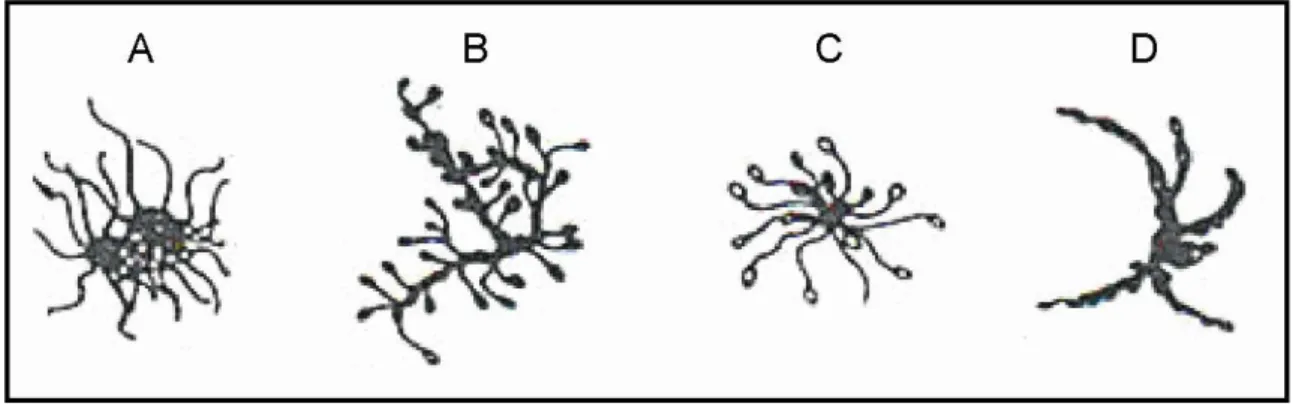
Normal sperm is characterized by an oval head with a long tail. Abnormal sperm has the defects of any body part (Fig. 3). Defects occurring on the head cause different shapes: large (giant), small (micro), elongated, irregular, amorphous, and then involve the acrosome
9 deficiency and the so-called bicephalic head. The defective mid-piece is asymmetric, bent, thin, thick, irregular or with cytoplasmic droplets. The defective tail is coiled, shortened, hairpined, broken, duplicated or with terminal droplets. Any defects may impair the ability of the sperm to reach and fertilize the oocyte. In general, sperm morphological abnormalities are related to congenital background, varicocele, high fever, infection or drug use (Gilbert, 2000).
10 Fig. 3 Examples of sperm morphological abnormalities, head, mid-piece, tail defects. (MFMER, 2013)
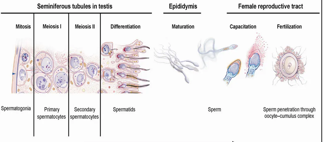
2.2.1 Spermatogenesis
Spermatogenesis (Fig. 4) is a complex process creating functional sperm, starting at puberty and ending with death. It occurs in the testis, progresses to the epididymis and takes approximately 64 days for completion. Effective maturation is conditioned by temperature, 2 °C lower than body temperature. This explains its location in external genitals. Sperm cells are then stored in the epididymides. The entire process is regulated by hormones (follicle stimulating hormone, luteinizing hormone, testosterone). It consists of three major steps: (1) the multiplication of spermatogonia by mitosis, (2) meiosis to reduce the chromosome number from diploid to haploid, (3) the successful transformation of the round spermatid into the so-called spermatozoa. The spermatogonial population is created from the germ cells in the testis. The population then fuses with the Sertoli cells by creating seminiferous cords. After multiplication, several types of spermatogonia are distinguished: type A and B. The subsequent meiosis reduces the chromosome number from a diploid to a haploid form of type B spermatogonia. Type A diploid spermatogonium divides into two diploid cells called primary spermatocytes. The newly developed cells migrate into seminiferous tubules to
11 undergo the meiosis by creating the secondary spermatocytes. The secondary spermatocytes are haploid. The next step reflects the forming of rounded spermatids. The differentiated spermatids mature in the epididymis into functional spermatozoa. There is an evidence of post-translational modifications that are considered to be essential for efficient spermatozoa. Some of them can activate capacitation directly in the epididymis or post-ejaculatory in the female reproductive tract. The cascade of modifications includes phosphorylation, glycosylation, proteolytic cleavage and methylation. A healthy man is usually able to produce up to 100 million of sperm/day. However, the concentration of 20 million/ml reflects, nowadays, the mean amount (Ulcova-Gallova, 2006; Warwick, 2006; Vacek, 2006; du Plessis et al., 2011).
12
13
2.2.2 Capacitation
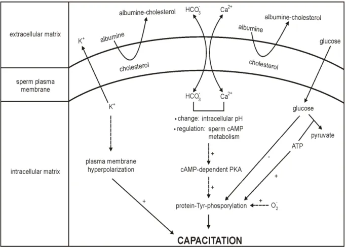
Sperm capacitation was independently discovered by Austin (1951) and Chang (1951). Capacitation is defined as the process, by which the sperm becomes able to undergo an acrosome reaction in the uterus, then to penetrate and fertilize the oocyte.
The main purpose of sperm capacitation (Fig. 5) consists of hyperactivation in order to reach the oocyte, particularly in the remodeling of sperm membrane with regards to the fusion with the oocyte (Sutovsky, 2011). Capacitation involves a sperm surface remodeling, protein phosphorylation, hyperpolarization, an increased membrane fluidity, internal Ca2+ concentration and pH. A number of different seminal factors have been shown to act as the initial factors of capacitation: fertilization promoting peptide, adenosine, calcitonin, heparin binding proteins and angiotensin II. Other participating messengers are derived from sperm-oocyte interaction. Other factors originate in the female reproductive tract such as leukocytes, progesterone, fertilization promoting peptide, cholesterol, sialic acid binding proteins, capacitation-associated tyrosine-phosphorylated proteins, heparin binding proteins and atrial natriuretic peptide (Storey, 1995; Kumar et al., 2008).
Capacitation requires a period of sperm incubation in the female tract, approximately 1 hour. It involves an increased metabolic activity, higher motility and the removal of the decapacitation factors from the sperm followed by the destabilization of the acrosomal sperm head membrane. This change involves the removal of steroids and seminal glycoproteins from sperm membrane. It allows greater binding between sperm and oocyte. Thanks to Ca2+ permeability,the binding is strengthened. An influx of Ca2+ results in sperm hyperactivation, which induces the higher cAMP level. Ca2+ and HCO3- play a critical role in the regulation of sperm function, most likely by acting as the enzyme effectors involved in signal transduction (Yanagimachi, 1994). Furthermore, the sperm adenylyl cyclase is significantly stimulated by HCO3-. The bicarbonate anionincreases an intracellular pH and has been suggested to act as an anion antiport with respect to Ca2+ (Okamura et al., 1985).
The major sperm sterol is cholesterol, among others e.g. desmosterol, desmosterol sulphate, cholesterol sulphate, cholesterol esters, cholestadienol. Additional cholesterol may be obtained from seminal plasma. Cholesterol moves from the sperm membrane to the acceptors and phospholipids into the sperm membrane. The ratio of cholesterol/phospholipids in a freshly ejaculated sperm is about 0.8. Albumin acts as a sink for the removal of cholesterol from the sperm plasma membrane. The loss is initially linear and leads to the exposure of a mannose receptor. Zona pellucida (ZP), an oocyte membrane composed of proteins (70%) and
14 saccharides (30%), contains mannose that interferes with sperm receptors. Cholesterol also inhibits the responsiveness and Ca2+/H+ exchange in the ionophores. It has been suggested that the increased cholesterol level has a role in male unexplained infertility since the concentration is about twice as high as in the fertile subjects. Cholesterol efflux corresponds to an increased level of tyrosine as well as proline protein phosphorylation. The sperm cholesterol level varies among individuals (Mitra and Shivaji, 2005; Jha et al., 2006).
Sperm protein tyrosine phosphorylation (PTP) has been considered to be the key signaling pathway. It is thought that it acts as a signal to alter mitochondrial function. Sperm PTP is dependent on the presence of calcium and bicarbonate ions. It is supported by ATP from glycolysis and is regulated through a protein kinase A (PKA) pathway. However, glucose is thought to inhibit PTP, which could be up-regulated by free radicals, most likely by superoxide anion. Moreover, the superoxide anion generation is also related to sperm lipid peroxidation, hyperactivation and viability (de Lamirande and Gagnon, 1993, Visconti et al., 1998).
Capacitation also involves the membrane hyperpolarization caused by K+ permeability. It has been speculated whether K+ throughput independently allows the recruitment of Ca2+. Subsequently, the membrane potential is increased in tens of mV (Zeng et al., 1995; Visconti et al., 1998).
An essential capacitation factor is a fertilization promoting peptide (FPP), a tripeptide (Glu-Glu-Pro) synthesized in the prostatic glands. FPP is present in SF and comes into contact with sperm after ejaculation. It becomes less active in the female genital tract due to vaginal acidic pH. It has a synergic stimulatory effect with adenosine that increases adenylyl cyclase activity in the sperm. Another seminal protein, semenogelin, appears to block sperm capacitation (Fraser and Osiguwa, 2004).
Sperm proteasome, located on the inner acrosomal membrane, is also involved in capacitation thanks to its proteolytic activity. It assists in protein removal during membrane remodeling, acrosome exocytosis by the degradation of membrane proteins in order to release the acrosomal matrix and to create the so-called acrosomal ghost. The sperm is then prepared to penetrate and fertilize the metaphase II-arrested oocyte (Sutovsky et al., 2004; Zimmerman and Sutovsky, 2009).
15
Fig. 5 Theoretical trans-membrane and intracellular signaling of sperm capacitation. PKA: protein kinase A, +/-: stimulating/inhibiting pathways, consequence, reaction, ion exchange. Based on de Lamirande and Gagnon (1993); Zeng et al. (1995); Visconti et al. (1998).
2.2.3 Acrosome reaction
Capacitated sperm becomes hyperactivated with regards to motility. Then, it is able to recognize the oocyte. The sperm membrane is destabilized after capacitation. It can be attached to ZP and undergo an acrosome reaction (AR). Gradual steps are mediated by an osmo-sensitive signal transducing mechanism. An oocyte-sperm fusion is coordinated by the carbohydrate-protein interaction. The carbohydrate-binding site on the sperm interacts with the oligosaccharide ligands of ZP (Topfer-Peterson et al., 2000).
AR (Fig. 6) is defined as the physiological release of acrosomal content. It occurs in the female genital tract following sperm capacitation. It is a precondition to penetrate and fuse the oocyte ZP and to undergo the fertilization event. The cholesterol removal, elevated internal levels of Ca2+ and increased pH during capacitation are thought to trigger the acrosomal exocytosis that reduces the sperm membrane and enables to perceive the oocyte. Acrosomal
16 disruption involves the intracellular/membrane modifications and hydrolytic/proteolytic enzyme release. The most involved enzymes are acrosin and hyaluronidase that are required for oocyte penetration. AR activation and acrosomal exocytosis display the new membrane domains representing the new antigenic targets of the female immune system (Breitbart and Spungin, 1997; Patrat et al., 2000).
AR inducing factors are zona pellucida glycoprotein 3 (ZP3) and progesterone, which is produced by the cumulus cells surrounding the oocyte. It has been shown that ZP binds at least to two sperm plasma membrane receptors, receptor activating phospholipase C (PLC) and tyrosine kinase receptor (TKR), which is coupled to PLC. Binding to a receptor activating PLC influences cAMP elevation and further PKA activation. Binding to TKR leads to the phosphorylation of the calcium transporter that activates phospholipase C (PLC ). Activated PLC catalyzes the hydrolysis of phosphatidylinositol 4,5-bisphoshate (PIP2) into inositol-triphosphate (IP3) and diacylglycerol (DAG). IP3 binds to a receptor on an outer acrosomal membrane, resulting in Ca2+ release. DAG stimulates phospholipase A (PLA) resulting in fusiogenic lipids (FL) and fatty acids (FA). These substances or their derivates, e.g. arachidonic acid, modulate the AR. The depolarization of plasma membrane by H+/Na+ ionophores increases intracellular pH and activates adenylyl cyclase, which stimulates cAMP elevation and PKA activation. H+/Na+ exchange also alkalizes the cytosol. The increased concentration of Ca2+ and increased pH allow membrane fusion, followed by the AR (Breitbart and Spungin, 1997; Patrat et al., 2000).
17
Fig. 6 Theoretical trans-membrane and intracellular signaling of sperm acrosome reaction. ZP3: zona pellucida glycoprotein, R1: coupled binding receptor, TKR R2: tyrosine kinase receptor, PLC 1: phospholipase C, i pH: increased intracellular pH, AC act.: activated adenylate cyclase,
PKA: protein kinase A, PIP2: phosphatidylinositol 4,5-bisphoshate, IP3: inositol-triphosphate, DAG: diacylglycerol, PLA: phospholipase A, R3: IP3 binding receptor, PD: protein derivates, FA: fatty acids, AR: acrosome reaction, binding, reaction, ion exchange,
18
2.2.4 Fertilization
During sexual intercourse, millions of sperm are deposited in the vagina. In approximately 8 seconds after ejaculation, acidic vaginal pH is changed into to basic. SF immediately cleaves cervical mucus to enable sperm to move. The speed of sperm depends on the menstrual cycle and varies from 0.2 to 0.4 mm/min. Sperm is able to survive for up to 12 h in the vagina. It has been proposed that 1 sperm out of approximately 5000 reaches the fallopian tube in 10 min (Ulcova-Gallova, 2006).
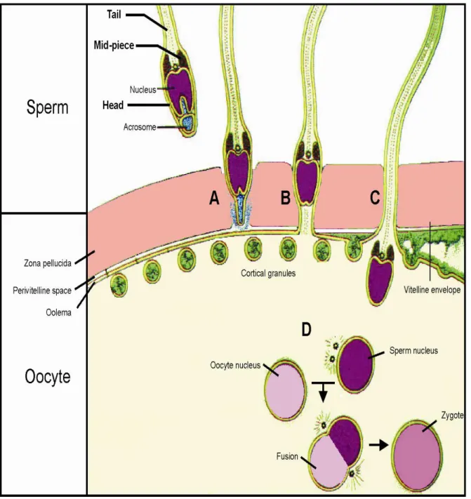
Fertilization is defined as the union of male and female gametes to form a zygote (Fig. 7). Sperm is able to fertilize only a secondary oocyte at metaphase II, thus an oocyte after the meiotic division. The crucial step of fertilization consists of ZP penetration. Sperm has to pervade the oocyte-cumulus complex to reach ZP. Sperm binds to the oocyte thanks to components such as fukoidin and lectins. Low pH is required for dissolving this plasma membrane, the so-called envelope that protects the oocyte against any physical damage. The penetration of approximately 10 µm layer depends on the activity of sperm acrosomal proteases and glycohydrolases. ZP is composed of three glycoprotein layers (ZP1-3) that differ in their length. ZP1-3 layers represent a heterogeneous complex of asparagine (N)- and serine/threonine (O)-linked oligosaccharides, creating a fibrogranular layer. Sperm interacts with ZP either through spermadhesin, which is a lectin containing galactose, mannose or mannose-6-phosphate, or through proacrosin, which binds to sulphates. Sperm also binds to an oviductal epithelium through spermadhesin, thus it participates in the formation of the sperm oviductal reservoir. Once the signaling mechanism is initiated, ZP receptor kinases and fertility antigen A1 are autophosphorylated. With regards to the hyperactivity of flagellating sperm tail and its acrosomal enzymes, ZP is digested. Oocyte and sperm membranes fuse. After ZP penetration, sperm interacts with a vitelline membrane, which implies the development of an embryo. As soon as the fusion has been started through the receptors, the electrical block is set up. The polarity of the oocyte membrane is reversed and the polyspermy is blocked. The oocyte then releases its cortical granules to initiate the zona reaction, thereby preventing multiple sperm penetration. Cortical follicles/granules fuse with the plasma membrane. The cortical enzymes dissolve and remove the sperm binding receptors, thanks to which the vitelline envelope separates from the plasma membrane. Perivitelline space is created to form the fertilization membrane. When the sperm head penetrates the ovum, the tail separates from the rest of the sperm. Only a sperm nucleus enters the cytoplasm to fuse with an oocyte nucleus. After the fusion, a zygote is created. The diploid zygote undergoes division
19 to the 2-, 4- and then to 8-cell stage. The next stage is a morula, from which a blastocyst is formed. The blastocyst is then implanted. To summarize, the above-described process depends on sperm motility, sperm survival in acidic vaginal pH, cervical mucus viscosity and the activated female immune sytem (Yanagimachi, 1994; Peterson, 1999; Topfer-Peterson et al., 2000).
Fig. 7 Schema of fertilization. A: acrosome reaction, B: fusion of oocyte-sperm membrane, C: cortical reaction, D: fusion of oocyte/sperm nuclei, A, B, C phases last 20-30 s. Based on Alberts et al. (2002).
20
3
FEMALE FACTOR IN REPRODUCTION
3.1
Oocyte
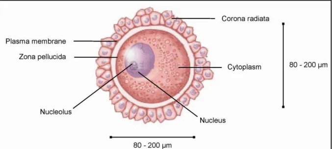
The oocyte (Fig. 8) is a circular cell that was first found and described by Karl Ernst Ritter von Baer in 1827. The oocyte is a female gametocyte also defined as a developing female germ cell. It is 80-200 µm in diameter with a viability of 12-24 h. It is protected by ZP and surrounded by the cumulus and corona cells. It is formed in an oogenesis process. It is rich in cytoplasm, containing a lot of yolk granules to nourish the cell at an early development (Mandelbaum, 2010). In comparison with sperm, the male gametocyte, the oocyte differs in many aspects (Tab. 3).
Fig. 8 Oocyte description. (http://ldysinger.stjohnsem.edu)
Tab. 3 Comparison of sperm and oocyte (Gilbert, 2000; Vacek, 2006)
Aspect Sperm Oocyte
size 55-65 µm 80-200 µm
size in body one of the smallest cell one of the largest cell shape straight, flagellated spherical
body parts – head, mid-piece, tail uniform
cytoplasm small amount large amount
nucleus condensed with no nucleoplasm bloated with nucleoplasm
centriole present absent
mitochondria compactly arranged scattered in cytoplasm motility motile (2 mm/min) immotile
21 Tab. 3 Comparison of sperm and oocyte (continuation; Gilbert, 2000; Vacek, 2006)
Aspect Sperm Oocyte
viability 72 h 12-24 h
genesis spermatogenesis oogenesis
site of creation process testis ovary
chromosomes 23 23
genesis beginning puberty fetal period
genesis end death menopause
limitation of production millions in 3 days 1 cell in 28 days
parent cells unlimited number can become sperm limited number can become oocyte optimal temperature 2 °C less than body temperature body temperature
3.1.1 Oogenesis
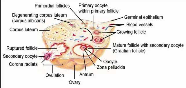
Oogenesis is the female equivalent of male spermatogenesis, and describes the development of an immature ovum (egg cell). The egg cells are produced in the ovary (Fig. 9) in several stages (Fig. 10). Primary follicles contain a primary oocyte and begin to produce the sex hormone estrogen. A primary oocyte carries two sets of chromosomes. In the first meiotic division, the primary oocyte is unevenly divided into a secondary oocyte (daughter cell) and the first polar body. The first polar body is an apoptotic cell that cannot be fertilized by sperm and does not contribute to zygote, embryo or fetus development. The secondary oocyte contains most of the original cytoplasm, is haploid and is bigger than the first polar body. The secondary follicle contains a secondary oocyte and produces estrogen and progesterone. As a result, the Graafian follicle develops. This follicle is named after its discoverer, Regnier de Graaf. It contains the secondary oocyte with a formed ZP. The secondary oocyte undergoes a second meiotic division into an ovum and a second polar body. In reality, the secondary oocyte is arrested at a metaphase of secondary meiotic division (metaphase II) until fertilization (chapter 2.2.4 Fertilization) takes place. Arrested at metaphase II, the secondary oocyte is released during ovulation by follicle rupture and is trapped by the linings of fallopian tube, which travels toward the uterus. The ruptured follicle undergoes transformation into a corpus luteum, a structure of approximately 2 cm (diameter). The corpus luteum is formed during the luteal phase of menstrual cycle, contains carotenoids and acts as an endocrine structure by producing progesterone. The secreted progesterone is necessary for the decidualization of endometrium. Decidualization involves several significant changes in the endometrium to implant the embryo. As the corpus luteum degenerates, a corpus albicans is developed (Gilbert, 2000; Vacek, 2006; Gellersen et al., 2007).
22 Fig. 9 Oogenesis, ovarian surface cut. (http://ldysinger.stjohnsem.edu)
23
3.2
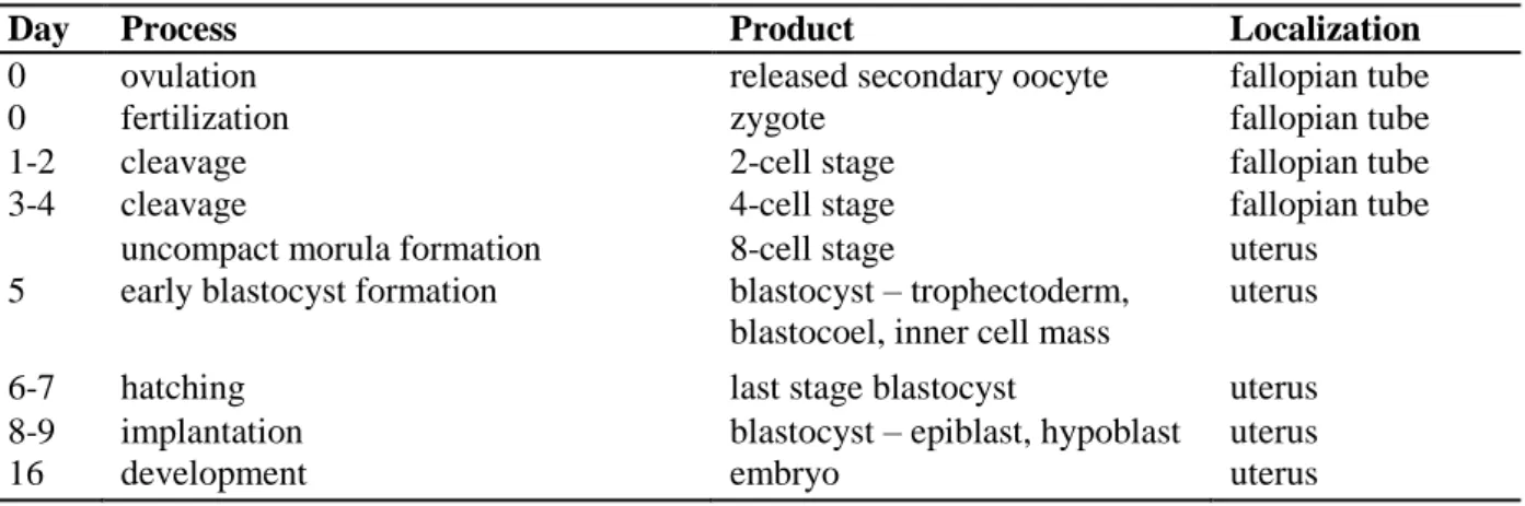
Embryo development
Sperm is able to fertilize the metaphase II-arrested oocyte (chapter 2.2.4 Fertilization) within 12-24 h after ovulation. After their fusion, a zygote is formed. Two-, four- and then eight-cell stages turn into a morula that represents the earliest phase of embryo development (Tab. 4). The journey of morula to uterus lasts approximately 4 days. The following step consists of a blastocyst formation. The trophoblast differentiates, then digests the uterine cells and burrows into the wall of the uterus. The blastocyst thus tries to attach itself to the uterine wall, and is implanted into the higher part of uterus. Implantation is conditioned by numerous endometrial changes (decidualization) that enable the acceptance of the blastocyst into uterus. Endometrial decidualization is a post-ovulatory and pregnancy-independent process. It remodels the endometrium and is based on the secretory transformation of the uterine glands, influx of uterine NK cells, vascular remodeling and stimulated glycogen accumulation in stromal cell cytoplasm. The decidualized stromal cells regulate trophoblast invasion and moderate potential oxidative stress and maternal immune response. Maternal macrophage activity is suppressed based on progesterone regulation and anti-idiotypic antibodies. Implantation also activates placenta development. The placenta produces progesterone and estrogen to maintain pregnancy. It serves as a vessel for the exchange of various compounds. Carbon dioxide, urea, water, and hormones are all transferred from fetus to mother. In the reverse direction, oxygen, nutrients, water, hormones, vitamins, and minerals are transported from mother to fetus. During the second trimester of pregnancy, the fetus is formed and its development continues up to the end of the third trimester (Gilbert, 2000; Vacek, 2006; Gellersen et al., 2007).
Pregnancy may be complicated by pre-eclampsia (PE), a disorder affecting both the mother and the unborn child. This life-threatening syndrome is the fatal culprit of perinatal morbidity/mortality, abortions and premature births. It is suggested that an inadequate trophoblast invasion leads to a reduced placental perfusion at the end of the first trimester, which is followed by endothelial dysfunction and clinical manifestation by the end of the second/during the third trimester. So far, the etiology is unknown and the diagnosis is still very complicated. The clinical manifestation varies since PE is associated with several risk factors such as obesity, molar pregnancy (a non-viable fertilized ovum is implanted into the uterus), hypercoagulation, formation of clots or diabetes mellitus. PE includes the manifestation of symptoms such as hypertension, proteinuria, edema, hyperuricemia. Additional PE symptoms including hemolysis, elevated liver enzymes and low platelet count are referred to as HELLP syndrome (Brazdova et al., submitted 2014a). PE syndrome is
24 considered to be a two-stage disease. The reduced placental permeability is associated with the first stage, causing abnormal implantation and fetal placental release into the maternal circulation. As a result, the maternal immune system creates antibodies against the trophoblast. The second phase consists of the maternal reaction to this condition. It is characterized by inflammation and the endothelial dysfunction of parental cells (Redman and Sargent, 2000).
Tab. 4 Embryo development (Vacek, 2006)
Day Process Product Localization
0 ovulation released secondary oocyte fallopian tube
0 fertilization zygote fallopian tube
1-2 cleavage 2-cell stage fallopian tube
3-4 cleavage 4-cell stage fallopian tube
uncompact morula formation 8-cell stage uterus
5 early blastocyst formation blastocyst – trophectoderm, blastocoel, inner cell mass
uterus
6-7 hatching last stage blastocyst uterus
8-9 implantation blastocyst – epiblast, hypoblast uterus
25
4
INFERTILITY
The World Health Organization defines infertility as a disease of the reproductive system by the failure to achieve a clinical pregnancy after 12 months or more of regular unprotected sexual intercourse (Rowe et al., 1993). Infertility has been reported to be one of the most prevalent chronic health disorders regarding couples of any age (Smith et al., 2003).
Over the last three decades, the number of documented infertility cases has increased. Decreased fertility is associated with other health issues, age, life style and environment. The agent of infertility for a couple is the male partner about 40% of the time, the female partner about 40% of the time , and about 20% of the time the fertility problems are shared by both the man and the woman. Factors contributing to infertility involve congenital, hormonal, morphological and immunological disorders. Infertility may be also related to other diseases such as severe avitaminosis, severe renal impairment, cancer and cachexia due to malnutrition or tumor (Doherty and Clark, 2006).
The main disorders involved in infertility include pathologic spermiogram, ovulation problems/anovulation, tubal diseases, pelvic adhesion/endometriosis, cervical factors and idiopathic reasons – i.e. unexplained infertility. However, a single specific cause of infertility is never certainly diagnosed (Gleicher and Barad, 2006; Siristatidis and Bhattacharya, 2007).
4.1
Unexplained infertility
Unexplained infertility (UI) is diagnosed to a couple when the standard investigations including semen analyses and tests of ovulation and tubal potency do not provide specific results or do not provide evidence of any abnormality (Crosignani et al., 1993). Several reports (Crosignani et al., 1993; Gleicher and Barad, 2006; Siristatidis and Bhattacharya, 2007) suggest that the diagnosis of UI is subjective and that UI is often misdiagnosed for endometriosis, tubal infertility, premature ovarian ageing and immune infertility. The prevalence of UI reaches up to 30% of infertile couples with regards to standard investigation.
Severe endometriosis affects the fertility potential. Mild endometriosis is not, however, associated with infertility in the absence of secondary organic disruption. It has been reported that approximately 20% of infertile females suffer from tubal disease, either distal or peritubal (Smith et al., 2003; Gleicher and Barad, 2006). Follicle number is genetically dependent.
26 Female subfertility caused by poor ovarian reserve is declared when the remaining follicle amount represents a fraction of the original value. This state becomes crucial at the age of 30. The critical point of ovaries is reached at the age of 38-40 when the follicle number decreases to approximately 25 000. At the age of 50, the amount reduces to approximately 1000 follicles (Nikolaou and Templeton, 2003). In some women, the so-called poor ovarian response has been noticed when the ageing ovary produces fewer follicles, follicles growth is poor, and follicular atresia occurs (Siristatidis and Bhattacharya, 2007).
Immunological tolerance plays a key role in UI. Molecular and cellular endometrial deficiency resulting in an implantation failure can be related to UI since the natural immunosuppression does not prevent maternal immune rejection. T regulatory cells (Treg) are believed to protect the fetus from an immune attack. Treg cells function in immune tolerance. The diminished production of Treg from naïve T helper (Th) cells is determined based on the expression of transcription factor Foxp3 (Agnello et al., 2003; Sumerset et al., 2004). UI is not only linked to Treg differentiation, and thus to immune suppression failure, but also to its recruitment into an implantation site. This is caused by the reduced expression and insufficient function of lymphocyte and chemotactic agents present in uterus. Since Treg differentiation is regulated by TGF , the idiopathic infertility may be related to a reduced availability of this factor. A lack of TGF results in insufficient Treg induction. Diminished CD4+CD25+ Treg population, the lower expression of Foxp3 and the failure of lymphocyte adherence and chemotaxis seem to play a negligible role in primary cause of UI (Bommireddy and Doetschman, 2004; Jasper et al., 2006).
4.2
Immune infertility
During the early phase of the primary immune response after exposure to an antigenic agent, IgM antibodies are produced. The so-called antibody switch into IgG and IgA is induced at the late phase of primary immune response or after repeated exposure to the same antigen (Batard et al., 1993). After chronic antigen exposure, IgG1 and IgG4 become the predominantly produced subclasses of IgG isotype. IgG4 is a unique antibody acting as an anti-inflammatory immunoglobulin and a blocking antibody toward IgE. It yet remains unclear whether IgG4 is a protective or a sensitizing antibody (Aalberse and Schuurman, 2002; Guma and Firestein, 2012). Some studies (Schroeder and Cavacini, 2010;Tamayo et al., 2012) proposed that IgG1 and IgG3 are, in general, induced in response to antigens of a protein