HAL Id: tel-02338412
https://tel.archives-ouvertes.fr/tel-02338412
Submitted on 30 Oct 2019HAL is a multi-disciplinary open access archive for the deposit and dissemination of sci-entific research documents, whether they are pub-lished or not. The documents may come from teaching and research institutions in France or abroad, or from public or private research centers.
L’archive ouverte pluridisciplinaire HAL, est destinée au dépôt et à la diffusion de documents scientifiques de niveau recherche, publiés ou non, émanant des établissements d’enseignement et de recherche français ou étrangers, des laboratoires publics ou privés.
Systems Level Analysis of Immune Cell Subsets and
Intercellular Communication Networks in Human Breast
Cancer
Floriane Noël
To cite this version:
Floriane Noël. Systems Level Analysis of Immune Cell Subsets and Intercellular Communication Networks in Human Breast Cancer. Quantitative Methods [q-bio.QM]. Université Paris-Saclay, 2018. English. �NNT : 2018SACLS418�. �tel-02338412�
Systems level analysis
of immune cell subsets and
intercellular communication
networks in human breast cancer
Thèse de doctorat de l'Université Paris-SaclayPréparée à l’Institut Curie École doctorale n°582 Cancérologie : biologie – médecine – santé (CBMS) Spécialité de doctorat: Aspects moléculaires et cellulaires de la biologie
Thèse présentée et soutenue à Paris, le 29 Octobre 2018, par
Floriane NOËL
Composition du Jury :Fabrice ANDRÉ
PUPH, Institut Gustave Roussy (– UMR 981) Président
Jenny VALLADEAU-GUILEMOND
Chargée de Recherche, Cancer Research Center Lyon
(– UMR INSERM 1052) Rapporteur
Antonio RAUSELL
Chargé de Recherche, Institut Imagine
(– Clinical Bioinformatics lab) Rapporteur
Ioannis XENARIOS
Professor, Swiss Institute of Bioinformatics Examinateur
Alexandre ESCARGUEIL
PU, Sorbonne Université Examinateur
Vassili SOUMELIS
MCUPH1, Institut Curie (– U932 Immunité et Cancer) Directeur de thèse
NNT : 2 0 1 8 SAC L S4 1 8
"Adaptation is the key to survival."
Acknowledgements
Dans un premier temps, je tiens à remercier les membres de mon jury de thèse : mes deux rapporteurs Dr Jenny Valladeau-Guilemond et Dr Antonio Rausell, ainsi que mes examinateurs Pr Fabrice André, Pr Ioannis Xenarios et Pr Alexandre Escar-gueil, pour avoir accepter d'évaluer ces trois années de travail. Je remercie également la faculté Paris Saclay ainsi que le ministère de la Recherche et l'Ecole Doctorale CBMS pour m'avoir donné l'opportunité d'effectuer cette thèse.
Je tiens à remercier mon directeur de thèse, Pr Vassili Soumelis, pour m'avoir accueillie au sein de son équipe, pour m'avoir guidée, conseillée et fait confiance sur ce projet. J'ai pu apprendre à son contact la rigueur et l'intégrité scientifique, l'autonomie et la persévérance, des compétences qui me seront utiles dans ma vie de chercheuse mais également personnelle.
Je remercie bien entendu Paula qui ma permis d'en apprendre tant sur les DCs. Elle a été mon mentor sur ce projet, elle m'a fait découvrir limmunologie et m'a guidée tout au long du projet DC et Cancer. Nous avons formé un super duo et ce fut une collaboration tellement enrichissante, mille merci !!!
Un très grand merci à Maude pour sa bienveillance au quotidien, pour nos dis-cussions scientifiques (ou pas ;)), mais également pour son soutien et son aide si précieuse.
nou-veaux : Philémon, Lilith, Caroline, Charlotte, Camille, Coline, Sarantis, Arturo, Léa, Élise, Ève, Max, Solana, Lucia, Alix, Ares, Marie, Carolina, Marine, Omar, Antonio, Rabie, Salvatore, FX, Clémence. Merci à tous pour ces fantastiques an-nées avec vous, à partager votre expertise, votre bonne humeur, à raconter des blagues ou les anecdotes du labo, merci pour tous ces délicieuses recettes partagées (et oui! Nous avons quelques chefs pâtissiers incognito dans l'équipe ;)).
Je tiens à remercier Christel, sans qui je ne serai peut-être pas arrivée dans l'U932, ainsi que tous les bioinfos de l'unité. Merci pour votre aide, vos précieux conseils, votre soutien et pour le partage de votre expérience.
Je remercie également Dr Sebastian Amigorena et tous les membres de l'unité U932. L'environnement que représente l'U932 est scientifiquement et humainement telle-ment enrichissant, ce fut pour moi un bonheur de partager mon quotidien de doc-torante avec chacun d'entre vous.
Je remercie les personnes avec qui j'ai collaboré notamment Nolwenn Lucas et Joël LeMaoult.
Merci à Alexandre de Brevern, pour ses mails humoristiques, pour son amitié pré-cieuse, et ses conseils avisés.
Spéciale dédicace à la team BTS, Adeline, Garance, Camille, Frédo et Romain avec qui j'ai découvert la vie de labo et la recherche.
A tous les amis du badminton, merci de m'avoir supportée et encouragée. On forme une équipe, une famille, vive le LVLM. J'ai une attention particulière pour Virginie, Sam, Alex, Caro, Félix, “les Cuistôts”, promis je n'apporterai plus mon ordinateur pour travailler lors de nos soirées ;).
belle-famille. Ils ont été un soutien inébranlable. Ils ont toujours cru en moi, et m'ont appris à ne jamais abandoner. Je suis heureuse et fière de leur présenter ce travail.
Contents
Synthèse 11 List of Abbreviations 17 Preamble 19 List of Figures 21 1 Introduction 23 1.1 Communication . . . 251.1.1 What does communication refer to? . . . 25
1.1.1.1 A General definition of communication . . . 25
1.1.1.2 Interest of communication . . . 27
1.1.2 Factors impacting communication . . . 29
1.1.3 Network representation of communication . . . 31
1.1.4 A model of network: cellular microenvironment . . . 33
1.1.4.1 Diversity of cellular microenvironments at the phys-iological state: role of the tissue type . . . 33
1.1.4.2 Physiology versus pathology . . . 34
1.2 Human Breast Cancer . . . 36
1.2.1 Factors of incidence . . . 36
1.2.2 Breast cancer subtypes and inter-tumor heterogeneity . . . 36
1.2.2.1 Classification . . . 36
1.2.2.2 Diversity of behavior and outcome . . . 38
1.2.4 Tumor microenvironment . . . 40
1.2.5 Inflammatory environment . . . 42
1.3 Antigen presenting cells . . . 45
1.3.1 Monocytes and macrophages . . . 45
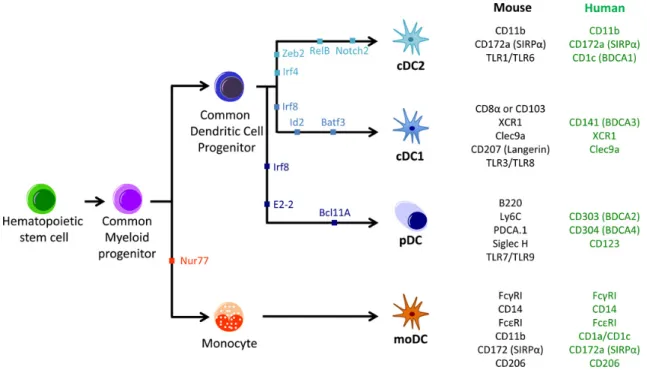
1.3.2 Dendritic cells . . . 46
1.3.2.1 Notion of subset . . . 47
1.3.2.2 Classical DCs . . . 49
1.3.2.3 Plasmacytoid pre-DC . . . 50
1.3.2.4 Emerging subsets of DC . . . 51
1.3.3 Inflammatory DC (Monocyte-derived inflammatory DC) . . . 52
1.3.4 Plasticity of APC . . . 52
1.3.5 Communication in TME . . . 53
1.4 How can we study the communication between cells in the TME? . . 55
1.4.1 Challenges . . . 55
1.4.2 Bioinformatics to study cell-to-cell communication . . . 56
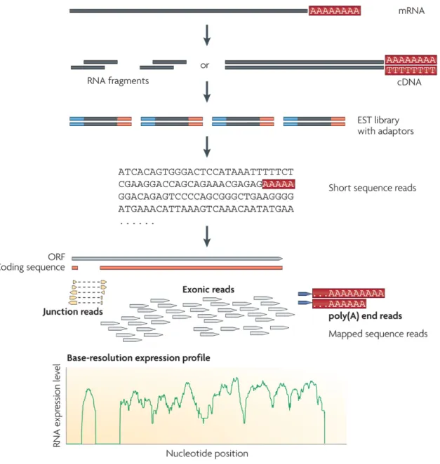
1.4.2.1 Transcriptomic profiles, information providers . . . . 56
1.4.2.2 Tools to study communication . . . 61
2 Objectives of the thesis 65 2.1 First objective: identify subsets of DCs and infer their biological func-tions in breast cancer using RNA-seq transcriptional profiles. . . 67
2.2 Second objective: reconstruct intercellular communication networks . 68 3 Results 69 3.1 Article 1: Adjustment of dendritic cells to the breast-cancer microen-vironment is subset specific . . . 71
3.2 ICELLNET: Reconstruction of intercellular communication networks using transcriptomic profiles . . . 127
4 General discussion et perspectives 165 4.1 Breast tumor-infiltrating APC subsets characterization . . . 167
4.3 Myeloid cells and interferon, a potential therapeutic axis in TNBC? . 170
4.4 Relevance of using transcriptomic data . . . 172
4.5 Single-cell RNA-seq technology . . . 172
4.6 Complexity of intercellular communication, a challenge to study . . . 175
Annexes 199
A Article 1: collaborative work . . . 199
Synthèse
Les systèmes vivants sont des systèmes ouverts qui échangent constamment des in-formations et de l'énergie-matière avec leur environnement. La communication, qui peut être définie comme un échange d’informations entre deux systèmes ou sous-systèmes, est donc un élément essentiel de la vie. Il permet la coordination efficace des processus homéostatiques et l'adaptation à un environnement en constante évo-lution, y compris la réponse aux menaces internes ou externes ou les processus auto-curatifs. Les organismes multicellulaires sont structurés de manière hiérarchique, les cellules étant souvent considérées comme des unités fondamentales : les cellules s’organisent pour former des tissus, un ensemble de tissus forme des organes, qui for-ment eux-mêmes des organismes. Par conséquent, la communication intercellulaire est à la base de l’organisation d’ordre supérieur observée dans les tissus, les organes et les organismes. Il est essentiel de coordonner la fonction de divers types cellulaires impliqués dans des processus biologiques complexes, tels que l'embryogenèse, la for-mation et le renouvellement des tissus, la régulation hormonale, la réponse au stress, une réaction immunitaire efficace aux agents pathogènes microbiens et le remode-lage tissulaire au cours d'une inflammation et de la cicatrisation. La dérégulation dans la communication intercellulaire peut entraîner une pathologie due à l'échec des processus homéostatiques et/ou à une adaptation défectueuse face aux menaces environnementales. Comment les cellules s'adaptent-elles à un microenvironnement spécifique en fonction de la communication intercellulaire ? Est-ce qu'elles gardent leur identité ou adoptent un comportement spécifique ? Ces questions sont parti-culièrement pertinentes quand on étudie le système immunitaire dans le contexte de l'inflammation et du cancer. Les interactions entre les cellules cancéreuses et le microenvironnement tumoral (TME) jouent un rôle crucial dans le développement
et la progression de la tumeur. Le TME est un système hétérogène constitué de nombreuses protéines et cellules de types différents qui interagissent au sein d'un réseau complexe. En particulier, de nombreux types de cellules immunitaires sont recrutés et participent à la réponse anti-tumorale, mais également à l'inflammation et à l'immunosuppression favorisant la tumeur. Il a été démontré que les cellules dendritiques (DCs) dans le TME étaient liées à la fois à la progression de la tumeur et à la prévention. Les DCs jouent un rôle essentiel dans le déclenchement des réponses immunitaires adaptatives. Chez l'homme, différentes sous-populations de DCs ont été identifiées. Des études antérieures ont montré que les DCs infiltrent des tumeurs solides, notamment le cancer du sein. Cependant, l'impact du TME sur le comportement des sous-ensembles de DCs infiltrant des tumeurs humaines est mal connu.
Dans le premier chapitre de la thèse, j'ai présenté les concepts généraux de la communication et du micro-environnement, puis j'ai décrit le cancer du sein et son microenvironnement, qui représentent un réseau complexe de cellules qui interagis-sent dans un contexte inflammatoire. Par la suite, j'ai préinteragis-senté l'hétérogénéité des cellules présentatrices d'antigènes (APCs) qui infiltrent le TME du sein et leurs com-munications dans ce contexte. Enfin, je me suis concentrée sur les défis techniques et méthodologiques liés à l'étude de la communication cellulaire et sur les outils bioinformatiques que nous pouvons utiliser pour surveiller les communications in-tercellulaires.
Le deuxième chapitre de la thèse pose les hypothèses et objectifs de mon tra-vail. Ce projet part de l'hypothèse que le microenvironnement tumoral module les réseaux de communication intra et intercellulaires formés par les APCs. Ces mod-ifications auraient des conséquences sur l'interaction entre la tumeur et le système immunitaire de l'hôte et a fortiori sur sur le développement de la tumeur. Mal-heureusement, dans la littérature, il existe plusieurs limites concernant l'étude de APCs qui sont des cellules rares dans un contexte tissulaire. Les sous-populations d'APCs ont été caractérisées dans différents tissus (e.g. le sang, la rate, la peau), des maladies (e.g. cancers, maladies auto-immunes), des organismes (e.g. souris, humains). Cependant, dans le cancer du sein, seuls les macrophages et les cDC2 ont
été étudiés [Ojalvo, Whittaker, et al.2010; Wargo et al.2016] et aucune comparaison n'a été faite entre le tissu tumoral (T) et le tissu non malin appelé juxtatumoral (J), ni entre différents sous-types de cancer du sein. L'objectif général de ma thèse est de comprendre l'impact du microenvironnement tumoral sur les sous-populations de DCs par une analyse systémique. Dans la première partie de mon travail de thèse, je cherchais à identifier les sous-populations de DCs dans le microenvironnement de la tumeur du sein. Plus précisément, le projet s'est concentré sur l'identification et la caractérisation des fonctions biologiques de sous-populations de DCs isolées de tumeurs du sein de deux sous-types différents: Luminal (LBC) et Triple-Négatif (TNBC), dont le pronostic est le plus sombre. Les APCs étant des cellules rares dans le TME du sein, nous avons voulu définir les sous-ensembles de APCs infil-trant les tumeurs à une résolution supérieure à celle décrite dans la littérature. En utilisant la technologie de séquençage ARN, nous avons généré le profil moléculaire de ces cellules et avons voulu en déduire les fonctions biologiques. Dans un pre-mier temps, l'objectif de cette étude était de décrypter comment le TME modulait le profil de transcription des sous-populations dAPCs, en comparant les profils de transcription des APCs dans les tumeurs et les juxtatumeurs et en liant la variation de l'expression des gènes aux fonctions biologiques. Dans un deuxième temps, nous avons évalué l'impact de l'hétérogénéité de la tumeur mammaire sur les DCs et les monocytes/macrophages (Monomacs). Pour ce faire, nous avons comparé le profil de transcription des sous-types d'APCs, isolés de deux types de cancer du sein, LBC et TNBC. Enfin, comme nous avions étudié le microenvironnement de la tumeur du sein et son impact potentiel sur le réseau de communication des sous-populations d'APCs, nous nous sommes demandé quel était le lien entre la caractérisation de chaque population spécifique d'APCs et le résultat clinique. Existe-t-il des dif-férences au niveau de la population entre les résultats cliniques et les différents TME, en fonction du sous-type de cancer du sein ? À partir de l'extraction de listes de gènes spécifiques caractérisant chaque population dAPCs identifiée dans le TME du sein, nous avons cherché à relier les signatures des sous-types cellulaires dans les différents contextes au pronostic des patients. Dans une seconde partie, nous avons étudié les communications cellulaires afin de comprendre comment les
cellules intègrent les signaux provenant de leur environnement. Pour ce faire, nous avons cherché à créer un score de communication simple basé sur des profils tran-scriptomiques de cellules. Ce score pourrait être appliqué aux données de puces à ADN ainsi qu'aux données de séquençage ARN. Il fera partie d'un outil comprenant une base de données sur les interactions des ligands et des récepteurs organisée et triée manuellement et un ensemble de profils de transcription des cellules primaires
accessibles au public dans BioGPS [ cite mabbottexpression2013].
Dans le troisième chapitre, les résultats de ma thèse sont présentés en deux parties. Dans une première partie, je présente les résultats publiés en montrant que les APCs s'adaptent au TME du sein dune manière spécifique selon la sous-population. Dans une seconde partie, je présente le manuscrit en préparation décrivant le développement et l'application d'un score de communication basé sur les profils transcriptomiques. Le TME est composé d'une grande variété de types de cellules qui influencent la progression de la tumeur et l'évasion immunitaire. Les DCs sont des APCs qui peuvent s'infiltrer dans la plupart des types de cancer. Ils peuvent jouer un rôle protecteur dans l'immunité antitumorale mais, inversement,
ils peuvent également favoriser l'immunosuppression [DeNardo, Barreto, et al.2009;
Faget et al. 2012; Ghirelli et al. 2015]. L'influence du TME sur la diversité et la
plasticité de ces APCs reste peu explorée. Dans le cadre de ma thèse, j'ai analysé les profils de séquençage à grande échelle des APCs infiltrant des tumeurs dans 8 échantillons de cancer du sein luminal (LBC) et 4 triples-négatifs (TNBC), en étroite collaboration avec Paula Michea, chercheuse post-doctorante au laboratoire. Sur la base d'analyses précédemment effectuées au sein du laboratoire et d'études publiées sur des sous-types de DCs humaines sur d'autres tissus, tels que le sang périphérique ou la peau, nous avons étudié quatre sous-populations de DCs et les macrophages au niveau phénotypique et transciptomique dans le cancer du sein. En comparant les transcriptomes de ces APCs provenant d'échantillons tumoraux et de tissus non cancéreux (juxtatumoraux) des mêmes patients, nous avons identifié des signatures géniques spécifiques à la tumeur pour chaque sous-population d'APCs, liées à des fonctions biologiques distinctes telles que la migration cellulaire chez les pDC. De plus, nous avons observé des différences substantielles entre les profils des
APCs infiltrant les TNBC et les LBC, révélant ldu microenvironnement tumoral et pas seulement l'empreinte tissulaire ou l'ontogenèse sur le comportement des APC. Il est intéressant de noter que la signature pDC était liée à une meilleure survie sans maladie dans les patients LBC, mais pas chez les patients TNBC, ce qui im-plique que le résultat associé à la signature pDC dépend du contexte. En conclusion, nous avons constaté que la reprogrammation transcriptionnelle d'APC infiltrant une tumeur est spécifique à un sous-type, ce qui suggère une interaction complexe en-tre l'ontogénie et l'empreinte tissulaire dans le conditionnement de la diversité des DCs dans le TME. Les signatures que nous avons générées sont particulièrement pertinentes pour l'identification de l'activation de voies biologiques et de nouveaux biomarqueurs dans les sous-types d'APCs.
Les résultats de la seconde partie de mon étude sont présentés sous la forme d'un manuscrit qui sera bientôt finalisé pour soumission. Il est intitulé “ńICELLNET: Reconstruction des réseaux de communication intercellulaires à l'aide de profils tran-scriptomiquesż”. Pour ce travail collaboratif, j'ai été impliqué dans le développe-ment d'une approche systémique basée sur la transcriptomique pour reconstruire des réseaux de communications intercellulaires. En effet, la communication inter-cellulaire est essentielle pour transférer des informations entre des cellules dotées de fonctions et de capacités de détection différentes. La communication intercellulaire coordonne les activités de divers types de cellules nécessaires aux processus com-plexes tels que l'embryogenèse, le remodelage tissulaire au cours de l'inflammation et la cicatrisation des plaies, ainsi que les réponses immunitaires. Actuellement, il n'existe pas de méthode systématique pour reconstruire la communication in-tercellulaire de manière qualitative et quantitative. Dans cette étude, nous avons développé ICELLNET, un outil intégrant des informations sur les interactions lig-and/récepteur, ainsi que des données d’expression génique spécifiques à une cellule et représentant des aspects quantitatifs et qualitatifs de la communication cellule à cellule sous forme de cartes de connectivité. ICELLNET peut être automatique-ment appliqué à n'importe quel profil transcriptomique au niveau de la population cellulaire afin d'estimer et de quantifier sa communication avec plus de 12 autres types de cellules. Nous avons appliqué cette méthode aux cellules tumorales, aux
cellules immunitaires innées et adaptatives (e.g., DC, cellules T, cellules B, NK), aux cellules épithéliales et stromales. En analysant un ensemble de données orig-inal de cellules dendritiques humaines générées de novo, nous avons identifié et validé expérimentalement l'IL-10 en tant que régulateur majeur de la connectivité intercellulaire des DCs au niveau systémique. Notre approche visant à évaluer la connectivité cellulaire peut constituer un outil précieux pour évaluer l'impact d'un contexte spécifique sur la communication entre cellules, en particulier dans un mi-croenvironnement inflammatoire tel que le cancer. Dans les perspectives futures, les applications d'ICELLNET pourraient apporter des informations biologiques impor-tantes et aider à orienter les manipulations pharmacologiques.
Dans la section discussion générale, je confronte mes résultats aux connaissances actuelles et expose les perspectives futures de ce travail. Dans un premier temps, j'ai discuté de la pertinence de caractériser les sous-populations d'APCs dans le cancer du sein et du positionnement de ce travail par rapport à la littérature. J'ai examiné l'impact de l 'hétérogénéité du cancer sur les communications cellulaires. En ce qui concerne les résultats biologiques que j'ai obtenus, j'ai discuté de la signature interféron trouvée dans les TNBC. De plus, j'ai souhaité examiner la pertinence d'utiliser des données transcriptomiques pour étudier la communication intercellu-laire et l'impact du microenvironnement sur le comportement celluintercellu-laire. J'ai in-clu des perspectives futures sur l'intérêt d'utiliser, dans ce domaine, une nouvelle technologie basée sur le séquençage d'ARN en cellule unique. Enfin, j'ai discuté de l'intérêt et de la complexité de la compréhension de la communication inter-cellulaire et des futurs développements pouvant être réalisés pour améliorer l'outil ICELLNET.
Enfin, en annexe, j'ai inclu deux manuscrits en préparation pour lesquels j'ai collaboré. Le premier décrit des îlots de DCs plasmacytoïdes dans la leucémie myélomonocytaire chronique. Le second est une étude de l'inhibition d'une pop-ulation de lymphocytes T CD8+ cytotoxiques par le point de contrôle immunitaire HLA-G.
List of abbreviations
TME: Tumor MicroenvironmentDC: Dendritic Cell
APC: Antigen Presenting Cell
MHC: Major Histocompatibility Complex Th: T helper
IL: Interleukin IFN: Interferon BC: Breast Cancer
LBC: Luminal Breast Cancer
TNBC: Triple-Negative Breast Cancer T: Tumor tissue
J: Juxtatumor tissue HR: Hormone Receptors ER: Estrogen Receptor PR: Progesterone Receptor
HER2: Human Epidermal Growth Factor 2 NK: Natural Killer
CSC: Cancer Stem Cell ECM: Extracellular Matrix
MDSCs: Myeloid Derived Suppressor Cells PD-L1: Programmed Cell Death 1 Ligand PD-1: Programmed Cell Death 1
pDC: plasmacytoid Dendritic Cell cDC: classical Dendritic Cell
Monomacs: Monocytes/macrophages TLR: Toll-like Receptor
PAMP: Pathogen-Associated Molecular Pattern PRR: Pattern Recognition Receptor
CLR: C-type Lectin Receptor
TAM: Tumor-Associated Macrophage DNA: Desoxyribonucleic Acid mRNA: messenger Ribonucleic Acid cDNA: complementary DNA RNA-seq: RNA sequencing
scRNA-seq: single-cell RNA sequencing DEG: Differentially Expressed Gene TGF-β: transforming growth factor-β NF-κB: nuclear factor βB
Preamble
Living systems are open systems constantly exchanging information and energy-matter with their environment. Communication, which can be defined as an infor-mation exchange between two systems or subsystems, is thus an essential part of life. It allows the efficient coordination of homeostatic processes, and the adaptation to an ever-changing environment including internal or external threat response or self-curative processes.
Multicellular organisms are structured in a hierarchical manner, with cells often being viewed as fundamental units: cells get organized to form tissues, multiple tis-sues form organs, which themselves form organisms. Hence, cell-cell communication is at the basis of the higher-order organisation observed in tissues, organs, and organ-isms. It is critical to coordinate the function of diverse cell types involved in complex biological processes, such as embryogenesis, tissue formation and renewal, hormonal regulation, response to stress, efficient immune reaction to microbial pathogens, and tissue remodelling during inflammation and wound healing. Dysregulation in cell-to-cell communication can lead to pathology through the failure of homeostatic processes, and/or the defective adaptation to environmental threats.
How cells adapt to a specific microenvironment depending on cell-to-cell commu-nication? Do they keep their identity or adopt a specific behavior? These questions are particularly relevant when studying the immune system in the context of inflam-mation and cancer. Interactions between cancer cells and the tumor microenviron-ment (TME) play a critical role in tumor developmicroenviron-ment and progression. The TME is a heterogeneous system, which consists of numerous proteins and cells of different type interacting within a complex network. In particular, many immune cell types are recruited and participate in anti-tumor response, but also in tumor-promoting
inflammation and immunosuppression. It has been shown that dendritic cells (DCs) within the TME were related to both tumor progression and prevention. DCs play a critical role in triggering adaptive immune responses. In human, different subsets of DCs have been identified. Previous studies reported that DCs infiltrate solid tumors, and particularly breast cancer. However, little is known about the impact of the TME on the behavior of DC subsets infiltrating human tumors.
As a framework of my study, I will introduce the general concepts of communica-tion and microenvironment, then I will focus on breast cancer and its microenviron-ment which represent a complex network of cells that interact in an inflammatory context. Subsequently, I will introduce the heterogeneity of Antigen Presenting Cells (APCs) that infiltrate breast TME, and their communications in this context. Finally, I will focus on the technical and methodological challenges of studying cel-lular communication and the bioinformatic tools we can use to monitor cell-to-cell communications.
The results will be presented in two sections. In a first part, I will present our published results showing that APCs adjust to the breast TME in a subset-specific manner. In a second part, I will present a manuscript in preparation on the development and application of a communication score based on cell transcriptomic profiles.
In the general discussion section, I will confront my results to the current knowl-edge and expose future perspectives of this work.
Finally, in the appendix, I will include two manuscripts in preparation for which I collaborated. The first one describe plasmacytoid DC islands in chronic myelomono-cytic leukemia. The second one is a study of the inhibition of a cytotoxic population
List of Figures
1.1 Figure 1 . . . 26 1.2 Figure 2 . . . 27 1.3 Figure 3 . . . 42 1.4 Figure 4 . . . 44 1.5 Figure 5 . . . 48 1.6 Figure 6 . . . 49 1.7 Figure 7 . . . 54 1.8 Figure 8 . . . 58 1.9 Table 1 . . . 59 1.10 Figure 9 . . . 61 4.1 Figure 10 . . . 174Chapter 1
Introduction
1.1 Communication
1.1.1 What does communication refer to?
1.1.1.1 A General definition of communication
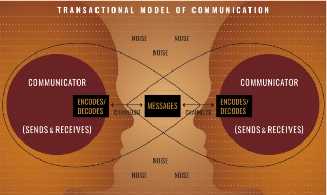
Communication is an important concept at the level of human being and it is sim-ply defined by the transmission of a message or an information between entities or groups. The players of communication are represented by a transmitter of the mes-sage and a recipient. They can be two individuals, groups of individuals, entities or societies. A government communicating information to the population is one illus-tration of communication between two entities. Communication is also defined as a process by which information is exchanged between individuals through a common system of symbols, signs, or behavior. In this definition, the emphasis is put on the use of the same system of symbols. Two individuals can discuss and exchange infor-mation using the same language. In order for the receiving individual to understand
and interpret the information, he has to be able to decode it (Figure 1.1). Coding
and decoding processes of symbol systems can make communication more complex. In cryptology, a lot of methods and algorithms to encrypt data or messages coexist. A key to decode the message is required, in order to be understandable by the en-tity receiving the information. Coding and decoding messages are used to create a specific communication between two entities. One example of tools used to decipher crucial communications during World War II was the ancestor of computer created by Pr. Alan Turing, a british mathematician. His device enabled to decode messages encrypted by Nazis from the enigma machine and is considered as the ancestor of computer science. The methods to communicate between human beings are numer-ous and have evolved through time, from cave painting to the internet nowadays. Major forms of communication use writing (e.g. books, letters) or speaking (e.g. direct speaking, phone, radio). Another interesting form of communication implies representation, images as painting, sculpture or even sign language. Evolution of technologies and science helped to develop different ways of communication. In par-ticular, the comprehension and use of waves allowed us to convey messages by sonar,
radio, television and phone.
Figure 1.1: Schema representing a communication model. From National
Commu-nication Association (www.natcom.org).
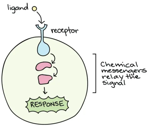
At the level of the cell, we observe the same phenomenon of communication. One cell can communicate with an another by sending chemicals signals that will be sensed and processed by a receiving cell to trigger a specific response or mechanism
(Figure1.2). Cells can sense a great diversity of signals from the extracellular
envi-ronment, such as growth factors, cytokines, danger signals, cell-to-cell contact and
extracellular vesicles [Niel, D’Angelo, and Raposo 2018]. This signals are hundreds
of distinct molecules, the majority being proteins and forming the words of the cell communication language. In cell biology, different types of signaling are described depending on the distance between the sending and the receiving cells. Paracrine signaling for short distance signaling, endocrine signaling for long distance signal-ing, autocrine signalsignal-ing, and direct signaling across gap junctions are the four types of signaling used by cells in multicellular organisms. To sense and process the in-formation, cells require decoding mechanisms. To detect the chemical signals, the receiving cell express specific receptors localized at their membrane or inside the cytoplasm or the nucleus. Once the ligand is attached to the receptor, the message
is transduced following complex signaling pathways inside the cell (Figure 1.2).
Figure 1.2: Message transduction at the cellular level, interaction between ligand
and receptor. From Introduction to cell signaling,https://www.khanacademy.org.
1.1.1.2 Interest of communication
Communication is essential to human life in many ways. First of all, the use of com-munication can derive from a need for interacting and coordinating peoples actions in order to survive and grow. As Aristotle said, “Man is, by nature, a social animal”
[Aristotle2018]. Following this concept, humans are born to live in cities, and better
exploit their potential via social interactions. Communities thrive around commu-nication of a diversity of information between people, enabling to organize groups of individuals with rights and rules. Communication promotes social interactions which are key to human evolution. Social interactions and cooperation enhance the development of intelligence not only in humans but also in other species [McNally,
Brown, and Jackson2012]. Communication plays a role in the enhancement and
ex-pansion of societies, especially via education. Communication of emotions via art or entertainment is important for the psychological development of humans helping to
avoid stress and anxiety and improve productivity and stability. In Africa, a study showed that communication between members of a community promotes active citi-zen participation and initiatives to the development of the communities [Adedokun,
Adeyemo, and Olorunsola 2010]. Throughout time, communication methods have
evolved to improve the efficiency to convey information. We witness a fast evo-lution of communication technologies, and nowadays, thanks to the new digital
technologies, the world is interconnected [E. Williams2011]. This evolution of
com-munication gave an easier and faster access to information, knowledge, and a faster transmission of information between individuals. More people are connected even if they are far away from each other thanks to the phone, internet, and social media replacing mailing post and telegraphy. Fast access to information and connection between individuals with different culture, origin, and experience enhance sharing and improvement of the world’s knowledge that can be then applied in various disciplines such as agro-industries, politics, entertainment, economy or justice. In science, one of the most important parts of the work is to communicate about the research and results to spread knowledge and information by means of conferences, publications, and posters.
In cell biology, communication is essential to development, growth, survival, maintenance, and defense of the individual cell but also for the development of
mul-ticellular organisms [Niklas and S. A. Newman 2013]. Depending on the sensed
signal, different responses are initiated by the receiving cells and impact their fate. Cell-to-cell interactions are crucial in the coordination of organism development and several signaling pathways are involved in and are responsible for most of the animal development: Hedgehog (Hh), wingless-related (Wnt), transforming growth factor-β (TGF-β), receptor tyrosine kinase (RTK), Notch, Janus kinase (JAK), signal trans-ducer and activator of transcription (STAT) and nuclear hormone pathways [Barolo
and Posakony 2002]. In the developed organism, intercellular communication
coor-dinates the activities of multiple cell types required for complex processes such as immune response, growth, and homeostasis. When cells are damaged, they are also able to sense intracellular signals such as DNA in the cytosol and trigger mechanisms of cell death (e.g. apoptosis, autophagy). Another interest of communication
be-tween cells is the complex signaling network to enable cell migrations which are crit-ical for immune cell trafficking, wound healing, and stem cell homing, among other processes. Immune cell crosstalks play a role in establishing central tolerance and
preventing autoimmunity. Indeed, in the thymus, when CD4+CD8+T cells recognize
the complex formed by an external peptide and the major histocompatibility com-plex (MHC) molecule presented on cortex thymic epithelial cells, they receive critical
survival signals and differentiate into CD4+ and CD8+ T cells. Then, they undergo
a step of negative selection in the medulla where the autoreactive T cells which
rec-ognize self-antigens presented by DCs are eliminated [Takaba and Takayanagi2017].
Cell-to-cell communication is essential to trigger an immune response and depends on the stimuli that activate immune cells. An impair in cell-to-cell communication can lead to the development of severe pathology. For instance, a lack of a specific receptor such as interferon-gamma receptor (IFNGR) in macrophages cause a rup-ture of communication. Cells do not receive the immune defense signals anymore
which induce an increase sensibility to mycobacteria infection [Newport et al.1996].
1.1.2 Factors impacting communication
Considering the diversity of communication methods, messages and responses, it becomes evident that independent factors impact interactions between individuals. In sociology, different theories point out the cultural context as a major factor in-fluencing communication and the efficacy of the message transmission. The theory introduced by anthropologist Edward T. Hall exposed that two types of culture, “low” and “high” contexts, are opposed and play a role in communication [Hall
1976]. “Low context” is defined by an explicit communication whereas “high
con-text” is characterized by implicit communication, with the use of ambiguity where facial expression and the way of speaking can change the meaning of words. There-fore, low-context individuals, who are not used to read between lines, are less able to fully understand the messages transmitted by high-context individuals. This can be nefast for social interactions and a fortiori for the development of the society especially in diplomatic exchanges. A second interesting hypothesis by Sapir and Whorf shows that culture significantly affects how people think and communicate.
More precisely, the language which is one of the bases of a culture and brings to-gether people strongly affects the way of thinking. Ciaccio and Bormann studied the influence of color terms on the behavior of Italian and German speakers [Ciaccio
and Bormann 2013]. They demonstrated that the judgments of colors boundaries
was influenced by the language which validate the hypothesis of Sapir and Whorf. Environmental factors (e.g. pollution) or physical factors (e.g. the intensity of a signal, the speed of the transmission) can affect the effectiveness of communication or alter the signal ending in a communication break. The clarity of the message is important for the comprehension between individuals. A study has revealed the nefaste impact of traffic noise on communication between frogs. The noises were masking the perception of acoustic communication signals preventing male frogs from communicating efficiently with female and it leads to a decrease of
reproduc-tion [Bee and Swanson 2007]. Personal history and previous communications can
drive the way of thinking and interpreting information facilitating or complexifying coding and decoding processes. Internet and social media increase communication between people by simplifying interactions, increasing speed of connection between people all around the world and allowing the spread of all kind of information. But it raises questions on the quality of communication: is the information trustable since it is easy to spread any kind of information? Due to the multiplicity of con-nections and exchanges, are the communication effective? This questions highlight the complexity of communication networks which are impacted by several factors in a positive or negative manner.
Looking at the cellular level, cell signaling can be impaired by factors acting directly on cells, altering the transmission or the reception of messages. Thera-peutic agents can be used as receptor blockade mechanisms mimicking the ligand but without carrying the message that would have induced a response from the sensing cell. Communication can also be altered by genetic mutation destabilizing gene expression and response to stimuli. Stimuli such as Ultraviolet radiation (UV) provoke genetic mutation that can alter the expression of key genes, inducing skin
cancer [Seebode, Lehmann, and Emmert2016]. Mutated cells use a communication
and survive. Cells can communicate and answer to stimuli differently according to their type and origin. If cells exhibit a plastic phenotype, they can sense external stimuli such as communication signals and adapt their future communication within the cellular environment (e.g. stress, UV, cigarette smoke, diet, culture medium). Given different stimuli and environments, a cell can differentiate into several states. A stimulus or a combination of stimuli sensed by one cell type can impact its com-munication with other cell types inducing various responses. For example, DCs have been identified as the main drivers of T helper (Th) polarization in 1999 [Rissoan
et al.1999]. However Th cells integrate numerous signals to specify their phenotypes
[Zygmunt and Veldhoen2011]. A large number of Th subsets have be defined based
on the diversity cytokines patterns produced by Th cells [Raphael et al.2015]. These
results reveal the intrinsic complexity of the Th differentiation process as a central communication system integrating multiple signals coming from DCs and
produc-ing a large diversity of Th responses [Grandclaudon et al. n.d.]. The environment
is a major factor impacting the signaling. Inflammation is triggered when innate immune cells detect infection or tissue injury. Changes occur in the inflamed envi-ronment such as the presence of cytokines impacting communication and behavior of non-immune cells. This peculiar microenvironment will be further described in section 1.4.2.
1.1.3 Network representation of communication
The organization of multiple entities through communication is a complex system that researchers try to understand. To study complex system such as cell-to-cell
communication, networks are powerful tools to use [M. E. J. Newman 2003]. In
mathematics, a network or graph is a set of nodes that are connected together by connections called edges or links. Two types of networks are distinguishable: directed and undirected. The first one is characterized by links indicating the direc-tion in which the informadirec-tion circulates. If all edges are bidirecdirec-tional, or undirected, the network is an undirected network. Representation of networks is often used in different fields of application. As examples we can cite connections between in-dividuals on social media, the internet, financial networks or biological networks.
In the case of a communication network, nodes describe entities communicating and edges monitor the transmission of messages. Putting communication into the perspective of a network enables to organize knowledge on cell interactions into a systemic view. Cell-to-cell communication networks comprise both intra- and intercellular processes. Several studies focusing on intracellular communication
net-works are found in the literature and describe metabolic netnet-works [Jeong et al.2000],
gene-regulatory networks [Thompson, Regev, and Roy2015], or networks of
protein-protein interactions [Hooda and Kim 2012]. These networks can model the signal
transduction processes inside the cell and the response induced by the message. In-tercellular networks model the interactions between different cell types. However, compared with intracellular signal transduction networks, the functions and engi-neering principles of cell-to-cell communication networks are less understood. Many studies have addressed cross-talks between a given pair of cell types [Ferlazzo and
Morandi2014; Haan, Arens, and Zelm 2014; Hivroz et al. 2012]. Most of the time,
communication process is considered a linear signaling cascade, such as immune
cas-cades [Ghirelli et al.2015; Y.-J. Liu et al.2007] involving the exchange of one
infor-mation signal at each step. Some studies have focused their purpose on specific cases
of communication such as the cytokines interleukin-2 (IL-2) [Fuhrmann et al.2018],
interferon-gamma (IFNγ) [Helmstetter et al. 2015], or tumor necrosis factor alpha(
(TNF-α) [Paszek et al. 2010; Tay et al. 2010]. This view has several limitations:
1) it does not consider the possibility that one given cell type could communicate with multiple cell types concomitantly within the same microenvironment [Bindea
et al. 2013; C. Q. F. Wang et al. 2013], 2) it does not consider the multiplicity of
information signals possibly sent by one cell to another, 3) it does not integrate the complex and constant rewiring and cell state modifications in the system following exchange of information, 4) it provides limited mechanistic insight into the com-plexity of multicellular pathophysiological processes, 5) as a consequence, it is very limited in predicting the effects of physiological or pharmacological perturbations in higher order multicellular systems.
To study cell-to-cell communication network, it is important to define and char-acterize the microenvironment of cells to model their interaction and behavior.
In-deed, the microenvironment impacts cell communication as culture impacts human communication.
1.1.4 A model of network: cellular microenvironment
1.1.4.1 Diversity of cellular microenvironments at the physiological state: role of the tissue type
Within an organism, each cell exists in the context of a complex extracellular mi-croenvironment. Different types of tissues across the human body have been defined such as nervous tissue, muscle tissue, epithelial tissue and connective tissue. Within a given tissue, microenvironmental factors and extracellular matrix proteins coop-erate to provide both the biochemical signals and structural constraints that are required to influence intracellular programs of gene expression and further the cellu-lar behaviors in the tissue in question. Various cell populations are described having tissue-dependent functions creating a specific cellular environment. This is the case for certain populations of immune cells. Studies have shown that T-cell primed by tissue-specific dendritic cells (DCs) can change their specific functions if they are
re-primed by other tissue-specific DCs [Mora and Andrian2006]. Natural killer cells
(NKs) are a type of lymphocyte that identify infected or transformed cells through a complex range of activating and inhibitory receptors that regulate direct and in-direct killing mechanisms. They migrate from peripheral blood to peripheral organs through cytokines-mediated signals. However, studies have highlighted the existence
of tissue-specific subpopulations of NKs [Shi et al. 2011]. Tissue-specific NK cells
are found in different tissues across the body. Studies suggest that subpopulations of tissue-specific NK cells may undergo phenotypic changes under inuence of the microenvironment, but also differentiate in situ from tissue-resident hematopoietic
progenitor cells [Lysakova-Devine and O’Farrelly 2014]. Macrophages are immune
cells present in most tissues in vertebrates. They are best known for their phagocytic role in immunity, but they can also function as an important source of growth factors for other cell types within tissues. Tissue-resident macrophages are heterogeneous populations in terms of phenotype and function. According to the location they
re-side, tissue-resident macrophages display specific functions which are important for
normal tissue homeostasis [Ginhoux and Guilliams2016; Gosselin et al.2014; Okabe
and Medzhitov2016]. Similarly, signaling factors derived from tissue environments
play key roles in promoting the ontology and phenotype of the residing macrophage
populations [Okabe and Medzhitov 2016].
1.1.4.2 Physiology versus pathology
In addition to the specificity of tissue microenvironment, one key factor to think of when studying cellular environment is the physiological or pathological context. Steady state and inflammation have a different impact on communication between cells creating a specific microenvironment. Inflammation is a state of the microen-vironment due to the establishment of an adaptive immune response after pathogen infection, external injuries or an effect of chemicals or radiations. Inflammation re-flects a complicated, multifactorial, and multidimensional process, in which acute and chronic inflammation are differentiated. Acute inflammation is a short-term process occurring in response to tissue injury appearing within minutes or hours. It is characterized by five main signs: pain, redness, loss of function, swelling and heat. Inflammation follows several steps independently of the stimulus initiating the immune response. First, cell surface pattern receptors recognize detrimental stimuli that lead to activation of inflammatory pathways such as NFκB or MAPK pathways. Then, inflammatory markers, inflammatory cytokines, proteins, or enzymes, are re-leased and inflammatory cells are recruited in the microenvironment [L. Chen et al.
2017]. The last step is the resolution of the issue by tissue repair and remodeling by
monocytes. This is made possible by the switching from pro-inflammatory to anti-inflammatory signals in the anti-inflammatory environment, promoting the recruitment
of monocytes and inhibiting recruitment of neutrophils [Medzhitov 2008]. In the
case of infection, if the acute inflammatory response fails to eliminate the pathogen, the inflammatory process persists with the presence of macrophages and T cells in
the tissue and a chronic inflammatory state occurs [Medzhitov 2008]. The chronic
inflammatory process that plays a central role in some of the most challenging dis-eases, including cancers, rheumatoid arthritis, heart disdis-eases, diabetes, asthma, and
even Alzheimers. Complex genetic and environmental interactions contribute to the development of chronic inflammatory diseases. Autoimmunity is characterized by dysregulation of the adaptive immune system as well as the pathogenic role of innate immunity and is associated with several chronic inflammatory diseases. Studies have shown the importance of microbiota in the development of autoimmunity
[Yurkovet-skiy, Pickard, and Chervonsky 2015] but also the genetic impact of several
autoim-mune diseases [Zenewicz et al.2010]. Chronic inflammation is thought to promote
cancer development. Today, between 5% and 10% of cancer cases are thought to be triggered by mutation and up to 15% by inflammation; the origin of the 80% left is
still unknown [Brücher and Jamall 2014]. Cancer is a complex and heterogeneous
disease affecting several cell populations in many localization and tissues. The tu-mor microenvironment (TME) is a complex network not only composed of malignant cells but also stromal cells. Communications among tumor and stromal cells create a distinct cellular environment that plays a significant role in tumor development and progression. In solid tumors, fibroblasts in the TME secreting chemokines and growth factors contribute to tumor growth and affect the extracellular matrix
envi-ronment that helps tumor to progress [Allen and Jones 2011]. Studies have shown
the impact of metabolism in TME, particularly hypoxia that induce angiogenesis,
and invasion [Allen and Jones2011]. Since it is an inflamed environment, we can find
immune cells infiltrating the TME. Leukocyte infiltration of solid tumors was first described in the 1800s by Virchow. Proinflammatory cytokines, chemokines, and adhesion molecules, which regulate the recruitment of leukocytes, are frequently ob-served in the TME. Some leukocytes including cytotoxic T cells and NK cells have
a pro-inflammatory and anti-tumor role [DeNardo, Andreu, and Coussens 2010;
Gavin P. Dunn, Old, and R. D. Schreiber 2004] whereas other leukocytes such as
regulatory T cells and macrophages play an anti-inflammatory and pro-tumoral role promoting cancer immune evasion and cancer progression [DeNardo, Andreu, and
Coussens2010].
For my thesis work I was interested in studying communication processes in one particular network which is breast cancer microenvironment.
1.2 Human Breast Cancer
1.2.1 Factors of incidence
Breast cancer is the second most common cancer worldwide with nearly 1.7 million
new cases in 2012 and is the first cause of mortality by cancer among women (http:
//globocan.iarc.fr/Default.aspx). In the literature, many factors are known to have an incidence on the risk to develop breast cancer. Some mutations, particularly in BRCA1/2, EGFR, and p53 genes result in an increased risk of occurrence of breast
cancer [M.-C. King et al. 2003; Malkin et al. 1990; Sun et al. 2017]. However, it
concerns only a small proportion of tumors, less than 30% of breast cancers. On the other hand, exposure to endogenous hormones (estrogen) increases the risk of breast
cancer occurrence [Travis and Key2003]. During the last decades, many groups have
pointed out the higher risk of developing breast cancer induced by using exogenous hormones such as hormone replacement therapies (HTR). Moreover, the relative risk of breast cancer in current users increases with increasing duration of use of
HRT [Li et al. 2003]. Additionally, environmental signals play a role in modifying
the incidence of breast cancer. Danaei et al. have studied the impact of several environmental factors (e.g. cigarette smoke, diet, obesity) on the incidence of cancers worldwide. They showed that alcohol use, overweight and obesity, and physical inactivity have a joint incidence on 21% of all breast cancer deaths worldwide [Danaei et al.2005].
The diversity of factors involved in the appearance of breast cancer is a first observation of the complexity of this disease. Another important layer is the het-erogeneity of breast cancer subtypes.
1.2.2 Breast cancer subtypes and inter-tumor heterogeneity
1.2.2.1 Classification
Breast cancer has been suggested to be a heterogeneous disease, and multiple classi-fications exist to better characterize this disease and improve treatments and care of the patients. The first classification of breast cancer relies on the histopathological
status of the disease. It is divided into more than 20 types with the most impor-tant being invasive ductal carcinomas (IDCs), not otherwise specified (NOS), and invasive lobular carcinoma (ILC). The grade of the disease can also be taken into ac-count in the classification of breast cancer. Several scores measure the disease state such as Eston-Ellis grade, Nottingham prognostic index, or tumor, lymph nodes and metastasis status (TNM). They are based on measurement of the tumor growth
and development, or the lymph node invasion status [Sinn and Kreipe 2013; Viale
2012]. Based on the molecular and transcriptional profile of breast cancers, different
subtypes have been identified and correlated with clinical outcome [Koboldt et al.
2012; Prat, Pineda, et al. 2015; Viale 2012]. Six breast cancer subtypes have been
established based on expression of hormone receptors (HR) which are estrogen re-ceptor (ER) and progesterone rere-ceptor (PR), expression of HER2 (human epidermal growth factor 2), and Ki-67 protein immunoreactivity:
• Luminal A breast cancer is hormone-receptor positive (estrogen-receptor and/or progesterone-receptor positive), but negative for HER2 and have low level of Ki67 immunoreactivity. It is also characterized by a genomic stability.
• Luminal B breast cancer is hormone-receptor positive as Luminal A but is characterized by less genomic stability with some amplification (HER2), dele-tions and mutadele-tions (P53). It can be either HER2-positive or negative with high levels of Ki67 immunoreactivity.
• HER2enriched breast cancer has amplification of ERBB2 and many other genes. It is defined by positive expression of HER2 and no expression of the hormone receptors (ER, PR).
• Triple-Negative (TNBC) or Basallike breast cancer is defined based on the absence of expression of hormone receptors (ER, PR) and HER2. TNBC have a high genomic instability.
• Normal Breastlike group is similar normal breast epithelium in transcriptomic analyses.
• Claudinlow breast cancer is characterized by low expression of cell-to-cell com-munication proteins (claudins), no/low markers of luminal differentiation and a high expression of epithelial to mesenchymal transition (EMT) markers, im-mune response genes and cancer stem-cell markers. These tumors are only high grade and are less frequent (12-14% of cancers)
These breast cancer classifications highlight the heterogeneity of the disease, at multiple layers: localisation, grade, molecular profile. Additionally, they have been linked to distinct clinical outcome.
1.2.2.2 Diversity of behavior and outcome
Tumor complexity is due to the heterogeneity of the disease which impacts the
clini-cal behavior and outcome of the patients [Koren and Bentires-Alj2015]. The
molec-ular subtypes of breast cancer correlate with different clinical outcomes and response
to treatment [Prat, J. S. Parker, et al.2010; Prat, Pineda, et al.2015; Troester et al.
2004]. Troester et al. compared basal and luminal BC cell lines and showed that
molecular subtypes of BC have a subtype-specific response to chemotherapies which
was validated by in vivo data [Troester et al.2004]. PAM50, a 50-gene qPCR assay,
has been identified as a predictive marker of pathological complete response (pCR) regarding chemotherapy response. This predictive marker was shown to reflect the intrinsic molecular classification and its correlation to clinical outcome [Y.-R. Liu
et al.2016; Prat, Pineda, et al.2015].
Luminal A cancers are low-grade, tend to grow slowly and have the best progno-sis and long-term survival while luminal B cancers prognoprogno-sis is slightly worse. This difference of prognosis was suggested to be due to a variation in response to
estro-gen therapy between luminal A and B [Rivenbark, OConnor, and Coleman 2013;
Sørlie et al.2003]. Triple-Negative or basal-like breast cancers are more aggressive
with high rates of cell proliferation and have poor clinical outcomes. As they do not express hormone receptors neither HER2, herceptin and hormone therapies can-not be used. Patients with claudin-low breast cancer have poor recurrence-free and overall survival outcomes, this cancer not being responsive to chemotherapy
with a poor clinical outcome. As they are ER negative, they are not treated with
anti-estrogen receptor therapies. However, survival of HER2+ breast cancer
(HER2-enriched, Luminal B) improved thanks to herceptin-targeted therapy, in addition to
adjuvant chemotherapy [Cortés et al.2012; Mukai 2010].
Although the molecular classification of breast cancer help to characterize the disease and defined adapted therapies, the patient outcomes are disparate. The observed variation in treatment efficacy has been connected to heterogeneity in the cellular composition of individual tumors and significant heterogeneity in immune
composition is observed across subtypes as well as patients [Dushyanthen et al.2015;
García-Teijido et al. 2016]. This highlights the importance of taking into account
the molecular subtypes as well as the intra-tumoral heterogeneity when studying breast cancer networks and communications.
1.2.3 Intra-tumor heterogeneity
Two distinct but complementary theories describe the origin of tumor cells
hetero-geneity, the cancer stem cell (CSC) hypothesis [Meacham and Morrison 2013] and
the clonal evolution and selection model [Greaves and Maley 2012]. CSCs
orig-inate from single cells possessing specific characteristics regarding cell plasticity. Those cells undergo tumor-reprogramming processes via multiple molecular alter-ations through a specific hierarchy and have indefinite self-renew potential that drive tumor growth. These mechanisms drive temporal intra-tumor heterogeneity. The clonal evolution/selection model is based on clonal expansion by natural selection and adaptation to tissue microenvironments. The factors contributing to clonal ex-pansion promotes certain cellular characteristics allowing cancer cell proliferation in hypoxia environments. Depending on the local microenvironment, the clonal expan-sion wont be promoting the same clones, contributing to spatial heterogeneity. In the majority of the cases intra-tumor heterogeneity is clonal-based, however it has been shown in the literature that some areas of the tumor can be morphologically
distinct with different repertoires of genetic aberrations [Greaves and Maley 2012;
Martelotto et al.2014]. Intra-tumoral heterogeneity is a complex interplay between
develop-ment and evolution of breast cancer to metastasis. Several studies have revealed genetic differences between primary breast tumors and their metastases [Bonsing
et al. 2000; Kuukasjärvi et al. 1997; Pandis et al. 1998; Torres et al. 2007; C. Wu
et al.2009]. Genetic and epigenetic modifications can be caused by external factors
such as cigarette smoke, UV lights, chemotherapy agents and/or the microenviron-ment during the developmicroenviron-ment and growth of the tumor contributing to the temporal
heterogeneity of breast cancers [Martelotto et al. 2014]. Studying intra-tumor
het-erogeneity could have clinical benefits since we observe treatment failures due to therapeutic selection of cancer cells harboring resistance mechanisms [Turner and
Reis-Filho 2012].
Both inter-tumoral and intra-tumoral heterogeneity make breast cancer a com-plex disease. Tumor cells evolve in a specific microenvironment (including non-tumoral cells) that display specific signaling that can be hijacked by the tumor to
promote its progression and survival [Poli, Fagnocchi, and Zippo 2018].
1.2.4 Tumor microenvironment
TME is a complex network composed of cancer cells, stromal cells, endothelial cells, immune cells as well as components of the extracellular matrix (ECM). As described above, the TME shows high level of spatiotemporal heterogeneity which is partly due to alterations of the microenvironment. In normal breast, epithelial and stromal cells communications are essential to inhibit tumor growth and proliferation [Quail
and Joyce 2013]. However, in breast cancer, communication between cancer cells
and non-malignant cells infiltrating the TME promotes heterogeneity, growth and
proliferation of the disease [Quail and Joyce2013]. Understand the composition of
the tumor microenvironment and what are the interactions that promote develop-ment and resistance of the disease could help define better therapies. The TME is not only impacted by the presence of tumor cells, but it is also involved in the development of the disease, in different ways. Specific changes happen in the breast tumor microenvironment that regulate progression to invasion and metastasis, for instance increase of fibroblast proliferation and ECM remodeling [Bonnans, Chou,
tu-morigenesis.[Mao et al. 2013]. We also observe cell-to-cell signaling changes. Genes encoding for secreted proteins and cell surface receptors are found differentially ex-pressed in epithelial and stromal cells during breast tumor progression [Allinen et al.
2004]. Paracrine signaling takes place through secretion of soluble factors by cancer
cells, fibroblasts and other cells of the TME. Allinen et al. performed a molecular characterisation of breast cancer microenvironment. They compared normal epithe-lial and stromal cells to cancer epitheepithe-lial cells and infiltrating stromal cells. High expression of CXCL12 and CXCL14 by myoepithelial and myofibroblast were found in the TME. These chemokines are involved in cell proliferation, differentiation, mi-gration, and invasion of breast cancer cell lines. Several signaling pathways involved in the interplay between tumor infiltrating cells and cancer cells promote tumor growth, metastatic spread or even drug resistance. TGF-β signaling in breast TME plays an important part in tumorigenesis. It has implication in angiogenesis, recruit-ment of endothelial cells, monocytes and macrophages, and activation of fibroblasts. TGF-β also suppress T cell immunosurveillance and cytotoxic activity [Scheel et al.
2011; Taylor, Lee, and Schiemann2011]. Breast tumor microenvironment represents
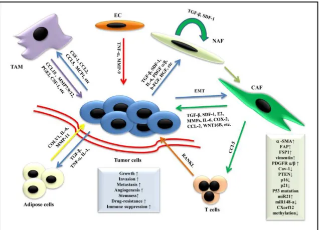
a social network where cells produce and interpret a diversity of signals promoting
cancer cells progression. Figure1.3 represent in a schematic view these interactions
between the cells of the breast environment. Cancer cells cross-talk with endothelial cells, fibroblasts and immune cells such as macrophages and T cells, using specific signaling including TGF-β, growth factors and inflammatory cytokines.
Figure 1.3: Schematic representation showing the role of stromal cells in microenvi-ronment and breast cancer progression. The tumor microenvimicroenvi-ronment is a dynamic composite of cells broadly categorized as multiple components of non-stromal and stromal cells, where tumor cells thrive. Stromal cells promote tumor growth, in-vasion, and metastasis by secreting multiple cytokines, chemokines, growth factors, etc. Moreover, tumor cells also affect the phenotype of stromal cells. From Mao et al.2013.
1.2.5 Inflammatory environment
At the beginning of cancer studies, the immune system was not considered as play-ing a role in cancer development neither on the clinical outcome of patients. In breast microenvironment, immune cells play a role of immunosurveillance, by killing potential cancer cells before they became a cancer. However, the immunosurveil-lance of immune cells put a selective pressure on cancer cells that develop resistance mechanisms and escape immune surveillance or generate an immunosuppressive
interaction network formed by the cells of the TME. Only recently, immune evasion has been recognized as a hallmark of BC which is enabled by three major char-acteristics being (epi)genetic modifications and clonal selection of cancer cells, and
tumor-promoting inflammation [Hanahan and Weinberg2011]. Now, it is well known
that the TME is composed of different immune cell populations such as T and B lymphocytes, natural killers (NK), and myeloid cells including macrophages, myeloid derived suppressor cells (MDSCs), and dendritic cells (DCs). Cellular crosstalk be-tween different leukocyte subsets infiltrating the breast cancer TME induces either pro- or antitumor functions driving immune-mediated anti- or pro-tumor activities
in the microenvironment [D. S. Chen and Mellman 2013; DeNardo, Andreu, and
Coussens 2010]. Distinct populations of tumor-infiltrating lymphoid and myeloid
cells have been linked to different prognosis in BC patients [Kroemer et al. 2015].
While breast tumor infiltration by CD8+ T cells was associated with patient
sur-vival and response to therapy [DeNardo, Brennan, et al.2011; Mahmoud et al.2011;
Seo et al.2013], regulatory CD4+FOXP3+ T cells support pro-tumor immunity and
are associated with a poor prognosis in some cases of breast carcinoma [Ibrahim
et al.2014; Yeong et al. 2017; Zhou et al.2017]. Myeloid cells localized in pre- and
malignant tissues release amount of cytokines, soluble factors and other inflamma-tory molecules. These signals contributing to tissue remodelling, angiogenesis, and
suppression of anti-tumor immunity [Stockmann et al.2014]. If MDSCs have been
characterized as regulator of the immune system [Gabrilovich and Nagaraj 2009],
they also play a role in cancer development and metastasis. MDSCs and cancer cells interaction via IL-6/STAT3 and NOTCH signaling induce CSCs development [Peng
et al. 2016]. Cancer cells also secrete molecules influencing the microenvironment
towards pro-tumoral and pro-inflammatory environment. Ghirelli et al described the activation of pDC via GM-CSF and IL-6 secretion by breast tumor cells that was
linked to a worse prognosis [Ghirelli et al.2015]. Over the past years, new therapies
have been developed targeting the immune system in cancer. As described by the concept of hot versus cold tumor, the diversity of TILs infiltration levels in tumors
may impact the efficacy of immunotherapies [L. Chen et al. 2017; Spranger 2016;
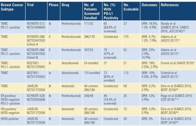
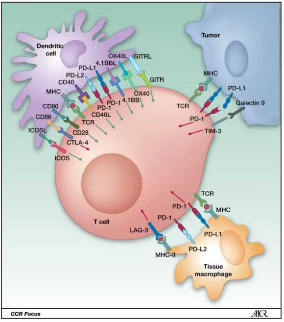
and high levels of TILs are correlated with increased expression of the checkpoint molecule programmed cell death 1 ligand (PD-L1). Immunotherapy treatments rely on therapeutic antibodies targeting immune checkpoint molecules that have co-stimulatory or co-inhibitory functions. Clinical trials on TNBC show some positive results. For instance, monotherapies targeting programmed cell death 1 (PD-1) and one of its ligand CD274 (PD-L1) which have an inhibitory interaction in metastatic
TNBC and showed between 4.7% and 23% of overall response rate (Figure1.4)[Tan
2018]. Despite some treatment successes, the response seen in patients is limited,
especially in other subtypes such as luminal, drawing attention to the need of better understanding the immune components of the TME.
Figure 1.4: Clinical trials of checkpoint inhibitors as monotherapy in metastatic
1.3 Antigen presenting cells
Antigen presenting cells (APCs) are key players of the immune system communi-cation/social interactions and infiltrate the tumor microenvironment. Professional APCs include dendritic cells (DCs), B cells, and macrophages [Parkin and Cohen
2001]. These peculiar cells are the sentinels of the body and have an extremely
important role as messenger of the immune system. They patrol many tissues and are able to trigger the adaptive immune response by presenting exogenous antigens through MHC class II molecules. This complex is then presented to T cells that recognize antigens via their TCR. These interactions lead to activation of T cells.
Here, we will focus on monocytes, macrophages and DCs which are mononuclear phagocytes distinguished on the basis of their morphology, function and origin.
1.3.1 Monocytes and macrophages
Monocytes are present in all vertebrates. In humans, these cells represent 10% of the nucleated cells in the blood. They arise from myeloid precursor cells in primary
lymphoid organs. Two main human monocyte subpopulations are defined as CD14+
and CD14lowCD16+ monocytes. The first category can be further subdivided into
distinct populations of CD14+CD16+ and CD14+CD16- monocytes that have
dif-ferential capacities to secrete key inflammatory cytokines upon in vitro stimulation
[Sánchez-Torres et al. 2001]. Monocytes and their progeny display various
physio-logical processes including both DC-like and macrophage-like activities. They are
able to promote angiogenesis and arteriogenesis [Ginhoux and Jung2014]. Upon
in-flammation, in tissues, monocytes can differentiate into tissue-resident macrophages and especially in cancer they can give rise to tumor associated macrophages [Wynn,
Chawla, and Pollard 2013].
Macrophages are myeloid immune cells that are characterized by avid phagocyto-sis. They are found in all tissues and have functions on various mechanisms such as development, tissue homeostasis, wound healing and tissue repair through immune
responses to pathogens [Wynn, Chawla, and Pollard2013]. Tissue macrophages have